Introduction
Hypopharyngeal squamous cell carcinoma (HSCC) is one
of the most aggressive malignancies of the upper aerodigestive
tract and is frequently characterized by early lymphatic metastasis
and a poor prognosis. At the population level, an estimated 86,257
new cases of hypopharyngeal cancer and 40,902 associated deaths
occurred worldwide in 2022. A recent systematic review and
meta-analysis further reported an annualized recurrence rate of
14.11 per 100 person-years for hypopharyngeal cancer (1,2). The
human HSCC cell line FaDu serves as a widely used in vitro
model due to its invasive phenotype and molecular characteristics
relevant to HSCC, including dysregulation of PI3K/AKT signaling,
cellular stress-response pathways and apoptosis-related signaling
(3–5). Currently, conventional therapeutic
modalities, including surgery, radiotherapy and cisplatin-based
chemotherapy, often result in suboptimal clinical outcomes, notable
treatment-associated toxicities and the development of therapeutic
resistance (2,5). Consequently, there is an urgent need
to develop novel multitargeted therapeutic strategies with reduced
toxicity for the treatment of this aggressive malignancy (3).
In the search for novel anticancer agents, natural
products have emerged as potential candidates for drug discovery
because of their diverse bioactive constituents and potential to
modulate pathways involved in cellular proliferation, survival,
oxidative stress and apoptosis (6,7).
Chrysanthemum indicum (CI) L. (wild chrysanthemum) has long
been used in Traditional East Asian Medicine, primarily for its
heat-clearing and detoxifying properties (8). Unlike Chrysanthemum morifolium
Ramat., which is traditionally used to relieve exterior syndromes
and exhibits comparatively weaker detoxifying activity, CI has
attracted increasing attention due to its broad pharmacological
potential (8–10). Results of previous studies have
demonstrated that CI exhibits diverse biological activities,
including anti-inflammatory, antioxidant, antimicrobial,
immunomodulatory, hepatoprotective and cardioprotective effects
(10,11). Notably, its bioactive constituents,
such as terpene-rich essential oils, have been reported to disrupt
cellular membrane integrity, increase membrane permeability and
ultimately impair cellular survival (11–13).
Emerging evidence has suggested that the antitumor
effects of numerous natural compounds are closely associated with
their ability to disrupt cellular redox homeostasis (14,15).
As cancer cells generally exhibit higher basal oxidative stress
compared with healthy cells, they may be particularly susceptible
to further oxidative damage induced through excessive accumulation
of reactive oxygen species (ROS) (14,16).
Certain natural products may act as pro-oxidants and induce acute
cytotoxic increases in intracellular ROS (15), thereby overwhelming cellular
antioxidant capacity, disrupting compensatory survival pathways and
promoting programmed cell death (14,16).
Despite its broad biological activities, the
potential therapeutic activity of CI against HSCC remains poorly
understood (12,13). In addition, although disruption of
intracellular redox homeostasis has been implicated in the
antitumor effects of numerous natural compounds, the molecular
mechanisms through which CI may alter redox homeostasis in FaDu
cells remain unclear (14). In
particular, it remains unknown whether CI-associated changes in
intracellular oxidative activity are accompanied by alterations in
important cellular survival pathways, such as the protein kinase
R-like endoplasmic reticulum kinase/eukaryotic translation
initiation factor 2 α kinase 3 (PERK/EIF2AK3) stress-response
pathway, which contributes to cellular adaptation to ER stress, and
the PI3K/Akt signaling pathway, which promotes tumor cell growth
and survival (14).
Therefore, the present study aimed to evaluate the
antitumor activity of crude CI extract in FaDu cells and
investigate the underlying molecular mechanisms. Specifically,
CI-associated changes in intracellular oxidative activity,
EIF2AK3/Akt signaling and expression of the anti-apoptotic protein
Bcl-2 were evaluated using western blotting. The present study
therefore aimed to provide preliminary mechanistic evidence for the
antitumor activity of CI in HSCC cells and establishes a basis for
further investigation of its therapeutic potential.
Materials and methods
Extraction method
Dried flower heads supplied as CI were purchased
from HORN GIN TRADING CO., LTD. Subsequently, 1,004.5 g dried
material was pulverized and extracted with 95% (volume/volume)
ethanol at room temperature for 24 h. The extraction procedure was
repeated three times. The combined filtrates were concentrated
under reduced pressure at ≤40°C using a rotary evaporator (60 rpm)
to obtain a dark brown crude extract. After drying, 245.35 g crude
extract was obtained, corresponding to an extraction yield of
23.93% (weight/weight), with a moisture content of 2.63%. The dried
extract was stored at −20°C until subsequent biological
experiments. Prior to cellular experiments, the dried CI extract
was dissolved in absolute ethanol to prepare a 100 mg/ml stock
solution and subsequently diluted in culture medium to the
indicated final concentrations. These defined extraction parameters
were maintained to ensure procedural consistency.
Botanical assessment of the plant
material
Plant material supplied as CI was subjected to
macroscopic examination, including assessment of its appearance,
color and odor. Microscopic analysis of tissue sections was
additionally performed using Fast Green staining to visualize
tissue morphology and Periodic Acid-Schiff (PAS) staining to detect
polysaccharide-containing structures and characterize anatomical
features. Plant tissue samples were fixed in 4% paraformaldehyde at
4°C overnight. Following fixation, the samples were washed twice
with 1X PBS for 30 min each on ice, dehydrated through a graded
ethanol series (10, 30, 50, 70, 85, 95 and 100%), cleared in xylene
and infiltrated with molten paraffin at 60°C. The tissues were then
embedded in paraffin and sectioned at a thickness of 4–8 µm using a
microtome. For Fast Green staining, paraffin-embedded sections were
deparaffinized twice in xylene for 5–10 min each and rehydrated
through a graded ethanol series (100, 95, 80 and 70%) to distilled
water. The sections were stained with 0.05% (w/v) Fast Green FCF
(Sigma-Aldrich; Merck KGaA) for 1 min at room temperature. The
sections were subsequently rinsed, dehydrated through a graded
ethanol series, cleared in xylene and mounted using a resin-based
mounting medium. For Periodic acid-Schiff (PAS) staining,
deparaffinized and rehydrated sections were stained using a PAS
staining kit (Muto Pure Chemicals Co., Ltd.). Briefly, the sections
were oxidized with 1% periodic acid for 5 min at room temperature,
rinsed thoroughly with distilled water and incubated with Schiff
reagent for 10 min at room temperature. The sections were then
washed under running tap water for 5 min, counterstained with
hematoxylin for 30 sec, dehydrated through a graded ethanol series,
cleared twice in xylene and mounted using a resin-based mounting
medium. PAS-positive polysaccharide-rich structures were identified
by their characteristic magenta staining. Stained sections were
examined and photographed using an Eclipse 4,000 light microscope
(Nikon Corporation) at ×400 magnification. Formal taxonomic
identification by a qualified botanist or taxonomist was not
performed, and no voucher specimen was deposited in a recognized
herbarium or institutional collection.
Cell culture
The human HSCC cell line FaDu was used as the in
vitro model in the present study. Cells were maintained in EMEM
supplemented with 10% FBS (Gibco; Thermo Fisher Scientific, Inc.)
and 1% penicillin-streptomycin solution (Gibco; Thermo Fisher
Scientific, Inc.) in a humidified incubator at 37°C with 5%
CO2. Upon reaching ~80% confluence, cell monolayers were
detached using 0.25% trypsin-EDTA (Gibco; Thermo Fisher Scientific,
Inc.) and routinely subcultured every 2–3 days to maintain
exponential growth.
Detection of intracellular oxidative
activity using flow cytometry
To quantitatively assess changes in intracellular
oxidative activity, FaDu cells were analyzed using the
cell-permeable, ROS-sensitive fluorescent probe
2′,7′-dichlorodihydrofluorescein diacetate (DCFH-DA; Cayman
Chemical Company) according to established redox biology protocols
(14,16). Briefly, FaDu cells were seeded in
6-cm culture dishes and allowed to adhere overnight at 37°C in a
humidified atmosphere containing 5% CO2, reaching ~80%
confluence prior to treatment. Cells were subsequently treated with
CI extract at concentrations of 1 or 2 mg/ml for 24 h.
Vehicle-control cells were treated with an equivalent volume of
absolute ethanol. For the ROS-scavenging control, a subset of cells
was pre-incubated with 5 mM N-acetylcysteine (NAC; Sigma-Aldrich;
Merck KGaA) for 1 h prior to CI treatment. Following the 24-h
treatment period, cells were harvested using 0.5% Trypsin-EDTA
without phenol red (cat. no. 15400054; Gibco; Thermo Fisher
Scientific, Inc.) for 5 min at 37°C and transferred to centrifuge
tubes. Cell pellets were washed twice with ice-cold PBS through
centrifugation 300 × g for 5 min, resuspended and incubated with 10
µM DCFH-DA for 30 min at 37°C in the dark. Fluorescence signals
were subsequently detected using a BD FACSLyric™ flow cytometer (BD
Biosciences). Cellular debris was excluded through appropriate
gating and a minimum of 10,000 events were acquired per sample.
Intracellular oxidative activity was quantified according to the
mean fluorescence intensity using FACSuite software (BD
Biosciences).
Western blotting
The expression levels of key proteins involved in
endoplasmic reticulum (ER) stress and cell survival pathways were
evaluated using western blotting. Following 24 h of CI treatment,
FaDu cells were harvested, washed with ice-cold PBS and lysed in
RIPA lysis buffer (Sigma-Aldrich; Merck KGaA) supplemented with 1%
protease and phosphatase inhibitor cocktail (Sigma-Aldrich; Merck
KGaA) on ice for 30 min. Total protein concentrations were
determined using a BCA protein assay kit. Equal amounts of protein
(30 µg per lane) were separated using 10% SDS-PAGE and transferred
onto PVDF membranes (MilliporeSigma; Merck KGaA). Nonspecific
binding sites were blocked with 5% BSA in TBS containing 0.1% Tween
20 (Sigma-Aldrich; Merck KGaA) for 1 h at room temperature. The
membranes were subsequently incubated overnight at 4°C with primary
antibodies against EIF2AK3 (cat. no. 24390-1-AP; 1:1,000;
Proteintech Group, Inc.), Bcl-2 (cat. no. 26593-1-AP; 1:1,000;
Proteintech Group, Inc.), phosphorylated (p)-Akt (cat. no. 4060;
1:1,000; Cell Signaling Technology, Inc.), total Akt (cat. no.
4691; 1:1,000; Cell Signaling Technology, Inc.) and GAPDH (cat. no.
AM4300; 1:1,000; Invitrogen; Thermo Fisher Scientific, Inc.), with
GAPDH used as the loading control. The membranes were washed three
times with TBST for 10 min each and incubated with the appropriate
HRP-conjugated secondary antibodies (Goat Anti-Mouse IgG cat. no.
115-035-003; and Goat Anti-Rabbit IgG; cat. no. 111-035-003;
1:5,000; Jackson ImmunoResearch, Inc.) for 1 h at room temperature.
Following three additional washes with TBST, immunoreactive protein
bands were visualized using an ECL detection kit (Revvity, Inc.)
and captured using a ChemiDoc MP digital imaging system (Bio-Rad
Laboratories, Inc.). Densitometric analysis was performed using
ImageJ (Version 1.53; National Institutes of Health) and the
relative expression levels of target proteins were normalized to
the corresponding GAPDH loading controls.
Cell viability assay (CCK-8)
The cytotoxic activity of CI extract was evaluated
using a CCK-8 assay (Sigma-Aldrich; Merck KGaA). To exclude
potential cytotoxic effects of the solvent, cells in the control
groups for all in vitro assays were treated with an
equivalent volume of absolute ethanol, with a maximum final
concentration of 2% (v/v), as the vehicle control. Preliminary
experiments determined that this ethanol concentration did not
induce marked cytotoxicity compared with untreated cells. FaDu
cells were seeded into 96-well flat-bottom plates at a density of
6×10³ cells/well and incubated overnight to allow cell
attachment. The culture medium was subsequently replaced with fresh
medium containing the indicated concentrations of CI extract, and
cells were incubated for an additional 48 or 72 h. At the indicated
time points, 10 µl CCK-8 reagent was added directly to each well,
followed by incubation for 1.5 h. Absorbance at 450 nm was measured
using a microplate reader (BioTek; Agilent Technologies, Inc.).
Cell viability was calculated relative to the vehicle control
group, which was designated as 100%. IC50 values were
calculated through nonlinear regression analysis of
concentration-response curves using GraphPad Prism (version, 10.0;
Dotmatics).
Statistical analysis
All in vitro experiments were performed using
three independent biological replicates (n=3). For the CCK-8 assay,
each condition was analyzed in triplicate wells within each
independent experiment. Comparisons between two groups were
performed using an unpaired Student's t-test, while comparisons
among multiple groups were performed using ANOVA followed by
Tukey's multiple comparisons test. For CCK-8 experiments involving
both concentration and time as independent variables, a two-way
ANOVA followed by Tukey's post hoc test was used. Data are
presented as the mean ± SD. P<0.05 was considered to indicate a
statistically significant difference.
Results
CI exhibits characteristic
morphological and anatomical features and yields a crude
extract
To characterize the morphological and anatomical
features of the raw material, dried flower heads of CI were
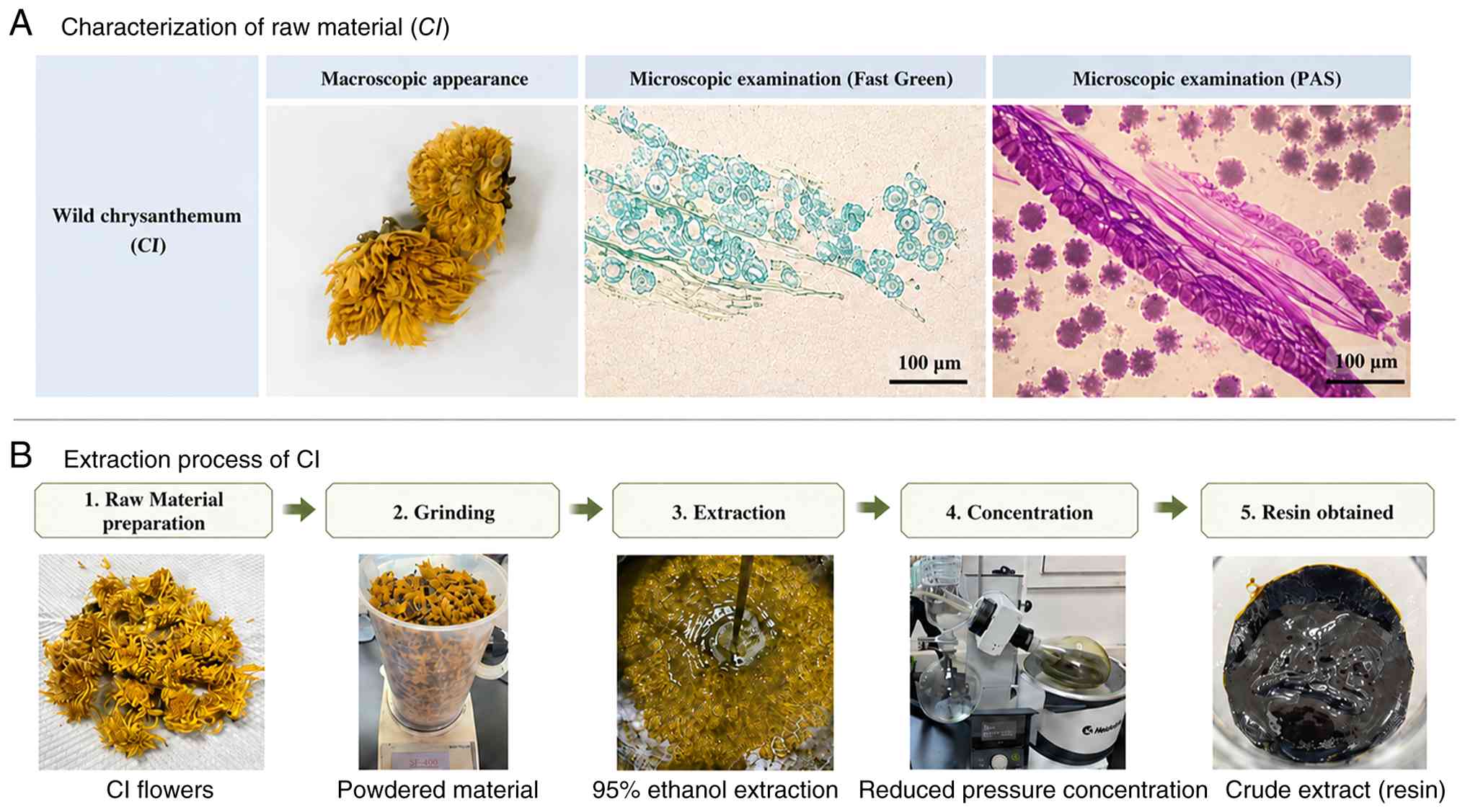
subjected to macroscopic and microscopic evaluation. Macroscopic
examination revealed a capitulum structure, comprising yellow
tubular central florets and outer ray florets, together with a
distinct aromatic odour, consistent with the morphological features
expected for the material supplied as CI (Fig. 1A). Histological cross-sections
stained with Fast Green and PAS further demonstrated the anatomical
architecture of the floral tissues. PAS staining delineated
polysaccharide-rich cell walls and secretory glandular trichomes in
magenta, whereas Fast Green provided contrasting visualization of
cytoplasmic components in green. Collectively, these findings
documented the morphological and anatomical characteristics of the
plant material (Fig. 1A). Following
morphological and histological assessment, 1,004.5 g pulverized
flower heads was extracted with 95% ethanol. Following evaporation
of the solvent under reduced pressure, a dark brown, viscous crude
extract was obtained (Fig. 1B). The
resulting crude extract was aliquoted and stored for subsequent
in vitro pharmacological and molecular experiments.
CI treatment increases intracellular
oxidative activity in FaDu cells
To characterize the early cellular response to CI
treatment, changes in intracellular oxidative activity were
evaluated using the cell-permeable fluorogenic probe, DCFH-DA.
Following cellular uptake, DCFH-DA is deacetylated through
intracellular esterases and subsequently oxidized to the
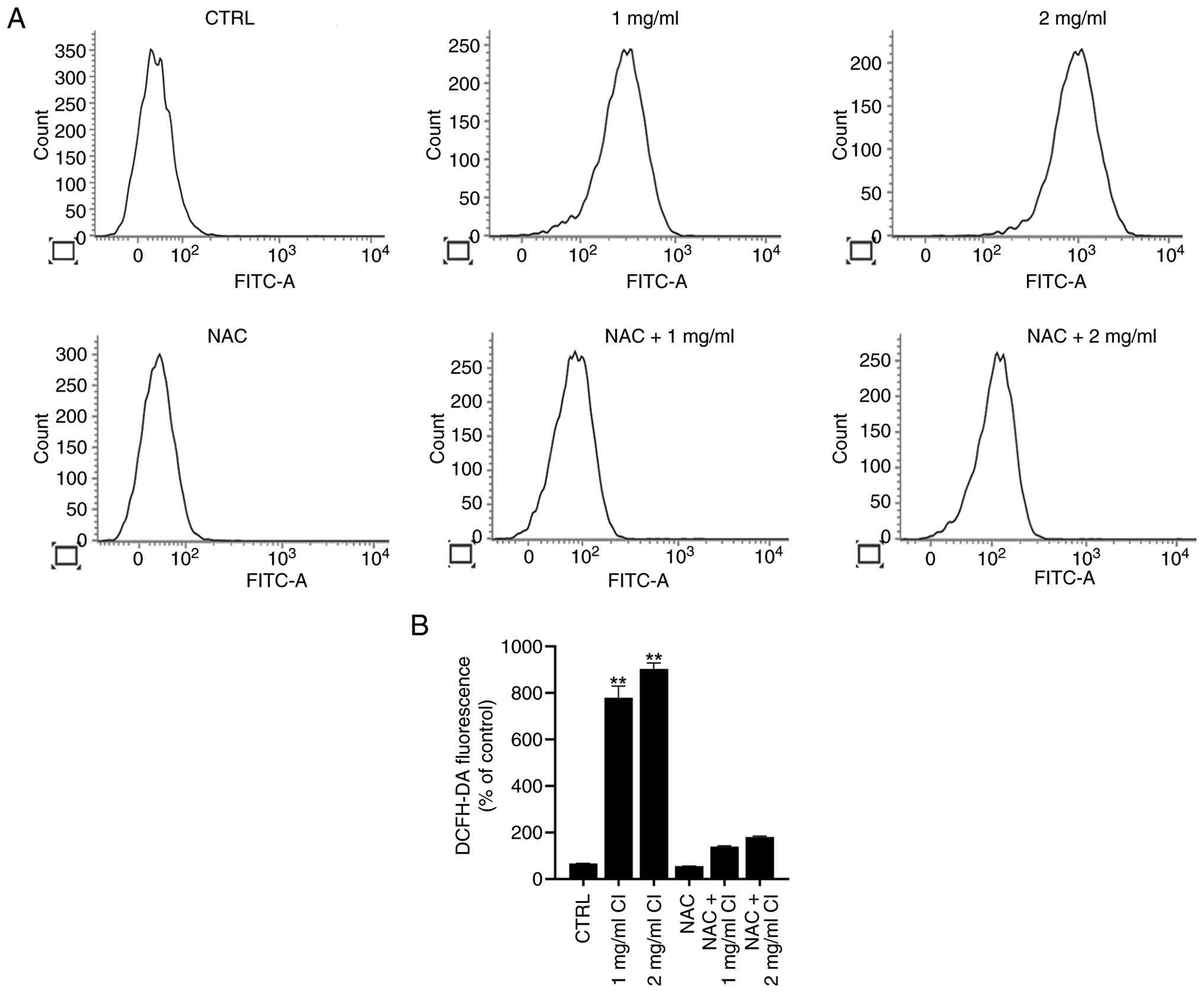
fluorescent compound DCF. Flow cytometric analysis revealed a
notable concentration-dependent rightward shift in green
fluorescence in FaDu cells treated with 1 or 2 mg/ml CI for 24 h
(Fig. 2A). Quantitative analysis
demonstrated that the relative mean fluorescence intensity was
significantly higher in CI-treated cells compared with
vehicle-treated controls (Fig. 2B),
indicating increased intracellular oxidative activity following CI
treatment (P<0.01; Fig. 2).
To determine whether the CI-induced increase in DCF
fluorescence was sensitive to antioxidant pretreatment, a rescue
experiment was performed using NAC, an antioxidant and ROS
scavenger. FaDu cells were pre-incubated with 5 mM NAC for 1 h
before CI treatment. NAC pretreatment markedly attenuated the
CI-induced increase in DCF fluorescence, reducing the fluorescence
signal toward baseline levels (Fig.
2). Collectively, these findings support the involvement of
increased intracellular oxidative activity in the cellular response
to CI treatment.
CI treatment reduces EIF2AK3
expression and Akt activation
To further characterize the molecular alterations
associated with CI treatment, proteins involved in ER stress
adaptation and cell survival were evaluated. FaDu cells were
treated with 1 or 2 mg/ml CI for 24 h, followed by western blotting
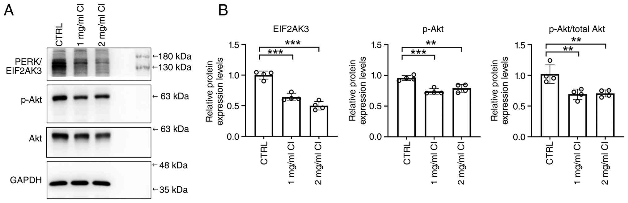
of whole-cell lysates. EIF2AK3, also known as PERK, exhibited a
significant concentration-dependent reduction following CI
treatment, as supported through densitometric semi-quantification
normalized to GAPDH (compared with control; Fig. 3). The activation status of Akt
signaling was subsequently evaluated. CI treatment significantly
decreased p-Akt levels and the p-Akt/total Akt ratio in FaDu cells
(compared with control; Fig. 3).
These findings indicated that CI treatment is associated with
reduced EIF2AK3 expression and decreased Akt activation in FaDu
cells.
CI treatment decreases Bcl-2 protein
levels
Given the observed alterations in EIF2AK3 expression
and Akt activation, the effect of CI treatment on the expression of
the anti-apoptotic protein Bcl-2 was subsequently investigated.
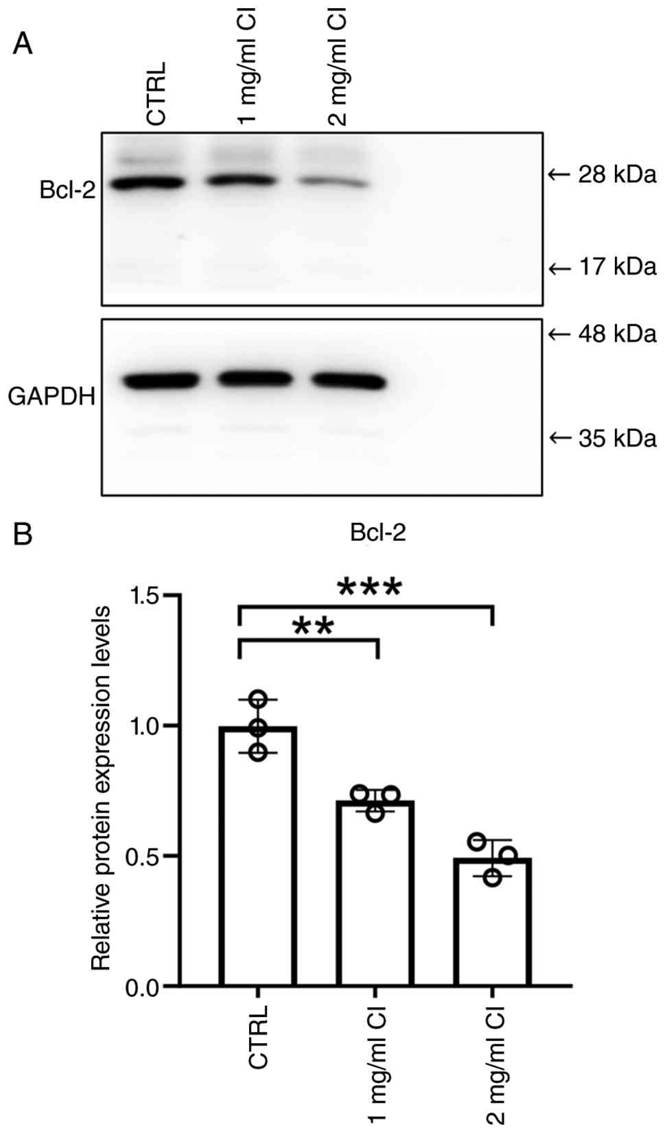
Western blotting of FaDu cells treated with 1 or 2 mg/ml CI for 24
h revealed a notable concentration-dependent reduction in Bcl-2
protein levels compared with vehicle-treated controls (Fig. 4A). Densitometric analysis further
determined the significant progressive reduction in Bcl-2
expression (Fig. 4B), indicating
that CI treatment is associated with decreased expression of this
anti-apoptotic protein.
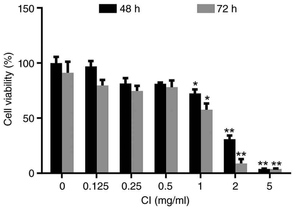
CI decreases FaDu cell viability
To determine whether the observed molecular
alterations were accompanied by changes in cell viability, FaDu
cells were evaluated using a CCK-8 assay. Cells were exposed to
increasing concentrations of CI, and cell viability was assessed
after 48 and 72 h of treatment. CI treatment notably reduced FaDu
cell viability in a concentration-dependent manner. Compared with
the vehicle control, CI significantly reduced cell viability at 1
mg/ml (P<0.05) and at 2 and 5 mg/ml (P<0.01) at both 48 and
72 h. The IC50 values of CI extract were 1.83 mg/ml at
48 h and 1.19 mg/ml at 72 h. The reduction in cell viability was
also time dependent, with 72 h of treatment producing a greater
decrease than 48 h of treatment relative to the vehicle control,
which was defined as 100% viability (Fig. 5). Collectively, these findings
demonstrated that CI decreases FaDu cell viability in a
concentration- and time-dependent manner and further support its
potential cytotoxic activity against HSCC cells.
Discussion
The present study investigated the antitumor
activity and underlying molecular mechanisms of crude CI extract in
human HSCC cells. CI treatment was shown to reduce FaDu cell
viability in a concentration- and time-dependent manner and was
accompanied by increased intracellular oxidative activity. Notably,
pretreatment with the antioxidant NAC markedly attenuated the
CI-induced increase in DCFH-DA fluorescence, supporting the
involvement of oxidative activity in the cellular response to CI
treatment.
The present study primarily focused on the human
HSCC cell line FaDu. Despite evaluation across multiple cell lines
perhaps providing a broader assessment of the antitumor activity of
CI, FaDu cells were selected as the in vitro model because
they exhibit characteristics relevant to aggressive HSCC (4,17).
FaDu cells display an invasive phenotype and molecular
heterogeneity that make them a useful model for investigating the
biology of advanced HSCC (2,5,17).
Clinically, hypopharyngeal cancer is associated with poor
prognosis, in part because of its propensity for early lymphatic
spread (2). Thus, the use of FaDu
cells provides an appropriate initial model for evaluating the
potential antitumor activity of CI. Despite this, validation in
additional HSCC cell lines will be necessary to determine the
generalizability of the present findings.
Molecular analyses demonstrated that decreased cell
viability following CI treatment was accompanied by a significant
concentration-dependent reduction in the anti-apoptotic protein
Bcl-2 (18,19). Bcl-2 is a key regulator of
mitochondrial apoptosis and contributes to the maintenance of
mitochondrial outer membrane integrity (18,19).
However, conventional assays directly assessing apoptosis, such as
Annexin V/PI flow cytometry or terminal deoxynucleotidyl
transferase dUTP nick-end labelling, were not performed in the
present study. Therefore, although the reduction in Bcl-2
expression together with decreased cell viability is consistent
with the potential involvement of apoptotic signaling, the present
findings did not establish apoptosis as the mechanism of CI-induced
cytotoxicity. Additional studies assessing apoptotic cell
populations, caspase activation and other markers of mitochondrial
apoptosis are required to demonstrate and quantify this
process.
An additional notable finding was the
concentration-dependent reduction in EIF2AK3 expression following
CI treatment. EIF2AK3 is an important component of the unfolded
protein response (UPR) and contributes to cellular adaptation under
conditions of ER and oxidative stress (20–22).
Accordingly, the observed reduction in EIF2AK3 expression may
reflect altered cellular stress adaptation following CI treatment.
However, changes in total EIF2AK3 expression alone are insufficient
to establish either inhibition or collapse of the UPR. Further
investigation of EIF2AK3 activation and additional canonical UPR
components, including inositol-requiring enzyme 1α, activating
transcription factor 6 and CCAAT/enhancer-binding protein
homologous protein, will therefore be necessary to determine how CI
affects ER stress signaling.
CI treatment also significantly decreased p-Akt
levels and the p-Akt/total Akt ratio, consistent with reduced Akt
activation. The PI3K/Akt pathway is frequently dysregulated in
squamous cell carcinomas and contributes to tumor cell
proliferation and survival (23,24).
Despite this, the present data demonstrate an association between
CI treatment and reduced Akt activation rather than a direct causal
role for Akt signaling in CI-induced cytotoxicity. Functional
experiments involving pharmacological or genetic modulation of Akt
signaling are therefore required to establish its mechanistic
contribution. Collectively, the reductions in EIF2AK3 and Bcl-2
expression and Akt activation, were associated with decreased FaDu
cell viability, suggesting that CI treatment affects a number of
stress- and survival-associated pathways (24,25).
A number of limitations within the present study
should be acknowledged. First, the experiments were conducted
exclusively using a single in vitro model, the FaDu cell
line, limiting the generalizability of the findings to other HSCC
models. FaDu represents a human papillomavirus (HPV)-negative
hypopharyngeal carcinoma model, and HPV-negative head and neck
cancers are generally associated with less favourable clinical
outcomes than their HPV-positive counterparts (26). Future studies incorporating
additional HPV-negative and HPV-positive models will be necessary
to determine whether the effects of CI vary according to HPV status
or tumor context. In addition, as a preliminary in vitro
investigation, the present study lacks in vivo efficacy,
toxicological and pharmacokinetic data. Therefore, the present
findings should be interpreted as preliminary evidence of
biological activity in FaDu cells and do not establish tumor
selectivity, a therapeutic window, systemic safety or tolerability.
Further evaluation using non-malignant cellular models and in
vivo toxicological and pharmacokinetic studies will be required
before translational conclusions can be drawn. In addition, formal
taxonomic authentication of the plant material by a qualified
botanist or taxonomist was not performed, and no voucher specimen
was deposited. Although macroscopic and histological assessments
were performed to characterize the plant material, these approaches
alone do not provide definitive species-level authentication.
The absence of formal taxonomic verification and a
voucher specimen limits retrospective traceability and independent
confirmation of the botanical material and should therefore be
considered when interpreting and reproducing the present findings.
Furthermore, although the extraction procedure was controlled with
respect to the amount of starting material, ethanol concentration,
extraction duration, number of extraction cycles, evaporation
temperature, extraction yield, moisture content and storage
conditions, comprehensive chemical standardization of the crude
extract was not performed. Specifically, chromatographic
fingerprinting and quantitative analysis of major constituents were
not performed for the extract used in the present study.
Consequently, chemical batch-to-batch consistency could not be
established, which limits reproducibility, comparison with extracts
prepared under different conditions and identification of the
constituents responsible for the observed biological effects.
Accordingly, the present findings should be interpreted as being
specific to the crude extract prepared under the conditions
described in the present study and should not be generalized to all
CI preparations. Future studies should aim to incorporate
comprehensive phytochemical profiling and quantitative
characterization of major constituents to improve chemical
standardization and batch-to-batch reproducibility. Finally, formal
physicochemical solubility profiling of the crude CI extract was
not performed. Therefore, parameters such as saturation solubility
and the physicochemical behavior of the extract in culture medium
remain undefined, which may affect the reproducibility and
interpretation of the concentration-response findings. Formal
solubility characterization should therefore be incorporated into
future studies.
In conclusion, the present findings demonstrated
that CI treatment increases intracellular oxidative activity and is
accompanied by reduced EIF2AK3 and Bcl-2 expression, decreased Akt
activation and reduced FaDu cell viability. The attenuation of
CI-induced DCFH-DA fluorescence through NAC pretreatment further
supports the involvement of oxidative activity in the cellular
response to CI. Collectively, these findings may provide
preliminary evidence supporting the potential antitumor activity of
CI in HSCC cells and warrant further mechanistic investigation
using additional cellular models and in vivo studies.
Acknowledgements
Not applicable.
Funding
The present study was supported by a grant from The National
Science and Technology Council (grant no. NSTC
115-2221-E-343-001).
Availability of data and materials
The data generated in the present study may be
requested from the corresponding author.
Authors' contributions
CWL was responsible for conceptualization,
methodology, investigation (plant material preparation and
histological assessment), data curation, data analysis and writing
the original draft. LCC contributed to conceptualization and
methodology. TTL contributed to the investigation (cell culture, CI
treatment and cell viability assays). HCL was responsible for
conceptualization, methodology, investigation (western blotting,
flow cytometric experiments and data acquisition), data curation,
data analysis, writing the original draft and supervision. CWL and
HCL confirm the authenticity of all the raw data. All authors read
and approved the final version of the manuscript.
Ethics approval and consent to
participate
Not applicable.
Patient consent for publication
Not applicable.
Competing interests
The authors declare that they have no competing
interests.
Glossary
Abbreviations
Abbreviations:
|
CI
|
Chrysanthemum indicum
|
|
DCFH-DA
|
2′,7′-dichlorodihydrofluorescein
diacetate
|
|
HPV
|
human papillomavirus
|
|
HSCC
|
hypopharyngeal squamous cell
carcinoma
|
|
NAC
|
N-acetylcysteine
|
|
PERK/EIF2AK3
|
protein kinase R-like endoplasmic
reticulum kinase/eukaryotic translation initiation factor 2 α
kinase 3
|
|
ROS
|
reactive oxygen species
|
|
UPR
|
unfolded protein response
|
References
|
1
|
Jang W, Kim S, Son Y, Kim S, Lee S, Kim
HJ, Jo H, Park J, Lee K, Lee H, et al: Global, regional, and
national burden of pharyngeal cancer and projections to 2050 in 185
countries: A population-based systematic analysis of GLOBOCAN 2022.
J Korean Med Sci. 40:e1772025. View Article : Google Scholar : PubMed/NCBI
|
|
2
|
Fang Y, Yi C, Sun R, Shen B, Yao M, Yang
Y, Ni Y, Liu Y, Li Z and Liu J: Global annualized recurrence rates
and dynamic risk profiles in head and neck cancer. Int J Cancer.
Jul 26–2026.(Epub ahead of print). doi: 10.1002/ijc.70675.
View Article : Google Scholar
|
|
3
|
Argiris A, Karamouzis MV, Raben D and
Ferris RL: Head and neck cancer. Lancet. 371:1695–1709. 2008.
View Article : Google Scholar : PubMed/NCBI
|
|
4
|
Rangan SR: A new human cell line (FaDu)
from a hypopharyngeal carcinoma. Cancer. 29:117–121. 1972.
View Article : Google Scholar : PubMed/NCBI
|
|
5
|
Leemans CR, Snijders PJF and Brakenhoff
RH: The molecular landscape of head and neck cancer. Nat Rev
Cancer. 18:269–282. 2018. View Article : Google Scholar : PubMed/NCBI
|
|
6
|
Newman DJ and Cragg GM: Natural products
as sources of new drugs from 1981 to 2014. J Nat Prod. 79:629–661.
2016. View Article : Google Scholar : PubMed/NCBI
|
|
7
|
Surh YJ: Cancer chemoprevention with
dietary phytochemicals. Nat Rev Cancer. 3:768–780. 2003. View Article : Google Scholar : PubMed/NCBI
|
|
8
|
Wang Y, Li Y, Guo W, Yang X, Qu J, Gao M,
Chen S, Dong J, Li Q and Wang T: Comparison of the chemical
components, efficacy and mechanisms of action of Chrysanthemum
morifolium flower and its wild relative Chrysanthemum
indicum flower against liver-fire hyperactivity syndrome of
hypertension via integrative analyses. Int J Mol Sci. 23:137672022.
View Article : Google Scholar : PubMed/NCBI
|
|
9
|
Youssef FS, Eid SY, Alshammari E, Ashour
ML, Wink M and El-Readi MZ: Chrysanthemum indicum and
Chrysanthemum morifolium: chemical composition of their
essential oils and their potential use as natural preservatives
with antimicrobial and antioxidant activities. Foods. 9:14602020.
View Article : Google Scholar : PubMed/NCBI
|
|
10
|
Liang Y, Liu T, Wang D and Liu Q:
Exploring the antimicrobial, anti-inflammatory, antioxidant, and
immunomodulatory properties of Chrysanthemum morifolium and
Chrysanthemum indicum: A narrow review. Front Pharmacol.
16:15383112025. View Article : Google Scholar : PubMed/NCBI
|
|
11
|
Zeng ZY, Li QQ, Huo YY, Chen CJ, Duan SS,
Xu FR, Cheng YX and Dong X: Inhibitory effects of essential oils
from Asteraceae plant against pathogenic fungi of Panax
notoginseng. J Appl Microbiol. 130:592–603. 2021. View Article : Google Scholar : PubMed/NCBI
|
|
12
|
Kim C, Kim MC, Kim SM, Nam D, Choi SH, Kim
SH and Ahn KS, Lee EH, Jung SH and Ahn KS: Chrysanthemum
indicum L. extract induces apoptosis through suppression of
constitutive STAT3 activation in human prostate cancer DU145 cells.
Phytother Res. 27:30–38. 2013. View
Article : Google Scholar : PubMed/NCBI
|
|
13
|
Yang HM, Sun CY, Liang JL, Xu LQ, Zhang
ZB, Luo DD, Chen HB, Huang YZ, Wang Q, Lee DY, et al:
Supercritical-Carbon dioxide fluid extract from Chrysanthemum
indicum enhances anti-tumor effect and reduces toxicity of
bleomycin in tumor-bearing mice. Int J Mol Sci. 18:4652017.
View Article : Google Scholar : PubMed/NCBI
|
|
14
|
Trachootham D, Alexandre J and Huang P:
Targeting cancer cells by ROS-mediated mechanisms: A radical
therapeutic approach? Nat Rev Drug Discov. 8:579–591. 2009.
View Article : Google Scholar : PubMed/NCBI
|
|
15
|
Gorrini C, Harris IS and Mak TW:
Modulation of oxidative stress as an anticancer strategy. Nat Rev
Drug Discov. 12:931–947. 2013. View
Article : Google Scholar : PubMed/NCBI
|
|
16
|
Dixon SJ and Stockwell BR: The role of
iron and reactive oxygen species in cell death. Nat Chem Biol.
10:9–17. 2014. View Article : Google Scholar : PubMed/NCBI
|
|
17
|
Mu Y, Sa N, Yu L, Lu S, Wang H and Xu W:
Epithelial cell adhesion molecule is overexpressed in
hypopharyngeal carcinoma and suppresses the metastasis and
proliferation of the disease when downregulated. Oncol Lett.
8:175–182. 2014. View Article : Google Scholar : PubMed/NCBI
|
|
18
|
Cory S and Adams JM: The Bcl2 family:
Regulators of the cellular life-or-death switch. Nat Rev Cancer.
2:647–656. 2002. View
Article : Google Scholar : PubMed/NCBI
|
|
19
|
Czabotar PE, Lessene G, Strasser A and
Adams JM: Control of apoptosis by the BCL-2 protein family:
Implications for physiology and therapy. Nat Rev Mol Cell Biol.
15:49–63. 2014. View
Article : Google Scholar : PubMed/NCBI
|
|
20
|
Bi M, Naczki C, Koritzinsky M, Fels D,
Blais J, Hu N, Harding H, Novoa I, Varia M, Raleigh J, et al: ER
stress-regulated translation increases tolerance to extreme hypoxia
and promotes tumor growth. EMBO J. 24:3470–3481. 2005. View Article : Google Scholar : PubMed/NCBI
|
|
21
|
Cubillos-Ruiz JR, Bettigole SE and
Glimcher LH: Tumorigenic and immunosuppressive effects of
endoplasmic reticulum stress in cancer. Cell. 168:692–706. 2017.
View Article : Google Scholar : PubMed/NCBI
|
|
22
|
Wang M and Kaufman RJ: The impact of the
endoplasmic reticulum protein-folding environment on cancer
development. Nat Rev Cancer. 14:581–597. 2014. View Article : Google Scholar : PubMed/NCBI
|
|
23
|
Asano T, Yao Y, Zhu J, Li D, Abbruzzese JL
and Reddy SA: The PI 3-kinase/Akt signaling pathway is activated
due to aberrant Pten expression and targets transcription factors
NF-kappaB and c-Myc in pancreatic cancer cells. Oncogene.
23:8571–8580. 2004. View Article : Google Scholar : PubMed/NCBI
|
|
24
|
Manning BD and Toker A: AKT/PKB signaling:
Navigating the network. Cell. 169:381–405. 2017. View Article : Google Scholar : PubMed/NCBI
|
|
25
|
Porta C, Paglino C and Mosca A: Targeting
PI3K/Akt/mTOR signaling in cancer. Front Oncol. 4:642014.
View Article : Google Scholar : PubMed/NCBI
|
|
26
|
Shi J, Wang L, Yao N, Sun L, Hu W, Li X,
Yang Y, Wang Y, Zhu W and Li B: The effect of HPV DNA and p16
status on the prognosis of patients with hypopharyngeal carcinoma:
A meta-analysis. BMC Cancer. 22:6582022. View Article : Google Scholar : PubMed/NCBI
|