Introduction
According to the latest data released by the
International Agency for Research on Cancer of the World Health
Organization, breast cancer has become the most common malignant
tumor among women globally (1). The
most frequent sites of distant metastasis are bone, liver, brain
and lung, whereas metastasis to the thyroid gland is uncommon
(2). The thyroid is a highly
vascularized endocrine organ with unique tissue characteristics,
including high blood flow and iodine concentration, which have been
proposed as potential factors contributing to its relatively low
susceptibility to secondary malignancies (3). Secondary thyroid cancers usually
originate from primary tumors in the kidney, gastrointestinal
tract, lung or skin, with only a minority arising from breast
cancer (4).
Secondary malignancies of the thyroid are uncommon.
Reported rates of thyroid metastasis vary considerably among
clinical studies and autopsy studies, with autopsy studies
reporting an incidence of 1.25–24%; breast cancer accounts for a
variable proportion of thyroid metastases across published studies
(5,6). The mechanisms underlying the relative
rarity of thyroid metastasis remain incompletely understood.
Despite the rich vascular supply of the thyroid, rapid arterial
blood flow, high oxygen tension and iodine-rich conditions have
been proposed to create an unfavorable environment for metastatic
implantation, whereas pre-existing thyroid abnormalities may
potentially alter the local microenvironment and facilitate
metastatic colonization (3,5,7,8). No
well-established patient-level risk factors specific to breast
cancer metastasis to the thyroid have been identified. Clinically,
thyroid metastasis may present as a focal nodule, diffuse gland
enlargement, cervical lymphadenopathy or an incidental imaging
abnormality. Diagnosis generally relies on integration of the
previous oncological history, imaging, cytological or histological
examination and immunohistochemical profiling (5,6).
Prognosis is influenced primarily by the biological behavior of the
primary tumor and the extent of systemic metastatic disease;
selected patients with isolated thyroid metastasis may benefit from
local treatment, whereas systemic therapy is generally favored in
patients with disseminated disease (5,6).
Due to the lack of specific clinical or imaging
features, differentiating between primary thyroid cancer and
metastatic breast cancer presents notable diagnostic challenges
(5). Immunohistochemistry remains a
reliable method for distinguishing these two entities. The present
study reports 2 cases of breast cancer metastasizing to the thyroid
and integrates them with a structured literature review to
summarize clinical characteristics, diagnostic considerations and
therapeutic strategies.
Case reports
Case 1
In December 2016, a 48-year-old woman was diagnosed
with right-sided breast cancer and underwent modified radical
mastectomy with axillary lymph node dissection at Weifang People's
Hospital (Weifang, China). Pathological examination revealed grade
II invasive ductal carcinoma (IDC) with ductal carcinoma in
situ (~10%), measuring 3×2.5×1.4 cm, with perineural invasion,
lymphovascular emboli and nipple involvement. Axillary lymph nodes
showed metastasis (8/20). Immunohistochemistry revealed: Estrogen
receptor (ER; 3+, 95%), progesterone receptor (PR; 2+, 30%), HER2
(2+, fluorescence in situ hybridization non-amplified),
Ki-67 (30%), cytokeratin 5/6 (CK5/6; focal +) and E-cadherin (+).
Histopathological and immunohistochemical examinations were
performed as part of routine diagnostic procedures in the hospital
pathology department. Owing to the retrospective nature of the
present study and the long interval since some of the original
examinations, complete technical details of the historical
immunohistochemical procedures, including antibody catalogue
numbers, dilutions, incubation conditions and detection-system
parameters, could not be fully retrieved from the archived
laboratory records. The immunohistochemical results for both cases
reported in the present study were obtained from the available
pathology reports and archived clinical records.
The patient received four cycles of epirubicin (90
mg/m2 i.v., d1, q3w) plus cyclophosphamide (600
mg/m2 i.v., d1, q3w) followed by four cycles of
docetaxel (100 mg/m2 i.v., d1, q3w), radiotherapy (total
dose 50 Gy in 25 fractions over 5 weeks) and long-term endocrine
therapy with anastrozole (1 mg once daily) from June 2017 to April
2025 (94 months), followed by regular follow-up.
During routine follow-up in April 2025, the patient
had no clinically notable local compressive symptoms, such as
dysphagia, dyspnea or hoarseness. Detailed thyroid function test
results were not available in the archived medical records. Thyroid
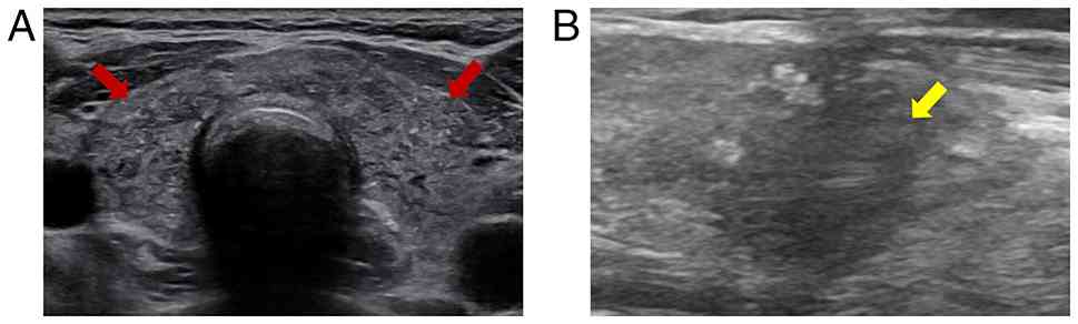
and cervical lymph node ultrasonography revealed diffuse thyroid
parenchymal changes with multiple punctate hyperechoic foci and
bilateral cervical lymphadenopathy. Some cervical lymph nodes
showed loss of the normal fatty hilum and punctate hyperechoic
foci. The largest left cervical lymph node measured 7.1×4.7 mm, and
the largest right cervical lymph node measured 10.6×5.6 mm
(Fig. 1A). Fine-needle aspiration
of the bilateral thyroid lobes and cervical lymph nodes revealed
malignant cells (Fig. 2).
Fine-needle aspiration smears were prepared and stained with
hematoxylin and eosin (H&E) and Papanicolaou stain according to
the routine cytopathology procedures of the hospital pathology
department. As these examinations were performed as part of routine
clinical diagnostic practice, the original detailed fixation and
staining-protocol records were not retained and could not be
retrospectively retrieved. No mutation was detected in BRAF exon 15
using amplification refractory mutation system-PCR. Core needle
biopsy of the right thyroid lesion confirmed malignancy.
Immunohistochemical staining (Fig.
3) showed ER (3+, 90%), PR (2+, 15%), HER2 (2+, equivocal),
GATA-binding protein 3 (GATA3; +), thyroid transcription factor-1
(TTF-1; -), thyroglobulin (TG; focal +), calcitonin (−), CD56 (−)
and Ki-67 (55%). The focal TG staining was interpreted cautiously
because it may have originated from entrapped non-neoplastic
thyroid follicles rather than metastatic tumor cells. Taken
together with the clinical history of the patient and the remaining
immunophenotypic findings, the lesion was diagnosed as metastatic
breast carcinoma involving the thyroid.
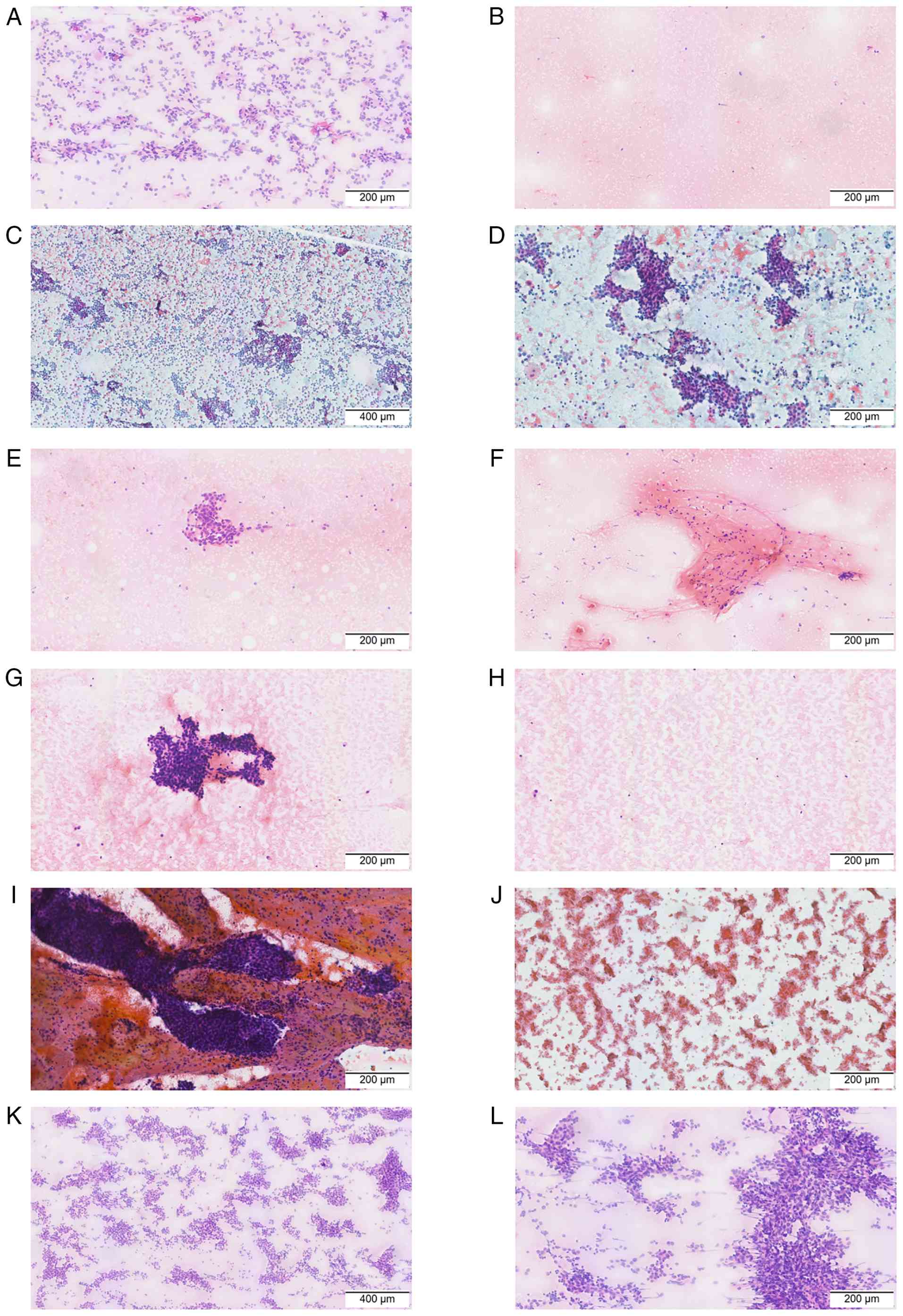 | Figure 2.Cytological findings from fine-needle
aspiration smears of bilateral thyroid lobes and bilateral cervical
lymph nodes in patient 1. (A) H&E staining in the positive area
of the left thyroid lobe. Scale bar, 200 µm. (B) H&E staining
in the negative area of the left thyroid lobe. Scale bar, 200 µm.
(C) Papanicolaou staining of the left thyroid lobe positive area.
Scale bar, 400 µm (no negative area observed in this region). (D)
Higher-magnification view of the Papanicolaou staining in the same
left thyroid lobe. Scale bar, 200 µm. (E) H&E staining of the
positive area of the left cervical lymph node. Scale bar, 200 µm.
(F) H&E staining of the negative area of the left cervical
lymph node. Scale bar, 200 µm. (G) H&E staining of the positive
area of the right thyroid lobe. Scale bar, 200 µm. (H) H&E
staining of the negative area in the right thyroid lobe. Scale bar,
200 µm. (I) Papanicolaou staining of the positive area in the right
thyroid lobe. Scale bar, 200 µm. (J) Papanicolaou staining in the
negative area of the right thyroid lobe. Scale bar, 200 µm. (K)
H&E staining of the positive area in the right cervical lymph
node. Scale bar, 400 µm (no negative area observed in this region).
(L) Higher-magnification view of the same right cervical
lymph-node. Scale bar, 200 µm. H&E, hematoxylin and eosin. |
 | Figure 3.Histopathological and
immunohistochemical findings from the core needle biopsy of the
right thyroid lesion in patient 1. (A) H&E staining. Scale bar,
200 µm. (B) H&E staining. Scale bar, 100 µm. (C) Estrogen
receptor strongly positive (3+, 90%). Scale bar, 200 µm. (D)
Progesterone receptor moderately positive (2+, 15%). Scale bar, 200
µm. (E) HER2 equivocal (2+). Scale bar, 200 µm. (F) Ki-67 labeling
index: 55% positive tumor nuclei. Scale bar, 200 µm. (G)
GATA-binding protein 3 positive. Scale bar, 200 µm. (H) Thyroid
transcription factor-1 negative. Scale bar, 200 µm. (I) Focal
thyroglobulin staining. Scale bar, 200 µm. H&E, hematoxylin and
eosin. |
Restaging examinations, including bone scintigraphy,
cranial MRI, and contrast-enhanced CT, showed no additional
metastatic sites beyond the thyroid and cervical lymph nodes.
Following multidisciplinary team discussion, thyroid and cervical
lymph node involvement was considered a manifestation of recurrent
hormone receptor-positive breast cancer after adjuvant endocrine
therapy. Because the patient had no clinically notable local
compressive symptoms, systemic endocrine-based therapy was
considered an appropriate initial management strategy, and
immediate thyroid surgery was not recommended. Systemic treatment
with dalpiciclib (125 mg p.o. q.d.; d1-21, q4w) plus fulvestrant
(500 mg i.m., cycle 1 d1, d15, then d1 q4w) was initiated in April
2025 as first-line systemic treatment for recurrent metastatic
disease and was ongoing at the time of manuscript preparation.
Follow-up ultrasonography in June 2025 showed an interval decrease
in the thyroid and cervical lymph node lesions, including a
reduction in the largest left cervical lymph node from 7.1×4.7 to
7.1×3.0 mm. In view of the continued absence of local symptoms and
the observed radiological disease control, systemic treatment and
surveillance were continued. The treatment timeline is shown in
Fig. 4A.
Case 2
A 40-year-old woman was diagnosed with right-sided
breast cancer in March 2013. The original 2013 pathology report and
immunohistochemical images could not be retrieved because of the
long interval since diagnosis; therefore, the historical
pathological and immunohistochemical information described below
was obtained from the available archived clinical record. After
four cycles of neoadjuvant chemotherapy with docetaxel, doxorubicin
and cyclophosphamide, they underwent modified radical mastectomy
and axillary lymph node dissection in June 2013. The available
clinical record documented invasive breast carcinoma with mixed
ductal and lobular components; no metastasis was found in axillary
lymph nodes (0/10). Immunohistochemistry revealed: ER (+), PR (+),
HER2 (−), Ki-67 (10%), E-cadherin (−) and p120 (+). The patient
received four cycles of adjuvant docetaxel plus cyclophosphamide
chemotherapy (TC), radiotherapy, and 5 years of tamoxifen as
endocrine therapy.
In July 2022, the patient developed multiple bone
metastases and was treated with letrozole (2.5 mg p.o. q.d.) plus
palbociclib (125 mg p.o. q.d., d1-21, q4w) as first-line therapy.
Due to disease progression in June 2024, they switched to alpelisib
(300 mg p.o. q.d., continuous) plus fulvestrant (500 mg i.m., cycle
1 d1, d15, then d1 q4w) as second-line treatment, but the regimen
was discontinued after 21 days because of severe rash and secondary
diabetes. Subsequently, the patient enrolled in a clinical trial
receiving fulvestrant (500 mg i.m., cycle 1 d1, d15, then d1 q4w)
combined with a novel targeted agent (CDK4/6 inhibitor) and
denosumab (120 mg s.c. q4w) for ~11 months, until June 2025.
During follow-up in June 2025, ultrasonography
revealed a solid nodule in the lower pole of the right thyroid
lobe, classified as Chinese Thyroid Imaging Reporting and Data
System category 4C (9), with
punctate hyperechoic foci and multiple enlarged bilateral cervical
lymph nodes (Fig. 1B). Detailed
information regarding thyroid-related symptoms and thyroid function
test results was not available in the archived medical records.
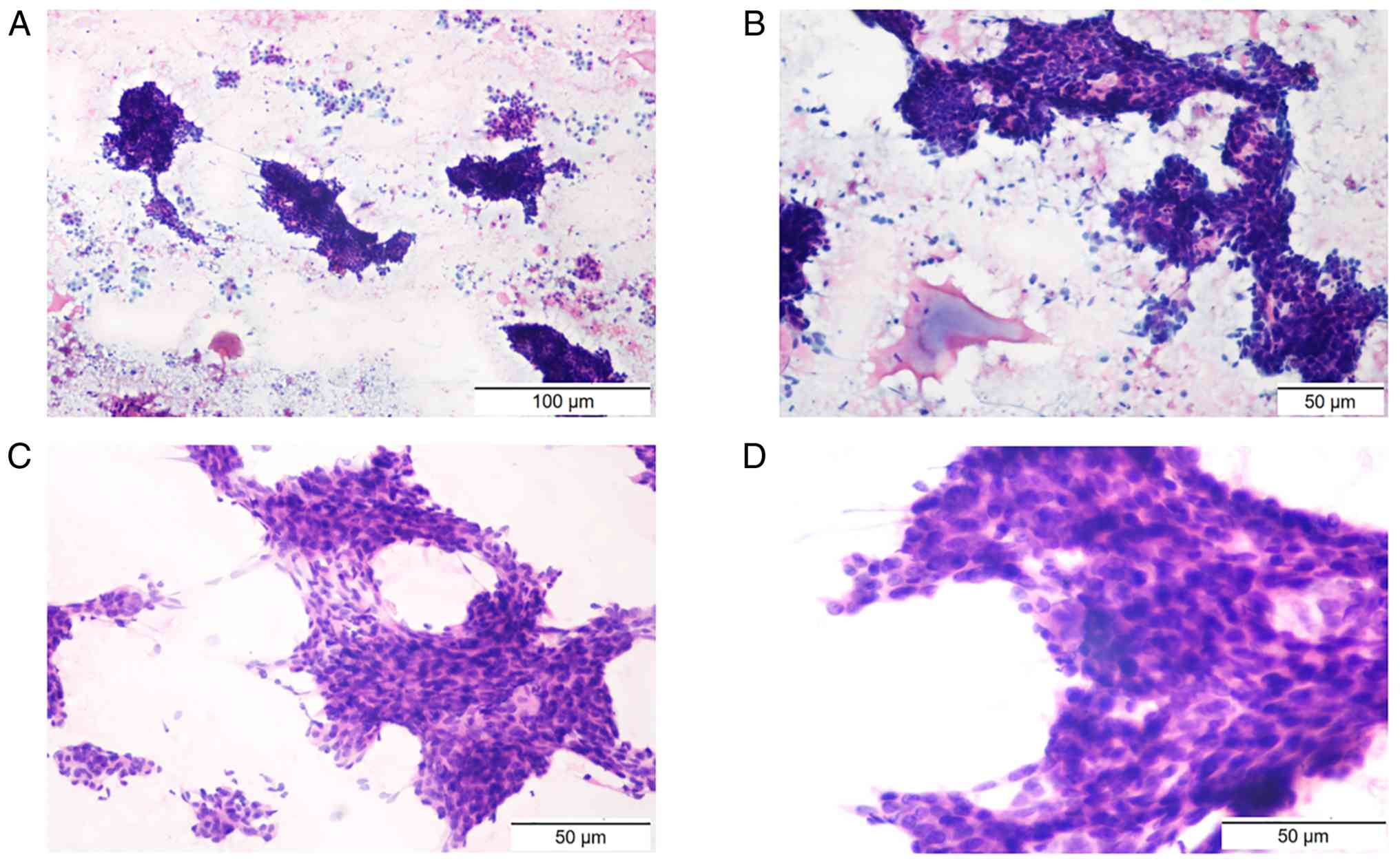
Fine-needle aspiration identified malignant cells morphologically
consistent with lymph node biopsy specimens (Fig. 5); BRAF mutation was absent. Right
cervical lymph node biopsy confirmed metastatic carcinoma.
Immunohistochemical analysis of the right cervical lymph node
specimen (Fig. 6) showed ER (1+,
35%), PR (2+, <1%), HER2 (IHC score 1+, classified as
HER2-negative), GATA3 (+), TG (−), TTF-1 (−), paired box gene 8
(PAX8; -) and CK7 (+). Due to the morphological concordance between
the thyroid aspirate and the cervical lymph node specimens,
together with the breast cancer history and the immunophenotype of
the lymph node metastasis of the patient, the thyroid lesion was
diagnosed as metastatic breast carcinoma.
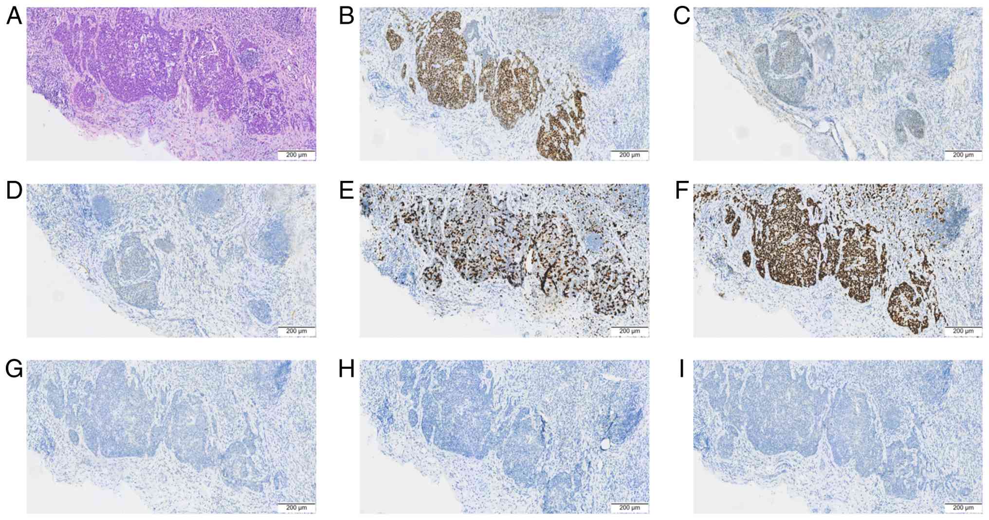 | Figure 6.Histopathological and
immunohistochemical findings of the right cervical lymph node
biopsy from patient 2. (A) Hematoxylin and eosin staining. Scale
bar, 200 µm. (B) Estrogen receptor, weakly positive (1+, 35%).
Scale bar, 200 µm. (C) Progesterone receptor, moderately positive
(2+, <1%). Scale bar, 200 µm. (D) HER2 immunohistochemical score
1+ (HER2-negative). Scale bar, 200 µm. (E) Ki-67 labeling index:
45% positive tumor nuclei. Scale bar, 200 µm. (F) GATA-binding
protein 3 positive. Scale bar, 200 µm. (G) Thyroid transcription
factor-1 negative. Scale bar, 200 µm. (H) Thyroglobulin, negative.
Scale bar, 200 µm. (I) Paired box gene 8, negative. Scale bar, 200
µm. |
Restaging PET/CT demonstrated hypermetabolic lesions
involving the bilateral cervical lymph nodes, bones, left adrenal
gland and right thyroid lobe, consistent with widespread metastatic
disease. Due to the presence of widespread extra-thyroid metastatic
disease and progression after multiple lines of endocrine-based
therapy, thyroid surgery was not considered likely to provide
meaningful survival or disease-control benefit. Systemic treatment
was therefore prioritized. Following further disease progression
during endocrine-based therapy, treatment was changed to exemestane
(25 mg p.o. q.d.), everolimus (10 mg p.o. q.d.) and abemaciclib
(150 mg p.o. b.i.d.) in July 2025. Disease progression with liver
metastasis occurred in September 2025, and the patient was then
switched to nab-paclitaxel (200 mg i.v., d1, q3w) plus cisplatin
(30 mg i.v., d1-3, q3w) chemotherapy. At the time of manuscript
preparation, the patient remained on this treatment. The treatment
timeline for the patient is shown in Fig. 4B.
Literature review
A structured literature search was conducted in
PubMed (https://pubmed.ncbi.nlm.nih.gov/) to identify reports
published between January 1, 2010 and December 31, 2025. The search
was last updated on August 6, 2026. The following search strategy
was used: (‘Breast Neoplasms’ [Mesh] OR ‘breast cancer’
[Title/Abstract] OR ‘breast carcinoma’ [Title/Abstract] OR ‘mammary
carcinoma’[Title/Abstract]) AND (‘Thyroid Neoplasms/secondary’
[Mesh] OR ‘thyroid metastasis’ [Title/Abstract] OR ‘thyroid
metastases’ [Title/Abstract] OR ‘thyroid gland metastasis’
[Title/Abstract] OR ‘thyroid gland metastases’ [Title/Abstract] OR
‘metastasis to the thyroid’ [Title/Abstract] OR ‘metastases to the
thyroid’ [Title/Abstract] OR ((‘Thyroid Gland’ [Mesh] OR thyroid
[Title/Abstract]) AND (metastasis [Title/Abstract] OR metastases
[Title/Abstract] OR metastatic [Title/Abstract]))). No article-type
or relative publication-date filters were applied.
Reports were eligible if they described ≥1 patient
with pathologically confirmed breast cancer metastasis to the
thyroid gland and provided extractable case-level clinical
information. Reports of thyroid malignancies metastatic to the
breast, metastases from non-breast primary tumors, articles without
individual patient data, duplicate reports and reports lacking
sufficient evidence of thyroid involvement were excluded.
The database search, screening of titles, abstracts
and full texts, reviewing of eligibility, and checking extracted
data were performed by two authors. Any disagreements were resolved
through discussion and consensus. Data were extracted into a
standardized table, including publication year, patient age, breast
cancer histological type, receptor status, interval between the
initial diagnosis of breast cancer and thyroid metastasis, other
involved or metastatic sites, ultrasonographic findings, and
treatment. When the same patient appeared to have been reported in
more than one publication, only the report containing the most
complete information was retained. Variables that were not reported
were recorded as ‘not reported’ and excluded from the denominator
for the corresponding percentage calculation.
Receptor status was recorded as reported in each
source publication. Because the specimen source was not
consistently specified, receptor status was analyzed at the case
level. When discordant receptor results were reported among
different specimens from the same patient, the case was classified
as positive if at least one reported specimen was positive.
Ultrasonographic findings were categorized as focal nodular or
diffuse parenchymal patterns according to the descriptions in the
source publications. Microcalcifications were counted only when
explicitly reported.
Discussion
In the present structured literature review, 44
cases of breast cancer metastasis to the thyroid, including the 2
cases reported in the present study, were evaluated (5,10-42;
Table I). The mean patient age was
55.9 years, with a range of 34–82 years. IDC was the most
frequently reported histological type. Among the 32 cases with
available ER data, 23 were ER-positive (71.9%), while 21 of the 32
cases with available PR data were PR-positive (65.6%). Cases in
which receptor status was not reported were excluded from the
corresponding denominator. These findings indicate that hormone
receptor-positive tumors accounted for a substantial proportion of
the reported cases. However, in the absence of a comparator
population, these descriptive data do not establish that hormone
receptor-positive breast cancers have a specific propensity to
metastasize to the thyroid.
 | Table I.Demographic, histological and
receptor characteristics of 44 cases of breast cancer metastasis to
the thyroid included in the present analysis. |
Table I.
Demographic, histological and
receptor characteristics of 44 cases of breast cancer metastasis to
the thyroid included in the present analysis.
| First author,
year | Age, years | Breast cancer
histology | ER | PR | HER2 | Interval,
months | (Refs.) |
|---|
| Present study, Case
1 | 56 | IDC | Positive | Positive | Primary: IHC 2+,
FISH non-amplified; thyroid metastasis: IHC 2+ (equivocal) | 100 | - |
| Present study, Case
2 | 52 | Mixed IDC/ILC | Positive | Positive | Primary: Negative;
cervical LN metastasis: IHC 1+ (HER2-negative) | 147 | - |
| Jbali et al,
2025 | 63 | ILC | Positive | Negative | Negative | 25 | (12) |
| He et al,
2025 | 34 | IDC | Positive | Positive | Negative | 97 | (13) |
| Gvazava et
al, 2025 | 63 | IDC | Positive | Positive | Negative | 179 | (14) |
| Guan et al,
2025 | 55 | IDC | Positive | Positive | Negative | 100 | (15) |
| Gao et al,
2025 | 41 | IDC | Positive | Positive | Negative | 96 | (10) |
| Yousaf et
al, 2025 | 47 | Invasive breast
carcinoma | NR | NR | NR | 0 | (16) |
| Ramírez Stieben
et al, 2024 | 56 | IDC | Positive | Positive | Positive | 60 | (19) |
| Patrizio et
al, 2023 | 71 | IDC | Positive | Positive | Negative | 216 | (5) |
| Hoshi et al,
2023 | 58 | IDC | Positive | Positive | Negative | 156 | (17) |
| Bai et al,
2023 | 35 | IDC | Negative | Negative | Positive | 60 | (18) |
| Zhang et al,
2022 | 46 | NR | NR | NR | NR | 24 | (20) |
| Yang et al,
2022 | 62 | IDC | Negative | Negative | Positive | 60 | (21) |
| Wen et al,
2022 | 49 | IDC | Positive | Positive | NR | 42 | (22) |
| Gharib et
al, 2022 | 48 | NR | NR | NR | NR | 0 | (23) |
| Celik et al,
2022 | 78 | IDC with
neuroendocrine differentiation | Positive | Negative | Negative | 0 | (24) |
| Yu et al,
2021 | 59 | IDC with IMPC
component | Positive | Positive | Negative | 60 | (25) |
| Wang et al,
2021 | 54 | IDC | Negative | Negative | Positive | 22 | (26) |
| Kiziltan et
al, 2021 | 82 | IDC | Positive | Positive | Negative | 144 | (27) |
| Jung et al,
2021 | 59 | IDC | NR | NR | NR | 180 | (28) |
| Zhang et al,
2020 | 38 | IDC | Negative | Negative | Positive | 24 | (29) |
| Wang et al,
2020 | 58 | Mucinous carcinoma
with IDC | NR | NR | NR | 156 | (30) |
| Lee et al,
2020 | 64 | IDC | Positive | Positive | NR | 5 | (31) |
| Raveendrannair
et al, 2019 | 36 | IDC | NR | NR | NR | 0 | (32) |
| Pakuła et
al, 2019 | 66 | Invasive breast
carcinoma | NR | NR | NR | 240 | (33) |
| Durmo et al,
2019 | 72 | IDC | Positive | Positive | Negative | NR | (34) |
| Pensabene et
al, 2018 | 64 | ILC | Positive | Positive | Negative | 6 | (35) |
| Zhou et al,
2017 | 48 | Invasive breast
carcinoma | Negative | Positive | Positive | 87 | (36), Case 1 |
| Zhou et al,
2017 | 59 | IDC | Negative | Negative | NR | 27 | (36), Case 2 |
| Zhou et al,
2017 | 57 | Signet-ring cell
carcinoma | Positive | Negative | NR | 108 | (36), Case 3 |
| Zhou et al,
2017 | 67 | IDC | Negative | Negative | NR | 71 | (36), Case 4 |
| Zhou et al,
2017 | 48 | IDC | Negative | Negative | NR | 121 | (36), Case 5 |
| Zhou et al,
2017 | 52 | IDC | Positive | Positive | Positive | 6 | (36), Case 6 |
| Zhou et al,
2017 | 69 | Invasive breast
carcinoma | NR | NR | NR | 57 | (36), Case 7 |
| Zhou et al,
2017 | 43 | Medullary
carcinoma | Negative | Negative | NR | 82 | (36), Case 8 |
| Plonczak et
al, 2017 | 62 | IDC | Positive | Positive | Negative | 144 | (37) |
| Magers et
al, 2016 | 37 | IDC | Positive | Positive | Positive | 72 | (11) |
| Liu et al,
2014 | 47 | IDC | Left breast:
Positive; right breast: Positive | Left breast:
Positive; right breast: Negative | Left breast:
Positive; right breast: Negative | 48 | (38) |
| Nguyen et
al, 2013 | 67 | Invasive breast
carcinoma | NR | NR | NR | 48 | (39) |
| Lacka et al,
2012 | 54 | ILC | NR | NR | NR | 168 | (40) |
| Cihan et al,
2011 | 70 | IDC | Positive | Positive | Negative | 18 | (41) |
| Skowrońska-Jóźwiak
et al, 2010 | 49 | ILC | NR | NR | NR | 0 | (42), Case 1 |
| Skowrońska-Jóźwiak
et al, 2010 | 65 | Invasive breast
carcinoma | NR | NR | NR | 48 | (42), Case 2 |
Ultrasonographic information was available for 39
cases. A focal nodular pattern was reported in 23 cases (59.0%),
whereas diffuse parenchymal involvement was reported in 16 cases
(41.0%). Among the 16 cases with diffuse involvement, 12 (75.0%)
exhibited microcalcifications. The metastatic distribution,
ultrasonographic patterns and treatment approaches of the included
cases are summarized in Table II.
The 2 patients in the present study demonstrated these two
principal patterns: Diffuse thyroid involvement with punctate
hyperechoic foci in Case 1 and a focal thyroid nodule with punctate
hyperechoic foci in Case 2. Nevertheless, neither the nodular nor
the diffuse pattern is specific for metastatic disease, and
punctate echogenic foci may also be observed in primary thyroid
malignancies. These ultrasonographic findings should therefore be
interpreted in combination with the oncological history of the
patient, pathological findings and systemic disease status.
| Table II.Metastatic distribution,
ultrasonographic findings and treatment. |
Table II.
Metastatic distribution,
ultrasonographic findings and treatment.
| First author,
year | Other involved or
metastatic sites | Ultrasonographic
pattern | Reported treatment
following diagnosis of thyroid metastasis | (Refs.) |
|---|
| Present study, Case
1 | Cervical LNs | Diffuse
parenchymal; punctate echogenic foci | Fulvestrant plus
dalpiciclib | - |
| Present study, Case
2 | Cervical LNs, bone,
left adrenal gland | Focal nodular;
punctate echogenic foci | Exemestane,
everolimus, and abemaciclib; after progression, nab-paclitaxel plus
cisplatin | - |
| Jbali et al,
2025 | Supraclavicular
LNs, cervical LNs | Focal nodular | Radioactive iodine;
radiotherapy; chemotherapy; endocrine therapy | (12) |
| He et al,
2025 | Supraclavicular
LNs, cervical LNs | Diffuse
parenchymal | Surgery; endocrine
therapy; targeted therapy; levothyroxine replacement therapy | (13) |
| Gvazava et
al, 2025 | Bone, mediastinal
LNs | Diffuse
parenchymal | Surgery; endocrine
therapy; targeted therapy | (14) |
| Guan et al,
2025 | Axillary LNs, lung,
bone | Diffuse
parenchymal; microcalcifications | Endocrine therapy;
targeted therapy | (15) |
| Gao et al,
2025 | None | Focal nodular | Surgery; endocrine
therapy; targeted therapy | (10) |
| Yousaf et
al, 2025 | Axillary LNs,
cervical LNs, mediastinal LNs | NR | NR | (16) |
| Patrizio et
al, 2023 | Liver, bone,
peritoneum, infraclavicular LNs, paratracheal LNs | Focal nodular;
microcalcifications | Surgery;
chemotherapy; levothyroxine replacement therapy | (5) |
| Hoshi et al,
2023 | Cervical LNs | Focal nodular | Surgery | (17) |
| Bai et al,
2023 | Pleura, chest wall,
lung (carcinomatous lymphangitis), left breast, pancreas, adrenal
glands, stomach | Diffuse
parenchymal; microcalcifications | Chemotherapy;
targeted therapy | (18) |
| Ramírez Stieben
et al, 2024 | Supradiaphragmatic
LNs, bone, liver | Focal nodular | Surgery;
chemotherapy; targeted therapy | (19) |
| Zhang et al,
2022 | Axillary LNs,
brain | Diffuse
parenchymal | Chemotherapy;
targeted therapy | (20) |
| Yang et al,
2022 | Cervical LNs | Focal nodular;
microcalcifications | Chemotherapy;
targeted therapy | (21) |
| Wen et al,
2022 | Cervical LNs,
mediastinal LNs, bone | Diffuse
parenchymal; microcalcifications | Chemotherapy | (22) |
| Gharib et
al, 2022 | Liver, bone, lung,
pleura | NR | Chemotherapy;
targeted therapy | (23) |
| Celik et al,
2022 | Bone, lung,
mediastinal LNs | NR | Endocrine
therapy | (24) |
| Yu et al,
2021 | Cervical LNs,
mediastinal LNs | Focal nodular;
microcalcifications | Surgery;
chemotherapy; targeted therapy | (25) |
| Wang et al,
2021 | Cervical LNs, chest
wall | Diffuse
parenchymal; microcalcifications | Chemotherapy;
targeted therapy | (26) |
| Kiziltan et
al, 2021 | Bone, mediastinal
LNs | Focal nodular;
microcalcifications | Chemotherapy | (27) |
| Jung et al,
2021 | Bone, mediastinal
LNs, paraaortic area | Focal nodular | Chemotherapy | (28) |
| Zhang et al,
2020 | None | Focal nodular | Surgery | (29) |
| Wang et al,
2020 | None | Focal nodular | Surgery | (30) |
| Lee et al,
2020 | None | Diffuse
parenchymal; microcalcifications | Surgery | (31) |
| Raveendrannair
et al, 2019 | Lung | NR | Surgery | (32) |
| Pakuła et
al, 2019 | None | Focal nodular;
calcifications | Surgery;
chemotherapy; endocrine therapy | (33) |
| Durmo et al,
2019 | Bone | Focal nodular | NR | (34) |
| Pensabene et
al, 2018 | None | Focal nodular;
calcifications | Surgery; endocrine
therapy | (35) |
| Zhou et al,
2017 | Chest wall | Diffuse
parenchymal; microcalcifications | Chemotherapy | (36), Case 1 |
| Zhou et al,
2017 | Chest wall | Diffuse
parenchymal; microcalcifications | Chemotherapy | (36), Case 2 |
| Zhou et al,
2017 | Lung,
supraclavicular LNs | Diffuse
parenchymal; microcalcifications | Chemotherapy | (36), Case 3 |
| Zhou et al,
2017 | Axillary LNs | Diffuse
parenchymal; microcalcifications | Chemotherapy | (36), Case 4 |
| Zhou et al,
2017 | Lung | Focal nodular | Surgery | (36), Case 5 |
| Zhou et al,
2017 | None | Focal nodular | Surgery | (36), Case 6 |
| Zhou et al,
2017 | None | Diffuse
parenchymal; microcalcifications | Surgery | (36), Case 7 |
| Zhou et al,
2017 | Cervical LNs,
mediastinal LNs | Diffuse
parenchymal; microcalcifications | Chemotherapy | (36), Case 8 |
| Plonczak et
al, 2017 | Lung, bone | Focal nodular | Surgery; endocrine
therapy | (37) |
| Magers et
al, 2016 | Brain, bone | NR | NR | (11) |
| Liu et al,
2014 | Cervical LNs | Diffuse
parenchymal; microcalcifications | Targeted
therapy | (38) |
| Nguyen et
al, 2013 | None | Focal nodular | NR | (39) |
| Lacka et al,
2012 | Supraclavicular
LNs, bone, adrenal gland | Focal nodular | Surgery;
levothyroxine replacement therapy | (40) |
| Cihan et al,
2011 | Bone, liver | Focal nodular | Radiotherapy;
chemotherapy | (41) |
| Skowrońska-Jóźwiak
et al, 2010 | None | Focal nodular;
calcifications | Surgery;
levothyroxine replacement therapy | (42), Case 1 |
| Skowrońska-Jóźwiak
et al, 2010 | Lung, pleura | Focal nodular;
calcifications | NR | (42), Case 2 |
Potential routes of breast cancer metastasis to the
thyroid include hematogenous dissemination and retrograde lymphatic
spread. The thyroid is highly vascularized, yet clinically detected
metastases to this organ remain uncommon. It has been proposed that
rapid arterial blood flow, high oxygen tension and a relatively
high iodine concentration may create a microenvironment that is
unfavorable for the implantation and growth of circulating tumor
cells (7). Pathological alterations
of the thyroid gland have also been proposed to modify this
environment and potentially increase susceptibility to metastatic
colonization (8,43). However, these hypotheses are based
primarily on clinical observations and theoretical considerations,
and direct mechanistic evidence remains limited. The proposed
microenvironmental suppression of thyroid metastasis should
therefore be regarded as speculative.
Estrogen can influence thyroid physiology through
interactions with the hypothalamic-pituitary-thyroid axis,
thyroxine-binding globulin and ER signaling in thyroid tissue
(44–47). However, these physiological
interactions do not establish a causal mechanism linking estrogen
signaling to thyroid-specific metastasis. The predominance of
hormone receptor-positive tumors in the reported cases should
therefore be interpreted as a descriptive clinical observation
rather than evidence of a hormone-mediated thyroid tropism.
Breast cancer metastasis to the thyroid may present
as a thyroid nodule, diffuse thyroid enlargement, a neck mass,
dysphagia, dyspnea or hoarseness, but it may also be detected
incidentally in asymptomatic patients. The radiological appearance
may closely resemble that of a primary thyroid malignancy (10,48).
When thyroid involvement is clinically suspected, ultrasonography
is a useful initial imaging modality for characterizing thyroid and
cervical lymph node abnormalities (48). Targeted thyroid evaluation may be
appropriate in patients with breast cancer with thyroid-related
symptoms, palpable cervical abnormalities, suspicious incidental
imaging findings, cervical lymphadenopathy or established
metastatic disease. However, the available case-report-level
evidence does not support routine thyroid ultrasound screening for
all breast cancer survivors.
Distinguishing metastatic breast carcinoma from a
primary thyroid malignancy requires integration of the clinical
history, tumor morphology and immunohistochemical profile of the
patient. Primary thyroid carcinomas may display adenocarcinoma-like
architecture, while metastatic breast carcinoma may show acinar,
trabecular or solid growth patterns that overlap with those of
primary thyroid tumors (11).
Breast-derived tumors commonly express markers such as ER, PR,
GATA3 and gross cystic disease fluid protein-15, whereas
thyroid-derived tumors generally express TTF-1, PAX8 and TG
(49,50). Nevertheless, no single
immunohistochemical marker should be interpreted in isolation. In
Case 1, focal TG staining may have reflected entrapped
non-neoplastic thyroid follicles rather than TG expression by
metastatic tumor cells. The combined findings of GATA3 and hormone
receptor positivity, together with TTF-1 negativity and the
previous breast cancer history, supported a breast origin. In Case
2, GATA3 positivity and negative staining for TTF-1, PAX8 and TG
similarly supported metastatic breast carcinoma. HER2 results
should also be interpreted according to established criteria: An
immunohistochemical score of 1+ is classified as HER2-negative,
whereas a score of 2+ is equivocal and requires confirmatory in
situ hybridization to determine HER2 amplification status.
Management of breast cancer metastasis to the
thyroid should be individualized according to the extent of
metastatic disease, local symptoms, tumor biology, expected
survival, available systemic treatment options and
multidisciplinary assessment. Thyroid surgery may be considered in
selected patients with isolated or oligometastatic thyroid
involvement, technically resectable disease or clinically notable
local compressive symptoms (5,7,8,51,52).
By contrast, for patients with widespread extra-thyroid metastases,
systemic therapy generally takes priority because local thyroid
surgery is unlikely to control the overall disease course. In Case
1, thyroid and cervical lymph node involvement occurred without
evidence of additional distant metastases. The patient had no
clinically notable local compressive symptoms. Following
multidisciplinary discussion, endocrine-based systemic therapy with
dalpiciclib and fulvestrant was selected as the initial treatment
rather than immediate thyroid surgery. Subsequent ultrasonography
demonstrated a reduction in the thyroid and cervical lymph node
lesions, supporting continued systemic treatment and surveillance.
In Case 2, thyroid involvement occurred in the setting of extensive
metastatic disease involving the cervical lymph nodes, bones, and
left adrenal gland, with subsequent liver progression. In this
context, thyroid surgery was unlikely to provide meaningful overall
disease-control or survival benefit, and systemic therapy was
therefore prioritized. These contrasting cases illustrate that the
decision to perform thyroid surgery should consider local symptoms,
disease distribution, anticipated benefit from systemic treatment
and multidisciplinary assessment rather than thyroid involvement
alone.
Several limitations should be acknowledged. First,
the present analysis was based predominantly on published case
reports and small case series, which are susceptible to publication
and selection bias and may not represent the full clinical spectrum
of breast cancer metastasis to the thyroid. Second, clinical,
pathological, ultrasonographic and treatment data were incompletely
reported in several published cases. Detailed thyroid function test
results were not available in the archived medical records of
either patient, and detailed thyroid-related symptom documentation
was unavailable for Case 2. For Case 2, the original pathology
report and immunohistochemical images from the initial breast
cancer diagnosis in 2013 could not be retrieved because of the long
interval since diagnosis and the limited availability of historical
archived materials. Therefore, the historical histological and
immunohistochemical information for the primary breast tumor was
extracted from the available archived clinical record rather than
verified by direct review of the original pathology materials.
Percentage calculations were therefore based only on cases for
which the relevant variable was available, and cases with
unreported data were excluded from the corresponding denominator.
Third, the structured literature review was based on a PubMed
search and did not include a formal risk-of-bias or quality
appraisal; therefore, it should not be interpreted as a
PRISMA-compliant systematic review. Finally, the included cases
covered the period from 2010 to 2025, during which systemic
treatment for metastatic breast cancer evolved substantially with
the introduction of CDK4/6 inhibitors, PI3K inhibitors and
antibody-drug conjugates. Differences in treatment era, therapeutic
availability, disease burden and follow-up duration limit direct
comparisons of management strategies and clinical outcomes across
the reported cases.
In conclusion, thyroid metastasis from breast cancer
is rare and may present as either focal nodular or diffuse thyroid
involvement. Because its clinical and imaging findings overlap with
those of primary thyroid malignancies, diagnosis should be based on
integration of the oncological history, imaging findings,
cytological or histopathological examination, and
immunohistochemical profile of the patient. Clinicians should
maintain appropriate awareness of possible thyroid involvement in
patients with breast cancer with thyroid-related symptoms, cervical
abnormalities, suspicious imaging findings or established
metastatic disease. Treatment should be individualized according to
the extent of disease, local symptoms, tumor biology, patient
condition and multidisciplinary assessment. The available evidence
does not support routine thyroid screening for all breast cancer
survivors.
Acknowledgements
Not applicable.
Funding
Funding: No funding was received.
Availability of data and materials
The data generated in the present study may be
requested from the corresponding author.
Authors' contributions
YL conducted the database search and screened the
titles, abstracts, and full texts. WW reviewed the eligibility
decisions and checked the extracted data. YL designed the study and
wrote the manuscript. YL and WW confirmed the authenticity of all
the raw data. YL and WW gathered medical pictures and examined
patient information. YL and WW contributed to the study's
conceptualization, general design and quality assurance. All
authors read and approved the final version of the manuscript.
Ethics approval and consent to
participate
The present study was approved by the Ethics
Committee of Weifang People's Hospital (approval no.
KYLL20251226-2).
Patient consent for publication
Written informed consent was obtained from both
patients for the publication of these case reports and the
accompanying images.
Competing interests
The authors declare that they have no competing
interests.
References
|
1
|
Bray F, Laversanne M, Sung H, Ferlay J,
Siegel RL, Soerjomataram I and Jemal A: Global cancer statistics
2022: GLOBOCAN estimates of incidence and mortality worldwide for
36 cancers in 185 countries. CA Cancer J Clin. 74:229–263.
2024.PubMed/NCBI
|
|
2
|
Nolan E, Kang Y and Malanchi I: Mechanisms
of organ-specific metastasis of breast cancer. Cold Spring Harb
Perspect Med. 13:a0413262023. View Article : Google Scholar : PubMed/NCBI
|
|
3
|
Tang Q and Wang Z: Metastases to the
thyroid gland: What can we do? Cancers (Basel). 14:30172022.
View Article : Google Scholar : PubMed/NCBI
|
|
4
|
Goldfarb M and Freyer DR: Comparison of
secondary and primary thyroid cancer in adolescents and young
adults. Cancer. 120:1155–1161. 2014. View Article : Google Scholar : PubMed/NCBI
|
|
5
|
Patrizio A, Ferrari SM, Stoppini G,
Palmisano E, Elia G, Ragusa F, Paparo SR, Balestri E, Mazzi V,
Botrini C, et al: Thyroid metastasis from primary breast cancer. J
Clin Med. 12:27092023. View Article : Google Scholar : PubMed/NCBI
|
|
6
|
Nixon IJ, Coca-Pelaz A, Kaleva AI,
Triantafyllou A, Angelos P, Owen RP, Rinaldo A, Shaha AR, Silver CE
and Ferlito A: Metastasis to the thyroid gland: A critical review.
Ann Surg Oncol. 24:1533–1539. 2017. View Article : Google Scholar : PubMed/NCBI
|
|
7
|
Angelousi A, Alexandraki KI, Kyriakopoulos
G, Tsoli M, Thomas D, Kaltsas G and Grossman A: Neoplastic
metastases to the endocrine glands. Endocr Relat Cancer. 27:R1–R20.
2020. View Article : Google Scholar : PubMed/NCBI
|
|
8
|
Battistella E, Pomba L, Mattara G,
Franzato B and Toniato A: Metastases to the thyroid gland: Review
of incidence, clinical presentation, diagnostic problems and
surgery, our experience. J Endocrinol Invest. 43:1555–1560. 2020.
View Article : Google Scholar : PubMed/NCBI
|
|
9
|
Zhou J, Yin L, Wei X, Zhang S, Song Y, Luo
B, Li J, Qian L, Cui L, Chen W, et al: 2020 Chinese guidelines for
ultrasound malignancy risk stratification of thyroid nodules: The
C-TIRADS. Endocrine. 70:256–279. 2020. View Article : Google Scholar : PubMed/NCBI
|
|
10
|
Gao X, Li M, Hong J, Wu Y, Fu T and Song
D: Case report: A rare case of thyroid metastasis from breast
cancer. Front Oncol. 15:16928912025. View Article : Google Scholar : PubMed/NCBI
|
|
11
|
Magers MJ, Dueber JC, Lew M, Pang JC and
Davenport RD: Metastatic ductal carcinoma of the breast to the
thyroid gland diagnosed with fine needle aspiration: A case report
with emphasis on morphologic and immunophenotypic features. Diagn
Cytopathol. 44:530–534. 2016. View
Article : Google Scholar : PubMed/NCBI
|
|
12
|
Jbali S, Amri A, Dhaha M, Jeridi L,
Houcine Y and Kedous S: Double trouble: Tumor-to-tumor metastasis
of breast cancer into a thyroid papillary carcinoma - Case report
and review of literature. Int J Surg Case Rep. 132:1114802025.
View Article : Google Scholar : PubMed/NCBI
|
|
13
|
He S, Li J, Wang D, Nong Q, Li G, Yin Y
and Liu X: Thyroid metastases from breast cancer: A case report and
brief literature review. Front Oncol. 15:16143762025. View Article : Google Scholar : PubMed/NCBI
|
|
14
|
Gvazava N, Stevens TM and Rader RK: A case
of tumor-to-tumor metastasis: Breast carcinoma metastatic to
oncocytic carcinoma of thyroid. Kans J Med. 18:46–48. 2025.
View Article : Google Scholar : PubMed/NCBI
|
|
15
|
Guan Y, Ma Q, Zhang Y, Zhao J and Guo X:
Thyroid metastases from breast cancer case report and literature
review. Curr Cancer Drug Targets. 25:83–95. 2025. View Article : Google Scholar : PubMed/NCBI
|
|
16
|
Yousaf F, Zubair A and Ilyas H: Thyroid
under siege: A rare manifestation of breast cancer metastasis. J
Pak Med Assoc. 75:1009–1010. 2025. View Article : Google Scholar : PubMed/NCBI
|
|
17
|
Hoshi M, Jikuzono T, Suzuki S, Ishibashi
O, Kawamoto Y, Kure S, Kawamoto M, Ohashi R and Sugitani I: Use of
ultrasonographic shear wave measurements to diagnose thyroid
metastasis from breast carcinoma. J Nippon Med Sch. 90:398–403.
2023. View Article : Google Scholar : PubMed/NCBI
|
|
18
|
Bai X, Fang C, Liu B, Huagn J, Chen X, Xie
X, Zhang Q, Liu M, Liang J, Guo J, et al: Breast cancer metastases
to the thyroid and stomach: A case report. Oncol Lett. 26:3862023.
View Article : Google Scholar : PubMed/NCBI
|
|
19
|
Ramírez Stieben LA, Vargas MC, Polillo DC,
Lufft K, Saldías PR and Bedini I: Metastasis of breast cancer to
the thyroid gland. Medicina (B Aires). 84:741–745. 2024.PubMed/NCBI
|
|
20
|
Zhang K, Yu Y, Zang Y, Xu H, Lv B and Wang
Q: Case report: Unique ultrasound feature of thyroid metastases in
occult breast cancer. Front Oncol. 12:9702862022. View Article : Google Scholar : PubMed/NCBI
|
|
21
|
Yang Z, Cai X, Cao X, Zhao C and Lu H:
Metastatic breast cancer to the thyroid diagnosed by fine-needle
aspiration cytology: A case report and literature review. Human
Pathology Reports. 28:3006222022. View Article : Google Scholar : PubMed/NCBI
|
|
22
|
Wen W, Jiang H, Wen HY and Peng YL:
Metastasis to the thyroid gland from primary breast cancer
presenting as diffuse goiter: A case report and review of
literature. World J Clin Cases. 10:1106–1115. 2022. View Article : Google Scholar : PubMed/NCBI
|
|
23
|
Gharib M, Nosrati F and Vedad P: Occult
breast cancer metastasis to the thyroid gland: Case report. Clin
Case Rep. 10:e59982022. View Article : Google Scholar : PubMed/NCBI
|
|
24
|
Celik E, Ozturk T, Samanci NS, Demirci NS,
Akovali B and Demirelli FH: Breast cancer to thyroid gland: An
unconventional metastatic site. J Cancer Res Ther. 18:788–791.
2022. View Article : Google Scholar : PubMed/NCBI
|
|
25
|
Yu Y, Wang R, Deng J, Zhou J, Zhou H and
Wang J: Invasive micropapillary carcinoma of mixed breast cancer
metastasizing to the cervical region and thyroid: Report of a rare
case. Gland Surg. 10:3141–3146. 2021. View Article : Google Scholar : PubMed/NCBI
|
|
26
|
Wang Y, Zhou S, Yu B, Zhou P, Zhu J, Wei T
and Li Z: Case report and review of literature: Thyroid metastases
from breast carcinoma. Front Endocrinol (Lausanne). 12:6318942021.
View Article : Google Scholar : PubMed/NCBI
|
|
27
|
Kiziltan G, Bozdogan N and Ozaslan C:
Breast cancer metastasis into thyroid papillary carcinoma: A case
report. Breast J. 27:547–549. 2021. View Article : Google Scholar : PubMed/NCBI
|
|
28
|
Jung HK, Kim B and Ryu JH: Intrathyroid
metastasis of breast cancer: Sonographic findings. J Clin
Ultrasound. 49:488–491. 2021. View Article : Google Scholar : PubMed/NCBI
|
|
29
|
Zhang YY, Xue S, Wang ZM, Jin MS, Chen ZP,
Chen G and Zhang Q: Thyroid metastasis from breast cancer
presenting with enlarged lateral cervical lymph nodes: A case
report. World J Clin Cases. 8:838–847. 2020. View Article : Google Scholar : PubMed/NCBI
|
|
30
|
Wang M, Liu X, Wei B, Liu N, Li Q and Su
X: Mucinous breast carcinoma metastatic to thyroid gland: Report of
a case diagnosed by fine-needle aspiration cytology. Diagn
Cytopathol. 48:475–478. 2020. View
Article : Google Scholar : PubMed/NCBI
|
|
31
|
Lee DH, Yoon RG, An JK and Woo JJ:
Ultrasonographic features and the diagnostic role of core needle
biopsy at metastatic breast cancer in the thyroid gland: A case
report. Taehan Yongsang Uihakhoe Chi. 81:719–725. 2020.PubMed/NCBI
|
|
32
|
Raveendrannair AK, Mathews A, Varghese BT
and Jayasree K: Papillary carcinoma thyroid serving as recipient
tumor to carcinoma breast: A rare example of tumor-to-tumor
metastasis. Indian J Pathol Microbiol. 62:122–124. 2019. View Article : Google Scholar : PubMed/NCBI
|
|
33
|
Pakuła J, Stępień T and Kuzdak K:
Metastatic breast cancer in regrowth of thyroid lobe after subtotal
thyroidectomy - case report. Pol Przegl Chir. 92:1–3.
2019.PubMed/NCBI
|
|
34
|
Durmo R, Albano D and Giubbini R: Thyroid
metastasis from breast cancer detected by 18F-FDG PET/CT.
Endocrine. 64:424–425. 2019. View Article : Google Scholar : PubMed/NCBI
|
|
35
|
Pensabene M, Stanzione B, Cerillo I,
Ciancia G, Cozzolino I, Ruocco R, Condello C, Di Lorenzo G,
Giuliano M, Forestieri V, et al: It is no longer the time to
disregard thyroid metastases from breast cancer: A case report and
review of the literature. BMC Cancer. 18:1462018. View Article : Google Scholar : PubMed/NCBI
|
|
36
|
Zhou L, Chen L, Xu D, Shao Q, Guo Z and Ge
M: Breast cancer metastasis to thyroid: A retrospective analysis.
Afr Health Sci. 17:1035–1043. 2017. View Article : Google Scholar : PubMed/NCBI
|
|
37
|
Plonczak AM, DiMarco AN, Dina R, Gujral DM
and Palazzo FF: Breast cancer metastases to the thyroid gland - an
uncommon sentinel for diffuse metastatic disease: A case report and
review of the literature. J Med Case Rep. 11:2692017. View Article : Google Scholar : PubMed/NCBI
|
|
38
|
Liu YP, Tiu CM, Chou YH, Hsu CY, King KL,
Lai YC, Wang HK, Chiou HJ and Chang CY: Thyroid metastasis from
breast cancer presenting with diffuse microcalcifications on
sonography: A case report. J Clin Ultrasound. 42:430–432. 2014.
View Article : Google Scholar : PubMed/NCBI
|
|
39
|
Nguyen MS, Ginat DT, Giampoli EJ and
O'Connell AM: Breast metastases to thyroid gland. Ultrasound Q.
29:327–328. 2013. View Article : Google Scholar : PubMed/NCBI
|
|
40
|
Lacka K, Breborowicz D, Uliasz A and
Teresiak M: Thyroid metastases from a breast cancer diagnosed by
fine-needle aspiration biopsy. Case report and overview of the
literature. Exp Oncol. 34:129–133. 2012.PubMed/NCBI
|
|
41
|
Cihan YB, Deniz K and Yilmaz MS:
Synchronous parotid and thyroid gland metastases from breast
cancer. Breast Care (Basel). 6:133–135. 2011. View Article : Google Scholar : PubMed/NCBI
|
|
42
|
Skowrońska-Jóźwiak E, Krawczyk-Rusiecka K,
Adamczewski Z, Sporny S, Zadrożny M, Dedecjus M, Brzeziński J and
Lewiński A: Metastases of breast cancer to the thyroid gland in two
patients - a case report. Endokrynol Pol. 61:512–515.
2010.PubMed/NCBI
|
|
43
|
Garneau M, Alyzadneh E, Lal G and Rajan Kd
A: Metastatic disease to a concurrent thyroid neoplasm: A case
series and review of the literature. Head Neck Pathol. 17:447–459.
2023. View Article : Google Scholar : PubMed/NCBI
|
|
44
|
Moreira RM, Curty FH, Lisboa PC, Amaral D,
Ortiga-Carvalho TM and Pazos-Moura CC: Estrogen modulates
neuromedin B effects on thyrotropin and prolactin release in vitro.
Life Sci. 72:917–923. 2003. View Article : Google Scholar : PubMed/NCBI
|
|
45
|
Tahboub R and Arafah BM: Sex steroids and
the thyroid. Best Pract Res Clin Endocrinol Metab. 23:769–780.
2009. View Article : Google Scholar : PubMed/NCBI
|
|
46
|
Santin AP and Furlanetto TW: Role of
estrogen in thyroid function and growth regulation. J Thyroid Res.
2011:8751252011. View Article : Google Scholar : PubMed/NCBI
|
|
47
|
Luo SD, Chiu TJ, Chen WC and Wang CS: Sex
differences in otolaryngology: Focus on the emerging role of
estrogens in inflammatory and pro-resolving responses. Int J Mol
Sci. 22:87682021. View Article : Google Scholar : PubMed/NCBI
|
|
48
|
Debnam JM, Kwon M, Fornage BD,
Krishnamurthy S, Clayman GL and Edeiken-Monroe BS: Sonographic
evaluation of intrathyroid metastases. J Ultrasound Med. 36:69–76.
2017. View Article : Google Scholar : PubMed/NCBI
|
|
49
|
Wu Q, Li J, Zhu S, Wu J, Chen C, Liu Q,
Wei W, Zhang Y and Sun S: Breast cancer subtypes predict the
preferential site of distant metastases: A SEER based study.
Oncotarget. 8:27990–27996. 2017. View Article : Google Scholar : PubMed/NCBI
|
|
50
|
Baloch Z, Mete O and Asa SL:
Immunohistochemical biomarkers in thyroid pathology. Endocr Pathol.
29:91–112. 2018. View Article : Google Scholar : PubMed/NCBI
|
|
51
|
Cao J, Yu YE, Li NN, Wu YX, Shi JN and
Fang MY: Thyroid metastasis from non-small cell lung cancer. Int J
Clin Exp Pathol. 12:3013–3021. 2019.PubMed/NCBI
|
|
52
|
Zivaljevic V, Jovanovic M, Perunicic V and
Paunovic I: Surgical treatment of metastasis to the thyroid gland:
A single center experience and literature review. Hippokratia.
22:137–140. 2018.PubMed/NCBI
|