Introduction
According to 2022 global cancer statistics, lung
cancer remains the leading cause of oncological mortality,
accounting for 12.5% all cases of cancer-related mortality
worldwide (1). Lung cancer can be
primarily classified into small cell lung cancer (SCLC) and
non-SCLC (NSCLC). NSCLC represents ~85% of all cases and is
associated with poor long-term survival (2,3).
Histologically, NSCLC can be further categorized into lung
adenocarcinoma (LUAD), lung squamous cell carcinoma (LUSC) and
large cell carcinoma, with LUAD and LUSC collectively constituting
70% of all lung cancer diagnoses (4). Despite therapeutic advancements,
evolving from conventional surgery and cytotoxic chemotherapy to
targeted therapies and immunotherapies, the long-term prognosis for
patients with advanced NSCLC remains poor, with 5-year overall
survival (OS) rates historically at ~6% (2,5,6).
Autophagy is a conserved catabolic process essential
for maintaining cellular homeostasis under metabolic stress
(7). Emerging evidence indicates
that autophagy serves a complex, dual role in tumor development. In
a mouse model of KRAS-driven NSCLC, early inhibition of the
autophagy-related gene ATG5 was shown to activate
hypoxia-inducible factor 1-α-dependent ectonucleoside triphosphate
diphosphohydrolase 1 expression, which promoted extracellular ATP
hydrolysis and established an immunosuppressive microenvironment
(8). This inhibition notably
increased the size and number of proliferative lesions and adenomas
(9). By contrast, during later
stages of tumorigenesis, autophagy facilitates the
adenoma-to-adenocarcinoma transition (10). In KRASG12D and
BrafV600E mouse models, autophagy-related (ATG)7
deficiency caused the accumulation of dysfunctional mitochondria
and disrupted cellular metabolism, thereby impairing cancer cell
proliferation and driving tumor differentiation toward benign
oncocytomas (11–13). Collectively, these findings indicate
that the impact of autophagy on lung cancer progression is highly
context-dependent, involving both tumor microenvironmental
remodeling and cell-autonomous metabolic and differentiation
programs.
Malignant cells can actively remodel the TME through
autophagic flux (14,15). Lung cancer stem cells have been
reported to attenuate p53 activity, thereby upregulating zinc
finger E-box-binding homeobox 1 (ZEB1) expression and enhancing
cancer cell stemness. Furthermore, radiotherapy-induced cellular
stress can trigger cancer-associated fibroblasts (CAFs) into
releasing cytokines and reactive oxygen species (ROS), which
concurrently heighten autophagy activity (16). In the A549 and H1975 NSCLC cell
lines, TGFβ-1 has been documented to promote autophagy, thereby
driving the epithelial-to-mesenchymal transition (EMT) (17). Similarly, CAFs promote NSCLC
invasion and EMT through autophagy-dependent high mobility group
box 1 (HMGB1) secretion and subsequent NF-κB pathway activation;
blockade of autophagic flux in CAFs attenuates HMGB1 release and
diminishes their capacity to modulate EMT and metastasis-associated
gene expression (18). Within the
NSCLC microenvironment, the TME largely biases autophagy toward
pro-tumor, cytoprotective functions, such as survival, EMT and
immune evasion (8). However, under
specific stimuli, such as TMEM164-driven ferroptosis (19), the same autophagic machinery can be
redirected toward antitumor outcomes. Therefore, context-specific
targeting of the TME-autophagy axis, adjusted for specific cell
types and pathological stages, represents a promising therapeutic
strategy (20,21).
Xanthine oxidase (XO) and xanthine dehydrogenase
(XDH) are interconvertible isoforms of the enzyme xanthine
oxidoreductase (XOR). In normal mammalian cells, XOR is primarily
synthesized as XDH. However, it can be converted into XO through
oxidation of thiol groups or proteolytic cleavage (22,23).
XOR is ubiquitously expressed across various human tissues and
plasma, where it catalyzes the hydroxylation of a broad spectrum of
substrates, including purines, xanthine, aromatic heterocycles and
aromatic aldehydes (24). Its
central metabolic role involves the sequential oxidation of
hypoxanthine to xanthine and subsequently to uric acid. Whilst XDH
is the predominant intracellular form, it frequently converts into
XO in extracellular and circulatory environments (23). A notable functional distinction lies
in their electron acceptor specificity, whereby XDH utilizes
NAD+ to produce NADH, whereas XO primarily utilizes
O2 to generate superoxide (O2•−)
and hydrogen peroxide (H2O2) (24–26).
Whilst these ROS are essential for physiological signaling and
immune defense, their overproduction has been implicated in
ischemia-reperfusion injury and oncogenesis (22,27).
Consequently, XOR expression and activity are pivotal for
maintaining cellular redox homeostasis. Dysregulation of the XO/XDH
ratio in malignant cells induces chronic oxidative stress, which
may function as a stimulus for pro-tumor autophagy, as ROS activate
autophagy via AMP-activated protein kinase (AMPK)-dependent mTOR
suppression (28,29).
Preliminary clinical studies have demonstrated that
erythrocyte XO activity is markedly elevated in patients with both
NSCLC and SCLC compared with that in healthy cohorts. Specifically,
in advanced NSCLC, a positive association exists between
erythrocyte malondialdehyde levels and erythrocyte XO activity,
suggesting that XO is associated with the systemic oxidative stress
imbalance in pulmonary malignancies (30). In addition, immunohistochemical
analysis of specimens from 82 patients with NSCLC previously
revealed no significant association between XOR expression and
patient survival. Notably, within the subset of 34 XOR-positive
cases treated with adjuvant chemotherapy, lower XOR expression was
associated with improved survival (31). These data suggest a potential
relationship between XOR and NSCLC. However, the specific mechanism
by which XOR impacts NSCLC remains to be elucidated.
Mechanistic studies have begun to elucidate the
functional role of XOR in lung cancer. Wang et al (32) previously found that
Astragalus polysaccharides can inhibit XO-induced autophagy
in the A549 cell line. Febuxostat (FBX) is a
2-(3-cyano-4-isobutoxyphenyl)-4-methyl-1,3-thiazole-5-carboxylic
acid that has been characterized as a potent, orally active,
non-purine selective XOR inhibitor. It is typically clinically
indicated for the management of chronic hyperuricemia in gout
(33,34). Alfaifi et al (35) synthesized
FBX-[poly(lactic-co-glycolic acid); PLGA]-polyethylene glycol (PEG)
by encapsulating FBX into PLGA nanoparticles and subsequently
validated its cytotoxic efficacy in A549 LUAD cells. Despite these
findings, the specific role of XOR in NSCLC and its downstream
regulatory mechanisms remain incompletely elucidated.
The present study was designed to investigate the
role of XOR in NSCLC progression and to evaluate the therapeutic
potential of its pharmacological inhibitor, FBX. The specific aims
were: i) To compare XOR expression and enzymatic activity between
NSCLC cell lines and normal bronchial epithelial cells; ii) to
determine the impact of small interfering RNA (siRNA)-mediated XOR
knockdown on NSCLC cell viability, apoptosis, migration and
invasion; iii) to assess the effects of FBX-mediated
pharmacological XOR inhibition on NSCLC malignant phenotypes; and
iv) to elucidate the underlying molecular mechanisms, with a
particular focus on pro-survival autophagy pathways. Collectively,
these investigations aimed to establish XOR as a novel metabolic
target and to provide preclinical evidence supporting repurposing
of FBX for NSCLC treatment.
Materials and methods
Bioinformatics and data
acquisition
The Tumor Immune Estimation Resource (TIMER)
database (https://cistrome.shinyapps.io/timer/) was utilized to
evaluate XDH gene expression across various cancer types. The
relationship between XDH expression and OS or survival until first
progression (FP) in lung cancers was examined using the
Kaplan-Meier (KM) Plotter (https://kmplot.com/analysis/). Hazard ratios (HRs)
with 95% CIs and log-rank P-values were also calculated. The Gene
Expression Profiling Interactive Analysis (GEPIA; http://gepia2.cancer-pku.cn/) is an interactive
platform that provides data for 60,000 genes and 198,000 isoforms
from TCGA and Genotype-Tissue Expression (GTEx) projects, focusing
on RNA sequencing expression. The correlation analysis of XDH
expression and autophagy gene markers in LUAD and LUSC was
conducted in GEPIA 2 using TCGA and GTEx expression data, with the
Spearman method applied to calculate the correlation
coefficient.
Cell lines and culture
The human LUAD cell line (A549), the LUSC cell line
(SK-MES-1) and normal immortalized human lung epithelial cell type
(BEAS-2B) were purchased from Procell Life Science & Technology
Co., Ltd. All cell lines were authenticated and routinely tested
negative for mycoplasma contamination. All cell lines were grown in
DMEM/High-glucose culture medium (cat. no. C3113-0500; Shanghai
VivaCell Biosciences, Ltd.) supplemented with 10% FBS (cat. no.
10270-106; Gibco; Thermo Fisher Scientific, Inc.), 0.1 mg/ml
streptomycin and 100 U/ml penicillin (cat. No. C3420-0100; Shanghai
VivaCell Biosciences, Ltd.) at 37°C in an environment of 5%
CO2 and 95% humidity.
Gene silencing by siRNA
Cells plated at 60% density in 6-well plate were
transfected with 200 pmol of either XOR-specific siRNA or scrambled
negative control using jetPRIME (Polyplus-transfection SA) as per
the manufacturer's instructions. siRNA and transfection reagent
were diluted in buffer, mixed and incubated at room temperature for
10 min to form transfection complexes. The complexes were then
added to cells, which were subsequently maintained at 37°C in a
humidified incubator with 5% CO2 for 6 h, after which
the transfection medium was replaced with complete growth medium.
At 48 h post-transfection, samples were harvested for simultaneous
protein and transcript quantification, and all other functional
assays. Targeting sequences for the siRNA guide strands are as
follows: siRNA-NC sense, 5′-CGUACGCGGAAUACUUCGATT-3′ and antisense,
5′-UCGAAGUAUUCCGCGUACGTT-3′ and siRNA-XOR-sense,
5′-UUCCACCAUAGCAAAGGGCTT-3′ and antisense,
5′-GCCCUUUGCUAUGGUGGAATT-3′.
RNA extraction and reverse
transcription-quantitative PCR
Total RNA was extracted from A549 and SK-MES-1 cells
using a TransZol Up Plus RNA Kit (ER501-01-V2; TransGen Biotech
Co., Ltd.) according to the manufacturer's instructions. RNA
concentration and purity were assessed by spectrophotometry, and
samples with A260/A280 ratios between 1.8 and 2.1 were retained for
downstream analysis. Genomic DNA contamination was eliminated using
the gDNA removal reagent supplied in the Evo M-MLV Reverse
Transcription Kit (AG11705; Hunan Accurate Bio-Medical Technology
Co., Ltd.). First-strand cDNA synthesis was performed with the same
kit, which contains Evo M-MLV reverse transcriptase, reverse
transcription buffer, dNTPs and random hexamer primers. The reverse
transcription reaction was carried out at 37°C for 15 min, followed
by enzyme inactivation at 85°C for 5 sec. Quantitative PCR was
performed using the SYBR Green Premix Pro Taq HS qPCR Kit (ROX
Plus) (AG11718; Hunan Accurate Bio-Medical Technology Co., Ltd.)
and a QuantStudio 5 system (Applied Biosystems; Thermo Fisher
Scientific, Inc.). Each 20-µl reaction mixture contained 10 µl SYBR
Green master mix (containing SYBR Green I fluorophore, hot-start
Taq DNA polymerase, dNTPs and optimized reaction buffer), 0.4 µM
each of forward and reverse primers, and 0.2 µl diluted cDNA
template. The thermocycling conditions were as follows: Initial
denaturation at 95°C for 30 sec, followed by 40 cycles of 95°C for
5 sec and 60°C for 30 sec. Melting curve analysis was conducted to
verify primer specificity. Relative gene expression was calculated
using the 2−ΔΔCq method, as described by Livak and
Schmittgen (36). The following
primer pairs were used for quantitative PCR: β-actin forward,
5′-GCCGACAGGATGCAGAAGGAGA-3′ and reverse,
5′-CACACGGAGTACTTGCGCTCAG-3′; and XOR forward,
5′-GCTCTGAAAATCCCCACCTC-3′ and reverse,
5′-GGAGCCACTGGGATTCTTCTT-3′.
Pharmacological treatments
Cells (A549 and SK-MES-1) were pre-treated with 50
µM FBX (cat. no. SF1114; Beyotime Institute of Biotechnology), 100
nM Rapamycin (Rapa; cat. no. HY-10219; MedChemExpress), 2 nM
Bafilomycin A1 (BafA1; cat. no. HY-100558; MedChemExpress) or 8 mM
3-Methyladenine (3-MA; cat. no. S2767; Selleck Chemicals) for 12 h
at 37°C in a humidified incubator with 5% CO2 before
being exposed to starvation medium (FBS-free DMEM medium) (37). A concentration of 1 mM
N-Acetylcysteine (NAC; cat. no. HY-B0215; MedChemExpress) was used
to scavenge ROS in vitro for 4 h at 37°C in a humidified
incubator with 5% CO2. An equal volume of dimethyl
sulfoxide served as the vehicle control.
ELISA
To assess XOR activity in NSCLC cells, the XO ELISA
kit (cat. no. A002-1-1; Nanjing Jiancheng Bioengineering Institute)
was used according to the manufacturer's instructions. The assay
principle is as follows: XOR catalyzes the oxidation of
hypoxanthine to xanthine, concomitantly generating superoxide anion
radicals. In the presence of an electron acceptor and chromogenic
reagent, a purple-red complex is formed, where XOR activity is
quantified based on the intensity of this complex. Briefly, the
detached cells (cells were seeded at equivalent densities and
allowed to adhere overnight) were added to the reagents 1 to 4 and
incubated at 37°C for 20 min. After the addition of reagent 5, the
sample's absorbance was measured at 530 nm using a 96-well plate
reader.
Cell counting kit-8 (CCK-8) assay
The CCK-8 assay was performed using the CCK-8 kit
(cat. no. HY-K0301; MedChemExpress) following the manufacturer's
instructions. A549 and SK-MES-1 cells (1,000 cells per well) were
seeded in 96-well culture plates. Subsequently, all cells were
treated under starvation conditions. After 24, 48 and 72 h of
incubation at 37°C in a humidified incubator with 5%
CO2, 100 µl CCK-8 solution was added to each well. The
CCK-8 solution was then incubated with the cells for an additional
hour at 37°C in a humidified incubator with 5% CO2. A
spectrophotometer was used to measure the optical density at a
wavelength of 450 nm. The experiments were conducted in triplicate
for biological repeats.
Flow cytometry analysis
Following the instructions of the Annexin
V-mCherry/SYTOX Green cell apoptosis detection kit (cat. no.
C1070S; Beyotime Biotechnology), cells were treated with FBX for 36
h at 37°C in a humidified incubator with 5% CO2. After
treatment, the cells were washed with PBS and centrifuged. The
binding buffer was then added and the samples were centrifuged
again before being washed. Subsequently, the samples were stained
at room temperature for 30 min with Annexin V-mCherry and SYTOX
Green. The analysis of the samples was performed using the BD
FACSCantoTM II system (BD Biosciences), and the data were analyzed
with FlowJo software (version 10.8.1; BD Biosciences).
Colony formation assay
A549 and SK-MES-1 cells were seeded in a 6-well
plate at a density of 400 cells per well and incubated with FBX in
starvation medium for 15 days at 37°C in a humidified incubator
with 5% CO2. Afterward, the colonies were treated with
0.1% crystal violet at room temperature for 15 min, before the
stained areas were analyzed using ImageJ software (version 1.8.0;
National Institutes of Health).
Scratch cell migration assay
A549 and SK-MES-1 cells were cultured in complete
medium supplemented with 10% FBS in 6-well plates until they
reached 90% confluency. Following a 12-h pre-treatment with FBX at
37°C in a humidified incubator with 5% CO2, the cell
monolayer was disrupted using the tip of a sterile 100-µl pipette.
Detached cells were removed by washing with PBS, and the medium was
subsequently replaced with serum-free medium. Following scratching,
cells were maintained at 37°C in a humidified incubator with 5%
CO2. The wound area was imaged using an Mshot light
microscope camera (Guangzhou Mingmei Optoelectronic Technology Co.,
Ltd.). The distance of the wound gap was measured and calculated
using ImageJ software (version 1.8.0). The migration rate was
determined using the formula migration rate=[(initial gap at 0
h-final gap at 24 or 48 h)/initial gap at 0 h] ×100%. The
experiment was conducted three times and the results are presented
as mean ± SEM.
Transwell cell invasion assay
A549 and SK-MES-1 cells were treated with or without
FBX for 10 h at 37°C in a humidified incubator with 5%
CO2 and then plated into 24-well Transwell inserts (8-µm
pore size; cat. no. 3422; Corning, Inc.). The upper chamber was
coated with 80 µl (1 mg/ml) Matrigel matrix (cat. no. 356234;
Corning, Inc.) in serum-free medium for 30 min at 37°C in a
humidified incubator with 5% CO2. Subsequently, NSCLC
cells suspended in serum-free DMEM were seeded into the upper
chamber at a density of 60,000 cells per well. Each well of the
24-well plates contained 500 µl DMEM with 20% FBS. After 15 h of
incubation at 37°C in a humidified incubator with 5%
CO2, the cells that migrated to the lower face of the
inserts were fixed with 4% paraformaldehyde at room temperature for
10 min. The migrated cells at the bottom of the upper chamber were
stained with 0.1% crystal violet (cat. no. E607309; Sangon Biotech
Co., Ltd.) at room temperature for 2 h and imaged under a light
microscope at 100× magnification in 10 randomly chosen fields. The
number of migrated cells was quantified using ImageJ software
(version 1.8.0; National Institutes of Health).
Western immunoblot analysis
Proteins for western blotting were extracted using
RIPA lysis buffer (cat. no. 9806; CST Biological Reagents Co.,
Ltd.) with a Protease/Phosphatase Inhibitor Cocktail (cat. no.
5871s; CST Biological Reagents Co., Ltd.). Immunoblotting analysis
was conducted using the Bio-Rad Bis-Tris Gel system (Bio-Rad
Laboratories, Inc.) following the manufacturer's guidelines.
Protein concentrations were determined using a BCA protein assay
kit (cat. no. G2026-200T; Wuhan Servicebio Technology Co., Ltd.)
according to the manufacturer's instructions, and equal amounts of
protein (20 µg per lane) were loaded onto a Bio-Rad Bis-Tris Gel
(Bio-Rad Laboratories, Inc.). Primary antibodies were utilized to
detect the following proteins: Phosphorylated (p-)-mTOR (1:1,000;
cat. no. HA1224; GenXspan, Inc.), mTOR (1:1,000; cat. no. HL0418;
GenXspan, Inc.), XDH (1:1,000; cat. no. 55156-1-AP; Proteintech
Group, Inc.), Fibronectin (1:1,000; cat. no. 66042-1-lg;
Proteintech Group, Inc.), N-cadherin (1:1,000; cat. no. 22018-1-AP;
Proteintech Group, Inc.), E-cadherin (1:1,000; cat. no. 20874-1-AP;
Proteintech Group, Inc.), p62/SQSTM1 (1:1,000; cat. no. 66184-1-Ig;
Proteintech Group, Inc.), LC3 (1:1,000; cat. no. 14600-1-AP;
Proteintech Group, Inc.) and GAPDH (1:3,000; cat. no. 60004-1-Ig;
Proteintech Group, Inc.). Proteins were separated on a 7.5%
SDS-PAGE gel and transferred onto a PVDF membrane by
electrophoresis. The membrane was subsequently blocked with 5%
non-fat dry milk (Sangon Biotech Co., Ltd.) in TBS with 0.1%
Tween-20 at room temperature for 1 h. The proteins were incubated
with antibodies for 12 h at 4°C, followed by a 2-h incubation at
room temperature with an HRP-conjugated secondary antibody
(1:5,000; cat. nos. SA00001-1 and SA00001-2; Proteintech Group,
Inc.). Detection was performed using Pierce ECL Western Blotting
Substrate (Thermo Fisher Scientific, Inc.) and visualized on an
iBright CL1000 imaging system (Thermo Fisher Scientific, Inc.).
Protein bands were semi-quantified using ImageJ (version 1.8.0;
National Institutes of Health).
Fluorescent analysis of acidic
lysosome
Cells that reached 90% confluence were placed in
starvation medium (previously treated with FBX for 2 h) for
durations of 4 and 8 h at 37°C in a humidified incubator with 5%
CO2. Following this, the cells were stained with 50 nM
Lyso-Tracker Red at 37°C for 30 min. The resulting red fluorescence
intensities were immediately observed using a fluorescence
microscope (Axio Vert.A1; Zeiss AG) at 400X magnification with
consistent exposure time. The fluorescent areas were analyzed using
ImageJ software (version 1.8.0; National Institutes of Health).
Quantitative measurement of free
radical production
The production of intracellular ROS was assessed
according to the manufacturer's instructions (cat. no. S0033S;
Reactive Oxygen Species Assay Kit; Beyotime Institute of
Biotechnology). Briefly, intracellular ROS levels were evaluated
using DCFH-DA. The cells were cultured in 96-well plates, where
after treatment, they were exposed to 10 µM DCFH-DA at 37°C in a
CO2 incubator for 30 min, followed by two washes with
200 µl serum-free DMEM medium. The fluorescent intensities were
then quantified immediately using a fluorescent microplate reader
(Synergy H1 multi-mode plate reader, BioTek Instruments; Agilent
Technologies, Inc.) with an excitation wavelength of 488 nm and an
emission wavelength of 525 nm.
Statistical analysis
Every experiment was conducted three times under
identical conditions. Data are presented as means with standard
error of the mean and were statistically evaluated using GraphPad
Prism software version 7.01 (Dotmatics). Unpaired Student's t-test
was employed to compare the test and control sample values and
one-way ANOVA with Tukey's multiple comparisons test were used for
multiple groups comparisons. P<0.05 was considered to indicate a
statistically significant difference.
Results
XOR is overexpressed in NSCLC and
targetable by febuxostat
XO is generated from its precursor XDH by reversible
thiol oxidation or irreversible proteolytic cleavage. Therefore,
XDH expression levels serve as a reliable surrogate indicator of
total XOR capacity (23). To
evaluate the clinical and biological relevance of this pathway in
NSCLC, XOR expression was characterized in lung adenocarcinoma
(A549), squamous cell carcinoma (SK-MES-1) and relative to normal
lung epithelial cells line BEAS-2B. Western blot analysis
demonstrated that XOR had significantly greater expression in both
A549 and SK-MES-1 cells compared with that in BEAS-2B cells
(Fig. 1A). Similarly, the present
analysis of the TIMER public database confirmed that XDH expression
is markedly elevated in primary LUAD and LUSC tissues compared with
that in adjacent normal tissues (Fig.
1B). Furthermore, ELISA showed that XOR activity was
significantly higher in both NSCLC cell lines compared with that in
BEAS-2B cells (Fig. 1C).
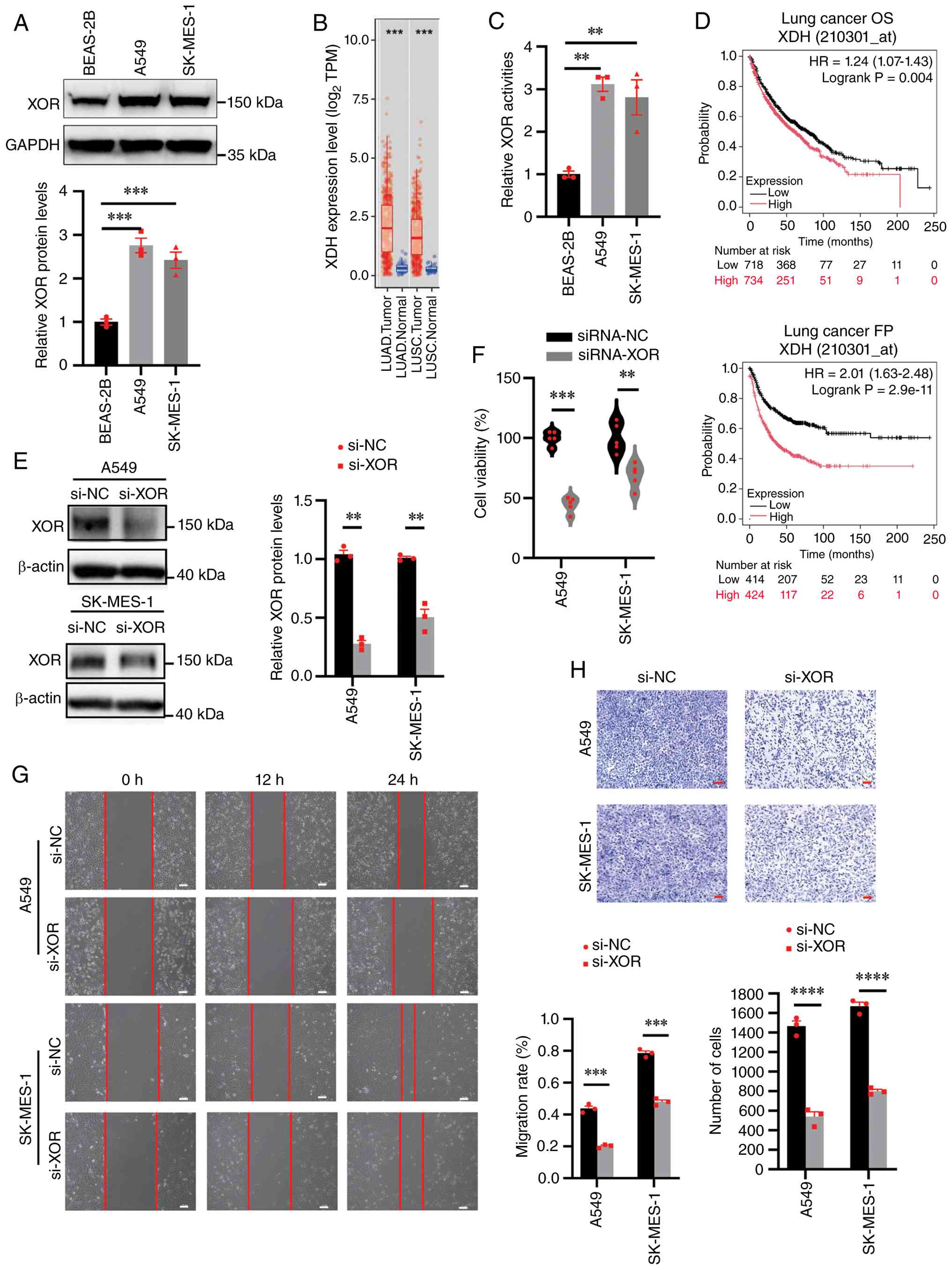 | Figure 1.XOR level positively associated with
NSCLC. (A) Expression of XOR is increased in NSCLC cells compared
with that in normal cells. (B) XDH expression levels in lung
cancers from The Cancer Genome Atlas database were determined by
the TIMER database. (C) Activity of XOR is increased in NSCLC cells
compared with that in normal cells. (D) Kaplan-Meier analysis of
survival according to XDH expression status in patients with lung
cancer using Kaplan-Meier plotter. High XDH expression was
associated with worse OS and survival until FP in lung cancer
cohorts (n=1,452 and n=838, respectively). (E) XOR protein levels
in A549 and SK-MES-1 cells after 48 h siRNA-transfection. (F) Cell
counting kit-8 cell proliferation assay evaluating the viability of
A549 and SK-MES-1 cells transfected with siRNA-NC or siRNA-XOR
after 72 h. (G) Cell migration was analyzed by the scratch assay.
A549 and SK-MES-1 cells transfected with siRNA-NC or siRNA-XOR were
seeded in 6-well plates and wounded by a 100 µl pipette tip. The
cells were then imaged at 12-h intervals for recording migration.
Scale bar, 20 µm. (H) A549 and SK-MES-1 cells transfected with
siRNA-NC or siRNA-XOR were seeded in a chamber covered with a
diluted Matrigel matrix. The invading cells were stained and
observed at ×20 magnification after 12 h. Scale bar, 20 µm. All the
values represent the mean ± SEM of three independent experiments.
****P≤0.0001, ***P≤0.001 and **P≤0.01. XOR, xanthine
oxidoreductase; NSCLC, non-small cell lung cancer; LUAD, lung
adenocarcinoma; LUSC, lung squamous cell carcinoma; XDH, xanthine
dehydrogenase; OS, overall survival; FP, first progression; siRNA
or si, small interfering RNA; NC, negative control; HR, hazard
ratio. |
To evaluate the prognostic significance of XOR in
lung cancer, the association between XDH expression and both OS and
survival until FP was analyzed using the KM Plotter database
(Fig. 1D). High XDH expression was
significantly associated with diminished OS (HR=1.24; 95% CI,
1.07–1.43) and markedly reduced FP survival (HR=2.01; 95% CI:
1.63–2.48). Collectively, these clinical data indicate that
elevated XOR is a negative prognostic indicator, suggesting that
XOR activity contributes to disease progression and an increased
risk of tumor recurrence following initial therapy, consistent with
previous reports (38–40).
To validate the role of XOR in NSCLC, XOR expression
was knocked down in NSCLC cell lines using siRNA. Significant
depletion of XOR was achieved at both protein and mRNA expression
levels in A549 and SK-MES-1 cells (Figs. 1E and S1A). XOR knockdown was found to
significantly reduce cell viability (Fig. 1F). Furthermore, wound healing and
Matrigel-Transwell invasion assays demonstrated impaired migratory
and invasive capacity upon XOR silencing (Fig. 1G and H).
As a Food and Drug Administration-approved XOR
inhibitor (41), the effects and
mechanisms of FBX in NSCLC were next investigated. In the
preliminary experiments of the present study, even at
concentrations up to 200 µM (data not shown), FBX failed to achieve
complete cell killing under nutrient-deprived conditions, where
A549 cells maintained ~40% viability, whilst in SK-MES-1 cells, 200
µM FBX showed an even weaker response (~50% viability). Therefore,
50 µM was adopted as the minimum concentration affecting cell
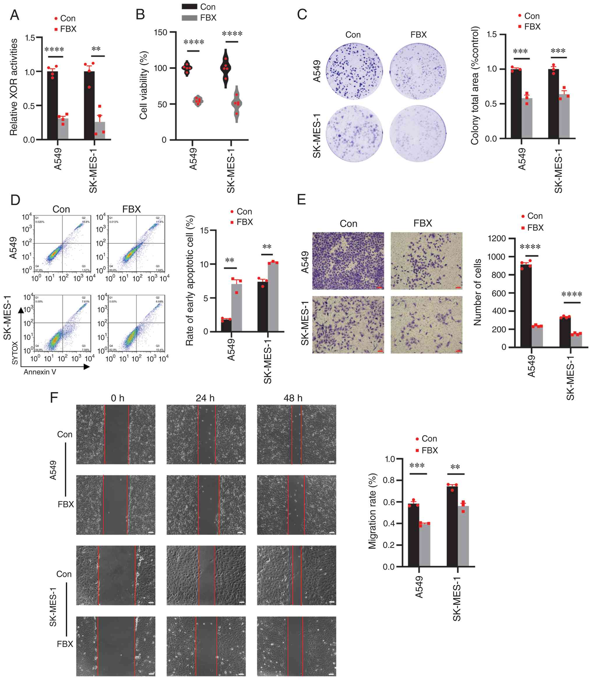
viability for subsequent experiments. ELISA revealed that 50 µM FBX
significantly reduced XOR enzymatic activity in both A549 and
SK-MES-1 cells (Fig. 2A),
accompanied by a significant decrease in cell viability under
starvation conditions (Fig. 2B),
suggesting that XOR inhibition effectively impairs NSCLC cell
proliferation. Clonogenic assays demonstrated that FBX
significantly compromised long-term cell survival, evidenced by a
substantial reduction in colony coverage area compared with that in
the control group after 15 days (Fig.
2C). Flow cytometric analysis revealed that FBX significantly
increased the proportion of early apoptotic cells without markedly
affecting the proportion of late apoptotic cells in both A549 and
SK-MES-1 cell lines (Fig. 2D). The
selective accumulation of early apoptotic cells may be attributed
to the moderate metabolic stress induced by serum deprivation.
Unlike complete nutrient starvation, the absence of serum alone may
be insufficient to propel cells through the full apoptotic cascade
into late apoptotic or secondary necrotic stages, thereby resulting
in a predominant early apoptotic phenotype (42). Consistent with the siRNA knockdown
results, Matrigel-transwell assays confirmed that FBX significantly
reduced the invasive potential of NSCLC cells (Fig. 2E). Scratch wound-healing assays
showed that FBX treatment significantly attenuated the migratory
capacity of both cell types (Fig.
2F). Collectively, these findings established that FBX can
exert potent antitumor effects by inhibiting proliferation,
inducing apoptosis and suppressing the migratory/invasive hallmarks
of NSCLC.
To explore the potential mechanisms underlying
FBX-induced phenotypic changes in NSCLC cells, intracellular ROS
levels were examined. In A549 cells, FBX treatment elicited a
significant increase in ROS within 30 min, irrespective of whether
cells were maintained under standard culture conditions or
nutrient-deprived conditions (Fig.
S2A). Notably, under starvation conditions, this ROS elevation
was substantially attenuated by pretreatment with NAC for 4 h
(Fig. S2B). Concordantly, NAC
significantly counteracted FBX-induced cytotoxicity in both A549
and SK-MES-1 cells (Fig. S2C).
These data indicate that FBX can disrupt cellular redox homeostasis
by XOR blockade, subsequently driving ROS-mediated proliferation
inhibition.
Effect of FBX on NSCLC cells is
associated with autophagy
Previous studies have established that FBX-mediated
XOR inhibition can induce intracellular ROS accumulation, a process
intricately associated with the regulation of autophagy (37,43).
In the present study, FBX had minimal impact on NSCLC cell
viability under standard culture conditions (Fig. S1B), yet markedly suppressed cell
viability under nutrient-deprived conditions (Fig. 2B), a condition known to induce
cytoprotective autophagy. Consequently, we hypothesized that FBX
modulate NSCLC phenotypes by suppressing autophagic activity. To
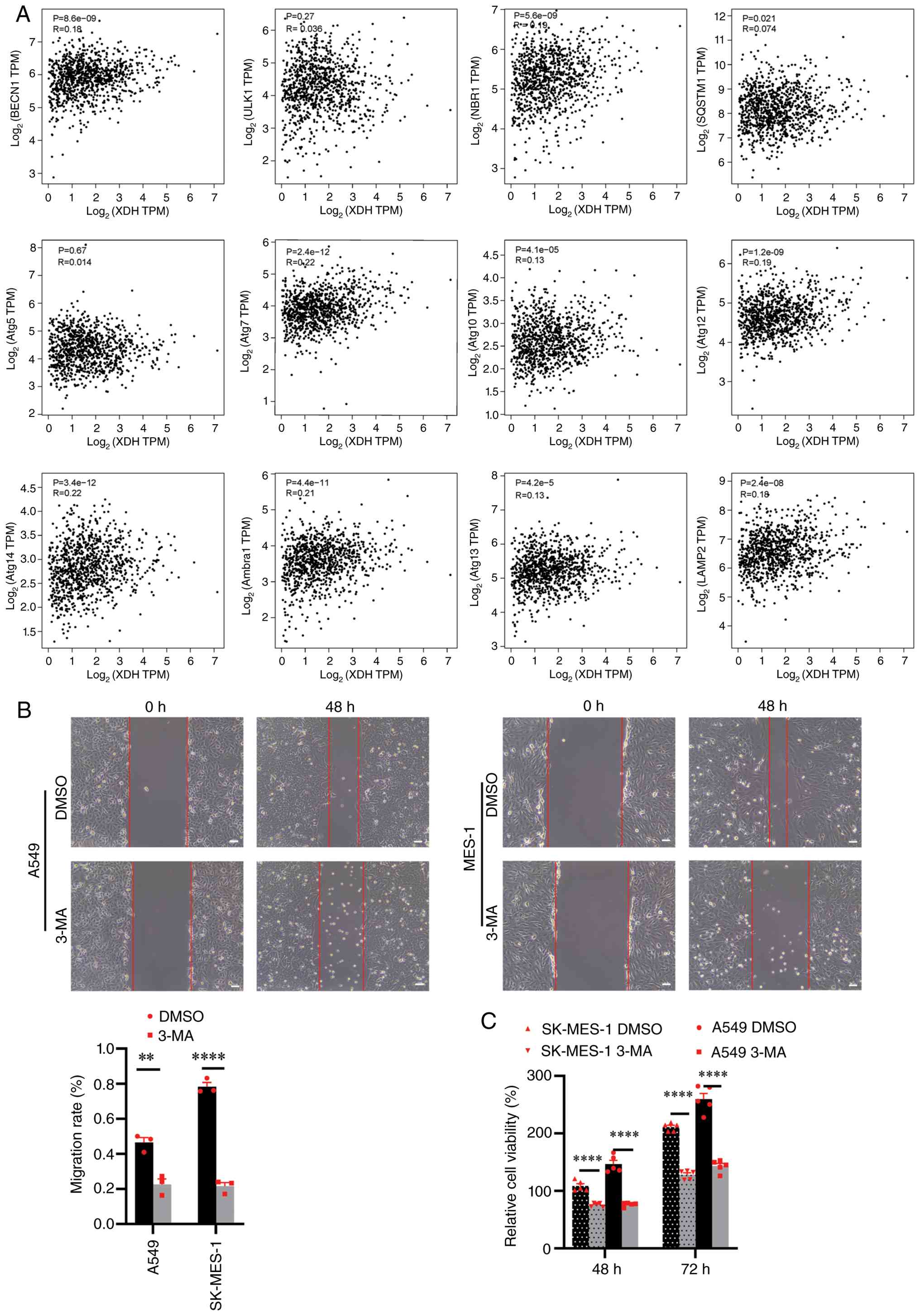
explore the clinical relevance of this association, the correlation
between XOR expression and key autophagy-related markers was
analyzed using the GEPIA2 database (integrating TCGA and GTEx
datasets). The analysis revealed that XOR expression positively
correlated with several essential autophagy regulators, including
Beclin-1 (BECN1, R=0.18), autophagy cargo receptor (R=0.19), ATG7
(R=0.22), ATG10 (R=0.13), ATG12 (R=0.19), ATG13 (R=0.13), ATG14
(R=0.22), autophagy and beclin 1 regulator 1 (R=0.21) and
lysosomal-associated membrane protein 2 (LAMP2; R=0.18; Fig. 3A). These data reveal weak but
statistically significant correlations between XOR signaling and
pro-survival autophagy markers in NSCLC. Whilst these associations
are consistent with a potential role in tumor maintenance and
immune evasion, further functional studies are warranted to
establish causal relationships.
To validate the relationships of autophagy and XOR,
3-MA, a class III PI3K inhibitor that blocks autophagosome
formation, was applied (44).
Consistent with the effects of FBX, 3-MA treatment significantly
reduced cell viability and migratory capacity in both A549 and
SK-MES-1 cells (Fig. 3B and C).
Since the formation of acidic autolysosomes is a
hallmark of late-stage autophagy flux, the acidophilic probe
Lyso-Tracker Red was utilized to monitor lysosomal fusion.
Following FBX treatment under starvation conditions, A549 cells
exhibited a significant reduction in red puncta and fluorescence
intensity at 4 and 8 h compared with controls (Fig. 4A and B), indicating an impairment of
autophagic flux.
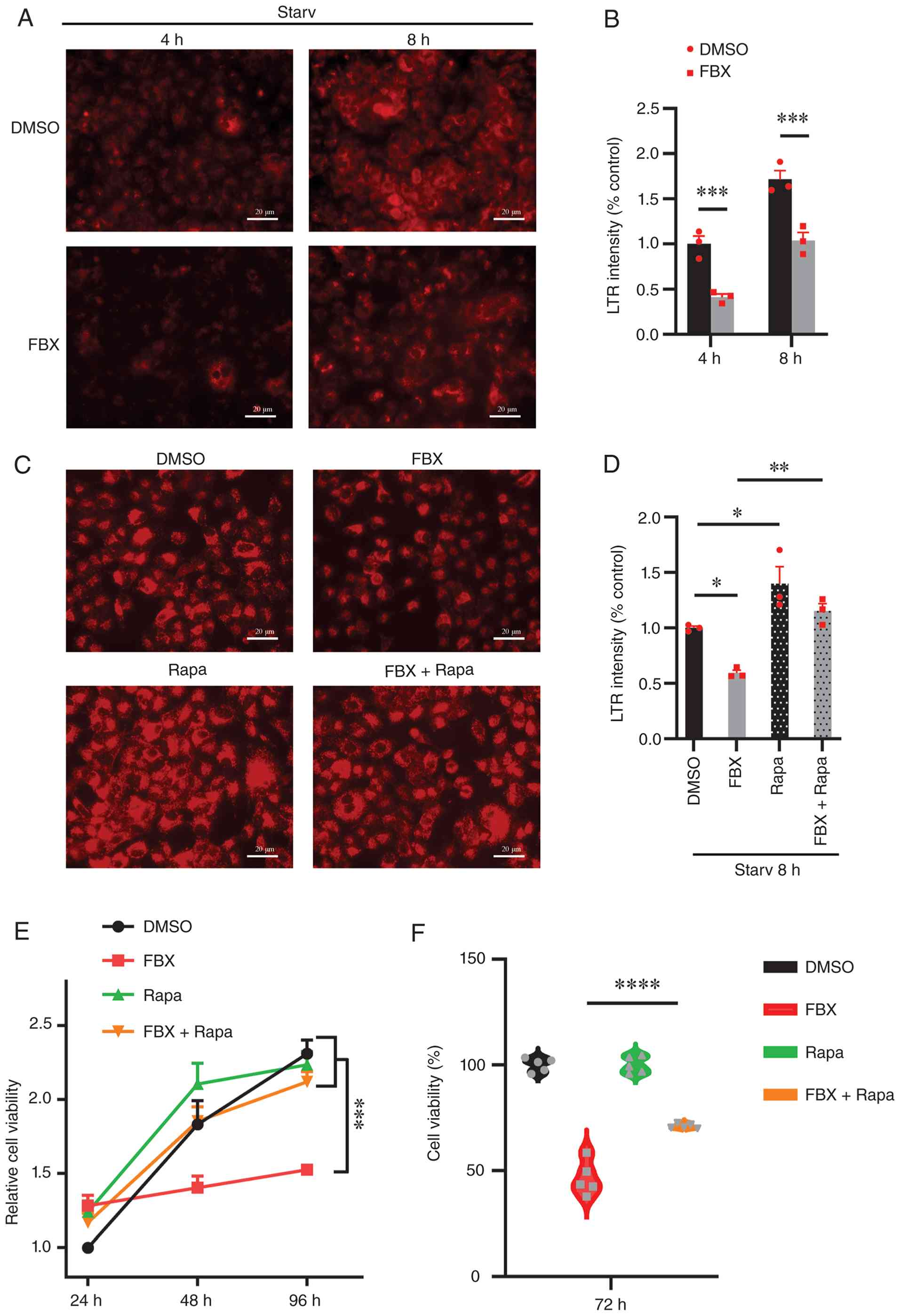 | Figure 4.Effect of FBX on non-small cell lung
cancer cells is associated with autophagy. (A) Assessment of
autolysosome formation in A549 pre-treated with FBX after
starvation at 4 and 8 h. Scale bar, 20 µm. (B) The relative LTR
fluorescent activity was quantified through ImageJ software. The
data are presented as the mean ± SEM of three separate experiments.
(C) Lysosomal activation and repression were analyzed using the
acidophilic probe LTR. A549 cells were pre-treated with FBX, Rapa
and FBX + Rapa after starvation at 8 h. Lysosomes/autolysosomes
were labeled with LTR and then subjected to fluorescent microscopy
analysis. Scale bar, 20 µm. (D) The relative LTR fluorescent
activity was quantified through ImageJ software. The data are
presented as the mean ± SEM of three separate experiments. Cell
counting kit-8 cell proliferation assay evaluating the viability of
A549 and SK-MES-1 cells pre-treated with FBX, Rapa and FBX + Rapa
at (E) 24, 48 and 96 h, and (F) 72 h. The data are presented as the
mean ± SEM of five separate experiments. ****P≤0.0001, ***P≤0.001,
**P≤0.01 and *P<0.05. FBX, febuxostat; Rapa, rapamycin;
LTR, Lyso-Tracker Red; Starv, starvation. |
Furthermore, the FBX-induced reduction in
autophagosome formation was fully rescued by Rapa, a specific mTOR
inhibitor and autophagy activator (Fig.
4C and D). Notably, cell proliferation assays demonstrated that
the anti-proliferative effect of FBX on A549 and SK-MES-1 cells was
significantly attenuated by Rapa-mediated autophagy induction
(Fig. 4E and F). Collectively,
these findings suggest that the anti-tumor efficacy of FBX is
mechanistically driven by the suppression of the autophagic
pathway.
FBX elicits changes in the expression
of autophagy signaling pathway components
To further elucidate the regulatory effect of FBX on
autophagy, the expression of key autophagic proteins in A549 cells
under starvation-induced stress was next assessed (Fig. 5A). The lipidation of LC3-I to LC3-II
serves as a key indicator of early autophagosome formation, whereas
p62/SQSTM1 acts as a selective autophagy receptor that is degraded
during functional autophagic flux (45). In control cells, LC3-II levels
initially decreased upon starvation but subsequently stabilized,
suggesting a continuous state of autophagosomes biogenesis. By
contrast, p62 expression increased immediately following starvation
and began to decline after 4 h, reflecting progressive autophagic
degradation in alignment with established starvation-response
models (46,47). Compared with the DMSO control, FBX
treatment under nutrient-deprived conditions for 4 h resulted in a
more pronounced decrease in LC3-II expression. Although p62 levels
also declined in the FBX group, the reduction was markedly slower
than that in the DMSO group, leading to greater p62 accumulation.
This simultaneous reduction in LC3 lipidation and accumulation of
p62 indicates that FBX impaired both autophagosome biogenesis and
lysosomal clearance capacity (Fig. S3D
and E). Furthermore, a significant reduction in Beclin-1
expression confirmed that FBX could disrupt the initiation of
starvation-induced autophagy (Fig.
S3A). Parallel to these autophagic changes, FBX treatment
downregulated the mesenchymal markers fibronectin (Figs. 5A and S3A) and N-cadherin (Fig. 5B), while upregulating the epithelial
marker E-cadherin (Figs. 5A and B,
and S3A). These reciprocal
alterations in EMT-associated proteins indicated that FBX inhibited
the mesenchymal phenotypes of NSCLC cells. These observations were
corroborated by 3-MA treatment, which similarly induced LC3-II
depletion and p62 accumulation whilst eliciting alterations in
fibronectin protein levels (Figs.
5C, and S3B and C).
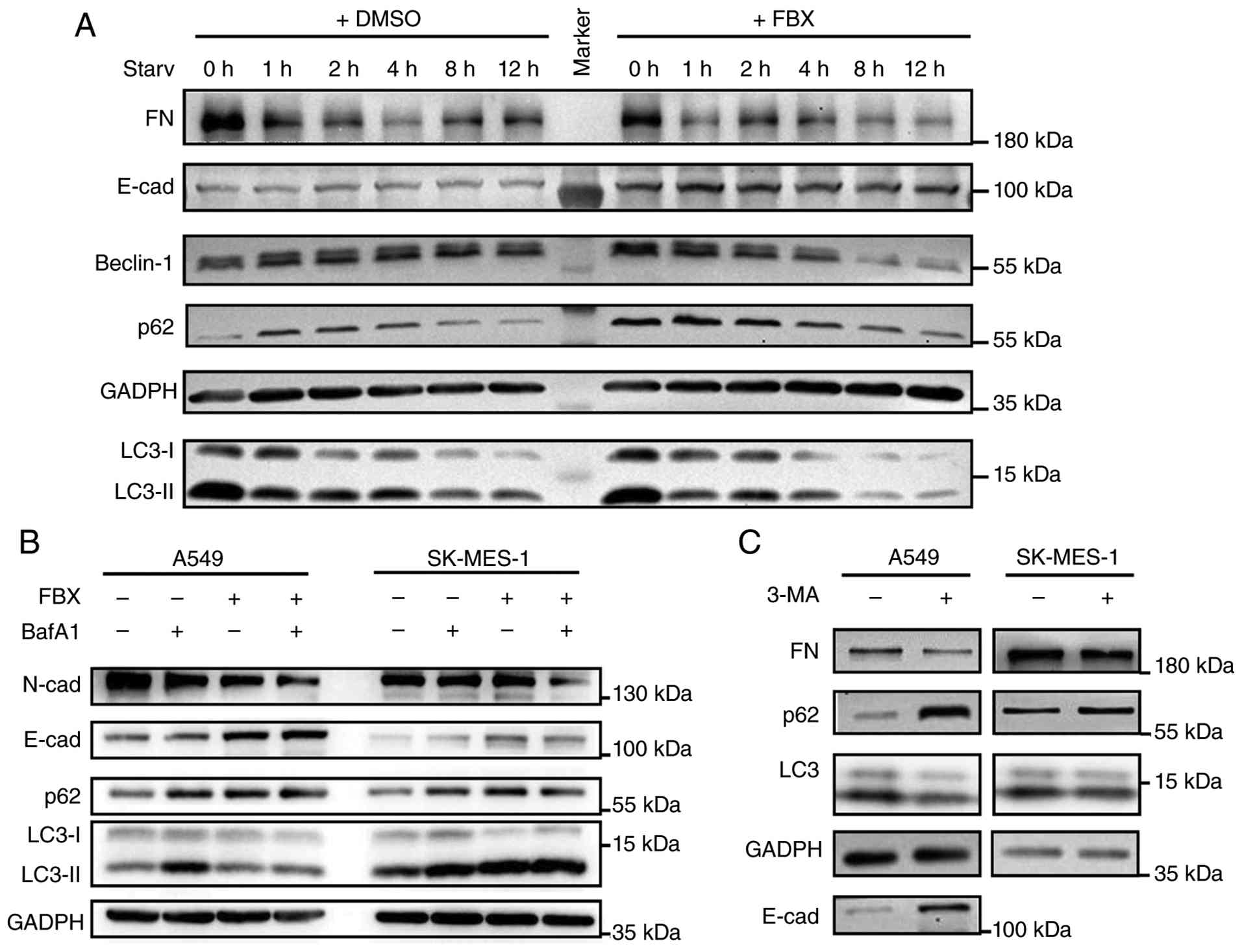 | Figure 5.FBX changed the gene expression of
autophagy signaling pathways in non-small cell lung cancer. (A) The
expression levels of FN, E-cadherin, Beclin-1, p62 and LC3 proteins
in A549 cells treated with FBX were analyzed by western blotting.
(B) The expression levels of N-cad, E-cad, p62 and LC3 proteins in
A549 and SK-MES-1 cells treated with FBX and/or BafA1 for 24 h in
starvation medium were analyzed by western blotting. (C) The
expression levels of FN, E-cad, p62 and LC3 proteins in A549 and
SK-MES-1 cells treated with 3-MA were analyzed by western blotting.
FN, fibronectin; Cad, cadherin; BafA1, bafilomycin A1; FBX,
febuxostat; 3-MA, 3-methyladenine. |
To further validate FBX-mediated autophagy
inhibition in NSCLC, cells were co-treated with BafA1 for 24 h in
starvation medium. BafA1 alone caused accumulation of p62 and
LC3-II in both A549 and SK-MES-1 cells, confirming blockade of
autophagosome-lysosomal degradation. Notably, FBX differentially
affected autophagic flux between the two cell lines (Fig. 5B). In SK-MES-1 cells, FBX induced
p62 and LC3-II accumulation comparable to BafA1. However, combined
treatment did not further increase these levels, indicating
near-maximal autophagy inhibition by BafA1. In A549 cells, FBX
elevated p62 without altering LC3-II, whereas BafA1 co-treatment
likewise failed to trigger additional LC3-II accumulation,
suggesting diminished autophagic flux. Collectively, these results
demonstrate that FBX can inhibit autophagy in NSCLC cells.
Based on a prior finding that the autophagy inducer
Rapa can reverse FBX-induced cytotoxicity in NSCLC cell lines
(Fig. 4E and F), it was then
hypothesized that FBX-mediated antitumor effects are
autophagy-dependent. To assess this, A549 cells were subjected to
starvation and treated with FBX (Fig.
6A; lane 8), Rapa (Fig. 6A;
lane 9) or a combination of FBX and Rapa (Fig. 6A; lane 10). Under nutrient-deprived
conditions, compared with the DMSO control (Fig. 6A; lane 6), FBX treatment induced p62
accumulation (Fig. S3F) and
reduced the LC3-II/LC3-I ratio (Fig.
S3G), with effects similar to those observed following 3-MA
treatment (Fig. 6A; lane 7).
Notably, the autophagy-inhibiting effect of FBX was attenuated by
Rapa-mediated autophagy induction (Fig.
6A; lane 10), as evidenced by a restoration of the LC3-II/LC3-I
ratio and a substantial decrease in p62 expression (Fig. S3F and G). However, these
alterations were not observed under standard culture conditions
(Fig. 6A; lanes 1–5). Similarly,
under nutrient-deprived conditions, FBX treatment increased p62
accumulation in SK-MES-1 cells without markedly affecting LC3-II
conversion (Figs. 6B and S3H), consistent with the aforementioned
observations (Fig. 5B).
Furthermore, rapamycin partially alleviated FBX-induced p62 buildup
(Fig. 6B; lane 8), further
supporting an autophagy-dependent mechanism. By contrast, under
standard culture conditions with sufficient nutrients, FBX exerted
minimal effects on LC3-II conversion and p62 turnover (Fig. 6B; lane 2), consistent with the low
basal autophagic flux observed in non-starved NSCLC cells.
Collectively, these results demonstrate that FBX acts as a potent
inhibitor of the autophagic pathway, where its phenotypic effects
can be partially rescued by pharmacological autophagy
activation.
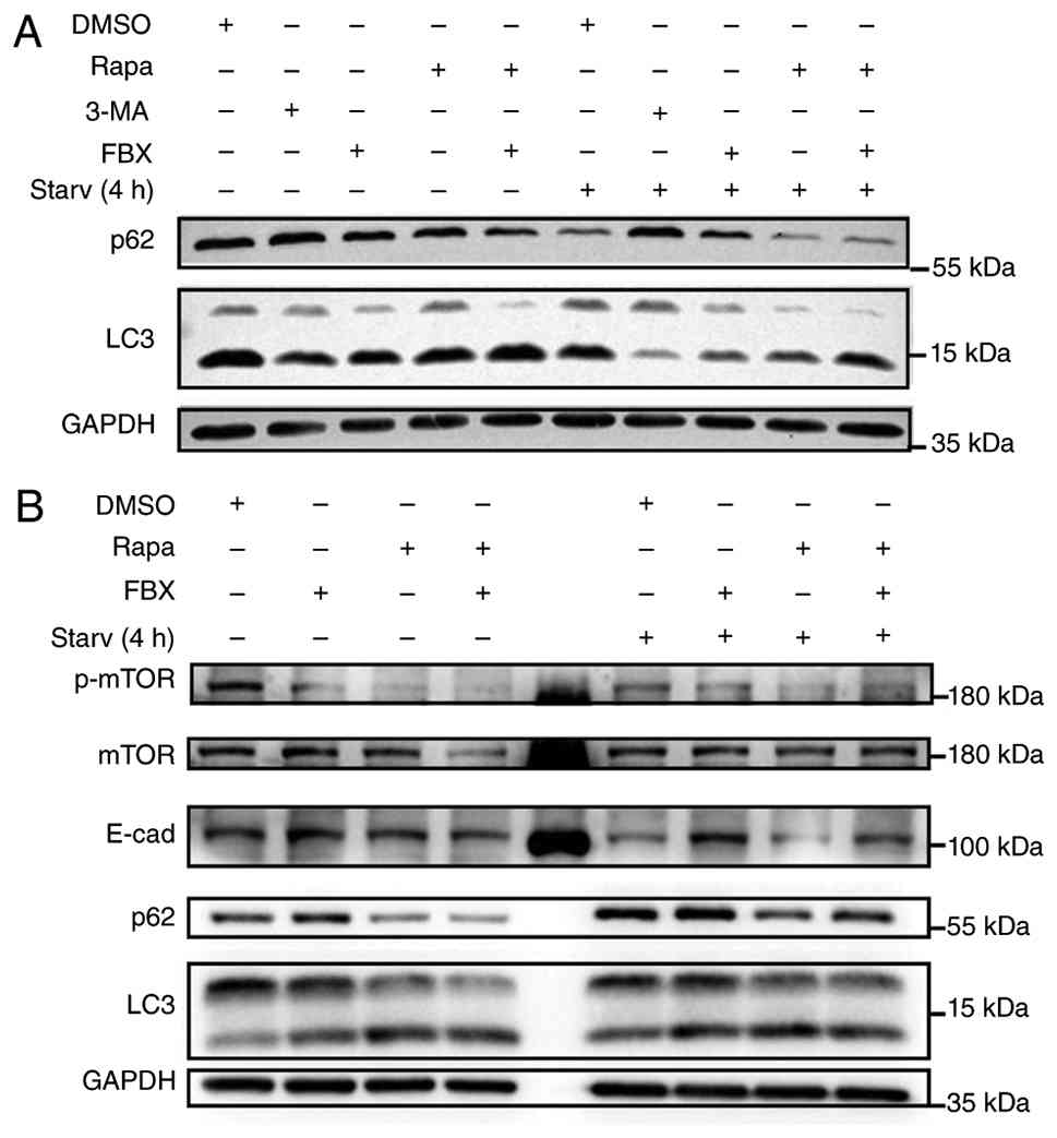 | Figure 6.Rapamycin neutralized the effect of
FBX on autophagy of non-small cell lung cancer cells. (A) The
expression levels of p62 and LC3 proteins in A549 cells pre-treated
with FBX, 3-MA and Rapa were analyzed by western blotting. (B) The
levels of p-mTOR, mTOR, E-cad, p62 and LC3 proteins in SK-MES-1
cells pre-treated with FBX and Rapa were analyzed by western
blotting. Rapa, rapamycin; FBX, febuxostat; Cad, cadherin; p-,
phosphorylated; 3-MA, 3-methyladenine; Starv, starvation. |
Discussion
In the present study, it was demonstrated that XOR
knockdown significantly impaired the proliferation and
invasive/migratory potential of NSCLC cells. This follows our
initial observation of elevated XOR levels in both NSCLC cell lines
and clinical tissue specimens in the present study. FBX markedly
reduced XOR activity and proliferation in NSCLC cells under
nutrient-deprived conditions. Furthermore, FBX treatment
significantly attenuated the migratory and invasive capacities of
NSCLC cells in vitro. Mechanistically, the anti-tumor
efficacy of FBX appears to be mediated through the suppression of
the autophagy signaling pathway. Notably, the induction of
autophagy through pharmacological activators abrogated the
tumor-suppressive effects of FBX. Beyond its primary role in
reducing serum urate levels, FBX has been proposed as a protective
agent against acute renal failure and tumor lysis syndrome
(48). The clinical utility of FBX
in oncological care is supported by both hepatoma rodent models and
malignant tumor multicenter clinical investigations (49,50).
These data suggest that FBX, an established gout therapeutic, may
warrant further preclinical investigation for potential repurposing
in malignant tumors, with a therapeutic profile that could prove
comparable or superior to the purine-analog inhibitor
allopurinol.
KM Plotter database analysis revealed that elevated
XOR expression is associated with shorter OS and shorter time to FP
survival in patients with NSCLC. However, this finding appears
inconsistent with a prior clinical study reporting reduced survival
in chemotherapy-treated patients with low XOR expression, as
determined by immunohistochemistry (IHC) of tumor tissues (31). These differences could be explained
by the different methodological approaches. First, the IHC-based
approach classified all unstained slides as XOR-negative (29/82
cases, 35%), whereas KM Plotter database mainly leveraged on
microarray sequencing data, which would likely categorize these
samples as low-expression rather than absent, potentially yielding
different outcomes. In addition, this previous study (31) utilized a selected subset. Amongst
the 53 IHC-positive cases, only 34 received adjuvant chemotherapy
and the final comparison only involved 14 low-vs. 20
high-expression samples. By contrast, the KM Plotter database is
comprised of >700 cases for OS and 400 cases for FP, utilizing
sequencing data with superior accuracy and statistical power.
Finally, the present experiments demonstrated significantly
elevated XOR activity in NSCLC cell lines compared with that in
normal lung epithelial cells. Both siRNA-mediated knockdown and
pharmacological inhibition of XOR by FBX exerted anti-tumor
effects, supporting an oncogenic role for XOR in NSCLC
progression.
The anti-tumor properties of FBX were first
characterized by Oh et al (51), who demonstrated that FBX can reduce
ROS production and MAPK phosphorylation, thereby inhibiting
metastasis in a murine model of hyperlipidemic breast cancer. A
subsequent study showed that the encapsulation of FBX in lipid
emulsions can markedly inhibit the proliferation of HCT116 human
colorectal cancer cells (52).
Further evaluations by Alfaifi et al (35) explored the cytotoxic potential of
FBX-PLGA-PEG nanoparticles in A549, whilst Burmaoglu et al
(53) reported that novel XO
inhibitors can markedly reduce the viability of breast cancer cells
MCF-7 and colon cancer cells Caco-2. The present study corroborated
with these aforementioned findings, confirming that FBX can exert
potent cytotoxicity in NSCLC by inhibiting XOR activity. In
addition, it was hypothesized that the FBX-mediated reduction in
NSCLC cell viability may primarily reflect impaired proliferation
rather than apoptosis alone, as the modest increase in apoptotic
rate shown in Fig. 2D cannot fully
account for the ~50% cytotoxic effect observed.
To the best of our knowledge, the present study
provided the first evidence that FBX can significantly diminish the
migratory and invasive abilities of NSCLC cells. Although FBX
significantly impaired NSCLC cell proliferation, which could
potentially confound longer-term wound healing assays, this
proliferative difference is negligible in short-term Transwell
invasion experiments (<24 h), where equal cell numbers are
seeded. According to pharmacokinetic studies of FBX (54), single doses of 40, 80 and 120 mg
achieve peak plasma concentrations of 1.6–2.8, 2.6–5.0 and 4.8–7.7
µg/ml, respectively, corresponding to 5–8.8, 8.2–17 and 15–24 µM.
Multiple-dose regimens showed no serious adverse events or drug
accumulation, demonstrating good tolerability in humans (55). These concentrations are comparable
with those used in the present in vitro experiments,
suggesting theoretical feasibility. However, to the best of our
knowledge, no clinical reports have yet described the use of FBX or
other XOR inhibitors in patients with NSCLC.
Wang et al (32) first established that exogenous XO
can trigger autophagy in A549 cells. The present database analysis
corroborates this by demonstrating a positive correlation between
XO expression and early autophagic regulators, including BECN1 and
ATG5, in addition to the lysosomal marker LAMP2, in patients with
lung cancer. Consequently, it was hypothesized that FBX can
attenuate autophagic flux in NSCLC cells by inhibiting XOR
enzymatic activity. Western blot analysis confirmed that FBX
differentially attenuates autophagy in NSCLC cells. Under
short-term (4 h) starvation, FBX suppressed LC3-II formation in
A549 cells, indicating reduced mature autophagosome biogenesis,
whereas this effect was absent in SK-MES-1 cells. Notably, p62
accumulated in both cell lines, suggesting that FBX primarily
blocks autophagic degradation in SK-MES-1 cells, whilst in A549
cells it may additionally inhibit autophagosome formation. This
differential inhibition became more pronounced following prolonged
(24 h) starvation. These differential effects suggest that FBX may
impede distinct stages of autophagy. Furthermore, the differential
rescue effects were evident in cell viability assays. Co-treatment
with Rapa and FBX restored A549 cell viability nearly to control
levels, whereas only partial rescue was observed in SK-MES-1 cells.
This further suggests that FBX may exclusively block autophagic
degradation in LUSC cells, while in LUAD cells it additionally
impairs autophagosome formation, a function specifically reversed
by Rapa. It is well established that the accumulation of ROS within
cells and mitochondria serves as a potent stimulus for autophagy
through the canonical AMPK/mTOR/Unc51-like kinase (ULK1) pathway
(56). Wang et al (57) reported that allopurinol can inhibit
XOR, thereby blocking gemcitabine-induced phosphorylation of
TGFβ-activated kinase 1 (TAK1), JNK, AMPK and ULK1, suppressing
gemcitabine-triggered autophagy and enhancing gemcitabine
cytotoxicity in HeLa and HT-29 cells. Therefore, FBX may similarly
inhibit autophagy through these pathways (58).
It is well established that XO generates superoxide
anion and hydrogen peroxide (H2O2) as
byproducts during its catalytic cycle. FBX-mediated XOR inhibition
attenuates ROS production, thereby conferring cyto-protection
(59). Huang et al (60) previously demonstrated that
inhibiting cathepsin S can trigger autophagy and subsequently
upregulate XO expression and activity, leading to ROS-mediated DNA
damage and cell death. In that model, XO blockade did abrogate ROS
production but did not prevent autophagy activation. By contrast,
the present findings indicate that FBX treatment can induce a rapid
increase in intracellular ROS and subsequently inhibit autophagy in
NSCLC cells. Comparable findings were observed in bladder cancer
(61), where combined FBX and
5-aminolevulinic acid (5-ALA) treatment markedly enhanced ROS
generation relative to 5-ALA monotherapy. Pretreatment with NAC
abrogated FBX-mediated ROS production and subsequently ameliorated
cell viability, indicating that ROS contributes to the FBX-induced
cytotoxic effect. Emerging evidence (62,63)
preliminarily suggests a complex reciprocal regulatory relationship
between XOR and ROS; however, the precise source of ROS remains
elusive.
Beyond the modulation of autophagy, the anti-tumor
efficacy of FBX may be governed by the functional balance between
XO and XDH. The natural compound Alternol has been found to enhance
the oxidative activity of XDH, thereby inducing apoptosis in
malignant cells (63). Furthermore,
Kusano et al (62) observed
that colorectal cancer and melanoma cells progressed more rapidly
in XO−/− mouse models compared with in XDH−/−
or wild-type cohorts. This accelerated growth was associated with
macrophages-derived ROS, since those in XO−/− mice
exhibited higher ROS levels, which promoted the expansion of
regulatory T cells in the TME. Notably, macrophage depletion
markedly attenuated tumor progression. Hypoxia upregulates XOR
activity and shifts the enzyme toward its oxidase form, rendering
XO the predominant source of ROS, primarily hydrogen peroxide,
during hypoxia and reoxygenation (64). Collectively, these findings
reinforce the hypothesis that targeted inhibition of XO activity
represents a viable therapeutic strategy for managing tumor growth,
especially in hypoxia conditions.
The present study has several limitations that
warrant future improvement. Cell proliferation assays (such as EdU
staining) should be performed to determine whether FBX primarily
suppresses proliferation, whereas classical apoptotic markers
[cleaved caspase-3, cleaved poly (ADP-ribose) polymerase or
BAX/BCL-2 ratio] should be assessed to investigate whether FBX can
induces programmed cell death. Genetic approaches, such as
knockdown or rescue of key autophagy genes (such as ATG5,
ATG7 or BECN1) are needed to validate FBX-mediated
autophagy modulation. In addition, signaling pathway components,
including p-mTOR, p-AMPK, p-ULK1, p-TAK1 and p-JNK, should be
examined to elucidate the molecular mechanism underlying
FBX-induced autophagy. Primary NSCLC cells or patient-derived
xenograft models should also be employed, with in vivo
experiments conducted to further validate the roles and mechanisms
of XOR and FBX in NSCLC. EMT markers, including N-cadherin,
Vimentin, Snail, Slug, ZEB1, MMP2 and MMP9, should be assessed to
clarify FBX-mediated EMT suppression. Ferroptosis markers should be
examined to explore whether ferroptosis can contribute to the
anti-tumor effects of FBX.
In summary, the present findings demonstrated that
XOR inhibition can suppress NSCLC cell viability and
invasion/migration by disrupting pro-survival autophagy. The
observation that rapamycin-mediated autophagy activation
effectively abrogates the anti-proliferative effects of FBX further
confirms the mechanistic dependency on this pathway. Collectively,
these data identify XOR participates in the metabolic regulation of
lung cancer progression and suggest that XOR-targeted therapies
warrant further evaluation as a novel strategy for the treatment of
NSCLC.
Supplementary Material
Supporting Data
Acknowledgements
The authors would like to acknowledge Professor Lin
WenChu (Longgang District People's Hospital of Shenzhen, Shenzhen,
China) for critically refining the logical structure and Dr Jia Liu
(Longgang District People's Hospital of Shenzhen, Shenzhen, China)
for inspiring the initial conceptual framework of the present
study. In addition, the authors would like to thank Dr Celina
Juliano (PhD; Faculty of College of Biological Science, University
of California, Davis, USA) for editing the language.
Funding
The present study was funded by the Longgang Medical and Health
Science and Technology Project (grant no. LGKCYLWS2020045).
Availability of data and materials
The data generated in the present study may be
requested from the corresponding author.
Authors' contributions
XHY contributed to the study methodology
(development and design of experimental procedures), performed
validation (verification of experimental results) and investigation
(conducting the experiments), wrote the original draft (preparation
of the initial manuscript), and participated in review and editing
(critical revision of the manuscript). MS and YWX performed formal
analysis (statistical and computational analysis of data) and data
curation (management, organization and maintenance of research
data). XHY and MS confirm the authenticity of all the raw data. All
authors read and approved the final version of the manuscript.
Ethical approval and consent to
participate
Not applicable.
Patient consent for publication
Not applicable.
Competing interests
The authors declare that they have no competing
interests.
References
|
1
|
Bray F, Laversanne M, Sung H, Ferlay J,
Siegel RL, Soerjomataram I and Jemal A: Global cancer statistics
2022: GLOBOCAN estimates of incidence and mortality worldwide for
36 cancers in 185 countries. CA Cancer J Clin. 74:229–263.
2024.PubMed/NCBI
|
|
2
|
Herbst RS, Morgensztern D and Boshoff C:
The biology and management of non-small cell lung cancer. Nature.
553:446–454. 2018. View Article : Google Scholar : PubMed/NCBI
|
|
3
|
Goldstraw P, Chansky K, Crowley J,
Rami-Porta R, Asamura H, Eberhardt WE, Nicholson AG, Groome P,
Mitchell A, Bolejack V, et al: The IASLC lung cancer staging
project: Proposals for revision of the TNM stage groupings in the
forthcoming (Eighth) edition of the TNM classification for lung
cancer. J Thorac Oncol. 11:39–51. 2016. View Article : Google Scholar : PubMed/NCBI
|
|
4
|
Duma N, Santana-Davila R and Molina JR:
Non-Small cell lung cancer: Epidemiology, screening, diagnosis, and
treatment. Mayo Clin Proc. 94:1623–1640. 2019. View Article : Google Scholar : PubMed/NCBI
|
|
5
|
Imyanitov EN, Iyevleva AG and Levchenko
EV: Molecular testing and targeted therapy for non-small cell lung
cancer: Current status and perspectives. Crit Rev Oncol Hematol.
157:1031942021. View Article : Google Scholar : PubMed/NCBI
|
|
6
|
Clarke MA, Barker CG, Nicholls A, Handler
MP, Pickard L, Shah AZ, Walter D, De Braekeleer E, Banerji U,
Choudhary JS, et al: Virtual tumors enable prediction of
personalized therapeutic combinations for Non-small cell lung
cancer. Cancer Res. 86:4705–4722. 2026. View Article : Google Scholar : PubMed/NCBI
|
|
7
|
He L, Zhang J, Zhao J, Ma N, Kim SW, Qiao
S and Ma X: Autophagy: The last defense against cellular
nutritional stress. Adv Nutr. 9:493–504. 2018. View Article : Google Scholar : PubMed/NCBI
|
|
8
|
Rao S, Yang H, Penninger JM and Kroemer G:
Autophagy in non-small cell lung carcinogenesis: A positive
regulator of antitumor immunosurveillance. Autophagy. 10:529–531.
2014. View Article : Google Scholar : PubMed/NCBI
|
|
9
|
Rao S, Tortola L, Perlot T, Wirnsberger G,
Novatchkova M, Nitsch R, Sykacek P, Frank L, Schramek D, Komnenovic
V, et al: A dual role for autophagy in a murine model of lung
cancer. Nat Commun. 5:30562014. View Article : Google Scholar : PubMed/NCBI
|
|
10
|
Wang J, Liu D, Sun Z, Ye T, Li J, Zeng B,
Zhao Q and Rosie Xing H: Autophagy augments the self-renewal of
lung cancer stem cells by the degradation of ubiquitinated p53.
Cell Death Dis. 12:982021. View Article : Google Scholar : PubMed/NCBI
|
|
11
|
Guo JY and White E: Autophagy is required
for mitochondrial function, lipid metabolism, growth, and fate of
KRAS(G12D)-driven lung tumors. Autophagy. 9:1636–1638. 2013.
View Article : Google Scholar : PubMed/NCBI
|
|
12
|
Strohecker AM, Guo JY, Karsli-Uzunbas G,
Price SM, Chen GJ, Mathew R, McMahon M and White E: Autophagy
sustains mitochondrial glutamine metabolism and growth of
BrafV600E-driven lung tumors. Cancer Discov. 3:1272–1285. 2013.
View Article : Google Scholar : PubMed/NCBI
|
|
13
|
Guo JY, Karsli-Uzunbas G, Mathew R, Aisner
SC, Kamphorst JJ, Strohecker AM, Chen G, Price S, Lu W, Teng X, et
al: Autophagy suppresses progression of K-ras-induced lung tumors
to oncocytomas and maintains lipid homeostasis. Genes Dev.
27:1447–1461. 2013. View Article : Google Scholar : PubMed/NCBI
|
|
14
|
Bustos SO, Antunes F, Rangel MC and
Chammas R: Emerging autophagy functions shape the tumor
microenvironment and play a role in cancer progression-Implications
for Cancer Therapy. Front Oncol. 10:6064362020. View Article : Google Scholar : PubMed/NCBI
|
|
15
|
Chavez-Dominguez R, Perez-Medina M,
Lopez-Gonzalez JS, Galicia-Velasco M and Aguilar-Cazares D: The
double-edge sword of autophagy in cancer: From tumor suppression to
Pro-tumor activity. Front Oncol. 10:5784182020. View Article : Google Scholar : PubMed/NCBI
|
|
16
|
Yan Y, Chen X, Wang X, Zhao Z, Hu W, Zeng
S, Wei J, Yang X, Qian L, Zhou S, et al: The effects and the
mechanisms of autophagy on the cancer-associated fibroblasts in
cancer. J Exp Clin Cancer Res. 38:1712019. View Article : Google Scholar : PubMed/NCBI
|
|
17
|
Alizadeh J, Glogowska A, Thliveris J,
Kalantari F, Shojaei S, Hombach-Klonisch S, Klonisch T and Ghavami
S: Autophagy modulates transforming growth factor beta 1 induced
epithelial to mesenchymal transition in non-small cell lung cancer
cells. Biochim Biophys Acta Mol Cell Res. 1865:749–768. 2018.
View Article : Google Scholar : PubMed/NCBI
|
|
18
|
Ren Y, Cao L, Wang L, Zheng S, Zhang Q,
Guo X, Li X, Chen M, Wu X, Furlong F, et al: Autophagic secretion
of HMGB1 from cancer-associated fibroblasts promotes metastatic
potential of non-small cell lung cancer cells via NFκB signaling.
Cell Death Dis. 12:8582021. View Article : Google Scholar : PubMed/NCBI
|
|
19
|
TA AL: Deciphering the role of TMEM164 in
autophagy-mediated ferroptosis and immune modulation in non-small
cell lung cancer. Cell Immunol. 409–410. 1049152025.
|
|
20
|
Wu H, Ren J, Gong Z, Zhang Y, Zhang H,
Chen W, Zhao C and Fang T: Exploring relationships between tumor
mutation burden with immunity and prognosis in lung adenocarcinoma
based on weighted gene co-expression network analysis. Precision
Med Sci. 14:168–180. 2025. View Article : Google Scholar
|
|
21
|
Wu R, Kadier K, Xu S, Yuan P, Yang Y, Xu
P, Huang X, Zhou S, Guo SB, Zhang H, et al: Mapping heterogeneous
molecular subtypes of circadian misalignment underlying lung
adenocarcinoma risk. Life Conflux. 1:e1372025. View Article : Google Scholar
|
|
22
|
Harrison R: Structure and function of
xanthine oxidoreductase: Where are we now? Free Radic Biol Med.
33:774–797. 2002. View Article : Google Scholar : PubMed/NCBI
|
|
23
|
Harrison R: Physiological roles of
xanthine oxidoreductase. Drug Metab Rev. 36:363–375. 2004.
View Article : Google Scholar : PubMed/NCBI
|
|
24
|
Pacher P, Nivorozhkin A and Szabo C:
Therapeutic effects of xanthine oxidase inhibitors: Renaissance
half a century after the discovery of allopurinol. Pharmacol Rev.
58:87–114. 2006. View Article : Google Scholar : PubMed/NCBI
|
|
25
|
Aslan M, Ryan TM, Adler B, Townes TM,
Parks DA, Thompson JA, Tousson A, Gladwin MT, Patel RP, Tarpey MM,
et al: Oxygen radical inhibition of nitric oxide-dependent vascular
function in sickle cell disease. Proc Natl Acad Sci USA.
98:15215–15220. 2001. View Article : Google Scholar : PubMed/NCBI
|
|
26
|
Enroth C, Eger BT, Okamoto K, Nishino T,
Nishino T and Pai EF: Crystal structures of bovine milk xanthine
dehydrogenase and xanthine oxidase: Structure-based mechanism of
conversion. Proc Natl Acad Sci USA. 97:10723–10728. 2000.
View Article : Google Scholar : PubMed/NCBI
|
|
27
|
Berry CE and Hare JM: Xanthine
oxidoreductase and cardiovascular disease: Molecular mechanisms and
pathophysiological implications. J Physiol. 555:589–606. 2004.
View Article : Google Scholar : PubMed/NCBI
|
|
28
|
Filomeni G, De Zio D and Cecconi F:
Oxidative stress and autophagy: The clash between damage and
metabolic needs. Cell Death Differ. 22:377–388. 2015. View Article : Google Scholar : PubMed/NCBI
|
|
29
|
Gao Q: Oxidative stress and autophagy. Adv
Exp Med Biol. 1206:179–198. 2019. View Article : Google Scholar : PubMed/NCBI
|
|
30
|
Kaynar H, Meral M, Turhan H, Keles M,
Celik G and Akcay F: Glutathione peroxidase,
glutathione-S-transferase, catalase, xanthine oxidase, Cu-Zn
superoxide dismutase activities, total glutathione, nitric oxide,
and malondialdehyde levels in erythrocytes of patients with small
cell and non-small cell lung cancer. Cancer Lett. 227:133–139.
2005. View Article : Google Scholar : PubMed/NCBI
|
|
31
|
Kim AW, Batus M, Myint R, Fidler MJ, Basu
S, Bonomi P, Faber LP, Wightman SC, Warren WH, McIntire M, et al:
Prognostic value of xanthine oxidoreductase expression in patients
with non-small cell lung cancer. Lung Cancer. 71:186–190. 2011.
View Article : Google Scholar : PubMed/NCBI
|
|
32
|
Wang X, Li Y, Liu D, Wang Y and Ming H:
Astragalus polysaccharide inhibits autophagy and regulates
expression of autophagy-related proteins in lung cancer A549 cells
induced by xanthine oxidase. Xi Bao Yu Fen Zi Mian Yi Xue Za Zhi.
35:619–624. 2019.(In Chinese). PubMed/NCBI
|
|
33
|
Tayar JH, Lopez-Olivo MA and
Suarez-Almazor ME: Febuxostat for treating chronic gout. Cochrane
Database Syst Rev. 11:CD0086532012.PubMed/NCBI
|
|
34
|
Joshi G, Sharma M, Kalra S, Gavande NS,
Singh S and Kumar R: Design, synthesis, biological evaluation of
3,5-diaryl-4,5-dihydro-1H-pyrazole carbaldehydes as non-purine
xanthine oxidase inhibitors: Tracing the anticancer mechanism via
xanthine oxidase inhibition. Bioorg Chem. 107:1046202021.
View Article : Google Scholar : PubMed/NCBI
|
|
35
|
Alfaifi MY, Shati AA, Elbehairi SEI, Fahmy
UA, Alhakamy NA and Md S: Anti-tumor effect of PEG-coated PLGA
nanoparticles of febuxostat on A549 non-small cell lung cancer
cells. 3 Biotech. 10:1332020. View Article : Google Scholar : PubMed/NCBI
|
|
36
|
Livak KJ and Schmittgen TD: Analysis of
relative gene expression data using real-time quantitative PCR and
the 2(−Delta Delta C(T)) method. Methods. 25:402–408. 2001.
View Article : Google Scholar : PubMed/NCBI
|
|
37
|
Yu L, McPhee CK, Zheng L, Mardones GA,
Rong Y, Peng J, Mi N, Zhao Y, Liu Z, Wan F, et al: Termination of
autophagy and reformation of lysosomes regulated by mTOR. Nature.
465:942–946. 2010. View Article : Google Scholar : PubMed/NCBI
|
|
38
|
Konno H, Minamiya Y, Saito H, Imai K,
Kawaharada Y, Motoyama S and Ogawa J: Acquired xanthine
dehydrogenase expression shortens survival in patients with
resected adenocarcinoma of lung. Tumour Biol. 33:1727–1732. 2012.
View Article : Google Scholar : PubMed/NCBI
|
|
39
|
Liu H, Zhu H, Shi W, Lin Y, Ma G, Tao G,
Gong W, Zhao Q, Du M, Wang M, et al: Genetic variants in XDH are
associated with prognosis for gastric cancer in a Chinese
population. Gene. 663:196–202. 2018. View Article : Google Scholar : PubMed/NCBI
|
|
40
|
Chen MM, Guo W, Chen SM, Guo XZ, Xu L, Ma
XY, Wang YX, Xie C and Meng LH: Xanthine dehydrogenase rewires
metabolism and the survival of nutrient deprived lung
adenocarcinoma cells by facilitating UPR and autophagic
degradation. Int J Biol Sci. 19:772–788. 2023. View Article : Google Scholar : PubMed/NCBI
|
|
41
|
Hair PI, McCormack PL and Keating GM:
Febuxostat. Drugs. 68:1865–1874. 2008. View Article : Google Scholar : PubMed/NCBI
|
|
42
|
White EZ, Pennant NM, Carter JR, Hawsawi
O, Odero-Marah V and Hinton CV: Serum deprivation initiates
adaptation and survival to oxidative stress in prostate cancer
cells. Sci Rep. 10:125052020. View Article : Google Scholar : PubMed/NCBI
|
|
43
|
Carretero-Fernández M, Cabrera-Serrano AJ,
Sánchez-Maldonado JM, Ruiz-Durán L, Jiménez-Romera F,
García-Verdejo FJ, González-Olmedo C, Cardús A, Díaz-Beltrán L,
Gutiérrez-Bautista JF, et al: Autophagy and oxidative stress in
solid tumors: Mechanisms and therapeutic opportunities. Crit Rev
Oncol Hematol. 212:1048202025. View Article : Google Scholar : PubMed/NCBI
|
|
44
|
Wu YT, Tan HL, Shui G, Bauvy C, Huang Q,
Wenk MR, Ong CN, Codogno P and Shen HM: Dual role of
3-methyladenine in modulation of autophagy via different temporal
patterns of inhibition on class I and III phosphoinositide
3-Kinase. J Biol Chem. 285:10850–10861. 2010. View Article : Google Scholar : PubMed/NCBI
|
|
45
|
Jiang P and Mizushima N: LC3- and
p62-based biochemical methods for the analysis of autophagy
progression in mammalian cells. Methods. 75:13–18. 2015. View Article : Google Scholar : PubMed/NCBI
|
|
46
|
Aydemir S, Yildirim Z, Bara B, Dogan E and
Bozok V: Differential regulation of STING expression and cisplatin
sensitivity by autophagy in non-small cell lung cancer cells. Med
Oncol. 42:2272025. View Article : Google Scholar : PubMed/NCBI
|
|
47
|
Han JH, Kim YK, Kim H, Lee J, Oh MJ, Kim
SB, Kim M, Kim KH, Yoon HJ, Lee MS, et al: Snail acetylation by
autophagy-derived acetyl-coenzyme A promotes invasion and
metastasis of KRAS-LKB1 co-mutated lung cancer cells. Cancer Commun
(Lond). 42:716–749. 2022. View Article : Google Scholar : PubMed/NCBI
|
|
48
|
Takai M, Yamauchi T, Fujita K, Lee S,
Ookura M, Kishi S, Urasaki Y, Yoshida A, Iwasaki H and Ueda T:
Controlling serum uric acid using febuxostat in cancer patients at
risk of tumor lysis syndrome. Oncol Lett. 8:1523–1527. 2014.
View Article : Google Scholar : PubMed/NCBI
|
|
49
|
Tamura K, Kawai Y, Kiguchi T, Okamoto M,
Kaneko M, Maemondo M, Gemba K, Fujimaki K, Kirito K, Goto T, et al:
Efficacy and safety of febuxostat for prevention of tumor lysis
syndrome in patients with malignant tumors receiving chemotherapy:
A phase III, randomized, multi-center trial comparing febuxostat
and allopurinol. Int J Clin Oncol. 21:996–1003. 2016. View Article : Google Scholar : PubMed/NCBI
|
|
50
|
Konishi M, Pelgrim L, Tschirner A,
Baumgarten A, von Haehling S, Palus S, Doehner W, Anker SD and
Springer J: Febuxostat improves outcome in a rat model of cancer
cachexia. J Cachexia Sarcopenia Muscle. 6:174–180. 2015. View Article : Google Scholar : PubMed/NCBI
|
|
51
|
Oh SH, Choi SY, Choi HJ, Ryu HM, Kim YJ,
Jung HY, Cho JH, Kim CD, Park SH, Kwon TH and Kim YL: The emerging
role of xanthine oxidase inhibition for suppression of breast
cancer cell migration and metastasis associated with
hypercholesterolemia. FASEB J. 33:7301–7314. 2019. View Article : Google Scholar : PubMed/NCBI
|
|
52
|
Fahmy UA, Aldawsari HM, Badr-Eldin SM,
Ahmed OAA, Alhakamy NA, Alsulimani HH, Caraci F and Caruso G: The
encapsulation of febuxostat into emulsomes strongly enhances the
cytotoxic potential of the drug on HCT 116 colon cancer cells.
Pharmaceutics. 12:9652020. View Article : Google Scholar
|
|
53
|
Burmaoglu S, Ozcan S, Balcioglu S, Gencel
M, Noma SAA, Essiz S, Ates B and Algul O: Synthesis, biological
evaluation and molecular docking studies of bis-chalcone
derivatives as xanthine oxidase inhibitors and anticancer agents.
Bioorg Chem. 91:1031492019. View Article : Google Scholar : PubMed/NCBI
|
|
54
|
Luo Z, Nan F, Miao J, Chen Z, Li M and
Liang M: Pharmacokinetics and bioequivalence of two formulations of
febuxostat 40-Mg and 80-Mg tablets: A randomized, open-label, 4-Way
crossover study in healthy Chinese male volunteers. PLoS One.
11:e01506612016. View Article : Google Scholar : PubMed/NCBI
|
|
55
|
Jordan A and Gresser U: Side effects and
interactions of the xanthine oxidase inhibitor febuxostat.
Pharmaceuticals (Basel). 11:512018. View Article : Google Scholar : PubMed/NCBI
|
|
56
|
Poillet-Perez L, Despouy G,
Delage-Mourroux R and Boyer-Guittaut M: Interplay between ROS and
autophagy in cancer cells, from tumor initiation to cancer therapy.
Redox Biol. 4:184–192. 2015. View Article : Google Scholar : PubMed/NCBI
|
|
57
|
Wang J, Hu Y, Liu P and Xu X: Xanthine
oxidoreductase mediates genotoxic drug-induced autophagy and
apoptosis resistance by uric acid accumulation and TGF-β-activated
kinase 1 (TAK1) activation. FASEB J. 37:e227232023. View Article : Google Scholar : PubMed/NCBI
|
|
58
|
Wang Y, Lu X, Tan J, He J, Yin H, Chen P,
Wu H, Luo Y and Li M: The cGAS-STING pathway: Insights into
regulatory mechanisms, disease dysregulation, and therapeutic
development. Life Conflux. 2:e3002025. View Article : Google Scholar
|
|
59
|
Fukui T, Maruyama M, Yamauchi K, Yoshitaka
S, Yasuda T and Abe Y: Effects of febuxostat on oxidative stress.
Clin Ther. 37:1396–1401. 2015. View Article : Google Scholar : PubMed/NCBI
|
|
60
|
Huang CC, Chen KL, Cheung CHA and Chang
JY: Autophagy induced by cathepsin S inhibition induces early ROS
production, oxidative DNA damage, and cell death via xanthine
oxidase. Free Radic Biol Med. 65:1473–1486. 2013. View Article : Google Scholar : PubMed/NCBI
|
|
61
|
Khatami F, Sharifkazemi H, Taheri D,
Oskouie IM, Mirzaei A, Mashhadi R, Nikoofar P and Aghamir SMK:
Evaluating the impact of febuxostat on 5-Aminolevulinic Acid-based
photodynamic therapy in bladder cancer cell lines. Photodiagnosis
Photodyn Ther. 56:1052862025. View Article : Google Scholar : PubMed/NCBI
|
|
62
|
Kusano T, Ehirchiou D, Matsumura T, Chobaz
V, Nasi S, Castelblanco M, So A, Lavanchy C, Acha-Orbea H, Nishino
T, et al: Targeted knock-in mice expressing the oxidase-fixed form
of xanthine oxidoreductase favor tumor growth. Nat Commun.
10:49042019. View Article : Google Scholar : PubMed/NCBI
|
|
63
|
Xu H, Li C, Mozziconacci O, Zhu R, Xu Y,
Tang Y, Chen R, Huang Y, Holzbeierlein JM, Schöneich C, et al:
Xanthine oxidase-mediated oxidative stress promotes cancer
cell-specific apoptosis. Free Radic Biol Med. 139:70–79. 2019.
View Article : Google Scholar : PubMed/NCBI
|
|
64
|
Griguer CE, Oliva CR, Kelley EE, Giles GI,
Lancaster JR Jr and Gillespie GY: Xanthine oxidase-dependent
regulation of hypoxia-inducible factor in cancer cells. Cancer Res.
66:2257–2263. 2006. View Article : Google Scholar : PubMed/NCBI
|