Introduction
DNA polymerase ε (POLE) mediates DNA replication and
repair (1,2). POLE proofreading-deficient variant
(POLE-pd) tumors exhibit an ultra-high tumor mutation burden (TMB)
(3,4) and a T-cell-rich microenvironment
(4), predicting anti-programmed
cell death protein 1 (PD-1) efficacy in mismatch repair-proficient
(pMMR) tumors. However, non-proofreading POLE mutations confer no
such benefit (4). POLE-pds occur in
only 0.6–1.7% of colorectal cancers (CRCs) (4,5).
The present report describes a patient with
previously treated metastatic CRC (mCRC) harboring a POLE P286R
mutation who achieved a durable complete clinical response (cCR)
following treatment with penpulimab (AK105; an anti-PD-1 IgG1
antibody) (6,7) combined with anlotinib (a
multi-targeted angiogenesis inhibitor) (8). Notably, during the course of
treatment, the patient developed an unusual presentation of a
ureteral fistula with persistent and unexplained ascites as the
initial symptom. This is a rare complication that is particularly
challenging to diagnose and may be easily overlooked in clinical
practice. To the best of our knowledge, the current case represents
the first description of a ureteral fistula presenting
predominantly as ascites, with successful conservative management
and >2.5 years of sustained remission.
Case report
A 52-year-old male patient was admitted to Peking
Union Medical College Hospital (Beijing, China) in October 2021 due
to retroperitoneal lymph node metastasis following right-sided
colon cancer resection. The patient had initially undergone
laparoscopic radical resection for right colon cancer at Cao County
People's Hospital (Weifang, China) in April 2020. Histopathological
examination revealed moderately differentiated adenocarcinoma
(pT4N0M0) and immunohistochemical analysis confirmed the pMMR
status. From May to October 2020, the patient received eight cycles
of adjuvant chemotherapy with oxaliplatin combined with
capecitabine. During routine follow-up in October 2021, imaging
studies identified a metastatic lymph node at the anterior aspect
of the inferior vena cava, with involvement of the mid-ureter at
the level of the L4 vertebral body (Fig. S1A-C).
Next-generation sequencing (NGS) was performed on
tumor tissue using a 41-gene panel (catalogue no. LK105; Guangzhou
Burning Rock Medical Laboratory Co., Ltd.) on the Illumina NovaSeq
6000 platform (Illumina, Inc.). Clean reads were aligned to the
human reference genome GRCh37/hg19. Variants were called using
VarDict v1.5.1 (VarDict; http://github.com/AstraZeneca-NGS/VarDict) and
annotated using BrasAnnotation v4.3.0 (Guangzhou Burning Rock
Medical Laboratory Co., Ltd.). The assay achieved a mean sequencing
depth of ≥500×, with a Q30 ratio of ≥80%. The POLE P286R mutation
was identified along with 29 additional somatic mutations, while
microsatellite stability (MSS) was confirmed. Detailed NGS
methodology is provided in Tables
SI and SII and the
Supplementary material.
From October to December 2021, the patient received
four cycles of 5-fluorouracil, leucovorin and irinotecan combined
with bevacizumab. However, restaging imaging revealed disease
progression (Fig. S1D),
accompanied by mild dilation of the right renal pelvis and proximal
ureter, suggesting possible ureteral obstruction secondary to tumor
progression.
The patient was subsequently enrolled in a clinical
trial (NCT04970914) at Peking Union Medical College Hospital in
March 2022. The patient received two cycles of penpulimab (200 mg
on day 1 of each 21-day cycle) plus anlotinib (12 mg daily on days
1–14) overall in March and April 2022. Restaging imaging after the
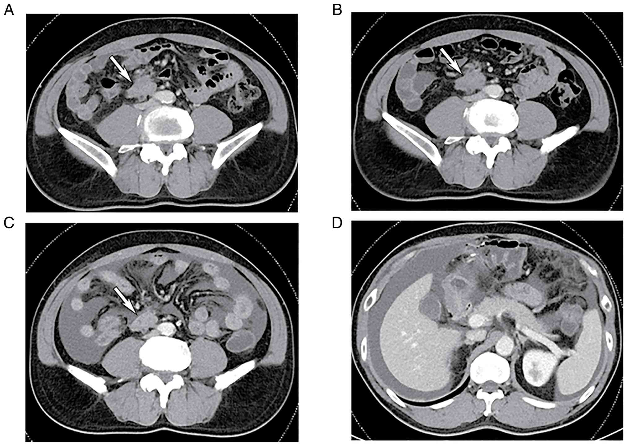
second cycle demonstrated stable disease (Fig. 1A and B). The third cycle was
administered later in April 2022. In early May 2022, the patient
developed transient, self-resolving mild abdominal pain and dysuria
without an identifiable cause. Subsequent laboratory tests revealed
mildly elevated serum creatinine levels (130 µmol/l in mid-May and
127 µmol/l in late May; reference range, 59–104 µmol/l), although
urinalysis was unremarkable. Renal ultrasound demonstrated
persistent mild dilation of the right renal pelvis and proximal
ureter (data not shown). The fourth treatment cycle was
administered as scheduled in May 2022. By early June 2022, renal
function had normalized, but ultrasound revealed new-onset mild
right hydronephrosis and moderate ascites (data not shown; findings
extracted from the patient's medical file). In early June 2022, the
patient reported abdominal distension, prompting therapeutic
abdominal paracentesis. The ascitic fluid was initially hemorrhagic
and became light yellow and clear 2 days later. Cytological
examination of the ascites was negative for malignancy on three
separate occasions. A CT scan in mid-June 2022 confirmed moderate
ascites with omental thickening (Fig.
1C and D); the previously noted mild hydronephrosis showed no
significant worsening compared with prior imaging. At that time,
despite a slightly smaller metastatic lymph node, the ascites
remained unexplained; anlotinib was withheld for one cycle, while
penpulimab was continued given the known sensitivity of POLE-pd
tumors to immunotherapy. The fifth cycle of penpulimab monotherapy
was administered in late June 2022, but persistent unexplained
ascites requiring recurrent drainage and delay of antitumor
treatment led to the patient's study withdrawal in July 2022.
During routine follow-up CT in early September 2022,
a fistula between the tumor and right ureter was identified. The
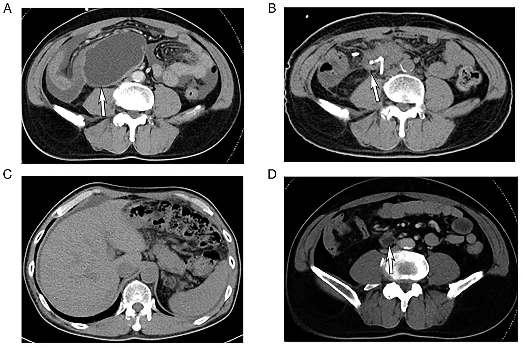
fistula formed a well-defined cystic mass (Fig. 2A), which was subsequently confirmed
by positron emission tomography (PET)-CT. To manage this
complication, drainage tubes were inserted into both the fistula
sac and peritoneal cavity in mid-October 2022. Biochemical analysis
revealed that the creatinine levels of the fistula sac and ascites
were 3,282/l and 232 µmol/l, respectively. The corresponding serum
creatinine level at that time was 69 µmol/l, yielding an
ascites-to-serum creatinine ratio of 3.4:1, which strongly
suggested urinary leakage. Following failed ureteral stent
implantation, a right nephrostomy was performed two days later. The
patient's abdominal distension was completely resolved, and both
the fistula sac and ascites volume were markedly reduced 3 weeks
later (Fig. 2B and C), which
prompted removal of the drainage tubes. A CT scan in late March
2023 showed resolution of omental fibrous density (Fig. S2A-D), minimal residual ascites and
reduction of the fistula-associated cyst to 2×1 cm. The nephrostomy
tube was successfully removed in mid-May 2023.
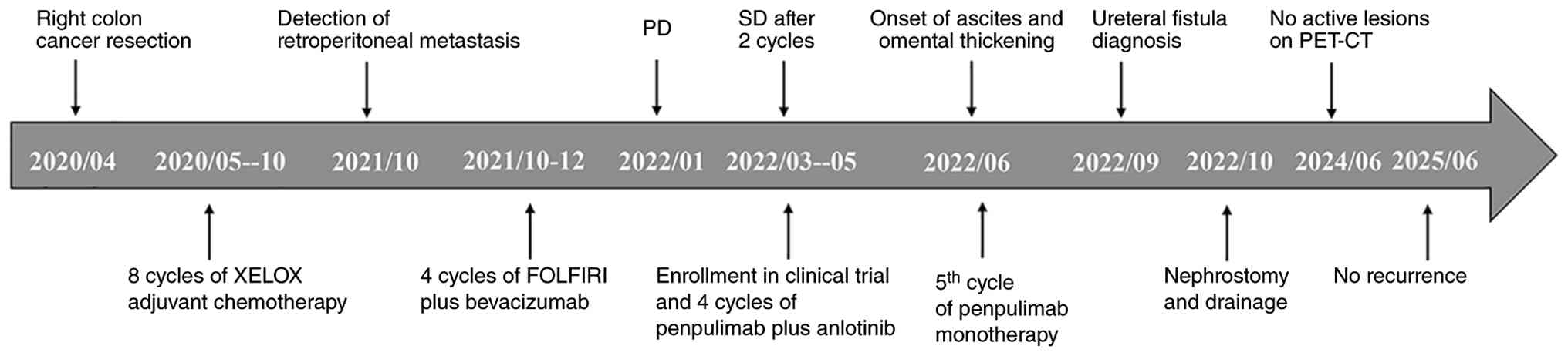
A detailed timeline of all major clinical events,
including surgery, systemic therapies, fistula diagnosis and
follow-up, is shown in Fig. 3.
During regular follow-ups every 3–6 months, the patient maintained
normal tumor markers with no evidence of disease recurrence on CT
scans. PET-CT in June 2024 revealed no active lesions (Fig. S3A-C). Based on these findings,
including complete radiographic resolution, normal tumor markers,
absence of symptoms and sustained remission for >12 months
without any antitumor therapy, the patient was assessed as having
achieved cCR. As of the submission of the manuscript, the last CT
scan in June 2025 confirmed no evidence of tumor and a residual
small cyst (Fig. 2D). No further
antitumor therapy was administered, and no additional intervention
was required for the residual cyst or the urinary tract. Renal
function remained normal during follow-up. The patient remained in
good general condition throughout the surveillance period.
Discussion
POLE exerts exonuclease activity via its
proofreading domain and deletes mispaired bases inserted during DNA
replication. POLE-pds lead to the accumulation of missense and
nonsense mutations, typically manifesting as an ultra-hypermutated
phenotype with a TMB exceeding 100 mutations/megabase (3,9). Such
extreme mutagenesis generates abundant neoantigens, which may
explain the enhanced clinical responsiveness to immune checkpoint
inhibitors (ICIs) observed in POLE-pd tumors (10,11).
The exonuclease domain contains several well-characterized
mutational hotspots that impair the proofreading capacity, with
P286R representing one of the most frequently documented pathogenic
variants in this critical functional domain (4,9).
POLE-pds exhibit low prevalence in mCRC, occurring
in <1% of cases (12). This
rarity may reflect the favorable prognosis associated with these
tumors following primary resection (13). Large-scale genomic analyses have
revealed that among 9,136 CRC specimens, only 2.4% harbored
alterations in either POLE or polymerases δ (POLD1), with a mere
0.4% exhibiting the characteristic ultra-hypermutated phenotype
(14). Notably, patients with
POLE-pd CRC exhibited a markedly elevated TMB compared with those
with microsatellite instability-high (MSI-H) tumors or POLE
variants outside the exonuclease domain (15).
Accumulating clinical evidence suggests that
patients with CRC with POLE-pds exhibit excellent responses to
ICIs. The first documented case was reported in 2017 and the
patient with treatment-refractory mCRC with the MSS phenotype and
POLE mutation exhibited a remarkable response to pembrolizumab
(16). Subsequent case reports
further supported these findings, demonstrating excellent
immunotherapy outcomes in patients with pathogenic POLE mutations
(17,18). In 2022, Wen et al (15) reported 4 patients with CRC with
liver metastasis who received toripalimab or sintilimab combined
with chemotherapy (with or without bevacizumab) after POLE
exonuclease domain mutations were detected. All 4 patients achieved
a pathological complete response after surgery despite showing only
a partial response on radiological evaluation. Recent larger
studies have further validated these findings. A global study of 27
patients with mCRC with POLE/POLD1 mutations leading to
proofreading deficiency revealed superior response rates to ICIs
and survival outcomes after ICI treatment compared with those of
MSI-H patients (3). In a
retrospective analysis (19), 22
consecutive patients with locally advanced CRC with MSI-H or
POLE-mutated bulky tumors (>8 cm) received neoadjuvant PD-1
blockade. Among the 2 patients with POLE mutations, 1 each achieved
pathological or clinical complete remission.
In the present case, the combination of anti-PD-1
therapy with anlotinib (a multi-target tyrosine kinase inhibitor
targeting VEGFR2/3, fibroblast growth factor receptor 1–4,
platelet-derived growth factor receptor α/β, c-Kit and Ret)
(20) may have produced synergistic
effects (21), leading to durable
complete remission for >2.5 years after treatment cessation.
However, this profound response was accompanied by an unusual
complication that posed significant diagnostic and therapeutic
challenges.
The development of the ureteral fistula in the
present patient is best understood as a multi-factorial event
rather than a direct and sole effect of immunotherapy. First, the
metastatic lesion already directly involved the mid-ureter at
baseline, structurally compromising the ureteral wall. Second, the
profound and rapid tumor necrosis induced by effective
immunotherapy disrupted the tumor-ureteral interface (22). Third, the anti-angiogenic effect of
anlotinib may have impaired local tissue repair and wound healing,
facilitating fistula formation (23,24).
Therefore, while immunotherapy was the primary driver of tumor
regression, the fistula was likely the result of combined
contributions from baseline invasion, treatment response and
anti-angiogenic therapy.
The diagnostic chain for urinary ascites in the
present case is instructive. The patient exhibited minimal urinary
tract symptoms, and early imaging studies, including ultrasound and
a CT scan, failed to provide evidence of fistula formation.
Initially, ascites and peritoneal thickening raised suspicion of
peritoneal carcinomatosis (ruled out by three negative cytologies)
or rare immune-related serositis (unlikely given the poor response
to treatment interruption). The key diagnostic evidence included:
i) Elevated creatinine levels in the fistula sac (3,282 µmol/l) and
ascites (232 µmol/l), with a serum creatinine level of 69 µmol/l,
yielding an ascites-to-serum ratio of 3.4:1, which was highly
suggestive of urinary leakage; ii) delayed CT imaging that
eventually visualized the distinct fistula sac; and iii) definitive
therapeutic confirmation, including complete resolution of ascites
following nephrostomy and fistula sac drainage. Early imaging
missed the fistula, potentially because it was initially small and
collapsed, with urine leaking directly into the peritoneal cavity
rather than forming a localized collection; the fistulous tract
only became radiologically detectable as it matured into a defined
cystic sac over time.
The choice of conservative management over surgical
repair was deliberate. Surgical intervention was considered
high-risk due to the following: i) Active immunotherapy and
anti-angiogenic effects potentially impairing wound healing; ii)
effective symptomatic control with drainage; and iii) achievement
of tumor remission, which made fistula repair unnecessary if the
urinary leak could be controlled. Nephrostomy combined with
continuous fistula sac drainage proved to be an effective and less
morbid approach, and this strategy may serve as a reference for
similar cases.
This rare complication, while presenting significant
diagnostic and therapeutic challenges, paradoxically reflects the
profound depth of the antitumor response induced by immunotherapy.
The occurrence of fistula formation in the present case aligns with
previous reports of fistula formation in rapidly responding tumors
(22,25). The patient's sustained remission for
>2.5 years without further therapy supports the growing
recognition that POLE-mutated CRC represents a distinct molecular
subtype with exceptional sensitivity to ICIs (3,15).
In conclusion, mCRC harboring a POLE-pd is highly
sensitive to immunotherapy. Rapid tumor regression after
immunotherapy may lead to the formation of a fistula between the
tumor and the adjacent organs. Ureteral fistulas presenting with
ascites as the primary symptom are rare and diagnostically
challenging. With the increasing clinical application of
immunotherapy, awareness of such rare complications is necessary
for timely diagnosis. Successful conservative management via
nephrostomy and drainage may provide a clinically actionable
approach for similar cases of ureteral fistulas.
Supplementary Material
Supporting Data
Supporting Data
Acknowledgements
Not applicable.
Funding
Funding: No funding was received.
Availability of data and materials
The raw sequencing data generated in the present
study may be found in the China National Center for Bioinformation
(CNCB-NGDC) under BioProject accession no. PRJCA073069 or at the
following URL: https://ngdc.cncb.ac.cn/omix/preview/SfmOFgG7WE.
Authors' contributions
JZ designed and supervised the study. JZ and XGo
analyzed patient data and wrote the manuscript. XGa contributed to
the acquisition of the medical images, and the analysis and
interpretation of the imaging data, and critically revised the
manuscript. All authors have read and approved the final version of
the manuscript. JZ, XGo and XGa confirm the authenticity of all the
raw data.
Ethics approval and consent to
participate
The present study was approved by the Human Research
Ethics Committee of Peking Union Medical College Hospital (Beijing,
China; approval no. HS-2986B) and the patient provided written
informed consent to participate in the clinical trial.
Patient consent for publication
Written informed consent was obtained from the
patient for the publication of all case information and medical
images included in the present article.
Competing interests
The authors declare that they have no competing
interests.
References
|
1
|
Pursell ZF, Isoz I, Lundström EB,
Johansson E and Kunkel TA: Yeast DNA polymerase epsilon
participates in leading-strand DNA replication. Science.
317:127–130. 2007. View Article : Google Scholar : PubMed/NCBI
|
|
2
|
Rayner E, van Gool IC, Palles C, Kearsey
SE, Bosse T, Tomlinson I and Church DN: A panoply of errors:
Polymerase proofreading domain mutations in cancer. Nat Rev Cancer.
16:71–81. 2016. View Article : Google Scholar : PubMed/NCBI
|
|
3
|
Ambrosini M, Rousseau B, Manca P, Artz O,
Marabelle A, André T, Maddalena G, Mazzoli G, Intini R, Cohen R, et
al: Immune checkpoint inhibitors for POLE or POLD1
proofreading-deficient metastatic colorectal cancer. Ann Oncol.
35:643–655. 2024. View Article : Google Scholar : PubMed/NCBI
|
|
4
|
Rousseau B, Bieche I, Pasmant E, Hamzaoui
N, Leulliot N, Michon L, de Reynies A, Attignon V, Foote MB,
Masliah-Planchon J, et al: PD-1 blockade in solid tumors with
defects in polymerase epsilon. Cancer Discov. 12:1435–1448. 2022.
View Article : Google Scholar : PubMed/NCBI
|
|
5
|
Hino H, Shiomi A, Kusuhara M, Kagawa H,
Yamakawa Y, Hatakeyama K, Kawabata T, Oishi T, Urakami K, Nagashima
T, et al: Clinicopathological and mutational analyses of colorectal
cancer with mutations in the POLE gene. Cancer Med. 8:4587–4597.
2019. View Article : Google Scholar : PubMed/NCBI
|
|
6
|
Huang Z, Pang X, Zhong T, Qu T, Chen N, Ma
S, He X, Xia D, Wang M, Xia M and Li B: Penpulimab, an
Fc-engineered IgG1 anti-PD-1 antibody, with improved efficacy and
low incidence of immune-related adverse events. Front Immunol.
13:9245422022. View Article : Google Scholar : PubMed/NCBI
|
|
7
|
Zheng Y, Zhu J, Xiong J, Jiang O, Wang H,
Xie Y, Zhou Y and Xu N: Phase 1b/2 study of penpulimab (AK105), an
antiprogrammed cell death-1 immunoglobulin G1 antibody, in advanced
or metastatic solid tumors (AK105-204). Cancer. 130:2180–2190.
2024. View Article : Google Scholar : PubMed/NCBI
|
|
8
|
Lin B, Song X, Yang D, Bai D, Yao Y and Lu
N: Anlotinib inhibits angiogenesis via suppressing the activation
of VEGFR2, PDGFRβ and FGFR1. Gene. 654:77–86. 2018. View Article : Google Scholar : PubMed/NCBI
|
|
9
|
Campbell BB, Light N, Fabrizio D, Zatzman
M, Fuligni F, de Borja R, Davidson S, Edwards M, Elvin JA, Hodel
KP, et al: Comprehensive analysis of hypermutation in human cancer.
Cell. 171:1042–1056.e10. 2017. View Article : Google Scholar : PubMed/NCBI
|
|
10
|
Samstein RM, Lee CH, Shoushtari AN,
Hellmann MD, Shen R, Janjigian YY, Barron DA, Zehir A, Jordan EJ,
Omuro A, et al: Tumor mutational load predicts survival after
immunotherapy across multiple cancer types. Nat Genet. 51:202–206.
2019. View Article : Google Scholar : PubMed/NCBI
|
|
11
|
Marabelle A, Fakih M, Lopez J, Shah M,
Shapira-Frommer R, Nakagawa K, Chung HC, Kindler HL, Lopez-Martin
JA, Miller WH Jr, et al: Association of tumour mutational burden
with outcomes in patients with advanced solid tumours treated with
pembrolizumab: Prospective biomarker analysis of the multicohort,
open-label, phase 2 KEYNOTE-158 study. Lancet Oncol. 21:1353–1365.
2020. View Article : Google Scholar : PubMed/NCBI
|
|
12
|
Antoniotti C, Korn WM, Marmorino F,
Rossini D, Lonardi S, Masi G, Randon G, Conca V, Boccaccino A,
Tomasello G, et al: Tumour mutational burden, microsatellite
instability, and actionable alterations in metastatic colorectal
cancer: Next-generation sequencing results of TRIBE2 study. Eur J
Cancer. 155:73–84. 2021. View Article : Google Scholar : PubMed/NCBI
|
|
13
|
Domingo E, Freeman-Mills L, Rayner E,
Glaire M, Briggs S, Vermeulen L, Fessler E, Medema JP, Boot A,
Morreau H, et al: Somatic POLE proofreading domain mutation, immune
response, and prognosis in colorectal cancer: A retrospective,
pooled biomarker study. Lancet Gastroenterol Hepatol. 1:207–216.
2016. View Article : Google Scholar : PubMed/NCBI
|
|
14
|
Mosalem O, Coston TW, Imperial R, Mauer E,
Thompson C, Yilma B, Bekaii-Saab TS, Stoppler MC and Starr JS: A
comprehensive analysis of POLE/POLD1 genomic alterations in
colorectal cancer. Oncologist. 29:e1224–e1227. 2024. View Article : Google Scholar : PubMed/NCBI
|
|
15
|
Wen L, Chen Z, Ji X, Fong WP, Shao Q, Ren
C, Cai Y, Li B, Yuan Y, Wang D, et al: Pathological complete
response to immune checkpoint inhibitor in patients with colorectal
cancer liver metastases harboring POLE exonuclease domain mutation.
J Immunother Cancer. 10:e0044872022. View Article : Google Scholar : PubMed/NCBI
|
|
16
|
Gong J, Wang C, Lee PP, Chu P and Fakih M:
Response to PD-1 blockade in microsatellite stable metastatic
colorectal cancer harboring a POLE mutation. J Natl Compr Cancer
Netw. 15:142–147. 2017. View Article : Google Scholar : PubMed/NCBI
|
|
17
|
Silberman R, F Steiner D, Lo AA, Gomez A,
Zehnder JL, Chu G and Suarez CJ: Complete and prolonged response to
immune checkpoint blockade in POLE-mutated colorectal cancer. JCO
Precis Oncol. 3:1–5. 2019. View Article : Google Scholar : PubMed/NCBI
|
|
18
|
Durando ML, Menghani SV, Baumann JL,
Robles DG, Day TA, Vaziri C and Scott AJ: Four-year disease-free
remission in a patient with POLE mutation-associated colorectal
cancer treated using anti-PD-1 therapy. J Natl Compr Cancer Netw.
20:218–223. 2022. View Article : Google Scholar : PubMed/NCBI
|
|
19
|
Li Y, Liang F, Li Z, Zhang X and Wu A:
Neoadjuvant immunotherapy for patients with microsatellite
instability-high or POLE-mutated locally advanced colorectal cancer
with bulky tumors: New optimization strategy. Clin Colorectal
Cancer. 24:18–31.e2. 2025.PubMed/NCBI
|
|
20
|
Sun Y, Niu W, Du F, Du C, Li S, Wang J and
Li L, Wang F, Hao Y, Li C and Li L: Safety, pharmacokinetics, and
antitumor properties of anlotinib, an oral multi-target tyrosine
kinase inhibitor, in patients with advanced refractory solid
tumors. J Hematol Oncol. 9:1052016. View Article : Google Scholar : PubMed/NCBI
|
|
21
|
Wang Z, Qin BD, Ye CY, Wang MM, Yuan LY,
Yao HS, Jiao XD, Liu K, Zhou WL, Qin WX, et al: Anlotinib plus
sintilimab as first-line treatment for patients with advanced
colorectal cancer (APICAL-CRC): An openlabel, single-arm, phase II
trial. Signal Transduct Target Ther. 10:3012025. View Article : Google Scholar : PubMed/NCBI
|
|
22
|
Greene C, Nakakura EK and Ko AH:
Gastrocutaneous fistula in a patient with locally recurrent
MSI-high colorectal cancer: Local complications arising from
therapeutic response to immune checkpoint blockade. Anticancer Res.
37:3679–3684. 2017.PubMed/NCBI
|
|
23
|
Ouchi R, Okada K, Usui K, Kurata N, Suzuki
S, Nagao M, Watanabe Y and Koyama K: Intestinal perforation in a
patient with colon cancer during treatment with regorafenib: A case
report and review of the literature. Tohoku J Exp Med. 254:207–211.
2021. View Article : Google Scholar : PubMed/NCBI
|
|
24
|
Deng P, Hu C, Li Y, Cao L, Yang H, Li M,
An J, Jiang J and Gu Q: Bronchial fistula: Rare complication of
treatment with anlotinib. Zhongguo Fei Ai Za Zhi. 23:858–865.
2020.PubMed/NCBI
|
|
25
|
Shinohara A, Miguchi M, Ikeda S, Yamauchi
M, Katayama S, Fujii T, Takemoto Y, Nakano R, Hashimoto M, Fujikuni
N, et al: Complete pathologic response after neoadjuvant
immunotherapy for mismatch repair-deficient colon cancer with
invading adjacent organs. Immunotherapy. 18:377–383. 2026.
View Article : Google Scholar : PubMed/NCBI
|