Introduction
Olfactory neuroblastoma (esthesioneuroblastoma) is a
rare malignant neuroectodermal neoplasm arising from the
neuroepithelium of the upper portion of the nasal cavity. Unlike
classic pediatric neuroblastoma, it primarily affects adolescents
and adults. The 5-year overall survival rate for patients with
olfactory neuroblastoma ranges between 60 and 80%, though prognosis
drops significantly for those presenting with advanced-stage or
metastatic disease (1). Olfactory
neuroblastoma accounts for 3–6% of all tumors of the nasal cavity.
Although typically orthotopic, <10% of cases correspond to
ectopic neuroblastomas, which originate in sites where olfactory
neural crest cells do not normally reside, presumably due to
aberrant migration during embryonic development. Among ectopic
locations, the paranasal sinuses (ethmoid, maxillary and sphenoid)
are the most common, and they are also reported in the nasal
septum, the sellar region and the nasopharynx. Tumors with ectopic
presentation are usually characterised by a more locally invasive
behaviour than their orthotopic counterparts (2).
Due to its neuroendocrine nature, olfactory
neuroblastoma possesses the ability to synthesise vasopressin.
Consequently, syndrome of inappropriate antidiuretic hormone
secretion (SIADH) has been identified as a paraneoplastic
manifestation in 2% of patients worldwide. However, this
paraneoplastic association increases markedly in ectopic variants,
affecting ≥18% of patients and manifesting concomitantly with the
tumor or preceding it by several years (3) Clinically, initial symptoms are usually
nonspecific and include unilateral nasal obstruction, persistent
rhinorrhea, epistaxis or visual and orbital disturbances in
advanced stages, coexisting with euvolemic hypoosmolar
hyponatraemia in functional cases (4).
The standard approach to olfactory neuroblastoma is
based on a multimodal regimen combining endoscopic or open surgery
with adjuvant radiotherapy, reserving chemotherapy for advanced or
initially unresectable stages. Despite therapeutic advances and an
overall 5-year survival rate of ~80%, long-term locoregional
recurrence rates remain high, occurring in ~30% of patients
(4).
A distinctive biological feature of this tumor is
its insidious course and its propensity for markedly late relapses,
even a decade after achieving complete remission, which warrants
prolonged and indefinite clinical and imaging surveillance.
Progression to the orbit or adjacent structures and high
histological grade (Hyams grades III and IV) notably worsen the
prognosis. In cases of recurrence or high-grade disease, the tumors
demonstrate high chemosensitivity, with cisplatin- and
etoposide-based regimens being the most widely validated first-line
systemic therapies (5).
The present case describes a rare tumor in adults
with its ectopic presentation in an unusual location, the right
sphenoid sinus. Furthermore, associated SIADH is identified in a
minimal percentage of patients with neuroblastoma. Finally,
although relapses typically occur several years later and show a
poor response to chemotherapy, the present case relapsed very late
(10 years) but exhibited a complete response to standard
chemotherapy with cisplatin and etoposide, representing an
exceptionally rare clinical scenario that is poorly documented in
the international medical literature.
Case report
A 35-year-old female patient was admitted to the
Department of Internal Medicine, Princess Marina Hospital
(Gaborone, Botswana) in December 2013. The patient had no notable
medical history; however, they reported a history of malaise,
muscle weakness, cramps and recurrent global headaches of ≥2 years
in duration. Clinical interview and physical examination revealed
right-sided nasal obstruction, dysosmia and dysgeusia of ≥1 year in
duration. Serum chemistry revealed sodium levels between 130–134
mmol/l during euvolemic states (normal range: 135–145 mmol/l),
leading to a definitive diagnosis of SIADH.
In January 2014, the patient underwent a
contrast-enhanced CT scan of the paranasal sinuses at initial
presentation. Fig. 1A shows a
sagittal view demonstrating a heterogeneous enhancing mass
occupying the right sphenoid sinus (5.3×4.2×4.4 cm), extending into
the ipsilateral ethmoid air cells, nasal cavity, and maxillary
sinus (green arrows). Fig. 1B shows
a sagittal view depicting the ectopic tumor location within the
sphenoid body and its relationship to the skull base, with no
evidence of macroscopic intracranial extension (green arrows).
Fig. 1C illustrates an axial view
showing an expansive mass in the right sphenoid sinus, which causes
bony wall opacification and remodeling, as well as a leftward
deviation of the nasal septum due to mass effect (green
arrows).
The contrast-enhanced CT scan of the neck showed a
symmetrical nasopharynx without well-defined lesions. The mucosal
surfaces of the upper aerodigestive tract, larynx and base of the
tongue appeared symmetrical and without notable findings. The
parotid and submandibular glands were normal and the thyroid gland
appeared symmetrical without well-defined nodules. The carotid and
jugular vessels, as well as the deep suprahyoid and infrahyoid
cervical spaces, showed no abnormalities. No suspicious cervical
lymph nodes were visualized. The central skull base was intact and
the cervical vertebrae, spinal cord and lung apices showed no
notable findings (data not shown).
An endoscopic incisional biopsy was performed
(January 2014), taking a small sample from the most accessible part
of the tumor in the nasal cavity, away from the cribriform plate
and the sphenoid sinus to avoid causing a cerebrospinal fluid leak.
Immunohistochemical staining showed positivity for synaptophysin,
S-100 and neuron-specific enolase (NSE), as well as a Ki-67 cell
proliferation index of 15%, while it was negative for CD99,
epithelial membrane antigen and cytokeratins (MNF116). This
information is based on the primary pathology report. Once the
diagnosis of olfactory neuroblastoma was confirmed, definitive
excision was performed.
In February 2014, after assessing the tumor's
relationship with the carotid artery and optic nerve, a transnasal
endoscopic surgical resection was performed. Given the absence of
intracranial involvement, the sphenoid ostium was directly
visualized and the tumor was excised while preserving the integrity
of the anterior skull base. A wide sphenoidotomy was then performed
to ensure a clear mucosa, along with reaming of the remaining bone
to remove any tumor nests, aiming for negative microscopic margins.
Serum sodium levels returned to normal 48 h after tumor
excision.
Microscopic examination allowed for the analysis of
multiple fragments of grayish-white tissue, measuring 3×2×1 cm in
total. Upon sectioning, the surface was solid and white, with a
lacy appearance and some bright white areas. A total of five
representative blocks were processed. Microscopic examination of
the sections revealed a tumor composed of irregular, circumscribed
tumor nests separated by fibrotic stroma, with some areas
exhibiting a diffuse pattern. Histological examination was
performed using standard hematoxylin and eosin staining on
formalin-fixed, paraffin-embedded tissue sections to evaluate
cellular morphology and architectural patterns. A total of five
representative tissue blocks were processed. Tissue sections (3–4
µm thickness) were deparaffinized in xylene (two changes, 10 min
each) and rehydrated through a descending graded ethanol series
(100, 95 and 70%, 5 min each) down to distilled water. Nuclear
counterstaining was carried out with Mayer's hematoxylin for 3–5
min, followed by rinsing in running tap water for 5 min to allow
blueing. Sections were briefly differentiated in 1% acid alcohol,
washed and counterstained with 1% eosin Y solution for 1–2 min for
cytoplasmic and stromal contrast. Finally, the slides were
dehydrated through ascending grades of ethanol, cleared in xylene
and mounted with synthetic resin under glass coverslips for
microscopic examination.
The neoplastic cells had small, round nuclei with
chromatin ranging from coarse to fine punctation and scant
cytoplasm. The tumor presented a fibrillar intercytoplasmic
background, and both Flexner and Homer-Wright rosettes were
identified (Fig. 2). Due to the
loss of architecture, nuclear pleomorphism, frequent necrosis and
high mitotic rates with sparse neuropil, it was classified as Hyams
grade III (4). The final
histopathological diagnosis was olfactory neuroblastoma (C72.2).
The diagnostic code corresponds strictly to the International
Classification of Diseases, Tenth Revision (ICD-10) nomenclature
for malignant neoplasms of the olfactory nerve. The absence of
microscopic dural invasion confirmed Kadish stage B (4).
Finally, treatment was completed with adjuvant
radiotherapy using intensity-modulated radiation therapy (IMRT), 60
Gy, which allowed the dose to be concentrated in the sphenoid body
and ethmoid air cells, protecting the optic nerves and optic
chiasm. Adjuvant radiotherapy commenced in March 2014 (6 weeks
postoperatively). A total dose of 60 Gy was delivered in 30 daily
fractions (2.0 Gy/session, 5 days per week), concluding in April
2014 after a 6-week treatment period.
No notable adverse effects were reported after
radiotherapy. Follow-up CT and MRI scans with contrast showed no
evidence of tumor lesions. The patient remained under
otolaryngology follow-up every 3 months for 2 years, then every 6
months for 3 years and annually thereafter. Blood tests and imaging
studies were repeated every 6 months for 5 years, including head
and neck CT or MRI, complete blood count and liver, kidney and
thyroid function tests every 6 months (Table SI).
However, in April 2024, the patient returned to the
Princess Marina Hospital (Gaborone, Botswana), presenting
spontaneously with persistent rhinorrhea and self-limiting
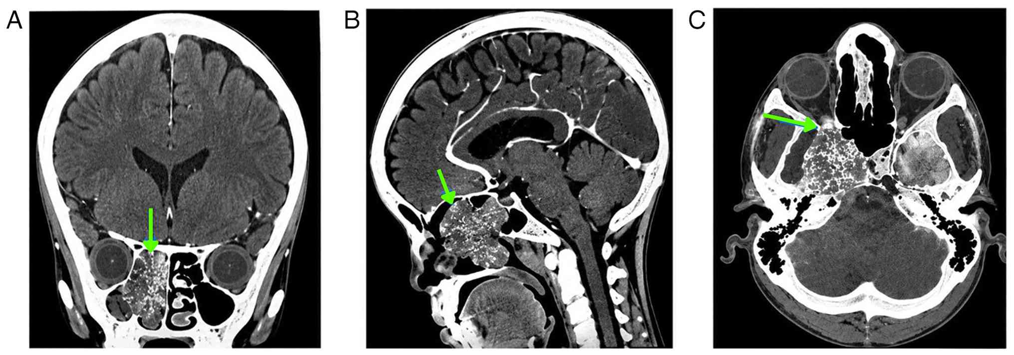
epistaxis. Fig. 3 presents
contrast-enhanced CT scans of the paranasal sinuses acquired in
April 2024, confirming tumor relapse. The images demonstrate a
complex, heterogeneous calcified mass, measuring 5.3×4.7 cm,
centered within the right nasal cavity. Coronal and sagittal views,
provided in Fig. 3A and B, reveal
extensive involvement of the ethmoid sinuses. Critically, these
views, combined with the axial image in Fig. 3C, show multi-compartmental direct
extension of the tumor. Specific features include significant
invasion and partial obliteration of the right maxillary antrum,
alongside clear encroachment into the medial aspect of the right
orbit, highlighting the locally aggressive nature of the
recurrence.
In May 2024, a late local recurrence was diagnosed.
Given the lack of available human and technological resources for
salvage surgery, the refusal of the patient to receive further
radiation therapy and considering the high risk of microscopic
metastases and the chemosensitivity of high-grade neuroendocrine
tumors, it was decided to administer 6 cycles of cisplatin [75
mg/m2 intravenously (IV), day 1] and etoposide (100
mg/m2 IV, days 1, 2 and 3) every 21 days, which began in
June 2024.
Despite the well-documented toxicity profile of the
cisplatin and etoposide regimen, which is often associated with
severe side effects, the good tolerance of the patient throughout
the 6 cycles resulted only in the occurrence of grade 1 nausea and
grade 2 myelosuppression. Both events were transient and of short
duration, allowing for effective management through the timely
initiation of supportive treatment. Following standard protocols,
the emetic symptoms were controlled with conventional antiemetics.
On day 1 (the highly emetogenic combination phase), intravenous
premedication consisted of a triple-agent antiemetic regimen
comprising a 5-HT3 receptor antagonist (ondansetron, 8
mg IV), a neurokinin-1 (NK1) receptor antagonist
(fosaprepitant, 150 mg IV) and dexamethasone (12 mg IV). For days 2
and 3 (the continuation of etoposide), daily prophylactic
antiemetic coverage included intravenous ondansetron (8 mg IV once
daily) along with oral dexamethasone (8 mg daily). Delayed emesis
protection on day 4 was maintained using oral dexamethasone (8 mg
once daily). Throughout all 6 treatment cycles, rescue medication
with metoclopramide (10 mg orally or IV as needed every 8 h) was
available, achieving effective emetic control with no grade ≥2
adverse gastrointestinal events reported. The haematological
toxicity responded favourably and predictably, avoiding
interruptions or dose reductions and reinforcing the success of the
therapeutic regimen. The patient demonstrated a favourable clinical
and radiological response, completing the last cycle in September
2024. They were subsequently monitored closely with clinical and
radiological follow-up every 12 weeks.
The most recent head and neck CT scan was performed
in December 2025, showing complete absence of the lesion described
as a recurrence in April 2024. The brain parenchyma, ventricles,
sulci and basal cisterns appeared normal, with no focal mass
lesions or abnormal enhancement after contrast administration; the
orbits and cranial bones also appeared normal. The neck, the
larynx, pharynx and postnasal drip were normal; the thyroid and
salivary glands appeared normal without notable cervical
lymphadenopathy. The infrahyoid muscles, prevertebral soft tissues
and cervical spine showed no abnormalities (Fig. 4). As of the date of the present
report (February 2026), the patient remained asymptomatic.
Discussion
Olfactory neuroblastoma is a rare malignant
neuroendocrine neoplasm that classically originates in the
olfactory neuroepithelium of the nasal cavity roof, specifically in
the cribriform plate, superior turbinate and superior nasal septum.
However, the biological behaviour of this entity becomes
considerably more complex when it occurs in ectopic locations, with
the sphenoid sinus being one of the most unusual anatomical
variants (5). In the present case,
the initial tumor mass predominantly occupied the right sphenoid
sinus. The presence of ectopic neuroepithelial tissue in the
paranasal sinuses is attributed to abnormalities in the
embryological migration of the olfactory placode or to the
persistence of pluripotent cell remnants along the craniofacial
developmental pathways. From a biological standpoint, ectopic
olfactory neuroblastomas tend to exhibit silent and expansive
growth due to the laxity of the air cavities that house them, which
explains why severe obstructive or neurological symptoms usually
manifest late, when the tumor has already reached critical
dimensions or caused notable local bone destruction (6). In the patient of the present study,
the Hyams grade III tumor showed marked local aggressiveness,
eroding adjacent structures and extending into the ethmoid air
cells and olfactory sulcus. However, despite its locally expansile
growth pattern, no microscopic dural invasion was identified at
initial presentation, indicating that the tumor remained confined
to the extracranial compartment.
In the context described, it is important to
emphasise that the histopathological characterisation of this
neoplasm should not be limited to the identification of structural
neuroendocrine markers, but rather requires the incorporation of
molecular prognostic tools, among which the Ki-67 cell
proliferation index is fundamental (7). The present case showed positivity for
synaptophysin, chromogranin, S-100, NSE and had a Ki-67 cell
proliferation index of 15%. It has been considered that the
omission of Ki-67 would have deprived the patient of a critical
factor for stratifying the biological behaviour of the tumor. An
elevated Ki-67, at thresholds of ≥10%, correlates with lower
progression-free survival, more locally invasive behaviour and a
greater propensity to develop local recurrences or distant
metastases, characteristics shared by high-grade ectopic variants
(7). In the present case,
classified as a Hyams grade III, the determination of Ki-67 served
as an indicator of tumor aggressiveness and additional
justification for its high sensitivity to cytotoxic regimens based
on cisplatin and etoposide after late relapse.
Furthermore, due to its ectopic location in the
sphenoid sinus and its morphology of small, round, blue cells, the
histopathological differential diagnosis required differentiation
from other malignant nasosinusal neoplasms. The absence of
immunoreactivity for cytokeratins (MNF116) and CD99 was the
cornerstone that allowed the exclusion of small cell neuroendocrine
carcinoma, which shows cytokeratin positivity in a paranuclear
droplet pattern, and Ewing sarcoma (diffuse and strong membranous
expression of CD99). Likewise, the negativity for cytokeratins
ruled out undifferentiated sinonasal carcinoma, an aggressive
entity that often overlaps morphologically with Hyams grade III,
but which retains epithelial markers. Furthermore, the positivity
for the S-100 protein in the periphery of the tumor nests, which
identifies the sustentacular cells, along with the background of
intercytoplasmic neuropil and the Homer-Wright and Flexner
rosettes, allowed it to be differentiated from an amelanotic
sinonasal melanoma or an alveolar rhabdomyosarcoma (4).
The Hyams grade III classification was based on an
evaluation of its cytological characteristics, which place this
neoplasm within the high-grade category. Unlike grades I and II,
where a distinct lobular architecture and prominent fibrovascular
stroma are preserved, the examination revealed a loss of
architecture with the presence of irregular tumor nests and areas
of diffuse or subtle growth patterns. Cytologically, the cells
exhibited moderate to severe nuclear pleomorphism with chromatin
variations and scant cytoplasm, accompanied by reduced background
neuropil compared to low-grade lesions. Finally, the determining
criteria for consolidating grade III corresponded to a high mitotic
rate and the presence of foci of tumor necrosis, parameters that
differentiate this stage from grade IV, where necrosis is
prominent, pleomorphism is extreme and neurofibrillary
differentiation or rosette formation disappears completely
(8).
In another context, it should be noted that the
clinical presentation of this tumor can be accompanied by
paraneoplastic syndromes due to its classic neuroendocrine
differentiation, with SIADH being one of the most notable and
clinically relevant systemic manifestations (9). In the present study, SIADH manifested
insidiously with a chronic history of malaise, muscle weakness,
cramps and recurrent global headaches, associated with persistently
low serum sodium levels in a euvolemic state. The ectopic secretion
of arginine vasopressin by these neoplastic cells disrupts water
homeostasis in the renal collecting tubules, promoting excessive
water reabsorption that culminates in dilutional hyponatraemia.
From a prognostic perspective, the coexistence of SIADH in patients
with skull base tumors can complicate perioperative management and
increase the risk of severe cerebral oedema if not carefully
managed (10). However, an
important clinical and diagnostic observation is that SIADH acts as
an indirect functional tumor marker; in the present study, the
normalization of serum sodium just 48 h after surgical resection
confirmed the success of tumor cytoreduction and the eradication of
the ectopic hormone production source.
Although SIADH and the associated dilutional
hyponatraemia were the principal neuroendocrine and electrolyte
disturbances of the patient at presentation, the management of
high-grade neuroendocrine tumors also requires consideration of
other metabolic complications associated with tumor progression and
cell lysis, such as paraneoplastic hypercalcaemia. Hypercalcaemia
in advanced or recurrent skull base malignancies can derive from
both local osteolysis due to bone invasion of the paranasal sinuses
and from tumor secretion of parathyroid hormone-related protein
(11). Complications arising from
uncontrolled hypercalcaemia are multisystemic and serious, ranging
from renal dysfunction mediated by the induction of nephrogenic
diabetes insipidus and vasoconstriction of the afferent arteriole,
to cardiac conduction abnormalities with shortening of the QT
interval and risk of ventricular arrhythmias. Likewise, at the
level of the central nervous system, hypercalcaemia exacerbates
neurological symptoms, inducing lethargy, progressive mental
confusion, profound neuromuscular weakness and, in key phases,
coma, which can mask or mimic symptoms derived from the tumor mass
at the base of the skull or from a possible orbital recurrence
(11).
Evidence indicates that the neoplasm in the present
study is distinguished from most carcinomas of the sinonasal region
by its propensity for very late recurrences, a biological feature
documented in the international literature. While common epithelial
neoplasms usually recur within the first 5 years after definitive
treatment, olfactory neuroblastoma can remain in a prolonged
latency state, with relapses reported even 10, 15 or 20 years after
initial management (12). In the
present case, the patient experienced a local recurrence 10 years
later, after successfully completing a combined regimen of
endoscopic surgery and adjuvant IMRT. The prevalence of recurrences
in long-term follow-up series ranges from 30–50% of cases, and a
marked proportion of these are classified as very late. This
behaviour underscores the inadequacy of 5-year follow-up protocols
for this disease, justifying the need for lifelong clinical and
imaging surveillance, alternating CT and MRI scans to detect tumor
reactivation in time.
Furthermore, the pathophysiology underlying such
prolonged tumor recurrence rests on contemporary concepts of tumor
latency and permissive microenvironments. Following the initial
multimodal treatment in 2014, which combined transnasal endoscopic
resection with bone drilling and 60 Gy radiotherapy, quiescent
tumor stem cells likely persisted in the deep microscopic margins
of the sinonasal region or in the surrounding connective tissue.
These cells possess advanced cellular DNA repair mechanisms and a
low replication rate, making them resistant to ionising radiation
and programmed cell senescence. Latent oncogenesis implies that
these cells enter a G0 cell cycle arrest, evading immune
surveillance through the downregulation of major histocompatibility
complex molecules and the expression of immune checkpoint ligands
(13). Over the course of a decade,
the progressive accumulation of new secondary somatic mutations,
genomic instability and local epigenetic changes, influenced by
post-radiation stromal fibrosis and chronic tissue hypoxia, could
have altered the biological balance, triggering tumor angiogenesis
and proliferative reactivation that may have ultimately given rise
to the new heterogeneous and calcified mass identified in 2024.
Finally, when comparing the clinical course of the
patient with reported recurrence cases, correlations and
discrepancies emerge. In the literature, disease-free survival in
late-relapse series averages between 7–12 years, placing the 10
years observed in the present case at the core of this
chronological range (11,12). Regarding the site of relapse, the
literature indicates that isolated local recurrences in the nasal
cavity and ethmoid sinuses are the most common, followed by
cervical lymph node metastases and distant spread to the lung, bone
and liver (13). The patient
presented with a destructive local recurrence involving the right
nasal cavity and ethmoid sinuses with extension to the right orbit,
an aggressive pattern of local spread consistent with historical
cases of high histological grade. Clinically, while numerous cases
present with neurological symptoms due to intracranial invasion,
the patient of the present study only exhibited persistent
rhinorrhea and self-limiting epistaxis, with no evidence of
extension to the anterior cerebral fossa in tomographic studies,
which gave them a more favourable risk profile in terms of
resectability, although with an ominous prognosis without timely
oncological intervention.
In the present case, the decision-making process
regarding the relapse was conditioned by logistical, technical and
structural limitations of the hospital, which unequivocally
determined its clinical course. Although guidelines suggest a
second salvage surgery for operable local recurrences (14), the reality at the time of diagnosis
presented a restrictive scenario; the hospital neither had the
specialized human resources with subspecialty in endoscopic skull
base surgery, nor the essential equipment (intraoperative
neuronavigation systems and endoscopy towers with instruments for
orbitomaxillary tumor reaming) necessary for a second transnasal
endoscopic resection and a wide salvage sphenoidotomy with secure
margins. Furthermore, the conditions of the public health network
and inter-institutional agreements made it impossible to refer the
patient to another healthcare centre that had these services. Given
this scenario and the refusal of the patient to undergo
re-irradiation, systemic chemotherapy became established as the
only real, viable and ethically justifiable alternative of active
oncological treatment to stop the progression of the disease.
The choice of platinum and epipodophyllotoxins was
based on the chemosensitivity of high-grade neuroendocrine tumors.
The use of 6 cycles of cisplatin and etoposide administered every
21 days replicated the salvage therapy protocols described in the
international medical literature for Hyams grade III/IV olfactory
neuroblastoma (15). Reviewing
historical series, the overall response rate to combination
chemotherapy in the context of recurrences ranges from 40–70%, with
complete response being a less frequent but documented event in
selected cases (16). The complete
tumor remission achieved, evidenced by the CT scan of August 2024
with the disappearance of the heterogeneous orbitonasal mass,
places the present case within the group of exceptional responders
to systemic cytotoxic agents. Analogous reported cases have shown
that macroscopic eradication of recurrent disease using
cisplatin/etoposide can offer prolonged progression-free survival,
provided that strict follow-up is maintained (16).
Upon confirmation of the recurrence, therapeutic
decision-making presented a highly complex challenge. Given the
impossibility of performing another surgical treatment and the
refusal of the patient to undergo another cycle of radiotherapy, a
comprehensive approach became essential. For these reasons, the
present case was discussed within a tumor board composed of a
clinical oncologist, a radiation oncologist, a radiologist, a
histopathologist, an internist, an otolaryngologist, a
maxillofacial surgeon, a social worker and a psychologist.
This deliberation allowed for the analysis not only
of biological criteria but also of the psychological and social
circumstances of the patient. The joint evaluation was the
cornerstone that ethically justified the choice of chemotherapy as
the viable alternative, achieving an optimal consensus that
resulted in the aforementioned outcome.
A limitation of the present study is that the
immunohistochemical evaluation described relies solely on the
original institutional pathology reports. Retrievable histological
image files or tissue slides were unavailable for direct
re-evaluation, which precluded independent verification of the
representative staining patterns.
In conclusion, the present case represents an
extraordinarily unusual and scientifically valuable manifestation
within nasosinusal neoplasms, describing an olfactory neuroblastoma
of ectopic origin in the right sphenoid sinus. The emergence of
this tumor outside its usual orthotopic zone in the nasal vault
poses complex diagnostic challenges, demonstrating a biological
behaviour of silent but locally aggressive growth that can be
confused with other malignant neoplasms of the skull base without
rigorous immunohistochemical confirmation.
The unique nature of this presentation is
underscored by its paraneoplastic association with SIADH. This
neuroendocrine dysfunction, triggered by the autonomous production
of vasopressin by ectopic tumor cells, served as an indirect
biological marker of treatment response, and its complete
biochemical resolution correlated directly with successful initial
surgical cytoreduction. The presence of SIADH underscores the need
for a multidisciplinary clinical approach to mitigate severe
metabolic risks during the course of the disease.
Finally, the most remarkable aspect of the present
case lies in the development of a late locoregional recurrence
after a decade of clinical and imaging latency, followed by
complete and sustained remission with cisplatin- and
etoposide-based salvage systemic chemotherapy. This scenario
documents the high chemosensitivity of high-grade variants to
standard cytotoxic regimens, even in the face of advanced relapses
with orbital invasion. This behaviour also deviates from
traditional short-term oncological follow-up protocols, justifying
the need for lifelong surveillance in these types of tumors.
Supplementary Material
Supporting Data
Acknowledgements
Not applicable.
Funding
Funding: Not applicable.
Availability of data and materials
The data generated in the present study are included
in the figures and/or tables of this article.
Authors' contributions
JEPR led the study, performed the final drafting and
handled manuscript submission. EASG selected the case and provided
the clinical data. MCGC performed the CT scan image analysis. MCMS
collaborated with data acquisition (histopathological data). GCNZ
drafted the manuscript and worked on data analysis and
interpretation. IEF and ALS conducted the literature review. IEF
made substantial contributions to the conception and design of the
work, as well as to data acquisition. ALS, in addition to
participating in data acquisition, contributed to the analysis and
interpretation of the data. JEPR and EASG confirm the authenticity
of all the raw data. All authors read and approved the final
version of the manuscript.
Ethics approval and consent to
participate
Not applicable.
Patient consent for publication
Written informed consent was obtained from the
patient for the publication of the images and associated medical
case information.
Competing interests
The authors declare that they have no competing
interests.
References
|
1
|
Kushner BH, Kramer K, LaQuaglia MP, Modak
S and Cheung NK: Neuroblastoma in adolescents and adults: The
Memorial Sloan-Kettering experience. Med Pediatr Oncol. 41:508–515.
2003. View Article : Google Scholar : PubMed/NCBI
|
|
2
|
Lui CG, Badash I, Tang L, Mark ME, Batra
PS and Wrobel BB: Ectopic olfactory neuroblastoma: systematic
review of a rare clinical entity among sinonasal tumours. J Neurol
Surg B Skull Base. 85:109–118. 2023.PubMed/NCBI
|
|
3
|
Heiland L and Heiland K: Functional
olfactory neuroblastoma inducing symptomatic SIADH. Ear Nose Throat
J. 102:NP518–NP521. 2023. View Article : Google Scholar : PubMed/NCBI
|
|
4
|
National Comprehensive Cancer Network
(NCCN), . NCCN Clinical Practice Guidelines in Oncology:
Neuroblastoma. NCCN; Plymouth Meeting, PA: 2026, https://www.nccn.org/professionals/physician_gls/pdf/neuroblastoma.pdf
|
|
5
|
Twist CJ, Naranjo A, Schmidt ML, Tenney
SC, Cohn SL, Meany HJ, Mattei P, Adkins ES, Shimada H, London WB,
et al: Defining risk factors for chemotherapeutic intervention in
infants with stage 4S neuroblastoma: A report from children's
oncology group study ANBL0531. J Clin Oncol. 37:115–124. 2019.
View Article : Google Scholar : PubMed/NCBI
|
|
6
|
Barnes L: Surgical Pathology of the Head
and Neck. 3rd Edition. CRC Press; Boca Raton, FL: 2009
|
|
7
|
Safi C, Spielman D, Otten M, Bruce JN,
Feldstein N, Overdevest JB and Gudis DA: Treatment strategies and
outcomes of pediatric esthesioneuroblastoma: A systematic review.
Front Oncol. 10:12472020. View Article : Google Scholar : PubMed/NCBI
|
|
8
|
Touihmi S, Horrane I and Rkain I: An
atypical Esthesioneuroblastoma of the sphenoid sinus: A case
report. Ann Med Surg (Lond). 85:2029–2033. 2023. View Article : Google Scholar : PubMed/NCBI
|
|
9
|
Plasencia YL, Cortés MB, Arencibia DM,
Dámaso TM, Contreras IL, Pino AO, Domínguez AC, Brier FL, Delgado
YG and Mogollón FJ: Esthesioneuroblastoma recurrence presenting as
a syndrome of inappropriate antidiuretic hormone secretion. Head
Neck. 28:1142–1146. 2006. View Article : Google Scholar : PubMed/NCBI
|
|
10
|
Turri-Zanoni M, Gravante G, Dalfino G,
Antognoni P, Locatelli D, Battaglia P and Castelnuovo P: Ectopic
primary olfactory neuroblastoma: Case series and literature review.
World Neurosurg. 158:e645–e653. 2022. View Article : Google Scholar : PubMed/NCBI
|
|
11
|
Kumaria A, Jahshan F, Paine S, Reed M,
Ingale HA and Ramakrishnan Y: Olfactory neuroblastoma limited to
sphenoid sinus. Br J Neurosurg. 37:237–240. 2023. View Article : Google Scholar : PubMed/NCBI
|
|
12
|
Hirunpat P, Panyaping T and Hirunpat S:
Olfactory neuroblastoma is not always located at the roof of the
nasal cavity. J Belg Soc Radiol. 108:392024. View Article : Google Scholar : PubMed/NCBI
|
|
13
|
Dulguerov P, Allal AS and Calcaterra TC:
Esthesioneuroblastoma: A meta-analysis and review. Lancet Oncol.
2:683–690. 2001. View Article : Google Scholar : PubMed/NCBI
|
|
14
|
Cranmer LD, Chau B, Rockhill JK, Ferreira
M Jr and Liao JJ: Chemotherapy in esthesioneuroblastoma/olfactory
neuroblastoma: an analysis of the surveillance epidemiology and end
results (SEER) 1973–2015 database. Am J Clin Oncol. 43:203–209.
2020. View Article : Google Scholar : PubMed/NCBI
|
|
15
|
Brisson RJ, Quinn TJ and Deraniyagala RL:
The role of chemotherapy in the management of olfactory
neuroblastoma: A 40-year surveillance, epidemiology, and end
results registry study. Health Sci Rep. 4:e2572021. View Article : Google Scholar : PubMed/NCBI
|
|
16
|
Porter AB, Bernold DM, Giannini C, Foote
RL, Link MJ, Olsen KD, Moynihan TJ and Buckner JC: Retrospective
review of adjuvant chemotherapy for esthesioneuroblastoma. J
Neurooncol. 90:201–204. 2008. View Article : Google Scholar : PubMed/NCBI
|