Introduction
Lung cancer represents the most frequently diagnosed
malignancy and the leading cause of cancerrelated mortality
worldwide. According to GLOBOCAN 2022, ~2.5 million new lung cancer
cases and 1.8 million lungcancerassociated deaths occur annually,
with nonsmallcell lung cancer (NSCLC) accounting for 80–85% of all
lung cancer diagnoses (1). It is
estimated one-third of patients with NSCLC have stage III locally
advanced disease (2). Most patients
with stage III NSCLC are ineligible for surgical resection,
primarily due to tumor extension to critical structures, such as
the mediastinum and major vessels, or preexisting comorbidities
(3). According to the National
Comprehensive Cancer Network guidelines, concurrent
chemoradiotherapy (CCRT) followed by consolidation immunotherapy is
the standard treatment for unresectable locally advanced NSCLC
(4). However, clinical outcomes
associated with this strategy remain suboptimal, characterized by
notably high locoregional recurrence (LRR) rates (31.8–45.7%)
(5,6), short progression-free survival times
(5.6–12 months) and low 5-year overall survival (OS) rates
(15.1–36.0%) (2,5,7).
Consolidation immunotherapy after chemoradiotherapy
has been reported to improve survival outcomes and has emerged as a
cornerstone in the management of patients with unresectable locally
advanced NSCLC (8–10). However, a previous study showed that
immunotherapy did not markedly improve intrathoracic disease
control, with LRR rates >38.5% (11). Furthermore, intrathoracic
progression has been observed more frequently in patients receiving
immunotherapy than in those in the placebo arm (80.6 vs. 74.5%)
(8). These findings indicate that
the most prominent benefit of immunotherapy lies in reducing the
risk of distant metastasis (DM). Therefore, improving local disease
control remains a primary focus of scientific research (12). Notably, locoregional control has
been reported to strongly correlate with OS (13). The rate of tumor control in patients
with NSCLC improves with increasing biological effect dose
delivered to the primary lesion (14). However, data from the Radiation
Therapy Oncology Group 0617 trial (15,16)
and multiple radiation dose-escalation studies (17–20)
have indicated that increasing radiation doses in conventional
radiotherapy regimens inevitably elevates radiation exposure to
organs at risk without improving OS in patients with locally
advanced NSCLC. Exposure of the lung and esophagus to high doses
contributes to elevated rates of acute and late toxicities, with
79.7–80.0% of patients developing severe adverse effects, including
grade ≥3 radiation-related adverse events (17.4–30.0%) and grade ≥3
respiratory toxicity (13.0–23.8%). Conversely, a previous study
showed that combining 3D interstitial high-dose-rate (HDR)
brachytherapy with intensity-modulated radiation therapy (IMRT) for
regional metastatic lymph nodes notably improved OS rates and
reduced toxicity profiles in patients with locally advanced
peripheral NSCLC (8).
Currently, tumor size, lymph node involvement and
specific histological features, such as tumor differentiation
grade, lymphovascular invasion and pleural invasion, are the most
recognized risk factors for poor outcomes. However, most of these
factors are based on clinical experience rather than rigorous
evidence. Accurate identification of patients at high risk of
recurrence is important for guiding optimal treatment selection.
Therefore, the present study aimed to identify risk factors for LRR
after CCRT in patients with driver mutation-negative unresectable
stage III NSCLC and to develop a risk stratification model to
identify patients at high risk for LRR. The findings of the present
study may provide clinical guidance for managing patients with
driver mutation-negative unresectable stage III NSCLC.
Materials and methods
Study design and population
For the present retrospective study, consecutive
patients diagnosed with NSCLC who were referred for genetic testing
at the Department of Oncology, The Second Affiliated Hospital of
Chongqing Medical University (Chongqing, China), between February
2020 and April 2025 were included. Medical records were
retrospectively accessed for this study from May 2025 to January
2026. The inclusion criteria were as follows: i) Histologically
confirmed mutant-negative NSCLC; and ii) histologically proven
stage III NSCLC, with variable imaging modalities adopted for
staging. A subset of patients received contrast-enhanced chest
computed tomography (CT), abdominal CT, brain magnetic resonance
imaging (MRI) and whole-body bone scintigraphy. The remaining
patients underwent whole-body fluorine-18 fluorodeoxyglucose
positron emission tomography (PET)-CT supplemented by
contrast-enhanced brain MRI to identify occult brain metastases.
Comprehensive integration of imaging findings and pathological
results confirmed stage III disease without DM. iii) All cases
required discussion by a multidisciplinary team, led by physicians
from the Departments of Medical Oncology, Pulmonology, Radiation
Oncology and Thoracic Surgery. The resectability of tumors was
comprehensively determined with additional input from pathology and
imaging specialists. iv) CCRT and immunotherapy consolidation
treatment. The detailed regimens of CCRT and immunotherapy are
presented in Table SI. Patients
who underwent lung surgery or other treatments were excluded from
the study. The staging was based on the 8th edition of the American
Joint Committee on Cancer/Union for International Cancer Control
(AJCC/UICC) staging system (21).
N substaging
N substaging data were collected for analysis as a
potential risk factor for LRR. Pretreatment CT/PET-CT and pathology
reports were reviewed to confirm unresectable stage III NSCLC. They
were further sub-staged as minimal N2/single station, and bulky
and/or multilevel N2, according to the AJCC/UICC 8th staging
criteria.
Genetic testing
Tissues were fixed in 10% neutralbuffered formalin
at room temperature for 12–24 h, embedded in paraffin, and
sectioned at 4 µm thickness for subsequent genetic testing.
Formalin-fixed paraffin-embedded tissue specimens from patients
with NSCLC were analyzed using the commercial MaiFeiJie®
Lung Cancer Multigene Joint Detection Kit (Guangzhou Maijing Gene
Medical Technology Co., Ltd.), covering 13 clinically actionable
genes: EGFR, HER2, BRAF, KRAS, neuroblastoma RAS viral oncogene
homolog, phosphatidylinositol-4,5-bisphosphate 3-kinase catalytic
subunit α, anaplastic lymphoma receptor tyrosine kinase (ALK),
c-ros oncogene 1 (ROS1), rearranged during transfection, MNNG HOS
transforming gene (MET), neurotrophic receptor tyrosine kinase
(NTRK)1, NTRK2 and NTRK3. The proprietary full-length primer and
probe sequences of the integrated multiplex panel are subject to
the confidential intellectual property restrictions of the
manufacturer. The primers, probes and internal loading control
sequences for the core target genes are publicly documented in a
series of authorized Chinese invention patents from the same
developer (22–25). All assays adopted unified
VIC-fluorescent labeled internal DNA/RNA loading controls for
reaction validity monitoring, consistent with the patented
detection systems. All standardized reverse
transcription-quantitative PCR/amplification refractory mutation
systems-PCR conditions were strictly implemented as described in
the Multigene Joint Detection Kit manual and patent protocols.
Tumor marker levels
Peripheral venous blood samples were collected after
completion of CCRT. Serum carcinoembryonic antigen (CEA) and
cytokeratin fragment antigen 211 (CYFRA211) were quantitatively
measured by electrochemiluminescence immunoassay using the cobas
e601 analyzer with matching Elecsys® kits (Roche
Diagnostics) at the Department of Clinical Laboratory, the Second
Affiliated Hospital of Chongqing Medical University. All detections
were performed strictly following the manufacturer's operating
instructions. The institutional reference ranges were 0–5.2 ng/ml
for CEA and 0–3.3 ng/ml for CYFRA211. These tumor-marker values
were used as indicators for surveillance of tumor recurrence and
progression.
Efficacy assessment after CCRT
Efficacy evaluation was performed 4–8 weeks after
CCRT completion, with systematic examination methods encompassing
chest and upper abdominal CT scans, bone scans, brain MRI and, in
select cases, PET-CT scans. According to the RECIST 1.1 criteria
(26), the efficacy evaluation
focused on changes in target and non-target lesions. Target lesions
were defined as measurable primary lung tumors and thoracic
regional (hilar and mediastinal) lymph nodes with a longest
diameter of ≥10 mm, whereas non-target lesions were defined as
non-measurable abnormalities, such as pleural thickening and
sub-centimeter lymph nodes. Treatment responses were classified as
complete response, partial response and stable disease.
Follow-up
For post treatment follow-up, chest and upper
abdominal CT, and brain MRI were performed 1–2 months after
treatment completion, at intervals of 3–6 months during the first 2
years and every 6–12 months thereafter (27). Bone scans, PET-CT scans and biopsies
were performed only under clinically necessary circumstances, such
as suspected disease progression. LRR was defined as any relapse
occurring within the ipsilateral hemithorax (excluding multiple
recurrent lesions in the ipsilateral lung) or involving regional
lymph nodes. Relapse detected in any other anatomical sites was
classified as DM (28). All
instances of ultimate disease progression, whether localized or
distant, were thoroughly documented.
Statistical analysis
Time from CCRT to the first locoregional relapse,
first DM and the date of last follow-up were recorded. Univariate
Cox regression analysis was performed to screen potential
prognostic factors, and variables with statistical significance in
univariate analysis were further included into multivariate Cox
regression to identify independent prognostic factors. Statistical
analyses were performed using R Studio (embedded R 4.5.1; Posit
Software, PBC) and SPSS 22.0 (IBM Corp.). A nomogram was
constructed to develop a prediction model, and receiver operating
characteristic (ROC) curves were plotted. Internal validation was
performed using bootstrap resampling with 1,000 iterations to
generate calibration and decision curve analysis (DCA) curves.
Timetoevent recurrence curves were plotted using the Kaplan-Meier
method, and subgroup comparisons were performed using the log-rank
test. Two-sided P<0.05 was considered to indicate a
statistically significant difference.
Results
Patient characteristics
A total of 1,044 patients were screened in the
present study. Among them, 134 consecutive patients met the
eligibility criteria and were included in the present study
(Fig. 1). Baseline demographic
characteristics of all included unresectable stage III
driver-gene-negative patients with NSCLC are summarized as follows:
A total of 84 male patients and 50 female subjects were included,
and the median age of the whole cohort was 68 years old, with an
age range from 48–81 years old. Table
I shows the clinicopathological characteristics and
treatment-related parameters. Of the 134 patients, 54 (40%) had
T1-2 disease and 80 (60%) had T3-4 disease. Furthermore, 68 (51%)
patients had normalized CYFRA21-1 levels after CCRT, whereas 66
(49%) patients did not.
 | Table I.Patient characteristics (n=134). |
Table I.
Patient characteristics (n=134).
|
Characteristics | Patients, n
(%) |
|---|
| Age, years |
|
|
≥60 | 110 (82) |
|
<60 | 24 (18) |
| Sex |
|
|
Male | 84 (63) |
|
Female | 50 (37) |
| Karnofsky
performance status |
|
|
90-100 | 130 (97) |
| 80 | 4 (3) |
| Tumor type |
|
|
Squamous cell carcinoma | 101 (75) |
|
Adenocarcinoma | 33 (25) |
| Pre-treatment T
stage |
|
|
T1-2 | 54 (40) |
|
T3-4 | 80 (60) |
| Pre-treatment N
stage |
|
|
N0-1 | 16 (12) |
|
N2-3 | 118 (88) |
| Smoking |
|
|
Yes | 91 (68) |
| No | 43 (32) |
| Carcinoembryonic
antigen returns to normal after concurrent chemoradiotherapy |
|
|
Yes | 85 (63) |
| No | 49 (37) |
| Cytokeratin
fragment antigen 21-1 returns to normal after concurrent
chemoradiotherapy |
|
|
Yes | 68 (51) |
| No | 66 (49) |
| Stable disease
after concurrent chemoradiotherapy |
|
|
Yes | 65 (49) |
| No | 69 (51) |
| Local
recurrence |
|
|
Yes | 78 (58) |
| No | 56 (42) |
| Distant
metastasis |
|
|
Yes | 65 (49) |
| No | 69 (51) |
| N
classificationa |
|
| Minimal
N2/single station | 69 (62) |
| Bulky
and/or multilevel N2 | 42 (38) |
Patterns of recurrence
During the present study, the median follow-up
duration was 20 months (range, 8–34 months). Of the 134 patients,
131 (97.7%) experienced relapse. Ultimately, 66 (49.2%) patients
had local recurrence only, 53 (39.5%) had DM only and 12 (8.9%) had
both. The locoregional and distant relapse sites are shown in
Table SII. Of note, the counts in
Table SII represent all patients
with locoregional or distant relapse (including overlapping cases
with concurrent events), rather than exclusive mutuallyexclusive
categories. Of the 131 patients who experienced relapse, 8 (6.1%)
received traditional Chinese medicine or palliative therapy due to
economic or personal reasons, while the remaining 123 patients
received firstline systematic or combined therapy. Detailed
postrelapse treatment information is summarized in Table SIII.
Risk factors and predictive nomogram
for LRR
An association between the T stage and LRR was
reflected by a median LLR of 23 months for T3-4 patients, while
median timetoLRR was not reached for T12 patients within the
followup period (Fig. 2A). Another
risk factor was the level of CYFRA21-1 after CCRT (23 vs. 26
months; Fig. 2B). The multivariate
analysis identified T3-4 stage disease [hazard ratio (HR), 16.187;
95% confidence interval (CI), 2.052–127.696; P=0.008] and abnormal
CYFRA21-1 level after CCRT (HR, 2.396; 95% CI, 1.296–4.430;
P=0.005) as independent risk factors for LRR (Table II).
| Table II.Prognostic factors for locoregional
recurrence. |
Table II.
Prognostic factors for locoregional
recurrence.
|
|
| Multivariable
analysis |
|---|
|
|
|
|
|---|
| Variable | Univariable
analysis (P-value) | HR (95% CI) | P-value |
|---|
| Age (≥60 vs. <60
years) | 0.127 |
|
|
| Sex (male vs.
female) | 0.695 |
|
|
| Karnofsky
performance status (90–100 vs. 80) | 0.694 |
|
|
| Pathological type
(squamous vs. adenocarcinoma) | 0.234 |
|
|
| Pre-treatment T
stage (T3-4 vs. T1-2) | 0.010 | 16.187
(2.052–127.696) | 0.008 |
| Pre-treatment N
stage (N0-1 vs. N2-3) | 0.575 |
|
|
| Smoking (yes vs.
no) | 0.330 |
|
|
| Carcinoembryonic
antigen returns to normal after concurrent chemoradiotherapy (not
normal vs. normal) | 0.560 |
|
|
| Cytokeratin
fragment antigen 21-1 returns to normal after concurrent
chemoradiotherapy (not normal vs. normal) | 0.011 | 2.396
(1.296–4.430) | 0.005 |
| N2 classification
(minimal N2/single station vs. bulky and/or multilevel N2) | 0.552 |
|
|
| Stable disease
after concurrent chemoradiotherapy (yes vs. no) | 0.186 | 1.008
(0.493–2.060) | 0.982 |
ROC curve analysis identified a total score cut-off
point of 100 to stratify patients into high- and low-risk groups.
Patients with a total score <100 were classified as low risk,
and those with a total score ≥100 were classified as high risk.
When stratified by nomogramderived risk groups, the median
timetoLRR was 23 months for the highrisk group; median timetoLRR
was not reached for the lowrisk group (Fig. 2C).
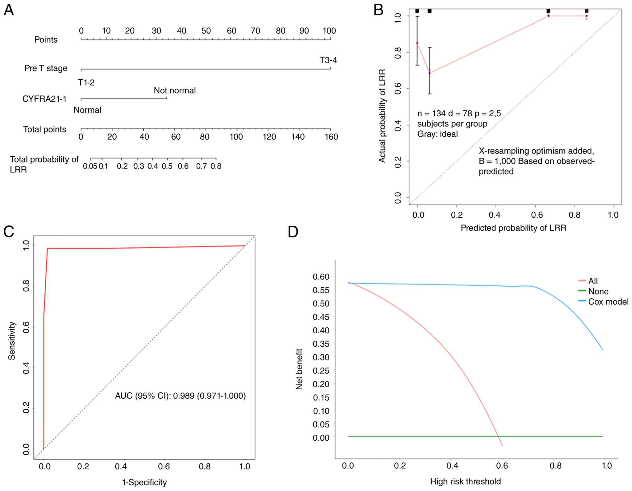
A nomogram predicting the cumulative incidence of
LRR was created by incorporating T stage and level of CYFRA21-1
after CCRT (Fig. 3A). The internal
validation showed that the nomogram had a concordance index of
0.986 (95% CI; 0.968–1.000) and was well calibrated (Fig. 3B). The area under the curve was
0.989 (95% CI; 0.971–1.000), sensitivity was 98.7% and specificity
was 98.2%, indicating that the model had strong discriminative
performance (Fig. 3C). The DCA
revealed that the net benefit rate of the clinical prediction model
was relatively high across all risk thresholds, indicating that the
model has high clinical practical value and performance (Fig. 3D).
Discussion
LRR is the most common mode of treatment failure
among patients with driver mutation-negative unresectable stage III
NSCLC treated with CCRT plus consolidation immunotherapy. The key
drivers of failure are tumor heterogeneity, subclinical residual
disease, insufficient local radiation dose, inherent
radioresistance of tumor cells and a defective antitumor immune
microenvironment (29–31). Currently, reliable tools to screen
individuals at risk of early LRR around the time of treatment
completion are lacking. Without risk stratification, standardized
one-size-fits-all protocols cannot achieve individualized
management. The present study investigated the patterns of
recurrence and risk factors for LRR in patients with driver
mutation-negative unresectable stage III NSCLC after CCRT. To the
best of our knowledge, this is the first large retrospective study
focusing on LRR status in this specific patient population.
The present study identified pretreatment bulky
tumors (T3-4) and persistently abnormal CYFRA21-1 levels after CCRT
as independent risk factors for LRR in patients with driver
mutation-negative unresectable stage III NSCLC after CCRT. These
findings are consistent with and extend existing evidence (32,33) on
LRR risk stratification in locally advanced NSCLC, providing
valuable insights into clinical decision-making.
Consistent with present findings, a previous study
demonstrated that the LRR rate remains as high as 40% in patients
with unresectable stage III NSCLC after receiving CCRT (34). Larger tumor volume or extensive
local invasion, such as chest wall or mediastinal invasion,
compromises the efficacy of CCRT. Mechanistically, bulky tumors are
prone to developing a hypoxic microenvironment, a well-recognized
mediator of radioresistance and chemoresistance (35). A total of 20–30% of radiotherapy and
chemotherapy treatment failures are attributed to tumor hypoxia
(36). Additional DNA repair
processes are triggered under hypoxic conditions. Hypoxia-inducible
factor 1 subunit α boosts the production of DNA-dependent protein
kinase, which is vital for repairing radiation-induced
double-strand breaks (36).
Additionally, T3-4 tumors often involve important anatomical
structures, such as the aorta or main bronchus. The definitive
radiation dose in standard CCRT is typically limited to 60–66 Gy to
avoid severe normal tissue toxicity, such as esophageal fistula,
which can compromise local disease control (8).
The detection of CYFRA21-1 in the blood is a
reliable indicator of NSCLC, and its concentration can be used to
monitor disease progression and treatment response. This biomarker
has high sensitivity and specificity for detecting NSCLC (37). In the present study, the level of
CYFRA21-1 gradually increased with lung cancer progression,
consistent with the results of a previous study (38). CYFRA21-1 has been reported to
notably positively correlate with stages I–III and negatively
correlate with stage IV (39). The
present study further confirmed that persistent abnormality of this
marker (>3.3 ng/ml) after CCRT independently predicted LRR (HR,
2.396; 95% CI, 1.296–4.430; P=0.005), which is highly consistent
with existing literature (39).
From a clinical practice perspective, CYFRA21-1 offers a distinct
advantage. Compared with CT or PET-CT, which may miss microresidual
lesions <1 cm, serial serum CYFRA21-1 testing (performed every 4
weeks) enables dynamic monitoring, thereby facilitating the
identification of high-risk patients before radiologically evident
recurrence.
Unlike previous studies that focused on risk factors
(40,41), such as N stage or programmed
death-ligand 1 expression, the present study is the first to
identify T3-4 tumors and persistently abnormal CYFRA21-1 levels
after CCRT as predictive risk factors for LRR through multivariate
analysis. The present study has notable clinical implications.
First, a visualized nomogram was developed to predict the risk of
LRR for driver mutation-negative unresectable stage III NSCLC after
CCRT. The model, constructed using easily obtainable clinical
variables, achieved favorable discrimination and calibration.
Second, this predictive tool supported individualized risk
stratification either before or immediately after CCRT, enabling
early identification of patients at risk of early LRR. Third, the
independent prognostic factors identified in the present analysis
can help improve clinicians understanding of the population
features associated with local treatment failure. This risk
stratification strategy may help clinicians implement
differentiated surveillance and therapeutic management for distinct
risk subgroups.
Intensified surveillance with shortened imaging
follow-up intervals is recommended for patients stratified as high
risk for LRR. Furthermore, these patients may be eligible for
innovative therapeutic strategies, such as local brachytherapy
combined with regional nodal IMRT and neoadjuvant chemotherapy
combined with immunotherapy. Brachytherapy offers unique and
complementary advantages in the multidisciplinary treatment
paradigm for NSCLC. A core strength of brachytherapy lies in its
steep dose gradient outside the target volume, thereby overcoming
the normal organ dose constraints associated with conventional
external beam radiotherapy within the thoracic cavity.
Endobronchial brachytherapy and radioactive seed implantation are
the primary brachytherapy modalities widely adopted in clinical
practice. Endobronchial brachytherapy can achieve curative intent
for early-stage tumors limited to the tracheobronchial lumen and
deliver effective palliative relief for patients with advanced
NSCLC complicated by central airway obstruction and refractory
hemoptysis (42). Intraoperative
seed implantation has been used as an adjuvant local therapy for
early NSCLC with high-risk surgical resection margins.
Particularly, CT-guided seed implantation assisted by 3D-printed
templates fulfills three critical clinical indications: Definitive
radical treatment for early-stage patients who cannot tolerate
surgical resection, a local dose-boost technique for locally
advanced lesions and salvage radiotherapy for recurrent tumors
after prior surgery or chemoradiotherapy failure (42). Consistent with these clinical merits
of interstitial brachytherapy, Xiang et al (8) conducted a phase II prospective trial
to assess the efficacy and safety of combined 3D interstitial HDR
brachytherapy and IMRT targeting regional metastatic lymph nodes in
patients with locally advanced NSCLC. During the entire follow-up
period, no grade ≥2 late radiation-related adverse events were
observed. The 2- and 5-year OS rates were 68.0 and 44.5%,
respectively. This combined regimen was associated with superior
survival outcomes and a favorable toxicity spectrum compared with
standard radiotherapy alone. Compared with previous trials
(43,44), the present study showed improved
survival and toxicity. Patients with any of the high-risk factors
(pretreatment T3-4 bulky tumors or persistently abnormal CYFRA21-1
levels after CCRT) may be considered for 3D interstitial HDR
brachytherapy.
Neoadjuvant chemotherapy combined with immunotherapy
may delay the administration of definitive radiotherapy. However,
systemic therapy marks the initiation of local treatment. In the
CheckMate-816 trial (45), the
pathological complete response rate reached 24% after 3 cycles of
nivolumab plus platinum-based chemotherapy, and the major
pathological response rate was 37%. In the AEGEAN (46), KEYNOTE-671 (47) and NADIM II studies (48), the pathological complete response
rates ranged from 18 to 37%, whereas the major pathological
response rates ranged from 30 to 53%. These findings indicate that
upfront systemic therapy does not compromise local control and
improves local efficacy in patients with unresectable disease.
Therefore, neoadjuvant therapy serves as one of the
‘efficacy-enhancing strategies’ for unresectable T3-4 stage III
NSCLC.
The present study has several limitations. First,
the retrospective single-center design may introduce selection
bias. The nomogram was only internally validated via bootstrap
resampling. Therefore, prospective multicenter external validation
is needed to verify its generalizability. Second, the prediction
model was constructed using routinely accessible clinical, imaging
and therapeutic variables. Peripheral blood biomarkers and
standardized physical status metrics were not included due to
limitations in the archived retrospective data. Incorporating these
parameters may enhance the performance of the model. Third, the
study population was limited to patients with driver
mutation-negative unresectable stage III NSCLC treated with CCRT
followed by consolidation immunotherapy, which may restrict
extrapolation to other NSCLC populations. Fourth, the optimal
CYFRA21-1 cutoff has not been established, and variability in
reference ranges across testing platforms may impede widespread
clinical application. Finally, genomic testing used a targeted
multigene PCR panel instead of whole-genome sequencing. Such
hotspot-focused assays fail to identify rare or unreported
structural variants outside the predefined gene regions. Further
studies with prospectively collected biomarker and physical status
data from subsequent cohorts are needed for external validation and
model refinement.
Collectively, the established nomogram and
identified risk factors facilitate risk stratification of LRR,
which may support individualized surveillance and optimized
therapeutic strategies for mutation-negative unresectable stage III
NSCLC after CCRT.
In conclusion, pretreatment T3-4 bulky tumors and
persistently abnormal CYFRA21-1 levels after CCRT are high-risk
factors for LRR in patients with driver mutation-negative
unresectable stage III NSCLC. High-risk patients may require
neoadjuvant therapy or 3D interstitial HDR brachytherapy to reduce
LRR. Further prospective studies are needed to validate these
findings and translate this risk stratification model into
effective interventions to improve survival outcomes.
Supplementary Material
Supporting Data
Acknowledgements
Not applicable.
Funding
The present study was supported by grants from the National
Natural Science Foundation of Chongqing (grant no.
CSTB2022NSCQ-MSX1239) and the National Reserve Talents Program in
the Field of Health of Chongqing (grant no. HBRC202404).
Availability of data and materials
The data generated in the present study may be
requested from the corresponding author.
Authors' contributions
YS and ZY were responsible for the study conception
and research design. ZY was involved in the establishment of
research framework and experimental workflows. JL and YQ performed
experimental verification and data reproducibility checks. JL
performed the statistical analysis and bioinformatics data
processing, the clinical sample collection and case data
acquisition. JL and YQ were responsible for dataset sorting,
cleaning and management. Code programming and computational tool
operation was performed by YQ. JL drafted the original manuscript
and revised the manuscript. YS provided overall study supervision
and project coordination and management. Funding was acquired by JL
and YS. YS and JL confirm the authenticity of all the raw data. All
authors read and approved the final version of the manuscript.
Ethics approval and consent to
participate
All experiments in the present retrospective study
were reviewed and approved by the Institutional Ethics Committee of
The Second Affiliated Hospital of Chongqing Medical University
[approval no. 2025(168)]. The Ethics Committee granted a formal
waiver of written informed consent from individual participants,
given that only retrospective archival clinical data were analyzed
in this research. The present work is an extended retrospective
sub-analysis under ethics approval no. 2025(168). While the
original ethical protocol primarily focused on stage IV
driver-negative NSCLC, the same treatment framework and archived
de-identified patient cohort also included unresectable stage III
cases, which were separately analyzed in the present study.
Patient consent for publication
Not applicable.
Competing interests
The authors declare that they have no competing
interests.
Glossary
Abbreviations
Abbreviations:
|
NSCLC
|
non-small cell lung cancer
|
|
CCRT
|
concurrent chemoradiotherapy
|
|
HR
|
hazard ratio
|
|
PET-CT
|
positron emission tomography-CT
|
|
DM
|
distant metastasis
|
|
OS
|
overall survival
|
|
LRR
|
locoregional recurrence
|
|
CYFRA21-1
|
cytokeratin fragment antigen 21-1
|
|
IMRT
|
intensity-modulated radiation
therapy
|
References
|
1
|
Bray F, Laversanne M, Sung H, Ferlay J,
Siegel RL, Soerjomataram I and Jemal A: Global cancer statistics
2022: GLOBOCAN estimates of incidence and mortality worldwide for
36 cancers in 185 countries. CA Cancer J Clin. 74:2292632024.
|
|
2
|
Mosmann MP, Borba MA, de Macedo FP,
Liguori Ade A, Villarim Neto A and de Lima KC: Solitary pulmonary
nodule and 18F-FDG PET/CT. Part 1: epidemiology, morphological
evaluation and cancer probability. Radiol Bras. 49:35–42. 2016.
View Article : Google Scholar : PubMed/NCBI
|
|
3
|
Remon J, Levy A, Gille R, Martel-Lafay I,
Bortolot M, Hendriks LEL, Faivre-Finn C, Leighl N, Reck M and Pérol
M: Unresectable stage III non-small-cell lung cancer: state of the
art and challenges. Nat Rev Clin Oncol. 23:22–39. 2026. View Article : Google Scholar : PubMed/NCBI
|
|
4
|
NCCN Clinical Practice Guidelines in
Oncology (NCCN Guidelines), . Non-Small Cell Lung Cancer. Version
2.2026.
|
|
5
|
Bradley JD, Hu C, Komaki RR, Masters GA,
Blumenschein GR, Schild SE, Bogart JA, Forster KM, Magliocco AM,
Kavadi VS, et al: Long-term results of NRG Oncology RTOG 0617:
Standard- versus high-dose chemoradiotherapy with or without
cetuximab for unresectable stage III non-small-cell lung cancer. J
Clin Oncol. 38:706–714. 2020. View Article : Google Scholar : PubMed/NCBI
|
|
6
|
De Ruysscher D, van Baardwijk A, Wanders
R, Hendriks LE, Reymen B, van Empt W, Öllers MC, Bootsma G, Pitz C,
van Eijsden L and Dingemans AC: Individualized accelerated isotoxic
concurrent chemo-radiotherapy for stage III non-small cell lung
cancer: 5-year results of a prospective study. Radiother Oncol.
135:141–146. 2019. View Article : Google Scholar : PubMed/NCBI
|
|
7
|
Goldstraw P, Chansky K, Crowley J,
Rami-Porta R, Asamura H, Eberhardt WE, Nicholson AG, Groome P,
Mitchell A, Bolejack V, et al: The IASLC lung cancer staging
project: Proposals for revision of the TNM stage groupings in the
forthcoming (eighth) edition of the TNM classification for lung
cancer. J Thorac Oncol. 11:39–51. 2016. View Article : Google Scholar : PubMed/NCBI
|
|
8
|
Xiang L, Ren PR, Li HX, Ye H, Pang HW, Wen
QL, Zhang JW, He LH, Shang CL, Yang BY, et al: Effect of
3-dimensional interstitial high-dose-rate brachytherapy with
regional metastatic lymph node intensity-modulated radiation
therapy in locally advanced peripheral non-small cell lung cancer:
5-year follow-up of a phase 2 clinical trial. Int J Radiat Oncol
Biol Phys. 115:347–355. 2023. View Article : Google Scholar : PubMed/NCBI
|
|
9
|
Wang Y, Zhang T, Huang Y, Li W, Zhao J,
Yang Y, Li C, Wang L and Bi N: Real-world safety and efficacy of
consolidation durvalumab after chemoradiation therapy for stage III
non-small cell lung cancer: A systematic review and meta-analysis.
Int J Radiat Oncol Biol Phys. 112:1154–1164. 2022. View Article : Google Scholar : PubMed/NCBI
|
|
10
|
Zhou Q, Chen M, Jiang O, Pan Y, Hu D, Lin
Q, Wu G, Cui J, Chang J, Cheng Y, et al: Sugemalimab versus placebo
after concurrent or sequential chemoradiotherapy in patients with
locally advanced, unresectable, stage III non-small-cell lung
cancer in China (GEMSTONE-301): Interim results of a randomised,
double-blind, multicentre, phase 3 trial. Lancet Oncol. 23:209–219.
2022. View Article : Google Scholar : PubMed/NCBI
|
|
11
|
Raben D, Rimner A, Senan S, Broadhurst H,
Pellas T, Dennis PA and Faivre-Finn C: Patterns of disease
progression with durvalumab in stage III non-small cell lung cancer
(PACIFIC). Int J Radiat Oncol Biol Phys. 105:6832019. View Article : Google Scholar
|
|
12
|
Catania C, Piperno G, Russo A, Greco C,
Agustoni F, Scotti V, Proto C, Sangalli C, Patani F, Santacaterina
A, et al: Open issues in the therapeutic management of unresectable
stage III NSCLC in the immunotherapy era. Crit Rev Oncol Hematol.
174:1036842022. View Article : Google Scholar : PubMed/NCBI
|
|
13
|
Machtay M, Paulus R, Moughan J, Komaki R,
Bradley JE, Choy H, Albain K, Movsas B, Sause WT and Curran WJ:
Defining local-regional control and its importance in locally
advanced non-small cell lung carcinoma. J Thorac Oncol. 7:716–722.
2012. View Article : Google Scholar : PubMed/NCBI
|
|
14
|
Machtay M, Bae K, Movsas B, Paulus R, Gore
EM, Komaki R, Albain K, Sause WT and Curran WJ: Higher biologically
effective dose of radiotherapy is associated with improved outcomes
for locally advanced non-small cell lung carcinoma treated with
chemoradiation: An analysis of the Radiation Therapy Oncology
Group. Int J Radiat Oncol Biol Phys. 82:425–434. 2012. View Article : Google Scholar : PubMed/NCBI
|
|
15
|
Videtic GM, Stephans KL, Woody NM, Reddy
CA, Zhuang T, Magnelli A and Djemil T: 30 Gy or 34 Gy? Comparing 2
single-fraction SBRT dose schedules for stage I medically
inoperable non-small cell lung cancer. Int J Radiat Oncol Biol
Phys. 90:203–208. 2014. View Article : Google Scholar : PubMed/NCBI
|
|
16
|
Bradley JD, Paulus R, Komaki R, Masters G,
Blumenschein G, Schild S, Bogart J, Hu C, Forster K, Magliocco A,
et al: Standard-dose versus high-dose conformal radiotherapy with
concurrent and consolidation carboplatin plus paclitaxel with or
without cetuximab for patients with stage IIIA or IIIB
non-small-cell lung cancer (RTOG 0617): A randomised, two-by-two
factorial phase 3 study. Lancet Oncol. 16:187–199. 2015. View Article : Google Scholar : PubMed/NCBI
|
|
17
|
Kong FM, Ten Haken RK, Schipper M, Frey
KA, Hayman J, Gross M, Ramnath N, Hassan KA, Matuszak M, Ritter T,
et al: Effect of midtreatment PET/CT-adapted radiation therapy with
concurrent chemotherapy in patients with locally advanced
non-small-cell lung cancer: A phase 2 clinical trial. JAMA Oncol.
3:1358–1365. 2017. View Article : Google Scholar : PubMed/NCBI
|
|
18
|
Schild SE, Hillman SL, Tan AD, Ross HJ,
McGinnis WL, Garces YA, Graham DL, Adjei AA and Jett JR; Mayo
Clinic and North Central Cancer Treatment Group, : Long-term
results of a trial of concurrent chemotherapy and escalating doses
of radiation for unresectable non-small cell lung cancer: NCCTG
N0028 (Alliance). J Thorac Oncol. 12:697–703. 2017. View Article : Google Scholar : PubMed/NCBI
|
|
19
|
Rodrigues G, Oberije C, Senan S, Tsujino
K, Wiersma T, Moreno-Jimenez M, Kim TH, Marks LB, Rengan R, De
Petris L, et al: Is intermediate radiation dose escalation with
concurrent chemotherapy for stage III non-small-cell lung cancer
beneficial? A multi-institutional propensity score matched
analysis. Int J Radiat Oncol Biol Phys. 91:133–139. 2015.
View Article : Google Scholar : PubMed/NCBI
|
|
20
|
Kong FM, Hu C, Ten Haken R, Xiao Y,
Matuszak M, Hirsh V, Pryma DA, Siegel BA, Gelblum DY, Hayman J, et
al: NRG-RTOG 1106/ACRIN 6697: A phase IIR trial of standard versus
adaptive (mid-treatment PET based) chemoradiotherapy for stage III
NSCLC-Results and comparison to NRG-RTOG 0617 (non-personalized RT
dose escalation). J Clin Oncol. 39 (Suppl 15):S85482021. View Article : Google Scholar
|
|
21
|
Amin MB, Edge SB, Greene FL, Byrd DR,
Brookland RK, Washington MK, Gershenwald JE, Compton CC, Hess KR,
Sullivan DC, et al: AJCC cancer staging manual. 8th edition.
Springer Verlag; New York: 2017, https://doi.org/10.3322/caac.21388
|
|
22
|
Guangzhou Mygene Medical Technology
Co.Ltd., . Detection primer, method and the kit of 29 kinds of EGFR
genetic mutations. China Patent CN108866186A. Filed June 12, 2018;
issued November 15. 2022.
|
|
23
|
Guangzhou Mygene Medical Technology
Co.Ltd., . ALK gene fusion detection primer, method and kit. China
Patent CN108728538B. Filed May 31, 2018; issued September 28.
2021.
|
|
24
|
Guangzhou Mygene Medical Technology
Co.Ltd., . ROS1 gene fusion detection primer, method and kit. China
Patent CN108531598B. Filed May 7, 2018; issued November 30.
2021.
|
|
25
|
Guangzhou Mygene Medical Technology
Co.Ltd., . Primer, system and kit for MET gene amplification
detection. China Patent CN110863051B. Filed December 13, 2019;
issued October 23. 2020.
|
|
26
|
Eisenhauer EA, Therasse P, Bogaerts J,
Schwartz LH, Sargent D, Ford R, Dancey J, Arbuck S, Gwyther S,
Mooney M, et al: New response evaluation criteria in solid tumours:
revised RECIST guideline (version 1.1). Eur J Cancer. 45:228–247.
2009. View Article : Google Scholar : PubMed/NCBI
|
|
27
|
Ettinger DS, Wood DE, Aisner DL, Akerley
W, Bauman JR, Bharat A, Bruno DS, Chang JY, Chirieac LR, DeCamp M,
et al: NCCN guidelines ® insights: Non-small cell lung
cancer, version 2.2023. J Natl Compr Canc Netw. 21:340–350. 2023.
View Article : Google Scholar : PubMed/NCBI
|
|
28
|
Guo W, Zhang T, Li R, Chen X, Pang J, Bao
H, Wu X, Shao Y, Qiu B, Gao S and He J: Molecular risk factors for
locoregional recurrence in resected non-small cell lung cancer.
Cancer Med. 12:15026–15036. 2023. View Article : Google Scholar : PubMed/NCBI
|
|
29
|
Shi X and Wang X, Yao W, Shi D, Shao X, Lu
Z, Chai Y, Song J, Tang W and Wang X: Mechanism insights and
therapeutic intervention of tumor metastasis: latest developments
and perspectives. Signal Transduct Target Ther. 9:1922024.
View Article : Google Scholar : PubMed/NCBI
|
|
30
|
Han Y, Ma L and Ma X: Tumor cells escape
from therapy-induced senescence: From molecular mechanisms to
targeted intervention. Biol Direct. Jul 18–2026.(Epub Online ahead
of print). View Article : Google Scholar
|
|
31
|
Shi Z, Hu C and Sun C: Radiation-Induced
alterations in cancer-associated fibroblasts: Drivers of tumor
radioresistance and therapeutic targets. Biomolecules. 16:7772026.
View Article : Google Scholar : PubMed/NCBI
|
|
32
|
Koh HW, Jung JJ, Kim HK, Moon HG, Han W,
Kim EK, Lee HB and Shin HC: Tumor volume as a predictor of
recurrence-free survival, but not overall survival, in early breast
cancer. Eur J Surg Oncol. 51:1100242025. View Article : Google Scholar : PubMed/NCBI
|
|
33
|
Xu S, Hu T, Zhou C, Wang M, Jiang J, Wang
Y and Wang C: The ratio of lymph node eluate/serum cytokeratin 21-1
is a potential predictor of lymph node metastasis in lung
adenocarcinoma. J Thorac Dis. 16:5709–5717. 2024. View Article : Google Scholar : PubMed/NCBI
|
|
34
|
Yan LF and Wu Y: Current status and
prospects of treatment for locally advanced non-small cell lung
cancer. Oncology Progress. 19:1957–1973. 2021.(In Chinese).
|
|
35
|
Devi D, Sharma K, Khan T, Rani H, Rana R,
Sharma H, Kaur P, Prajapati L, Rizwan A, Allieu M, et al:
Hypoxia-driven metabolic and molecular reprogramming: From tumor
microenvironment to therapeutic interventions. Pathol Res Pract.
276:1562712025. View Article : Google Scholar : PubMed/NCBI
|
|
36
|
Rakotomalala A, Escande A, Furlan A,
Meignan S and Lartigau E: Hypoxia in solid tumors: How low
oxygenation impacts the ‘Six Rs’ of radiotherapy. Front Endocrinol
(Lausanne). 12:7422152021. View Article : Google Scholar : PubMed/NCBI
|
|
37
|
Garcia-Valdecasas Gayo S, Ruiz-Alvarez MJ,
Gonzalez Gay D, Ramos-Corral R, Marquez-Lietor E, Del Amo N, Plata
MDC, Guillén-Santos R, Arribas I and Cava-Valenciano F: CYFRA 21-1
in patients with suspected cancer: Evaluation of an optimal cutoff
to assess the diagnostic efficacy and prognostic value. Adv Lab
Med. 1:202000052020.PubMed/NCBI
|
|
38
|
Ito M, Ariyasu R, Fujishima S, Iso H,
Nibuya K, Nemoto T, Sugai M, Tsugitomi R, Amino Y, Uchibori K, et
al: Prognostic value of serum CYFRA21-1 in locally advanced
non-small cell lung cancer undergoing durvalumab consolidation
therapy after chemoradiotherapy. Lung Cancer. 217:1094282026.
View Article : Google Scholar : PubMed/NCBI
|
|
39
|
Liu TC, Zheng MH, Zeng XY, Kang R,
Bahabayi A, Tuerhanbayi B, Lu SS and Liu C: Imbalance of
circulating follicular regulatory and follicular helper T cell
subpopulations is associated with disease progression and serum
CYFRA 21-1 levels in patients with non-small cell lung cancer. Curr
Med Sci. 44:102–109. 2024. View Article : Google Scholar : PubMed/NCBI
|
|
40
|
Kim S, Kim IH, Kim JY, Yun JK, Choi S, Kim
HR, Kim YH, Kim DK, Park SI and Lee GD: Refining N1 classification:
Prognostic validation of N1 subdivision in resected non-small cell
lung cancer. Lung Cancer. 218:1094742026. View Article : Google Scholar : PubMed/NCBI
|
|
41
|
Liu C, Yang G, Wang Z, Sun H, Fu C, Han D,
Zhao Q and Huang W: Concurrent chemoradiotherapy with or without
simultaneous integrated boost for unresectable stage III
non-small-cell lung cancer: A randomized phase II trial with
exploratory EGFR and PD-L1 analyses. Cancer Genet. 306-307:219–229.
2026. View Article : Google Scholar : PubMed/NCBI
|
|
42
|
Qiu B, Jiang P, Ji Z, Huo X, Sun H and
Wang J: Brachytherapy for lung cancer. Brachytherapy. 20:454–466.
2021. View Article : Google Scholar : PubMed/NCBI
|
|
43
|
Heinzerling JH, Mileham KF, Robinson MM,
Symanowski JT, Induru RR, Prabhu RS, Gant DA, Pal SE, Haggstrom DE,
Kim ES, et al: Phase II prospective trial of primary lung tumor
stereotactic body radiation therapy (SBRT) followed by concurrent
mediastinal chemoradiation and adjuvant immunotherapy for
locallyadvanced nonsmall cell lung cancer (NSCLC): Planned toxicity
analysis. J Clin Oncol. 38:e210472020. View Article : Google Scholar
|
|
44
|
Hepel JT, Leonard KL, Safran H, Ng T,
Taber A, Khurshid H, Birnbaum A; Brown University Oncology Research
Group; Wazer DE, ; DiPetrillo T: Stereotactic body radiation
therapy boost after concurrent chemoradiation for locally advanced
nonsmall cell lung cancer: A phase 1 dose escalation study. Int J
Radiat Oncol Biol Phys. 96:102110272016. View Article : Google Scholar
|
|
45
|
Mayenga M, Pedroso AR, Ferreira M, Gille
T, Pereira Catarata MJ and Duchemann B: The CheckMate 816 trial: a
milestone in neoadjuvant chemoimmunotherapy of nonsmall cell lung
cancer. Breathe (Sheff). 20:2400442024. View Article : Google Scholar : PubMed/NCBI
|
|
46
|
Heymach JV, Mitsudomi T, Harpole D,
Aperghis M, Jones S, Mann H, Fouad TM and Reck M: Design and
rationale for a phase III, double-blind, placebo-controlled study
of neoadjuvant durvalumab+chemotherapy followed by adjuvant
durvalumab for the treatment of patients with resectable stages II
and III non-small-cell lung cancer: The AEGEAN trial. Clin Lung
Cancer. 23:e247–e251. 2022. View Article : Google Scholar : PubMed/NCBI
|
|
47
|
Spicer JD, Gao S, Liberman M, Kato T,
Tsuboi M, Lee S-H, Chen K-N, Dooms C, Majem M, Eigendorff E, et al:
LBA56 Overall survival in the KEYNOTE-671 study of perioperative
pembrolizumab for early-stage non-small-cell lung cancer (NSCLC).
Ann Oncol. 34 (Suppl 2):S1297–S1298. 2023. View Article : Google Scholar
|
|
48
|
Provencio M, Nadal E, Insa A, García
Campelo R, Casal J, Dómine M, Massuti B, Majem M, Rodríguez-Abreu
D, Martínez-Martí A, et al: Perioperative chemotherap y and
nivolumab in non-small-cell lung cancer (NADIM): 5-year clinical
outcomes from a multicentre, single-arm, phase 2 trial. Lancet
Oncol. 25:1453–1464. 2024. View Article : Google Scholar : PubMed/NCBI
|