Introduction
Cervical cancer (CC) is one of the main causes of
cancer mortalities worldwide. Although the incidence of CC
continues to decline with the widespread vaccination of human
papillomavirus (HPV) vaccines and the popularization of CC
screening, there are currently >600,000 novel diagnoses and
340,000 mortalities worldwide each year, posing a serious threat to
the physical and mental health (1).
The progression from cervical intraepithelial neoplasia to invasive
CC is regulated by genes involved in mitosis, DNA repair, cell
proliferation, apoptosis and angiogenesis (2). However, >20% of patients experience
recurrence (3). However, the
specific mechanism that leads to the progression of CC remains to
be elucidated and to the best of our knowledge, there are currently
no effective biomarkers for the diagnosis, prognosis and recurrence
of CC. Although squamous cell carcinoma (SCC) antigen is a commonly
used CC marker, its specificity and sensitivity are not high,
particularly close monitoring of local recurrence in low-level
patients (normal range of SCC antigen; reference range, <1.5
ng/ml) is necessary (4,5), Therefore, there is an urgent need to
search for novel diagnostic and prognostic markers for CC in
clinical practice.
Ferroptosis is an iron dependent cell death
characterized by the accumulation of intracellular lipid peroxides
and mitochondrial reactive oxygen species (ROS). Previous studies
have demonstrated that ferroptosis serves a key role in a variety
of malignant tumors, such as pancreatic cancer and liver cancer
(6–9). The induction of ferroptosis is
gradually becoming a novel tumor mode in tumor cells and exploring
novel diagnostic and therapeutic methods may be beneficial. Xia
et al (10) identified that
an iron death inducer, talaroconvolin A, promotes iron death by
increasing ROS and upregulating the expression level of
arachidonate epidermal lipoxygenase 3. In addition, Wang et
al (11) reported that
LINC00336 is upregulated in cancer and reversed by inhibiting
ferroptosis. Other previous studies reported that ferroptosis
imbalance between tumor-associated macrophages and CC cells
involved in the progression of CC and resistance to cancer
chemotherapy and targeted therapy in CC (12,13).
Wang et al (14) reported
that the mitochondrial carrier 1 (MTCH1)-FOXO1-glutathione
peroxidase 4 (GPX4) signaling pathway was identified and it was
proposed that MTCH1 deficiency inhibits FOXO1 activation, leading
to downregulation of GPX4 transcription and ROS accumulation,
ultimately triggering iron death in CC cells. Chen et al
(15) demonstrated that the
combination of deferoxamine and oxaliplatin can reduce oxaliplatin
resistance in CC cells. These studies indicate that exploring the
mechanisms of ferroptosis is a potential target for the diagnosis
and treatment of CC in the future.
The present study screened differentially expressed
genes (DEGs) associated with ferroptosis in CC and adjacent tissues
using The Cancer Genome Atlas (TCGA) database. Further analysis of
the roles of stearoyl-CoA desaturase (SCD), transferrin receptor
(TFRC) and carbonic anhydrase 9 (CA9) in the prognosis and immune
infiltration of CC was performed, providing prognostic candidate
biomarkers for the diagnosis and immunotherapy of CC.
Materials and methods
Data preprocessing
Clinical phenotype and RNA-sequencing (seq) data
were obtained by searching cervical cancer in TCGA database
(https://portal.gdc.cancer.gov/projects/TCGA-CESC)
and transcript levels were estimated by the University of
California Santa Cruz Xena
[https://xenabrowser.net/datapages/?cohort=TCGA Cervical Cancer
(CESC)] platform (16). The present
study screened CC as the primary tumor and grouped it according to
cancer and para-cancer tissues, recording basic clinical and
pathological information of patients in the screening data.
TCGA data analysis
The expression difference P-value and the expression
fold-change (FC) value were calculated using the R package (version
3.2.1) DESeq2 (version 1.8.1) (17)
in and using adjusted P<0.05 and |log2FC|>1
(2-fold relationship), screened for DEGs. The differential heat map
and volcano map were further analyzed using the R package ‘heatmap’
(version 1.0.12; R Development Core Team) (18).
Gene ontology (GO) and kyoto
encyclopedia of genes and genomes (KEGG) analysis
The genes associated with ferroptosis were
downloaded, including ferroptosis driver, ferroptosis marker and
ferroptosis suppressor genes using the Ferroptosis database
(zhounan.org/FerrDb) (19). A Venn
diagram was drawn through the intersection of DEGs and ferroptosis
by using the R package ‘Venn diagram’ (version 1.6.20) (20). KEGG enrichment analysis was further
performed on cross genes using R package ‘Venn plots’ (version
1.6.20; significance threshold set to P≤0.05) (21) and bubble plots were created for
visualization using R package ‘ggplot2’ (version 3.3.3; R
Development Core Team) (22).
Gene set enrichment analysis
(GSEA)
GSEA was performed using the GenePattern tool with
version of 5.2 of the Molecular Signature Database (MSigDB;
broadinstitute.org/gsea/msigdb/index.jsp) (23). The GSEA involved estimating the
significance of enrichment scores (ES), correcting multiple
hypothesis tests and evaluating the significance of ES.
Gene set variation analysis
(GSVA)
Using the c2.cp.kegg. v7.2.symbols.gmt gene set
data, the CC data were transformed into ES values for GSVA using
R-package ‘gene set variation analysis’ (GSVA; version 1.34.0)
(24). Further differential
analysis was performed on the ES value matrix using the ‘limma’ R
software package (version 3.42.2; R Development Core Team) and
lastly, using the differential gene set, volcano and heat maps were
constructed to identify the key pathways of DEGs.
Protein-protein interaction (PPI)
network construction
The Search Tool for the Retrieval of Interacting
Genes/Proteins database (STRING-db.org/) was used to search for
interacting genes (25). The
database analyzed the PPI between CC and iron death-related genes
and set the required confidence level (comprehensive score) of the
PPI threshold to >0.15. A PPI network was drawn using Cytoscape
software (3.7.1) (26) and the top
30 key genes were extracted for subsequent analysis using the
‘Cytohubba’ plugin (27). Lastly,
ferroptosis-related key genes that affect the prognosis of CC were
identified (Fig. 1).
 | Figure 1.Schematic diagram of
ferroptosis-related key genes and pathways that affect the
prognosis of cervical cancer. DEGs, differentially expressed genes;
GSEA, Gene Set Enrichment Analysis; GO, Gene Ontology; KEGG, Kyoto
Encyclopedia of Genes and Genomes; PPI, protein-protein
interaction; SCD, stearoyl-CoA desaturase; TFRC, transferrin
receptor; CA9, carbonic anhydrase 9; OS, overall survival; DSS,
disease specific survival; PFS, progression-free survival. |
Prognosis and clinical association
analysis
RNA-seq spectra and clinical information of patients
with CC were obtained from TCGA to analyze the relationship with
the prognosis of patients with SCD, TFRC and CA9 expression using
the Kaplan-Meier method and log-rank test.
Database analysis
RNA-seq spectra and clinical information from
patients with CC were downloaded from TCGA database (https://portal.gdc.cancer.gov/projects/TCGA-CESC)
by searching cervical cancer to determine their association with
clinical case characteristics and prognosis of CC. The images of
human expression data were obtained from the Human Protein Atlas
database (proteinatlas.org/). SWISS-MODEL (28) was employed to construct a 3D
structure for SCD, TFRC and CA9 using homology modeling. The
sequence of SCD, TFRC and CA9 (UniProt: Q00417) was obtained from
the UniProt (29).
Immune infiltration analysis of
prognostic genes
The Cell-type Identification By Estimating Relative
Subsets Of RNA Transcripts (CIBERSORT) method (30), which is a computational approach
based on gene expression data used to quantify the relative
proportions of different immune cell subpopulations in tumor
tissues, was used for immune infiltration analysis of prognostic
genes. After standardizing the prognostic gene data associated with
CC necrosis, a signature matrix containing specific cell type
marker genes was then selected or constructed. Subsequently, the
gene expression data and signature matrix were input into CIBERSORT
software (V2.0.0) for computational analysis, estimating the
abundance of various immune cell subpopulations in the sample to
analyze the immune infiltration of the tumor.
Patients and sample preparation
The present study was approved by the Ethics
Committee of Tongji Medical College, Huazhong University of Science
and Technology (approval no. 2021-S353; Wuhan, China), and written
informed consent was obtained from all patients. A total of three
cases of CC tissue and adjacent tissues were obtained from Union
Hospital, Tongji Medical College, Huazhong University of Science
and Technology from September to December 2023 from patients who
underwent surgery before initiating antitumor therapy and who did
not receive radiotherapy, chemotherapy or antiangiogenic treatment.
The preparation of RNA was performed as previously described
(31).
RNA extraction and fluorescence
reverse transcription-quantitative PCR (RT-qPCR)
For RT-qPCR, total RNA was extracted by lysing the
aforementioned tissues in 1 ml RNAiso Plus reagent (cat. no. 9109;
Takara Biotechnology Co., Ltd.), ensuring complete homogenization.
Following centrifugation (12,000 × g; 4°C; 15 min), the supernatant
was removed and the RNA pellet was washed with 75% ethanol to
improve purities. RNA extraction and subsequent reverse
transcription were performed according to the manufacturer's
protocols of the PrimeScript™ RT Master Mix (cat. no.
RR036A; Takara Biotechnology Co., Ltd.) and TB Green®
Premix Ex Taq™ II (cat. no. RR820A; Takara Biotechnology
Co., Ltd.) kits. qPCR was carried out on the 7500 Real-Time PCR
System (cat. no. RR820A; Takara Biotechnology Co., Ltd.) using
standard operational guidelines. The conditions for RT-qPCR were as
follows: 95°C for 10 min, 40 cycles of 94°C for 30 sec, 60°C for 30
sec and 72°C for 30 sec, and a final extension of 10 min at 72°C.
Relative mRNA expression levels were quantified using the
2−ΔΔCq method (32) and
normalized against GAPDH as an internal control. The primer
sequences used for RT-qPCR are shown in Table I.
 | Table I.Primer sequences used for
RT-qPCR. |
Table I.
Primer sequences used for
RT-qPCR.
| Gene | Primer sequence
(5′-3′) |
|---|
| SCD | F:
CTACACAACCACCACCACCATCAC |
|
| R:
CTCTCATTTCAGGGCGGATGTCTTC |
| TFRC | F:
GGCTACTTGGGCTATTGTAAAGG |
|
| R:
CAGTTTCTCCGACAACTTTCTCT |
| CA9 | F:
CATCCTAGCCCTGGTTTTGG |
|
| R:
CCTTCTGTGCTGCCTTCTCAT |
| GAPDH | F:
GGTGTGAACCATGAGAAGTATGA |
|
| R:
GAGTCCTTCCACGATACCAAAG |
Prediction of drug-gene
interactions
Firstly, genes or gene products reported to interact
with TKIs were identified in the drug-gene interaction database
(DGIdb; v2.22; http://www.dgidb.org/). DGIdb
integrates existing resources to generate hypotheses on how mutant
genes may be targeted for therapy, which provides an interface for
a general search of genes for drug-gene interactions and
potentially drug-available genes (33). A large amount of drug-gene
interaction information was aggregated from different data sources,
providing data on the association between specific gene variations
and drug responses, which helps researchers quickly identify
potential drug targets and biomarkers. The prognostic genes
associated with CC necrosis were imported into DGIdb for analysis
to explore drugs or small molecule organic compounds associated
with it.
Cell culture
The human CC cell lines HeLa (cat. no. GDC0001) and
SiHa (GDC0042) were purchased from the China Center for Type
Culture Collection and cultured in RPMI-1640 medium (Gibco; Thermo
Fisher Scientific, Inc.) containing 10% FBS (both Gibco; Thermo
Fisher Scientific, Inc.). HeLa is a human cervical adenocarcinoma
cell line and SiHa is a human SCC cell line. All cells were
authenticated using short tandem repeat analysis and maintained in
a humid atmosphere at 37°C and 5% CO2.
Small-interfering RNA (siRNA)
transfection
The TFRC siRNA (forward:
5′-GGAUAUGGGUCUAAGUCUATT-3′; reverse: 5′-UAGACUUAGACCCAUUCTT-3′).
The cells were incubated for 48 h following transfection at 37°C.
and a universal negative control siRNA (forward,
5′-AAGACGUACGAGCUACUGCtt-3′; reverse, 5′-GCAGUAGCUCGUACGUCUUtt-3′;
cat. no. SIC001; Beijing Tsingke Biotech Co., Ltd.) were used for
transient silencing in CC cells (MilliporeSigma; Merck KGaA).
Lipofectamine® 3000 (Invitrogen; Thermo Fisher
Scientific, Inc.) was used for transient transfection with a final
siRNA concentration of 32 nM for 48 h. The transfected cells were
harvested and used for subsequent experiments.
Cell proliferation detection
The MTT assay kit (Beyotime Biotechnology) was used
to assess cell proliferation. Logarithmic growth phase cells were
seeded at a density of 1,000 cells per well in a 96-well plate,
cultured for 24, 48 and 72 h. The MTT solution (0.5 mg/µl) was
added and the plate was incubated at 37°C for 4 h. Subsequently,
100 µl of DMSO was added to separate the crystals and the plate was
stirred at 37°C for 20 min. The absorbance was measured at 490 nm
using an ELISA reader.
Wound healing and Transwell
assays
For the wound healing assay, cervical cancer cells
were seeded in a 6-well plate at a density of 3×10⁵
cells per well. After transfection with siRNA, the cells were
cultured for 48 h at 37°C. When the cell confluence reached 90–100%
and a dense monolayer was formed, the wound healing assay was
performed. The cell monolayers were gently scratched using a 200-µl
pipette tip and the cells were incubated in serum-free RPMI-1640
medium (Gibco; Thermo Fisher Scientific, Inc.) for 24 h at 37°C. To
remove any detached cells, the wells of the plate were gently
washed with fresh medium. The distance between edges of the wound
was measured under a light microscope (Olympus Corporation), and
multiple visual fields were selected for observing each well. After
24 h, the wound channel distance was measured again for analysis.
The wound area was measured using ImageJ software (version 1.48;
National Institutes of Health). Wound healing assay results were
presented as migration rate (%)=(initial wound area-wound area at
24 h)/initial wound area ×100.
For the Transwell assay, 200 µl serum-free medium
containing 5×10⁴ cells was seeded onto the upper surface
of the chamber without coating (migration assay) and 600 µl
complete medium was added to the lower chamber. After incubation at
37°C for 24 h, the cells were fixed by passing them through an 8 µm
pore membrane with 4% paraformaldehyde at 37°C for 30 min. Then,
the cells were stained with 0.1% crystal violet at 37°C for 5 min.
The number of cells in five random fields of view were counted for
each inserted segment to evaluate the migration of cancer cells at
a ×200 magnification. Each experiment was performed three
times.
Western blotting
The total protein was extracted as previously
described and protein blotting was performed (34). The primary antibodies used were
rabbit anti-TFRC (1:1,000; ProteinTech Group, Inc.; cat. no.
84766-4-RR) and mouse anti-GAPDH (1:1,000; Cell Signaling
Technology, Inc.; cat. no. #2118). GAPDH was used as the control.
The bands were quantified using Image J software (version 1.51;
National Institutes of Health).
Statistical analysis
R software (version 3.6.3; R Development Core Team)
was used to perform the statistical analyses and data were
presented as the mean ± SD. The expression levels of selected top
30 key genes and phenotypes (such as age, tumor stage, tumor
differentiation and lymph node metastasis) were analyzed using
univariate and multivariate Cox regression analyses. The median
expression level of the genes in all samples was used as the
boundary to divide patients into groups (above median, high
expression group; below median, low expression group). The
Kaplan-Meier method and log-rank test were used to analysis the
overall survival, disease-specific survival and progression-free
survival of patients with CC. Univariate and multivariate Cox
regression analyses were performed to identify survival-associated
genes. The association between SCD, TFRC and CA9 expression and
clinicopathological features were evaluated using the paired
Student's t-test test after a normality test. Spearman correlation
coefficients were calculated to assess the associations between
SCD, TFRC and CA9 expression and the infiltration levels of 24
immune cell types. Significant differences between different groups
were evaluated using Student's t-tests. P<0.05 was considered to
indicate a statistically significant difference and all statistical
tests were two-sided.
Results
Differences in TCGA data of CC
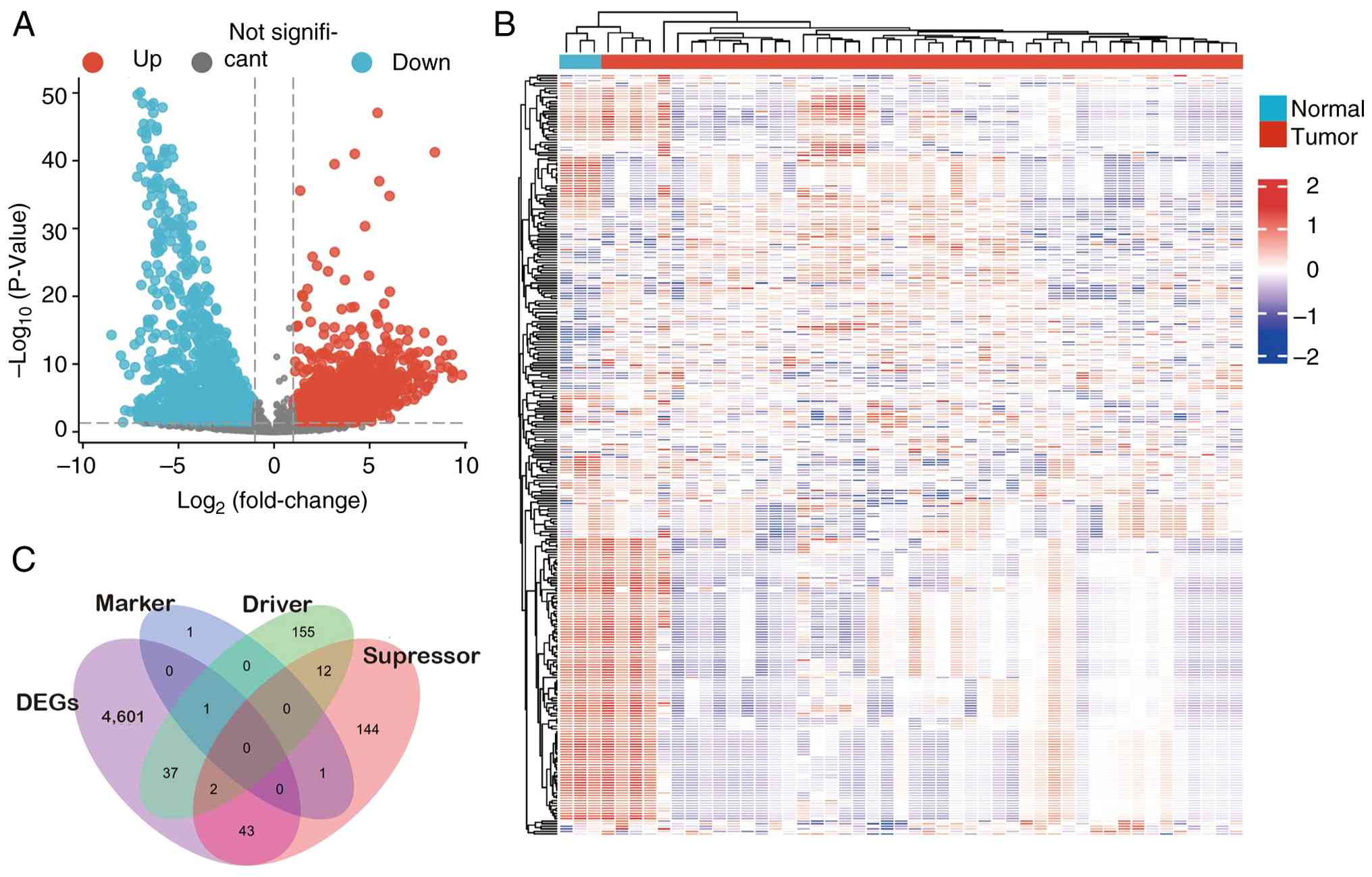
There was a total of 309 samples of CC and adjacent
RNA-seq data, including 3 samples of normal cervical tissue and 306
samples of CC tissue from TCGA. A total of 20,532 genes were
screened, including 4,683 DEGs (2,563 upregulated and 2,120
downregulated) (Fig. 2A and B). A
total of 214 human ferroptosis genes (driver, suppressor and marker
genes) were screened out. By screening for cross genes between DEGs
and ferroptosis genes, 80 potential CC ferroptosis genes were
ultimately obtained (Fig. 2C).
Cluster analysis
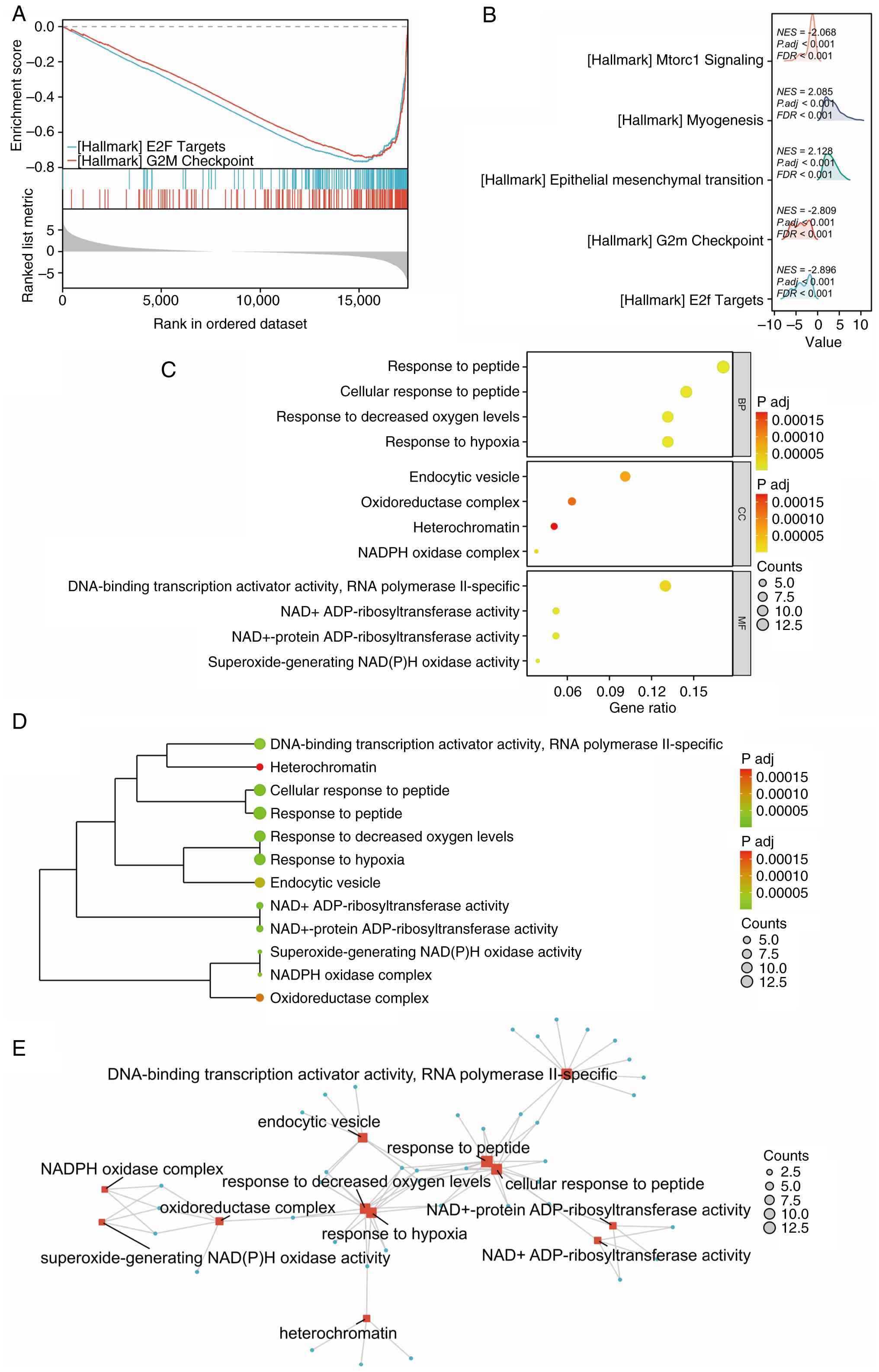
GSEA in TCGA cohort identified that the early region
2 binding factor (E2F) targets and G2M checkpoint were
significantly enriched using the DEGs between CC and cervical
paracancerous tissue (Fig. 3A). The
mountain map indicated that it is mainly enriched in ‘Mtorc1
signaling’, ‘myogenesis’, ‘Epithelial mesenchymal transition’, ‘E2F
targets’ and ‘G2M checkpoint’ between CC and normal
cervical tissue (Fig. 3B). GO and
KEGG enrichment (GSEA) analyses of 80 potential ferroptosis-related
genes of CC were performed and revealed that ‘response to peptide’,
‘response to hypoxia’ and ‘DNA-binding transcription activator
activity, RNA polymerase II-specific’ were significantly enriched.
(Fig. 3C-E).
PPI network
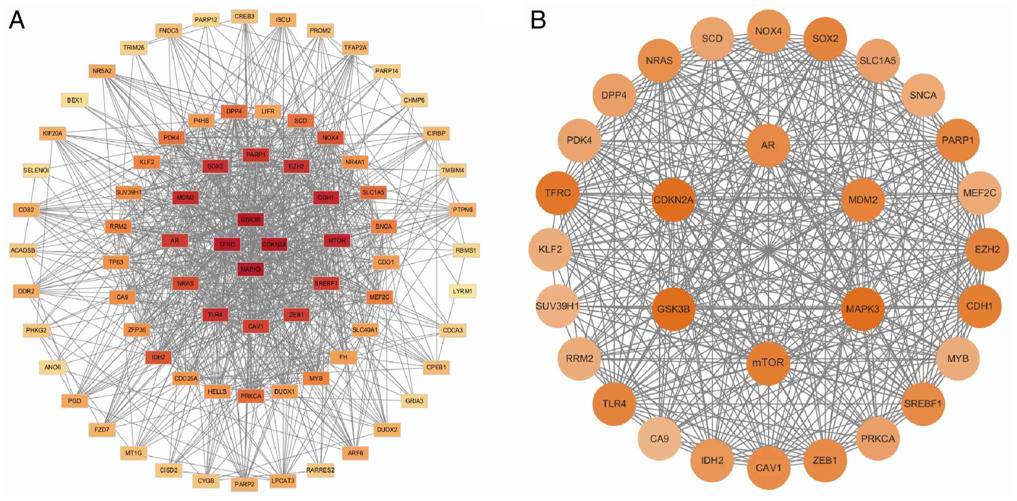
The PPI network analysis was performed on 80 key
genes associated with ferroptosis in CC and revealed that there
were a total of 911 interaction relationships (Fig. 4A). Among these genes, 30 genes were
confirmed as hub genes, which were murine double minute 2 (MDM2),
androgen receptor (AR), CDK inhibitor 2A (CDKN2A), GSK-3β,
neuroblastoma Ras viral oncogene homolog (NRAS), pyruvate
dehydrogenase kinase 4 (PDK4), Krüppel-like factor 2 (KLF2),
suppressor of variegation 3–9 homolog 1 (SUV39H1), toll-like
receptor 4 (TLR4), isocitrate dehydrogenase 2 (IDH2), caveolin-1
(CAV1), sterol regulatory element-binding transcription factor 1
(SREBF1), myeloblastosis (MYB), cadherin-1 (CDH1), myocyte-specific
enhancer factor 2C (MEF2C), MAPK3, solute carrier family 1 member 5
(SLC1A5), nuclear receptor subfamily 4 group A member 1 (NR4A1),
NADPH oxidase 4 (NOX4), SCD, dipeptidyl peptidase 4 (DPP4), TFRC,
CA9, zinc finger enhancer-box-binding homeobox 1 (ZEB1), protein
kinase C α (PRKCA), mTOR, enhancer of zeste homolog 2 (EZH2),
poly(ADP-ribose) polymerase 1 (PARP1), synuclein α (SNCA) and Sox2
(Fig. 4B).
Cancer and pan-cancer analysis
The prognostic analysis of the aforementioned 30
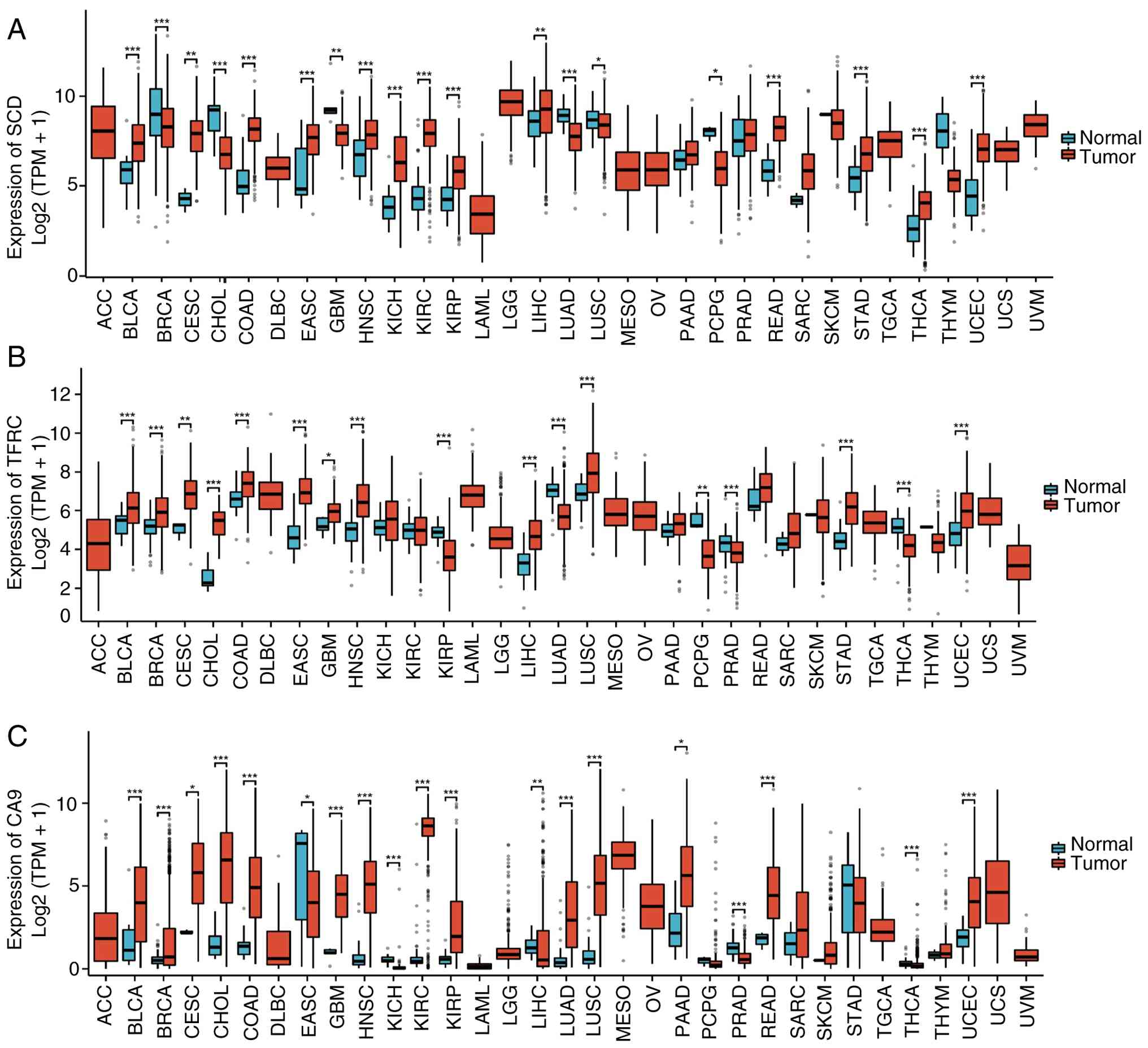
screened genes using TCGA dataset demonstrated that SCD, TFRC CA9
are generally highly expressed in CC. SCD was highly expressed in
most tumors but generally low expression in BRCA,
cholangiocarcinoma, lung adenocarcinoma (LUAD), lung SCC and
pheochromocytoma and paraganglioma (PCPG) (Fig. 5A). TFRC was highly expressed in most
tumors but generally low expression in LUAD, prostate
adenocarcinoma (PRAD), PCPG and thyroid carcinoma (Fig. 5B). CA9 was highly expressed in most
tumors but generally low expression in the esophageal adenosquamous
carcinoma and PRAD (Fig. 5C).
Prognostic analysis
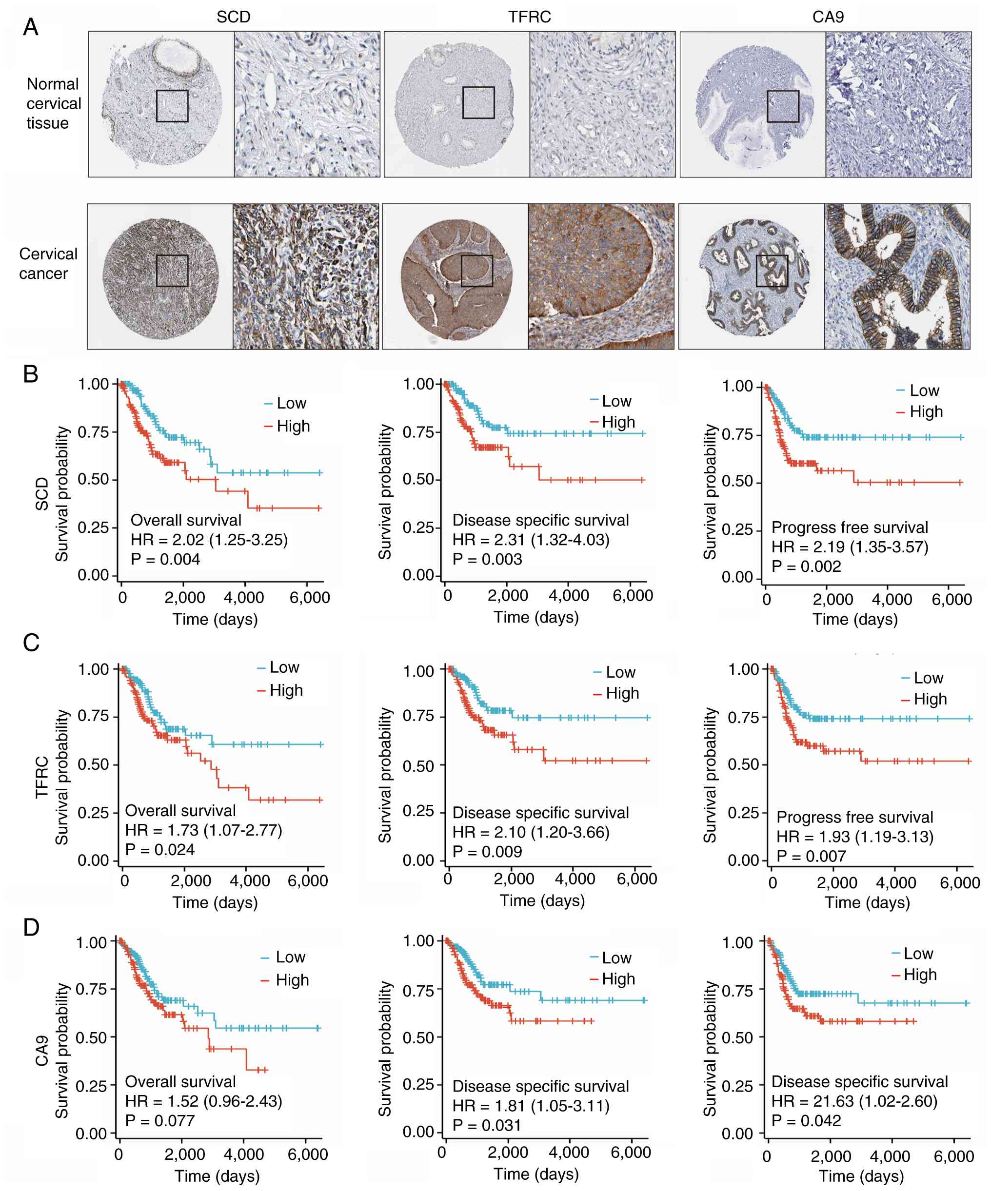
Using the HPA database, SCD, TFRC and CA9 was
demonstrated to have no or low expression in normal cervical
tissues but they were highly expressed in cervical SCC and
adenocarcinoma tissues, and they were expressed in both the nucleus
and cytoplasm (Fig. 6A). The
prognostic analysis of the aforementioned 30 genes screened using
TCGA dataset indicated that SCD, TFRC and CA9 are closely
associated with prognosis. The overall survival, disease-specific
survival and progress free survival of patients with CC with high
expression levels of SCD and TFRC were significantly reduced
(Fig. 6B and C). The
disease-specific survival and progress free survival of patients
with CC with high expression level of CA9 were significantly
reduced (Fig. 6D).
Immune infiltration analysis of
ferroptosis-associated prognostic genes in CC
The present study identified that the differences in
immune infiltration between the high and low expression groups of
SCD, TFRC and CA9 were mainly concentrated in naive B cells, memory
B cells, plasma cells, T cells, CD8 cells, naive CD4 T cells, CD4
memory resting cells, CD4 memory activated T cells, follicular
helper T cells, regulatory T cells, T cells γδ, resting natural
killer (NK) cells, activated NK cells, monocytes, M0 macrophages,
M1 macrophages, M2 macrophage, resting dendritic cells, activated
dendritic cells, resting mast cells and activated mast cells
(Fig. 7A-C). The heatmap displays
the Spearman correlation between the abundance of
immune-infiltrating cells and SCD, TFRC and CA9 expression of 24
types of tumor-infiltrating immune cells (Fig. 7D). SCD, TFRC and CA9 were most
closely correlated with central memory T cell, T helper (Th) 2 cell
and Th17, respectively (Fig.
7E-G).
Structural model diagram and drug-gene
interaction prediction based on ferroptosis-associated prognostic
genes in CC
Using the UniProt database (29), amino acid sequences of SCD, TFRC and
CA9 were retrieved and the SWISS-MODEL method (28) was used to predict the folding
structure models of SCD, TFRC and CA9 (Fig. 8A-C). In the DGIdb database
(http://dgidb.org/), the analysis of drug-gene
interactions revealed 148 drugs/compounds targeting SCD, TFRC and
CA9. The top 24 targeted drugs/compounds and their order based on
the ‘interaction group score’ (35)
in the DGIdb database are shown in Fig.
8D.
Verification of the key
ferroptosis-associated prognostic genes in CC in the signature
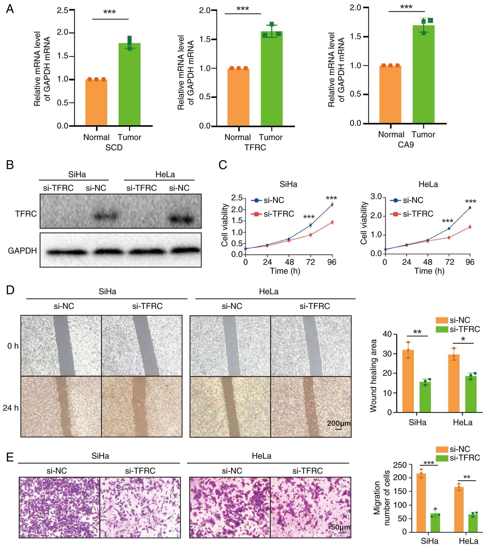
RT-qPCR further confirmed that compared with normal
cervical tissue, the expression levels of SCD, TFRC and CA9 were
significantly increased in CC (Fig.
9A). It has been reported that TFRC serves a key role in
epithelial ovarian cancer, breast cancer, liver cancer and other
tumors (36–38). Therefore, the present study focused
on further validation of hub genes, particularly TFRC. To validate
the biological function of TFRC in CC cells, TFRC was knocked down
in the present study using siRNA in SiHa and HeLa cells (Fig. 9B). MTT showed knockdown of TFRC
significantly reduced the proliferation of CC cells (Fig. 9C). The wound healing rate of SiHa
and HeLa cells was significantly inhibited by TFRC knockdown, as
indicated by the results of the wound-healing assay (Fig. 9D). The migration ability of SiHa and
HeLa cells was significantly inhibited in Transwell assays
following TFRC knockdown (Fig. 9E).
In summary, knocking down TFRC inhibited proliferation and
migration in CC cells in vitro.
Discussion
In 2021, cervical cancer ranked as the second most
common cause of cancer-related mortality in China (39). Although CC screening and the
promotion of HPV vaccines have reduced the incidence of CC
(40,41), >50% of CC is diagnosed as locally
advanced (42). Therefore, there is
an urgent need to screen and identify novel biomarkers for the
diagnosis and prognosis of CC in the future. Since the first
proposal of ferroptosis in 2012, numerous studies have reported its
key role in malignant tumors. Yang et al (43) reported that iron death-related genes
are closely associated with the prognosis of CC. In the present
study, bioinformatics methods were used to screen for DEGs based on
RNA-seq results of CC and identified 214 genes associated with
ferroptosis. Among them, 80 potential ferroptosis-related genes of
CC were identified by the intersection of ferroptosis-related genes
and DEGs, suggesting that differential genes in CC are closely
associated with ferroptosis.
GSEA, GO and KEGG analyses of 80 potential
ferroptosis-related genes showed CC enriched in ‘E2F targets’,
‘G2M checkpoint’, ‘response to peptide’, ‘response to
hypoxia’ and ‘DNA-binding transcription activator activity, RNA
polymerase II-specific’. PPI network analysis revealed that there
was a total of 911 interaction relationships and 30 genes were
confirmed as hub genes, which were MDM2, AR, CDKN2A, GSK-3β, NRAS,
PDK4, KLF2, SUV39H1, TLR4, IDH2, CAV1, SREBF1, MYB, CDH1, MEF2C,
MAPK3, SLC1A5, NR4A1, NOX4, SCD, DPP4, TFRC, CA9, ZEB1, PRKCA,
mTOR, EZH2, PARP1, SNCA, Sox2. The prognostic analysis of the
aforementioned 30 screened genes using TCGA dataset demonstrated
that SCD, TFRC and CA9 are closely associated with prognosis. The
present study further confirmed using RT-qPCR that compared with
normal cervical tissue, the expression levels of SCD, TFRC and CA9
were increased in CC, Furthermore, knocking down TFRC can inhibit
cell proliferation and migration in CC cells in vitro.
Han et al (44) revealed that insulin-like growth
factor 2 mRNA binding protein 3 (IGF2BP3) regulates SCD mRNA
N6-methyladenosine modification through the
IGF2BP3-methyltransferase-like 14 complex, thereby enhancing CC
proliferation, metastasis and lipid metabolism. Fang et al
(45) revealed that silencing TFRC
can limit cell proliferation and migration ability, prevent cell
cycle and promote apoptosis of CC cells. Previous studies have
confirmed that the alkaline sulfonamide S-1 (CA9 inhibitor) can
induce apoptosis in CC cells and hypoxia-induced immunosuppression
may be associated with the therapeutic resistance of locally
advanced CC, demonstrating CA9 is a key target for future treatment
of CC (46,47). Therefore, SCD, TFRC and CA9 may be
potential prognostic markers for CC.
The invasion of HPV into the body limits the
proliferation and activation of tumor-infiltrating T lymphocytes,
causing virus immune escape and gradually transforming normal
cervical tissue into malignancy, which is the main cause of CC
(48). The subtypes of
tumor-infiltrating T lymphocytes change with the occurrence and
development of CC, with various details (49). The changes and balance of cell
subtypes have different impacts on the progression of CC.
Infiltrating T lymphocytes may be an effective biomarker in
predicting the efficacy and prognosis of CC (50). The relationship between SCD, TFRC,
CA9 and immune cell infiltration were further investigated in the
present study. SCD, TFRC and CA9 were most closely correlated with
Central Memory T cell, T helper (Th) 2 cell and Th17, respectively
T lymphocytes serve a notable role in coordinating the immune
response of the human body to tumors. In the field of
immunotherapy, immune checkpoint inhibitors such as programmed cell
death protein-1 (PD-1) have demonstrated promising clinical
efficacy in various tumor treatments. It is key to note that as of
the end of 2021, according to the results of the KEYNOTE-826 trial,
pembrolizumab combined with chemotherapy with or without
bevacizumab has been approved for first-line treatment of
recurrent/metastatic CC. However, the overall response rate is
still moderate (~15%) (51,52). Therefore, a combination of novel
treatment methods and immunomodulatory targeted therapies is
warranted to improve the efficacy of existing cancer
immunotherapies in the future. The present study identified three
major apoptosis-related genes (SCD, TFRC and CA9) associated with
CC prognosis, searched the DGIdb database and obtained drug-gene
interaction results for three genes, SCD, TFRC and CA9, totaling 24
potential targeted drugs.
TFRC is a key membrane protein that serves a key
role in cellular iron uptake. Iron is essential for various
cellular processes, including growth and proliferation (7). Feng et al (53) reported that TFRC is overexpressed in
nasopharyngeal carcinoma and TFRC knockdown inhibits nasopharyngeal
carcinoma progression by suppressing the PI3K/AKT/mTOR signaling
pathway. In a previous study on pancreatic and colon cancer cells,
TFRC has been reported to regulate MAPK signaling pathway, which
helps improve cell viability and resistance to cell death (54). The present study demonstrated that
knocking down TFRC can inhibit cell proliferation and migration in
CC cells in vitro. Therefore, TFRC is a very promising
target, providing potentially novel ideas for the treatment of CC
in the future.
In summary, the present study identified the top 30
genes associated with ferroptosis in CC using bioinformatics
analysis and identified that SCD, TFRC and CA9 are closely
associated with the pathological T stage, lymph node metastasis and
poor prognosis of CC. In terms of immunity, SCD, TFRC and CA9 are
closely associated with T-cell depletion. Although the present
study identified genes associated with ferroptosis in CC, SCD, TFRC
and CA9 can serve as prognostic markers for CC and potential novel
targets for immunotherapy, and 24 potential targeted
drugs/compounds were predicted for these genes. The expression
levels of SCD, TFRC and CA9 were experimentally validated, which is
consistent with the present bioinformatics analysis. Furthermore,
knocking down TFRC can inhibit cell proliferation and migration in
CC cells in vitro.
There are several limitations of the present study
that should be considered when interpreting the results. First, the
present study mainly utilized in vitro experiments, further
in vivo experiments are warranted to confirm the effect of
ferroptosis-associated prognostic genes on the progression of CC.
Second, the limited number of clinical samples included may bias
the results and further clinical translation requires the inclusion
of more clinical samples to further confirm the present study
conclusions. Third, the mechanism of ferroptosis is still unclear
and requires further research to clarify, which will be a promising
direction for further investigation. Nevertheless, the present
study suggests that SCD, TFRC and CA9 are associated with the
prognosis of CC. The present study results have potentially laid
the foundation for the study of ferroptosis-associated prospective
genes in CC in the future.
Acknowledgements
Not applicable.
Funding
The present study was financially supported by the Hubei
Provincial Department of Science and Technology Project (grant no.
2023DJC144), the Xianning City Program of Science & Technology
(grant no. 2023ZRKX090), the transverse research and the Scientific
Research Foundation of Hubei University of Science and Technology
(grant no. 2023HX026), the Open Research Fund of Hubei Province Key
Laboratory of Precision Radiation Oncology (grant no.
2024ZLJZFL003) and Key Laboratory of Anesthesiology and
Resuscitation (Huazhong University of Science and Technology,
Ministry of Education; grant no. 2024MZFS010).
Availability of data and materials
The data generated in the present study may be
requested from the corresponding author.
Authors' contributions
LFW and ZXW conceived the study conceptualization.
LFW, GJT and ZXW designed the study methodology. Statistical
analysis was performed by LFW and HC. Data interpretation was
performed by ZXW, HC, GJT and LFW. LFW and GJT critically reviewed
and edited the manuscript. ZXW, HC, GJT and LFW confirm the
authenticity of all the raw data. All authors have read and
approved the manuscript.
Ethics approval and consent to
participate
The present study was performed according to the
principles expressed in the Declaration of Helsinki and was
approved by the Ethics Committee of Union Hospital, Tongji Medical
College, Huazhong University of Science and Technology (approval
no. 2021-S353; Wuhan, China). Written informed consent was obtained
from all patients.
Patient consent for publication
Not applicable.
Competing interests
The authors declare that they have no competing
interests.
References
|
1
|
Sung H, Ferlay J, Siegel RL, Laversanne M,
Soerjomataram I, Jemal A and Bray F: Global cancer statistics 2020:
GLOBOCAN estimates of incidence and mortalsity worldwide for 36
cancers in 185 countries. CA Cancer J Clin. 71:209–249.
2021.PubMed/NCBI
|
|
2
|
Balasubramaniam SD, Balakrishnan V, Oon CE
and Kaur G: Key molecular events in cervical cancer development.
Medicina (Kaunas). 55:3842019. View Article : Google Scholar : PubMed/NCBI
|
|
3
|
Koh WJ, Abu-Rustum NR, Bean S, Bradley K,
Campos SM, Cho KR, Chon HS, Chu C, Clark R, Cohn D, et al: Cervical
cancer, version 3.2019, NCCN clinical practice guidelines in
oncology. J Natl Compr Canc Netw. 17:64–84. 2019. View Article : Google Scholar : PubMed/NCBI
|
|
4
|
Yamaoka Y, Tamura S, Yamanoi K, Taki M,
Murakami R, Yamaguchi K, Hamanishi J and Mandai M: Clinical
significance of serum SCC levels before treatment for locally
advanced cervical squamous cell carcinoma. Anticancer Res.
44:2009–2019. 2024. View Article : Google Scholar : PubMed/NCBI
|
|
5
|
Shi V, Grover S, Huang Y, Thaker PH,
Kuroki LM, Powell MA, Mutch DG, Contreras JA, Schwarz JK, Grigsby
PW and Markovina S: Accuracy of surveillance serum squamous cell
carcinoma antigen for cervical cancer recurrence after definitive
chemoradiation. Int J Gynecol Cancer. 34:808–816. 2024. View Article : Google Scholar : PubMed/NCBI
|
|
6
|
Zhang C, Liu X, Jin S, Chen Y and Guo R:
Ferroptosis in cancer therapy: A novel approach to reversing drug
resistance. Mol Cancer. 21:472022. View Article : Google Scholar : PubMed/NCBI
|
|
7
|
Bu X and Wang L: Iron metabolism and the
tumor microenvironment: A new perspective on cancer intervention
and therapy (review). Int J Mol Med. 55:392025. View Article : Google Scholar : PubMed/NCBI
|
|
8
|
Li J, Mao S, Yang S, Lou Y, Zhao X, Chen
J, Bu Y, Lv B, Shi Q, Zhang Y, et al: Targeting JMJD6/PPARγ/GPX4
axis overcomes ferroptosis resistance and enhances therapeutic
efficacy in hepatocellular carcinoma. Oncogene. 44:4377–4390. 2025.
View Article : Google Scholar : PubMed/NCBI
|
|
9
|
Lin H, Zhu S, Chen Y, Lu J, Xie C, Liao C,
Huang X, Li G, Wu Y, Li Z, et al: Targeting cTRIP12 counteracts
ferroptosis resistance and augments sensitivity to immunotherapy in
pancreatic cancer. Drug Resist Updat. 81:1012402025. View Article : Google Scholar : PubMed/NCBI
|
|
10
|
Xia Y, Liu S, Li C, Ai Z, Shen W, Ren W
and Yang X: Discovery of a novel ferroptosis
inducer-talaroconvolutin A-killing colorectal cancer cells in vitro
and in vivo. Cell Death Dis. 11:9882020. View Article : Google Scholar : PubMed/NCBI
|
|
11
|
Wang M, Mao C, Ouyang L, Liu Y, Lai W, Liu
N, Shi Y, Chen L, Xiao D, Yu F, et al: Long noncoding RNA LINC00336
inhibits ferroptosis in lung cancer by functioning as a competing
endogenous RNA. Cell Death Differ. 26:2329–2343. 2019. View Article : Google Scholar : PubMed/NCBI
|
|
12
|
Xiaofei J, Mingqing S, Miao S, Yizhen Y,
Shuang Z, Qinhua X and Kai Z: Oleanolic acid inhibits cervical
cancer Hela cell proliferation through modulation of the ACSL4
ferroptosis signaling pathway. Biochem Biophys Res Commun.
545:81–88. 2021. View Article : Google Scholar : PubMed/NCBI
|
|
13
|
Wang C, Zeng J, Li LJ, Xue M and He SL:
Cdc25A inhibits autophagy-mediated ferroptosis by upregulating
ErbB2 through PKM2 dephosphorylation in cervical cancer cells. Cell
Death Dis. 12:10552021. View Article : Google Scholar : PubMed/NCBI
|
|
14
|
Wang X, Ji Y, Qi J, Zhou S, Wan S, Fan C,
Gu Z, An P, Luo Y and Luo J: Mitochondrial carrier 1 (MTCH1)
governs ferroptosis by triggering the FoxO1-GPX4 axis-mediated
retrograde signaling in cervical cancer cells. Cell Death Dis.
14:5082023. View Article : Google Scholar : PubMed/NCBI
|
|
15
|
Chen SJ, Kuo CC, Pan HY, Tsou TC, Yeh SC
and Chang JY: Desferal regulates hCtr1 and transferrin receptor
expression through Sp1 and exhibits synergistic cytotoxicity with
platinum drugs in oxaliplatin-resistant human cervical cancer cells
in vitro and in vivo. Oncotarget. 7:49310–49321. 2016. View Article : Google Scholar : PubMed/NCBI
|
|
16
|
Goldman MJ, Craft B, Hastie M, Repečka K,
McDade F, Kamath A, Banerjee A, Luo Y, Rogers D, Brooks AN, et al:
Visualizing and interpreting cancer genomics data via the Xena
platform. Nat Biotechnol. 38:675–678. 2020. View Article : Google Scholar : PubMed/NCBI
|
|
17
|
Love MI, Huber W and Anders S:
Moderatedestimation of fold change and dispersion for RNA-seq data
with DESeq2. Genome Biol. 15:5502014. View Article : Google Scholar : PubMed/NCBI
|
|
18
|
Ji Z, Fang Z, Dong X, Wang J, Wan X and
Yan A: Potential ferroptosis-related diagnostic and prognostic
biomarkers in laryngeal cancer. Eur Arch Otorhinolaryngol.
279:5277–5288. 2022. View Article : Google Scholar : PubMed/NCBI
|
|
19
|
Zhou N and Bao J: FerrDb: A manually
curated resource for regulators and markers of ferroptosis and
ferroptosis-disease associations. Database (Oxford).
2020:baaa0212020. View Article : Google Scholar : PubMed/NCBI
|
|
20
|
Chen H and Boutros PC: VennDiagram: A
package for the generation of highly-customizable Venn and Euler
diagrams in R. BMC Bioinformatics. 12:352011. View Article : Google Scholar : PubMed/NCBI
|
|
21
|
Kanehisa M and Goto S: KEGG: Kyoto
encyclopedia of genes and genomes. Nucleic Acids Res. 28:27–30.
2000. View Article : Google Scholar : PubMed/NCBI
|
|
22
|
Subramanian A, Tamayo P, Mootha VK,
Mukherjee S, Ebert BL, Gillette MA, Paulovich A, Pomeroy SL, Golub
TR, Lander ES and Mesirov JP: Gene set enrichment analysis: A
knowledge-based approach for interpreting genome-wide expression
profiles. Proc Natl Acad Sci USA. 102:15545–15550. 2005. View Article : Google Scholar : PubMed/NCBI
|
|
23
|
Choi W, Namkung J, Hwang I, Kim H, Lim A,
Park HJ, Lee HW, Han KH, Park S, Jeong JS, et al: Serotonin signals
through a gut-liver axis to regulate hepatic steatosis. Nat Commun.
9:48242018. View Article : Google Scholar : PubMed/NCBI
|
|
24
|
Hänzelmann S, Castelo R and Guinney J:
GSVA: Gene set variation analysis for microarray and RNA-seq data.
BMC Bioinformatics. 14:72013. View Article : Google Scholar : PubMed/NCBI
|
|
25
|
Szklarczyk D, Gable AL, Lyon D, Junge A,
Wyder S, Huerta-Cepas J, Simonovic M, Doncheva NT, Morris JH, Bork
P, et al: STRING v11: Protein-protein association networks with
increased coverage, supporting functional discovery in genome-wide
experimental datasets. Nucleic Acids Res. 47(D1): D607–D613. 2019.
View Article : Google Scholar : PubMed/NCBI
|
|
26
|
Shannon P, Markiel A, Ozier O, Baliga NS,
Wang JT, Ramage D, Amin N, Schwikowski B and Ideker T: Cytoscape: A
software environment for integrated models of biomolecular
interaction networks. Genome Res. 13:2498–2504. 2003. View Article : Google Scholar : PubMed/NCBI
|
|
27
|
Chin CH, Chen SH, Wu HH, Ho CW, Ko MT and
Lin CY: cytoHubba: Identifying hub objects and sub-networks from
complex interactome. BMC Syst Biol. 8 (Suppl 4):S112014. View Article : Google Scholar : PubMed/NCBI
|
|
28
|
Biasini M, Bienert S, Waterhouse A, Arnold
K, Studer G, Schmidt T, Kiefer F, Gallo Cassarino T, Bertoni M,
Bordoli L and Schwede T: SWISS-MODEL: Modelling protein tertiary
and quaternary structure using evolutionary information. Nucleic
Acids Res. 42((Web Server Issue)): W252–W258. 2014. View Article : Google Scholar : PubMed/NCBI
|
|
29
|
UniProt Consortium: UniProt: A hub for
protein information. Nucleic Acids Res. 43((Database Issue)):
D204–D212. 2015.PubMed/NCBI
|
|
30
|
Ding L, Bailey MH, Porta-Pardo E, Thorsson
V, Colaprico A, Bertrand D, Gibbs DL, Weerasinghe A, Huang KL,
Tokheim C, et al: Perspective on oncogenic processes at the end of
the beginning of cancer genomics. Cell. 173:305–320.e10. 2018.
View Article : Google Scholar : PubMed/NCBI
|
|
31
|
Nilsson J, Vallbo C, Guo D, Golovleva I,
Hallberg B, Henriksson R and Hedman H: Cloning, characterization,
and expression of human LIG1. Biochem Biophys Res Commun.
284:1155–1161. 2001. View Article : Google Scholar : PubMed/NCBI
|
|
32
|
Livak KJ and Schmittgen TD: Analysis of
relative gene expression data using real-time quantitative PCR and
the 2(−Delta Delta C(T)) method. Methods. 25:402–408. 2001.
View Article : Google Scholar : PubMed/NCBI
|
|
33
|
Griffith M, Griffith OL, Coffman AC,
Weible JV, McMichael JF, Spies NC, Koval J, Das I, Callaway MB,
Eldred JM, et al: DGIdb: Mining the druggable genome. Nat Methods
10: 1209–1210, 2013. Heydar H, Mansouri K, Norooznezhad M,
Norooznezhad F, Mohamadnia A and Bahrami N: Bevacizumab inhibits
angiogenic cytokines in head and neck squamous cell carcinoma: From
gene to the protein. Int J Hematol Oncol Stem Cell Res. 12:136–141.
2018.PubMed/NCBI
|
|
34
|
Gillette MA, Satpathy S, Cao S,
Dhanasekaran SM, Vasaikar SV, Krug K, Petralia F, Li Y, Liang WW,
Reva B, et al: Proteogenomic characterization reveals therapeutic
vulnerabilities in lung adenocarcinoma. Cell. 182:200–225.e35.
2020. View Article : Google Scholar : PubMed/NCBI
|
|
35
|
He F, Ma N, Midorikawa K, Hiraku Y, Oikawa
S, Zhang Z, Huang G, Takeuchi K and Murata M: Taurine exhibits an
apoptosis-inducing effect on human nasopharyngeal carcinoma cells
through PTEN/Akt pathways in vitro. Amino Acids. 50:1749–1758.
2018. View Article : Google Scholar : PubMed/NCBI
|
|
36
|
Cannon M, Stevenson J, Stahl K, Basu R,
Coffman A, Kiwala S, McMichael JF, Kuzma K, Morrissey D, Cotto K,
et al: DGIdb 5.0: Rebuilding the drug-gene interaction database for
precision medicine and drug discovery platforms. Nucleic Acids Res.
52(D1): D1227–D1235. 2024. View Article : Google Scholar : PubMed/NCBI
|
|
37
|
Yu H, Yang C, Jian L, Guo S, Chen R, Li K,
Qu F, Tao K, Fu Y, Luo F and Liu S: Sulfasalazine-induced
ferroptosis in breast cancer cells is reduced by the inhibitory
effect of estrogen receptor on the transferrin receptor. Oncol Rep.
42:826–838. 2019.PubMed/NCBI
|
|
38
|
Huang Y, Huang J, Huang Y, Gan L, Long L,
Pu A and Xie R: TFRC promotes epithelial ovarian cancer cell
proliferation and metastasis via up-regulation of AXIN2 expression.
Am J Cancer Res. 10:131–147. 2020.PubMed/NCBI
|
|
39
|
Muhammad J, Bajbouj K, Shafarin J and
Hamad M: Estrogen-induced epigenetic silencing of FTH1 and TFRC
genes reduces liver cancer cell growth and survival. Epigenetics.
15:1302–1318. 2020. View Article : Google Scholar : PubMed/NCBI
|
|
40
|
Ren W, Guo X, Liu Z, Wu Y, Peng R, Liu H
and Qi J: Burden of female-specific cancers in China from 1990 to
2021: A systematic analysis for the global burden of disease study
2021. Cancer. 131:e357122025. View Article : Google Scholar : PubMed/NCBI
|
|
41
|
Xia C, Dong X, Li H, Cao M, Sun D, He S,
Yang F, Yan X, Zhang S, Li N and Chen W: Cancer statistics in China
and United States, 2022: Profiles, trends, and determinants. Chin
Med J (Engl). 135:584–590. 2022. View Article : Google Scholar : PubMed/NCBI
|
|
42
|
Wei M, Zhou W, Bi Y, Wang H, Liu Y and
Zhang ZJ: Rising mortality rate of cervical Cancer in younger women
in Urban China. J Gen Intern Med. 34:281–284. 2019. View Article : Google Scholar : PubMed/NCBI
|
|
43
|
Ma Y, Di J, Bi H, Zhao Q, Qin T, Xu W, Liu
Z, Yi N, Zhao J, Zhou D, et al: Comparison of the detection rate of
cervical lesion with TruScreen, LBC test and HPV test: A real-world
study based on population screening of cervica cancer in rural
areas of China. PLOS One. 15:e02339862020. View Article : Google Scholar : PubMed/NCBI
|
|
44
|
Yang X, Yin F, Liu Q, Ma Y, Zhang H, Guo
P, Wen W, Guo X, Wu Y, Yang Z and Han Y: Ferroptosis-related genes
identify tumor immune microenvironment characterization for the
prediction of prognosis in cervical cancer. Ann Transl Med.
10:1232022. View Article : Google Scholar : PubMed/NCBI
|
|
45
|
Han C, Hu C, Liu T, Sun Y, Hu F, He Y,
Zhang J, Chen J, Ding J, Fan J, et al: IGF2BP3 enhances lipid
metabolism in cervical cancer by upregulating the expression of
SCD. Cell Death Dis. 15:1382024. View Article : Google Scholar : PubMed/NCBI
|
|
46
|
Fang X, Hu P, Gao Y, Chen C and Xu J:
Transferrin receptor modulated by microRNA-497-5p suppresses
cervical cancer cell malignant phenotypes. Adv Clin Exp Med.
33:273–282. 2024. View Article : Google Scholar : PubMed/NCBI
|
|
47
|
Koyuncu I, Tülüce Y, Slahaddin Qadir H,
Durgun M and Supuran CT: Evaluation of the anticancer potential of
a sulphonamide carbonic anhydrase IX inhibitor on cervical cancer
cells. J Enzyme Inhib Med Chem. 34:703–711. 2019. View Article : Google Scholar : PubMed/NCBI
|
|
48
|
Yoon M, Lee HK, Park EY, Kim JH, Lee JH,
Kim YS, Kim HJ, Kim H, Yoo CW, Lee S, et al: Randomized multicenter
phase II trial of prophylactic irradiation of para-aortic lymph
nodes in advanced cervical cancer according to tumor hypoxia:
Korean radiation oncology group (KROG 07-01) study. Int J Cancer.
151:2182–2194. 2022. View Article : Google Scholar : PubMed/NCBI
|
|
49
|
Meng L, Lu H, Li Y, Zhao J, He S, Wang Z,
Shen J, Huang H, Xiao J, Sooranna SR and Song J: Human
papillomavirus infection can alter the level of tumour stemness and
T cell infiltration in patients with head and neck squamous cell
carcinoma. Front Immunol. 13:10135422022. View Article : Google Scholar : PubMed/NCBI
|
|
50
|
Mbuya W, Mcharo R, Mhizde J, Mnkai J,
Mahenge A, Mwakatima M, Mwalongo W, Chiwerengo N, Hölscher M,
Lennemann T, et al: Depletion and activation of mucosal CD4 T cells
in HIV infected women with HPV-associated lesions of the cervix
uteri. PLoS One. 15:e02401542020. View Article : Google Scholar : PubMed/NCBI
|
|
51
|
Jiang L, Wu T, Qu X, Li S, Jiang Q, Ren T,
Liang J, Ding Y, Hua K, Tang Z and Qiu J: Targeting CD39 boosts
PD-1 blockade antitumor therapeutic efficacy via strengthening CD8
+ TILs function and recruiting B cells in cervical cancer. J
Nanobiotechnology. 23:4132025. View Article : Google Scholar : PubMed/NCBI
|
|
52
|
Chung HC, Ros W, Delord JP, Perets R,
Italiano A, Shapira-Frommer R, Manzuk L, Piha-Paul SA, Xu L,
Zeigenfuss S, et al: Efficacy and safety of pembrolizumab in
previously treated advanced cervical cancer: Results from the phase
II KEYNOTE-158 study. J Clin Oncol. 37:1470–1478. 2019. View Article : Google Scholar : PubMed/NCBI
|
|
53
|
Colombo N, Dubot C, Lorusso D, Caceres MV,
Hasegawa K, Shapira-Frommer R, Tewari KS, Salman P, Hoyos Usta E,
Yañez E, et al: Pembrolizumab for persistent, recurrent, or
metastatic cervical cancer. N Engl J Med. 385:1856–1867. 2021.
View Article : Google Scholar : PubMed/NCBI
|
|
54
|
Feng G, Arima Y, Midorikawa K, Kobayashi
H, Oikawa S, Zhao W, Zhang Z, Takeuchi K and Murata M: Knockdown of
TFRC suppressed the progression of nasopharyngeal carcinoma by
downregulating the PI3K/Akt/mTOR pathway. Cancer Cell Int.
23:1852023. View Article : Google Scholar : PubMed/NCBI
|
|
55
|
Miao C and Fan D: Identification of
differentially expressed genes and pathways in diquat and paraquat
poisoning using bioinformatics analysis. Toxicol Mech Methods.
32:678–685. 2022. View Article : Google Scholar : PubMed/NCBI
|