Introduction
Gastrointestinal (GI) malignancies, particularly
gastric, colorectal, pancreatic and biliary tract cancers,
collectively account for a substantial proportion of cancer-related
mortality worldwide; in 2022, colorectal, gastric and pancreatic
cancer accounted for ~17% of new cancer cases and 21% of cancer
deaths globally (1). A shared
anatomical feature of these tumors is their predilection for
hepatic spread: The liver, by virtue of its portal venous drainage,
is the most common site of distant metastasis from GI primaries,
and diffuse parenchymal infiltration frequently impairs hepatic
synthetic and excretory function (2). The resulting hyperbilirubinemia, when
caused by parenchymal tumor burden rather than biliary obstruction,
cannot reliably be managed by drainage procedures and instead
demands systemic therapy, yet this is precisely the patient group
excluded from nearly all registration trials.
Most pivotal Phase III trials restrict enrollment to
patients with serum bilirubin <1.5 times the upper limit of
normal (ULN). Patients with more severe dysfunction are therefore
underrepresented in the evidence base, and standard dosing
recommendations do not apply (3).
Clinicians are left to extrapolate from pharmacokinetic principles
and small retrospective series when making treatment decisions for
this population.
FOLFOX, which combines oxaliplatin with leucovorin
(folinic acid) and bolus and infusional 5-fluorouracil every 2
weeks, is an important chemotherapy backbone across GI malignancy.
The pharmacokinetic rationale for using oxaliplatin in hepatic
dysfunction rests on its predominantly non-hepatic elimination.
Oxaliplatin is rapidly cleared from plasma by non-enzymatic
biotransformation, with urinary excretion accounting for ~54% of
drug removal (4). By contrast
irinotecan requires hepatic bioactivation and detoxification of its
active metabolite SN-38, and to the taxanes, which depend on
hepatobiliary extraction and are contraindicated in
moderate-to-severe liver dysfunction. Fluoropyrimidines undergo
primary hepatic metabolism but have been used cautiously in
dose-adjusted regimens even in patients with impaired liver
function (5).
Several case reports and small series have
documented the feasibility of oxaliplatin- and
fluoropyrimidine-based regimens in patients with hyperbilirubinemia
from liver metastases. The largest published case series, by Quidde
et al (6), described 12
patients with GI cancer and a median bilirubin of 6.1 mg/dl who
received oxaliplatin combined with 5-fluorouracil and leucovorin;
bilirubin normalized or fell >50% within 8 weeks in half of the
patients, and median OS was notably longer in those who responded.
Comparable observations have been reported in individual case
reports of colorectal and gastric cancer (7,8).
Despite these encouraging reports, the available
evidence remains limited by small sample sizes, heterogeneous
treatment regimens and single-tumor-type focus. Real-world data
from a broader population of patients with GI cancer including
gastric, biliary, pancreatic and colorectal primaries managed with
individualized FOLFOX-based regimens are lacking. The present study
aimed to evaluate bilirubin response, toxicity and survival
outcomes in 27 patients with metastatic GI cancer and marked
hyperbilirubinemia who received individualized FOLFOX-based
chemotherapy and to investigate whether early bilirubin response
was associated with survival.
Materials and methods
Study design and patients
The present study was retrospective, including a
single-center cohort conducted at the Department of Medical
Oncology, Etlik City Hospital (Ankara, Türkiye). Patients treated
between July 2023 and March 2025 were identified through a review
of institutional electronic records and oncology databases. The
present study was approved by the institutional ethics committee
(approval no. AEŞH-BADEK2-2025-432) and conducted in accordance
with the Declaration of Helsinki. Informed consent was waived given
the retrospective design and anonymized data. Medical records were
accessed and data were extracted for research purposes between
October 1 and October 30, 2025, after ethics committee approval was
obtained on September 16, 2025.
Eligibility criteria
Patients were eligible if they were aged ≥18 years,
had histologically confirmed metastatic GI malignancy (gastric,
biliary tract, pancreatic or colorectal), had radiologically
evident liver metastases and had a serum total bilirubin ≥4.5 mg/dl
at the time of treatment initiation. This threshold was set above
the National Cancer Institute Organ Dysfunction Working Group
definition of severe hepatic impairment (bilirubin >3× ULN,
corresponding to 3.6 mg/dl using Etlik City Hospital ULN of 1.2
mg/dl) (9). Patients were included
whether or not a drainage procedure had been performed: Those
ineligible for biliary drainage and those with persistently
elevated bilirubin despite technically feasible drainage were both
eligible.
Patients were excluded if hyperbilirubinemia was
attributable to non-malignant hepatic conditions (viral hepatitis,
cirrhosis or drug-induced injury), or if chemotherapy had been
administered within 14 days before bilirubin elevation was
documented.
Treatment protocol
All patients received an individualized FOLFOX
backbone rather than a single standardized mFOLFOX6 protocol, given
variation in oxaliplatin dosing, 5-fluorouracil administration
route and dose modifications described below. Oxaliplatin was
administered intravenously at 60–85 mg/m2;
5-fluorouracil was given as a 46-h continuous infusion (2,400
mg/m2) or in bolus form according to individual
tolerability; and leucovorin was administered at 400–500
mg/m2. Cycles were repeated every two weeks. Monoclonal
antibodies (bevacizumab, cetuximab or panitumumab) were added at
the discretion of the treating physician based on molecular tumor
characteristics. Dose escalation was permitted in patients showing
biochemical improvement and acceptable toxicity.
Definitions and endpoints
Bilirubin response was defined as: i) A reduction of
≥50% in total bilirubin within 4–8 weeks of treatment initiation;
or ii) normalization of total bilirubin to ≤1.2 mg/dl within 12
weeks. Bilirubin at each nominal timepoint was measured within ±10
days of that timepoint. Patients were categorized as responders or
non-responders based on these criteria. Radiologic response was
assessed by Response Evaluation Criteria in Solid Tumors (RECIST)
v1.1. Toxicity was graded using Common Terminology Criteria for
Adverse Events (CTCAE) version 5.0 (10,11)
The primary endpoint was bilirubin response rate; secondary
endpoints were progression-free survival (PFS), overall survival
(OS) and treatment safety.
Statistical analysis
Continuous variables are presented as median with
ranges or interquartile ranges (IQR); categorical variables are
presented as frequencies and proportions. Changes in bilirubin over
time were assessed with the Wilcoxon signed-rank test, restricted
to patients with paired bilirubin measurements at baseline and the
corresponding follow-up assessment. PFS and OS were measured from
the date of initiation of FOLFOX-based chemotherapy to progression
or death, respectively; patients without the corresponding event
were censored at the date of last follow-up. PFS and OS were
estimated by the Kaplan-Meier method; differences between groups
were compared using the log-rank test. Cox proportional hazards
regression was used to examine potential predictors of PFS and OS;
variables with P<0.20 in univariate analysis were entered into
the multivariate model. To address potential immortal time bias
arising from classification according to week 4 bilirubin response,
post hoc week 4 landmark analysis was performed for both PFS and
OS. The week 4 landmark was defined as day 28 after treatment
initiation, and landmark response classification was based
exclusively on bilirubin measurements obtained on or before day 28.
For the PFS landmark analysis, only patients who were alive,
progression-free and under observation at week 4 were included; for
the OS landmark analysis, patients who were alive and under
observation at week 4 were included. Survival time was reset to
zero at the day 28 landmark. Kaplan-Meier curves were generated
according to week 4 bilirubin response (≥50 vs. <50% reduction
in total bilirubin), groups were compared using the log-rank test
and univariable Cox proportional hazards models were used to
estimate hazard ratios (HRs) and 95% confidence intervals (CIs).
Toxicities were compared between patients with Eastern Cooperative
Oncology Group Performance Status (ECPG PS) 0 and 1 using two-sided
Fisher's exact tests because of the small sample size and sparse
cell counts. Radiologic response categories and the prespecified
bilirubin-response endpoint, including each of its components, were
summarized descriptively as counts and percentages. All analyses
were performed in SPSS version 26.0 (IBM Corp.); a two-sided
P<0.05 was considered to indicate a statistically significant
difference.
Results
Patient characteristics
A total of 27 patients were included in the present
analysis The median age was 62 years (range 25–80), and 21 (77.8%)
were men. The most common primary site was gastric cancer (n=10,
37.0%), followed by biliary tract cancer (n=8, 29.6%), pancreatic
cancer (n=5, 18.5%) and colorectal cancer (n=4, 14.8%). At
treatment initiation, 16 patients (59.3%) had an ECOG PS of 0 and
11 (40.7%) had a status of 1. The median modified Charlson
comorbidity index was 8 (range 6–11). Liver metastasis was present
in all 27 patients (100%), and 8 patients (29.6%) had undergone
percutaneous transhepatic biliary drainage (PTBD) before or at the
time of diagnosis. All 20 tested patients had microsatellite-stable
disease, as assessed by immunohistochemistry. Targeted therapy was
administered only to patients with colorectal primaries, in whom it
was clinically indicated (bevacizumab, n=3; cetuximab, n=1); it was
not used in patients with gastric, biliary or pancreatic primaries.
Baseline laboratory and clinical characteristics are summarized in
Table I.
 | Table I.Baseline patient and disease
characteristics. |
Table I.
Baseline patient and disease
characteristics.
| Characteristics | Patients (n=27) |
|---|
| Age, years | 62 (25–80) |
| Sex |
|
| Male | 21 (77.8) |
|
Female | 6 (22.2) |
| Primary tumor
site |
|
| Gastric
cancer | 10 (37.0) |
| Biliary
tract cancer | 8 (29.6) |
|
Pancreatic cancer | 5 (18.5) |
|
Colorectal cancer | 4 (14.8) |
| Eastern Cooperative
Oncology Group |
|
| Performance
Status |
|
| 0 | 16 (59.3) |
| 1 | 11 (40.7) |
| Modified Charlson
comorbidity index score | 8 (6–11) |
| Metastatic
sitea |
|
| Liver
metastasis | 27 (100.0) |
|
Peritoneal carcinomatosis | 7 (25.9) |
|
Pulmonary | 4 (14.8) |
| Bone/bone
marrow | 3 (11.1) |
| Vascular
invasion | 1 (3.7) |
| Microsatellite
instability status |
|
|
Microsatellite stable | 20 (74.1) |
|
Unknown | 7 (25.9) |
| Percutaneous
transhepatic biliary drainage at diagnosis or relapse |
|
| Yes | 8 (29.6) |
| No | 19 (70.4) |
| Baseline laboratory
values |
|
| Total
bilirubin, mg/dl | 6.9 (5.0–18.2) |
| Direct
bilirubin, mg/dl | 5.4 (3.0–13.4) |
|
Hemoglobin, g/dl | 10.3 (7.0–13.1) |
| White
blood cell count, ×103/µl | 9.2 (3.5–17.4) |
| Platelet
count, ×103/µl | 246.0
(39.0–577.0) |
Bilirubin response
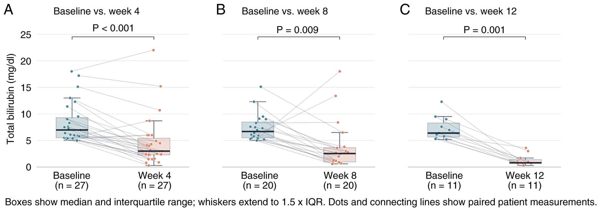
The median total bilirubin at baseline was 6.9 mg/dl
(IQR 5.5–9.5). Following initiation of FOLFOX, bilirubin levels
decreased significantly at each subsequent evaluation. At week 4,
the median total bilirubin was 3.0 mg/dl (IQR 2.3–5.4; P<0.001
vs. baseline). The decline continued at week 8, with a median of
2.5 mg/dl (IQR 1.0–3.7; P=0.009). By week 12, the median bilirubin
had fallen to 0.8 mg/dl (IQR 0.7–1.4; P=0.001), within the normal
range (Fig. 1). Bilirubin
measurements were available in 27 patients at baseline, 27 at week
4, 20 at week 8 and 11 at week 12; longitudinal comparisons were
restricted to patients with paired baseline and corresponding
follow-up measurements, giving paired sample sizes of 27, 20 and 11
at weeks 4, 8 and 12, respectively. Patients without a measurement
of a given timepoint had not remained on treatment or under
follow-up long enough to reach it, primarily due to death or
clinical progression. In total 2 patients died before the day 28
landmark, on days 18 and 26. Their last available bilirubin
measurements were retained in the descriptive week 4 analysis. Both
patients were classified as non-responders in the exploratory
overall-cohort Cox analyses as neither achieved a ≥50% reduction in
bilirubin. Both were excluded from the day 28 landmark analysis,
leaving 25 eligible patients (16 responders and 9 non-responders).
All landmark response classifications were based on bilirubin
measurements obtained on or before day 28.
Longitudinal bilirubin kinetics are illustrated in
Fig. 1. The prespecified composite
bilirubin-response endpoint was achieved by 21 of 27 patients
(77.8%); 20 patients (74.1%) achieved a ≥50% reduction in total
bilirubin within weeks 4–8 and 11 (40.7%) achieved normalization to
≤1.2 mg/dl within 12 weeks (Table
SI).
Treatment response and survival
Radiologic response was evaluable according to
RECIST v1.1 in 11 of 27 patients (40.7%); the remaining 16 patients
(59.3%) were non-evaluable because radiologic assessment was not
feasible before the first scheduled imaging evaluation, most often
due to clinical deterioration or death. Among the 11
RECIST-evaluable patients, no complete responses were observed, 8
(72.7%) achieved a partial response, 1 (9.1%) had stable disease
and 2 (18.2%) had progressive disease. Accordingly, among patients
who were RECIST-evaluable, the objective response rate (ORR) was
72.7% (8/11) and the disease control rate (DCR) was 81.8% (9/11)
(Table SI). Median PFS was 2.56
months (95% CI 1.81–3.65); PFS rates at weeks 4, 8 and 12 were
92.6, 59.3 and 48.1%, respectively. Median OS was 3.65 months (95%
CI 2.56–5.72); OS rates at weeks 4, 8 and 12 were 92.6, 66.7 and
51.9%, respectively. More than half of the cohort (51.9%) was alive
at 12 weeks (Table II). To address
potential immortal time bias arising from week 4 response
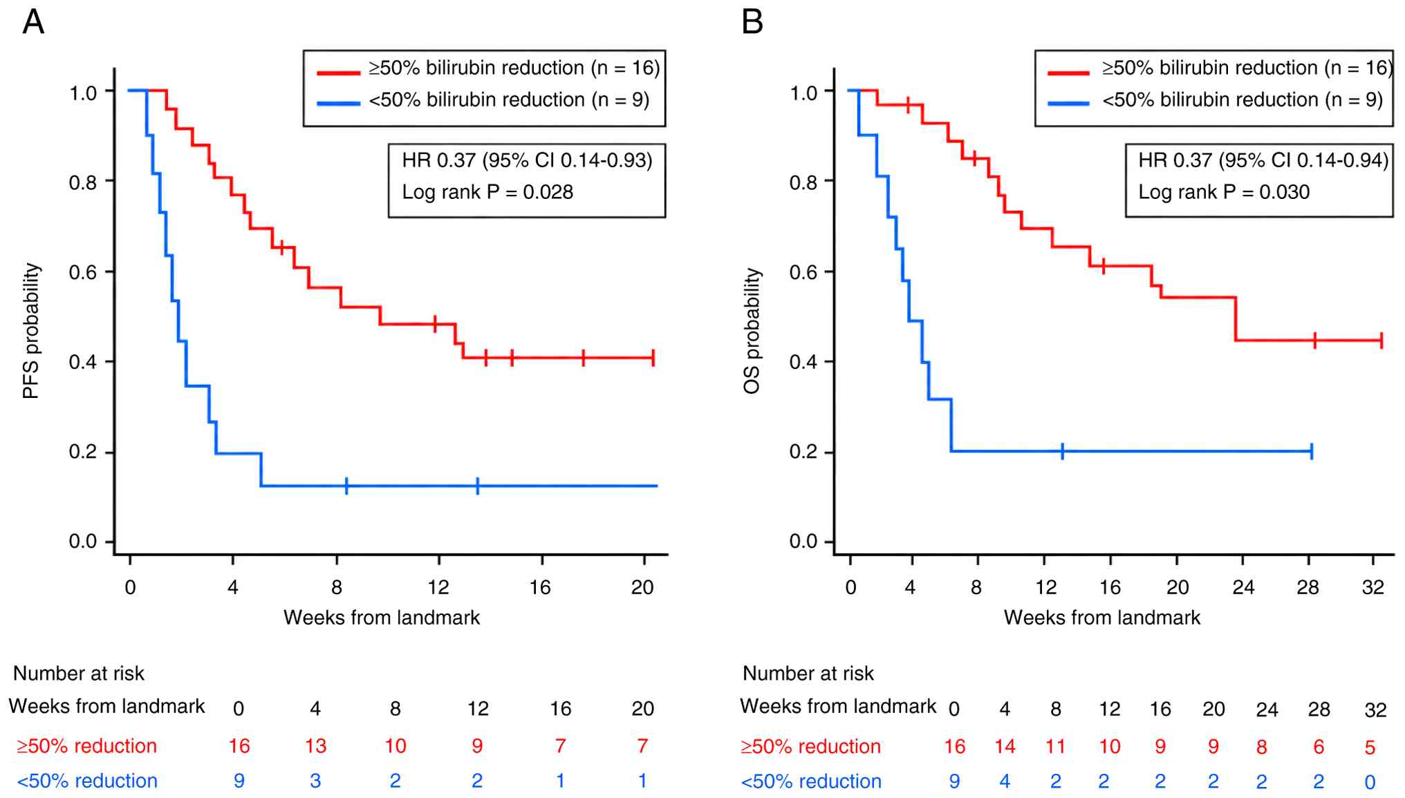
classification, post hoc week 4 landmark analysis was performed. In
the PFS landmark analysis (n=25 patients alive and progression-free
at week 4; 16 responders, 9 non-responders), median landmark PFS
was 2.89 months (95% CI 1.08–10.09) in responders vs. 0.53 months
(95% CI 0.13–3.58) in non-responders (univariable HR 0.37; 95% CI
0.14–0.93; log-rank P=0.028). In the OS landmark analysis (n=25
patients alive at week 4; 16 responders and 9 non-responders),
median landmark OS was 5.52 months (95% CI 1.28–14.24) in
responders vs. 0.53 months (95% CI 0.13–6.51) in non-responders
(univariable HR 0.37; 95% CI 0.14–0.94; log-rank P=0.030) (Fig. 2).
| Table II.Summary of survival outcomes (PFS and
OS). |
Table II.
Summary of survival outcomes (PFS and
OS).
| Outcome measure | PFS | OS |
|---|
| Median survival,
months (95% confidence interval) | 2.56 (1.81–3.65) | 3.65 (2.56–5.72) |
| Survival rate, n
(%) |
|
|
| At week
4 | 25 (92.6) | 25 (92.6) |
| At week
8 | 16 (59.3) | 18 (66.7) |
| At week
12 | 13 (48.1) | 14 (51.6) |
Toxicity and dose modifications
Toxicity was generally manageable, except for two
grade 5 acute kidney injury events (Table III). No patient developed grade 3
or 4 hepatotoxicity. Grade 1 hepatotoxicity was observed in seven
patients (25.9%) and grade 2 in 1 (3.7%). Grade 5 acute kidney
injury occurred in 2 patients (7.4%) and grade 2 nephrotoxicity in
1 (3.7%). Hematological toxicity of grade 2 was the most frequent
finding (7 patients, 25.9%); grade 3 hematological toxicity
occurred in 1 patient (3.7%). Overall, 8 patients (29.6%) received
treatment at full dose throughout; the remainder required dose
reductions ranging from 10–50%. The most common reduction level was
20% (n=8, 29.6%). Toxicity did not differ significantly between
patients with ECOG PS 0 and 1, although nephrotoxicity was observed
only among patients with ECOG PS 1 (27.3 vs. 0%; Fisher's exact
P=0.056; Table SII).
| Table III.Adverse events and dose modifications
during FOLFOX-based treatment. |
Table III.
Adverse events and dose modifications
during FOLFOX-based treatment.
| Event | Patients (n=27) |
|---|
| Hepatotoxicity |
|
| Grade
0 | 19 (70.4) |
| Grade
1 | 7 (25.9) |
| Grade
2 | 1 (3.7) |
| Grade
3/4 | 0 (0.0) |
| Nephrotoxicity |
|
| Grade
0 | 24 (88.9) |
| Grade
2 | 1 (3.7) |
| Grade
1/3/4 | 0 (0.0) |
| Grade
5 | 2 (7.4)a |
| Hematological
toxicity |
|
| Grade
0 | 15 (55.6) |
| Grade
1 | 4 (14.8) |
| Grade
2 | 7 (25.9) |
| Grade
3 | 1 (3.7) |
| Grade
4 | 0 (0.0) |
| Dose reductions |
|
| No
reduction | 8 (29.6) |
| 10%
reduction | 5 (18.5) |
| 20%
reduction | 8 (29.6) |
| 25%
reduction | 2 (7.4) |
| 30%
reduction | 1 (3.7) |
| 50%
reduction | 3 (11.1) |
Predictors of survival
On univariate Cox regression, a ≥50% reduction in
total bilirubin at week 4 was associated with significantly longer
PFS (HR 0.38, 95% CI 0.16–0.91; P=0.030) and OS (HR 0.37, 95% CI
0.15–0.91; P=0.030). On multivariate analysis, this association
persisted for both PFS (HR 0.30, 95% CI 0.12–0.76; P=0.011) and OS
(HR 0.29, 95% CI 0.11–0.76; P=0.012), although these estimates
derive from the overall cohort without landmark restriction and
should be interpreted alongside the aforementioned week 4 landmark
analysis, which was performed to address immortal time bias. ECOG
PS (1 vs. 0) was not significantly associated with PFS (univariate
HR 1.66, 95% CI 0.71–3.90; P=0.245) or OS (univariate HR 1.83, 95%
CI 0.77–4.37; P=0.175; multivariate HR 1.10, 95% CI 0.36–3.35;
P=0.862). No other variable reached statistical significance on
univariate or multivariate analysis (Tables IV and V).
| Table IV.Cox proportional hazards regression
analysis for progression-free survival. |
Table IV.
Cox proportional hazards regression
analysis for progression-free survival.
| Variable | Univariate HR (95%
CI) | P-value | Multivariate HR
(95% CI)a | P-value |
|---|
| Eastern Cooperative
Oncology | 1.66
(0.71–3.90) | 0.245 | - | - |
| Group Performance
Status (1 vs. 0) |
|
|
|
|
| Baseline total
bilirubin (>6.9 vs. | 0.92
(0.40–2.12) | 0.850 | - | - |
| ≤6.9 mg/dl) |
|
|
|
|
| ≥50% Bilirubin
reduction at week | 0.38
(0.16–0.91) | 0.030 | 0.30
(0.12–0.76) | 0.011 |
| 4 (yes vs. no) |
|
|
|
|
| Percutaneous
transhepatic biliary drainage procedure (yes vs. no) | 0.65
(0.26–1.66) | 0.369 | - | - |
| Dose reduction (yes
vs. no) | 1.21
(0.50–2.95) | 0.677 | - | - |
| Table V.Cox proportional hazards regression
analysis for overall survival. |
Table V.
Cox proportional hazards regression
analysis for overall survival.
| Variable | Univariate HR (95%
CI) | P-value | Multivariate HR
(95% CI)a | P-value |
|---|
| Eastern Cooperative
Oncology | 1.83
(0.77–4.37) | 0.175 | 1.10
(0.36–3.35) | 0.862 |
| Group Performance
Status (1 vs. 0) |
|
|
|
|
| Baseline total
bilirubin (>6.9 vs. ≤6.9 mg/dl) | 1.04
(0.45–2.41) | 0.933 | - | - |
| ≥50% Bilirubin
reduction at week 4 (yes vs. no) | 0.37
(0.15–0.91) | 0.030 | 0.29
(0.11–0.76) | 0.012 |
| Percutaneous
transhepatic biliary drainage procedure (yes vs. no) | 0.72
(0.28–1.85) | 0.495 | - | - |
| Dose reduction (yes
vs. no) | 1.34
(0.52–3.43) | 0.546 | - | - |
Discussion
The present retrospective series adds to the limited
body of evidence supporting oxaliplatin- and fluoropyrimidine-based
chemotherapy in patients with GI cancer and notable hepatic
dysfunction. Among 27 patients with a median baseline bilirubin of
6.9 mg/dl, a level at which most guidelines would preclude
cytotoxic therapy, FOLFOX produced a rapid and statistically
notable bilirubin reduction at each evaluated timepoint, with
median levels falling to within the normal range by week 12
(3,6). Grade ≥3 hepatotoxicity was not
observed despite the severity of hepatic impairment at
baseline.
The principal challenge in this population is the
near-total absence of prospective data. Most pivotal chemotherapy
trials restrict inclusion to patients with bilirubin <1.5 times
the ULN, so patients such as those enrolled in the present study
fall outside the evidence base almost by definition. The
pharmacokinetic argument for oxaliplatin rests on solid ground:
Unlike irinotecan, which depends on hepatic
UDP-glucuronyosyltransferase family1 member A1-mediated
detoxification of its active metabolite SN-38, oxaliplatin
undergoes rapid nonenzymatic hydrolysis and is eliminated primarily
by the kidney (4,12,13).
These pharmacokinetic properties make FOLFOX a rational candidate
where irinotecan- or taxane-based regimens would carry
substantially greater risk.
The present findings are consistent with those of
Quidde et al (6) who
described 12 patients with a median bilirubin of 6.1 mg/dl treated
with oxaliplatin/fluoropyrimidine combinations, half of whom
achieved bilirubin normalization or a >50% reduction within 8–12
weeks, with a median OS of 9.7 months in bilirubin responders vs.
3.0 months in non-responders. The absolute survival figures in the
present cohort (median OS 3.65 months) reflect a population with
more advanced disease burden and a higher proportion of
non-colorectal primaries; gastric, biliary and pancreatic cancers
tend to carry worse prognoses than metastatic colorectal cancer in
this setting. However, the pattern of bilirubin response was
strikingly similar, supporting the generalizability of these
observations across tumor types (7,8,14,15).
The ORR of 72.7% among RECIST-evaluable patients
warrants cautious interpretation, as only 11 of 27 patients (40.7%)
underwent formal radiologic assessment before death or clinical
deterioration. It should be interpreted alongside the bilirubin
response data: Improvements in bilirubin may in some cases reflect
antitumor activity reducing hepatic tumor burden, even in patients
who do not meet formal radiologic response criteria. Conversely,
bilirubin normalization without radiologic shrinkage may reflect
relief of biliary compression rather than treatment efficacy. The 8
patients who underwent PTBD are an important confounder in the
present study, as drainage lowers bilirubin independent of tumor
response. Bilirubin did not fully normalize in this subgroup
despite drainage, indicating that mechanical decompression alone
does not account for the full decline. PTBD status was not notably
associated with PFS or OS, but bilirubin response rates were not
compared by PTBD status and its contribution to the decline seen in
this subgroup cannot be excluded. Separating these mechanisms was
not possible in a retrospective design, and this distinction
warrants prospective investigation. Future prospective studies
should stratify bilirubin kinetics by PTBD status to disentangle
the relative contributions of mechanical decompression and
antitumor activity.
Dose reductions were required in more than
two-thirds of patients, reflecting the difficulty of delivering
standard doses in the context of hepatic impairment. The absence of
grade ≥3 hepatotoxicity despite these modifications supports a
cautious dose-escalation approach as tolerated, consistent with the
treatment algorithm proposed by Quidde et al (6) which recommends starting at 60–75% of
standard doses and escalating if early tolerance is acceptable.
Grade 5 acute kidney injury occurred in 2 patients,
both with normal baseline renal function. A patient developed acute
kidney injury during bacteremic sepsis while receiving a
nephrotoxic antibiotic. The other patient had advanced disease with
ascites and probable infection, with renal decline evident around
the time of treatment initiation. Both patients required renal
replacement therapy, and neither recovered renal function;
consequently, both patients died.
CTCAE v5.0 assigns grade 5 when death is associated
with the adverse event, irrespective of whether the event was the
primary cause of mortality (11).
In both patients, acute kidney injury was part of the multiorgan
failure preceding death, both remained dialysis-dependent until
death and a contribution of renal failure to death therefore cannot
be excluded. Both events accordingly have been classified as grade
5. This grading reflects the severity of the event and its
association to mortality, not its attribution to treatment. Whether
FOLFOX caused the renal injury is a separate question. Oxaliplatin
is cleared renally, so a contribution from treatment cannot be
excluded, but sepsis, hemodynamic compromise and, in one patient, a
concurrent nephrotoxic antibiotic are more likely explanations
(4). Hydration or volume status was
not systematically documented at the time of these events, the role
of FOLFOX in these two cases are regarded as uncertain rather than
established. Renal function should be monitored closely in this
population, particularly during concurrent infection (4).
An important exploratory finding in the present
study was an association between early bilirubin dynamics and
survival. A ≥50% reduction in total bilirubin at week 4 was
associated with longer PFS and OS in the overall cohort; however,
because response status is necessarily determined after a minimum
survival time, this raw association is susceptible to immortal time
bias. A post hoc week 4 landmark analysis, restricted to patients
alive and progression-free at week 4, confirmed a statistically
significant association for both PFS, indicating the association is
unlikely to be solely an artifact of immortal time bias, although
residual confounding cannot be excluded given the small sample
size. This pattern is consistent with the survival advantage
observed in bilirubin responders in the Quidde et al
(6) study and raises the
hypothesis-generating possibility that early hepatic response may
reflect tumor sensitivity to FOLFOX in this population; however,
this remains exploratory and has not been validated as an
independent predictor or surrogate endpoint. However, the
mechanisms underlying bilirubin reduction in this setting are not
uniform. In some patients, declining bilirubin may reflect genuine
antitumor activity with reduction of hepatic tumor burden, whereas
in others it may result from relief of biliary compression
independent of tumor response, a distinction that radiologic
assessment alone cannot always resolve. The observation that a
proportion of bilirubin responders did not meet formal RECIST
response criteria suggests that hepatic functional improvement may,
in some cases, occur independently of measurable tumor shrinkage,
although this cannot be confirmed retrospectively. Prospective
studies incorporating paired imaging, hepatic function testing and
bilirubin kinetics would be needed to dissect these mechanisms and
validate early bilirubin response as a formal surrogate endpoint.
While this finding requires validation in larger cohorts, it has
potential clinical utility: Patients achieving rapid bilirubin
reduction within the first month may be candidates for treatment
continuation or dose escalation, whereas those without early
response may warrant earlier reassessment of goals of care.
Several limitations of the present study deserve
acknowledgment. The retrospective design introduces the possibility
of selection bias, as patients who received FOLFOX in this setting
may have been those judged by their treating clinicians to have
sufficient performance reserve. The association between week 4
bilirubin response and survival is susceptible to immortal time
bias, as patients had to survive to week 4 to be classified as
responders. Although a post hoc week 4 landmark analysis yielded a
similar direction of association, this analysis was restricted to
univariable comparisons given the limited number of events in the
landmark subgroup (25 patients for both PFS and OS), and residual
immortal time bias or unmeasured confounding cannot be excluded;
early bilirubin response should therefore not yet be regarded as a
validated independent predictor or surrogate marker of survival.
The sample size of 27 patients limits power for subgroup analyses
and multivariate modeling. This is especially relevant for the
multivariate Cox models, given the limited number of events
relative to the covariates included; the resulting estimates should
be interpreted with caution.
Bilirubin data were not available for all patients
at all timepoints, 11 of 27 had week 12 assessments and 20 of 27
had week 8 assessments, because patients who died or progressed
earlier were, by definition, excluded from later evaluation. This
survival-conditioned sampling likely favors more favorable
trajectories and may overstate the durability of bilirubin
normalization at weeks 8 and 12; the week 4 findings, for which
bilirubin measurements were available in all 27 patients, are less
susceptible to this bias. Similarly, radiologic response data were
available only for a subset able to undergo imaging before death or
clinical deterioration; the reported ORR and DCR are therefore
based on the RECIST-evaluable subgroup only, and incomplete
radiologic assessment limits their interpretation and
generalizability. Finally, the present study used a single-center
cohort, and the distribution of tumor types and baseline
characteristics may differ from those of Western or East Asian
populations.
Despite these limitations, the present study adds
real-world data to a clinical scenario where physicians frequently
face treatment decisions without adequate evidence. The consistency
of findings with prior case series across different tumor types and
institutional contexts strengthens the overall argument for FOLFOX
feasibility in this population (7,8,14,15).
Prospective registries or multicenter retrospective analyses with
standardized bilirubin response definitions and structured toxicity
monitoring are the next logical step.
Overall, FOLFOX-based chemotherapy yielded rapid and
sustained reductions in bilirubin levels across a heterogeneous
population of patients with GI cancer with liver metastasis-related
hyperbilirubinemia, without grade ≥3 hepatotoxicity. A ≥50%
reduction in total bilirubin by week 4 was associated with longer
PFS and OS in exploratory analyses, including a week 4 landmark
analysis performed to address immortal time bias; these present
findings are hypothesis-generating and require prospective
validation before early bilirubin response can be considered a
validated prognostic marker. In patients with ECOG PS 0–1 and no
irreversible non-malignant hepatic disease, this regimen appears
feasible and may confer clinical benefit. These findings provide
preliminary evidence supporting consideration of FOLFOX in patients
who would otherwise be referred for symptom-directed care without
anticancer therapy, pending validation in larger prospective
studies in this underserved population.
Supplementary Material
Supporting Data
Acknowledgements
Not applicable.
Funding
Funding: No funding was received.
Availability of data and materials
The data generated in the present study may be
requested from the corresponding author.
Authors' contributions
KB, KBY, OS and DMK conceived and designed the
study. KB, GCU and DMK analyzed and interpreted the data. EY, KBY,
OS and EH interpreted data. KB wrote the manuscript. KBY, OS and
DMK supervised the study. KB and GCU confirm the authenticity of
all the raw data. All authors read and approved the final
manuscript.
Ethics approval and consent to
participate
The present study was approved by the Institutional
Ethics Committee of Etlik City Hospital (Ankara, Türkiye) (approval
no. AEŞH-BADEK2-2025-432) and was conducted in accordance with the
Declaration of Helsinki.
Patient consent for publication
Not applicable.
Competing interests
The authors declare that they have no competing
interests.
References
|
1
|
Sung H, Ferlay J, Siegel RL, Laversanne M,
Soerjomataram I, Jemal A and Bray F: Global cancer statistics 2020:
GLOBOCAN estimates of incidence and mortality worldwide for 36
cancers in 185 countries. CA Cancer J Clin. 71:209–249.
2021.PubMed/NCBI
|
|
2
|
Benson AB, D'Angelica MI, Abbott DE, Anaya
DA, Anders R, Are C, Bachini M, Borad M, Brown D, Burgoyne A, et
al: Hepatobiliary cancers, version 2.2021, NCCN clinical practice
guidelines in oncology. J Natl Compr Canc Netw. 19:541–565. 2021.
View Article : Google Scholar : PubMed/NCBI
|
|
3
|
Synold TW, Takimoto CH, Doroshow JH,
Gandara D, Mani S, Remick SC, Mulkerin DL, Hamilton A, Sharma S,
Ramanathan RK, et al: Dose-escalating and pharmacologic study of
oxaliplatin in adult cancer patients with impaired hepatic
function: A National cancer institute organ dysfunction working
group study. Clin Cancer Res. 13:3660–3666. 2007. View Article : Google Scholar : PubMed/NCBI
|
|
4
|
Graham MA, Lockwood GF, Greenslade D,
Brienza S, Bayssas M and Gamelin E: Clinical pharmacokinetics of
oxaliplatin: A critical review. Clin Cancer Res. 6:1205–1218.
2000.PubMed/NCBI
|
|
5
|
Twelves C, Glynne-Jones R, Cassidy J,
Schüller J, Goggin T, Roos B, Banken L, Utoh M, Weidekamm E and
Reigner B: Effect of hepatic dysfunction due to liver metastases on
the pharmacokinetics of capecitabine and its metabolites. Clin
Cancer Res. 5:1696–1702. 1999.PubMed/NCBI
|
|
6
|
Quidde J, Azémar M, Bokemeyer C, Arnold D
and Stein A: Treatment approach in patients with hyperbilirubinemia
secondary to liver metastases in gastrointestinal malignancies: A
case series and review of literature. Ther Adv Med Oncol.
8:144–152. 2016. View Article : Google Scholar : PubMed/NCBI
|
|
7
|
Fakih MG: 5-fluorouracil leucovorin and
oxaliplatin (FOLFOX) in the treatment of metastatic colon cancer
with severe liver dysfunction. Oncology. 2004.67:222–224. 2004.
View Article : Google Scholar : PubMed/NCBI
|
|
8
|
Hwang SJ, Park JW, Lee SD, Kim GJ, Sin CH,
Nam SH and Kim BS: Capecitabine and oxaliplatin (XELOX) for the
treatment of patients with metastatic gastric cancer and severe
liver dysfunction. Korean J Intern Med. 21:252–255. 2006.
View Article : Google Scholar : PubMed/NCBI
|
|
9
|
Ramanathan RK, Egorin MJ, Takimoto CH,
Remick SC, Doroshow JH, LoRusso PA, Mulkerin DL, Grem JL, Hamilton
A, Murgo AJ, et al: Phase I and pharmacokinetic study of imatinib
mesylate in patients with advanced malignancies and varying degrees
of liver dysfunction: A study by the National cancer institute
organ dysfunction working group. J Clin Oncol. 26:563–569. 2008.
View Article : Google Scholar : PubMed/NCBI
|
|
10
|
Eisenhauer EA, Therasse P, Bogaerts J,
Schwartz LH, Sargent D, Ford R, Dancey J, Arbuck S, Gwyther S,
Mooney M, et al: New response evaluation criteria in solid tumours:
Revised RECIST guideline (version 1.1). Eur J Cancer. 45:228–247.
2009. View Article : Google Scholar : PubMed/NCBI
|
|
11
|
Freites-Martínez A, Santana N,
Arias-Santiago S and Viera A: Using the common terminology criteria
for adverse events (CTCAE-version 5.0) to evaluate the severity of
adverse events of anticancer therapies. Actas Dermosifiliogr (Engl
Ed). 112:90–92. 2021.(In English, Spanish). PubMed/NCBI
|
|
12
|
Jerremalm E, Wallin I and Ehrsson H: New
insights into the biotransformation and pharmacokinetics of
oxaliplatin. J Pharm Sci. 98:3879–3885. 2009. View Article : Google Scholar : PubMed/NCBI
|
|
13
|
Mathijssen RH, van Alphen RJ, Verweij J,
Loos WJ, Nooter K, Stoter G and Sparreboom A: Clinical
pharmacokinetics and metabolism of irinotecan (CPT-11). Clin Cancer
Res. 7:2182–2194. 2001.PubMed/NCBI
|
|
14
|
Walia T, Quevedo JF, Hobday TJ, Croghan G
and Jatoi A: Colorectal cancer patients with liver metastases and
severe hyperbilirubinemia: A consecutive series that explores the
benefits and risks of chemotherapy. Ther Clin Risk Manag.
4:1363–1366. 2008. View Article : Google Scholar : PubMed/NCBI
|
|
15
|
Kasi PM, Thanarajasingam G, Finnes HD,
Villasboas Bisneto JC, Hubbard JM and Grothey A: Chemotherapy in
the setting of severe liver dysfunction in patients with metastatic
colorectal cancer. Case Rep Oncol Med. 2015:4201592015.PubMed/NCBI
|