Introduction
Colorectal cancer is the fourth most commonly
diagnosed malignancy in the United States (1) and continues to show a steadily
increasing incidence in Korea (2).
The molecular pathogenesis of CRC has been extensively
characterized, with genetic alterations involving tumor suppressor
genes such as APC, KRAS, TP53, and DCC playing pivotal roles in
tumor initiation and progression (3). Beyond genetic mutations, alterations
in epithelial cell–cell adhesion structures are critically involved
in tumor invasion and metastasis. In particular,
epithelial–mesenchymal transition (EMT), a process whereby
epithelial cells acquire mesenchymal characteristics and enhanced
migratory capacity, is considered a key step in malignant
progression (Fig. 1) (4,5).
 | Figure 1.Schematic overview of the mechanisms
linking colorectal cancer progression, EMT, p53 and β-catenin. CRC
progression is driven by genetic alterations, including APC, KRAS
and TP53 mutations, which contribute to tumor initiation and
progression. EMT is characterized by the loss of epithelial
features and the acquisition of mesenchymal traits, promoting tumor
invasion and metastasis. p53 suppresses EMT via the miR-34 pathway
through the regulation of Snail and E-cadherin. β-catenin exhibits
distinct functional roles depending on its subcellular
localization, contributing to cell-cell adhesion at the membrane
and in the cytoplasm, while acting as a transcriptional
co-activator in the nucleus via Wnt signaling. These mechanisms
provide the basis for investigating the prognostic significance of
p53 expression and β-catenin localization in colon and rectal
cancer. APC, adenomatous polyposis coli; chr, chromosome; CRC,
colorectal cancer; EMT, epithelial-mesenchymal transition; KRAS,
Kirsten rat sarcoma viral oncogene homolog; miR, microRNA; TP53,
tumor protein p53. |
Multiple transcriptional regulators and adhesion
molecules participate in EMT, among which p53 and β-catenin have
been widely studied. p53, a tumor suppressor protein encoded by the
TP53 gene on chromosome 17, regulates DNA damage repair and
apoptosis (6–8). Aberrant p53 expression or functional
loss is observed in approximately 40–50% of CRC cases. Emerging
evidence suggests that p53 also modulates EMT through pathways such
as miR-34, suppressing EMT-related transcription factors including
Snail1 and indirectly maintaining E-cadherin-mediated cell adhesion
(9,10).
β-catenin was originally identified as a component
of adherens junctions, mediating intercellular adhesion through
interaction with E-cadherin at the cell membrane. It is now
recognized as a central regulator of the Wnt signaling pathway
(11). In normal epithelial cells,
β-catenin is predominantly localized to the membrane and cytoplasm;
however, upon Wnt pathway activation or oncogenic mutation,
β-catenin accumulates in the nucleus, where it functions as a
transcriptional co-activator of oncogenic target genes (12). Aberrant β-catenin activation can
induce EMT and promote tumor invasion and metastasis, and altered
β-catenin expression or subcellular localization has been
associated with prognosis in various malignancies (13–15).
Previous studies have reported that reduced
cytoplasmic β-catenin expression is associated with poorer survival
in patients with gastrointestinal cancers, whereas the prognostic
significance of nuclear β-catenin expression and p53 status remains
inconsistent (16). These findings
suggest that the prognostic implications of β-catenin may differ
according to its subcellular localization and tumor site (17–20).
Therefore, the present study aimed to evaluate the prognostic
significance of p53 expression and β-catenin intensity, with
particular emphasis on subcellular localization, in patients with
colon and rectal cancer.
Materials and methods
Patient population
Tumor specimens from patients who underwent curative
resection for colorectal cancer at Gyeongsang National University
Hospital between 2000 and 2007 were retrospectively reviewed. Among
these, 171 patients with sufficient clinical data and available
tumor tissue were included in the analysis. The patients ranged in
age from 23 to 90 years (median, 64 years). This study was approved
by the Institutional Review Board of Gyeongsang National University
Changwon Hospital (approval number: 2025-06-011).
Immunohistochemical analysis of p53
and β-catenin
Immunohistochemistry (IHC) was performed on
formalin-fixed, paraffin-embedded tissue sections. For detection of
β-catenin, a primary antibody from Boster (PA1212-1) was used at a
dilution of 1:100 (0.5 µg/ml). For p53 detection, an antibody from
Abcam (ab32049) was used at a dilution of 1:50 (0.5 µg/ml).
Heat-mediated antigen retrieval using citrate buffer (pH 6.0) was
performed prior to immunostaining. Appropriate positive and
negative controls were included in each staining run in accordance
with the institutional pathology laboratory's routine
quality-control procedures. Archival formalin-fixed,
paraffin-embedded breast tissue routinely maintained by the
Department of Pathology was used as a negative control for
β-catenin immunohistochemistry. This control tissue was not
obtained specifically for the present study or from patients in the
study cohort.
p53 positivity was defined as moderate to strong
nuclear staining in ≥10% of tumor cells. β-catenin expression was
evaluated separately in the nucleus and cytoplasm. Nuclear
β-catenin expression was assessed semiquantitatively by two
experienced pathologists based solely on the intensity of nuclear
staining and was scored as 0 (absent), 1+ (weak; faint brown
staining), 2+ (moderate; distinct brown staining), or 3+ (strong;
intense dark brown staining). The proportion of positively stained
tumor cells was not considered in the nuclear scoring system.
Cytoplasmic β-catenin staining was classified as negative (weak
staining in <5% of cells), weak positive (moderate staining in
5–29% of cells), or strong positive (strong staining in ≥30% of
cells) (Fig. S1). The
immunohistochemical scoring system used in this study was adapted
from established protocols in gastrointestinal oncology to ensure
reproducibility. To assess interobserver agreement, IHC slides were
independently evaluated by two blinded pathologists. The Cohen's
kappa coefficient (κ) was 0.82 (95% CI: 0.75–0.89), indicating
excellent interobserver agreement. Discordant cases were jointly
reviewed using a multi-head microscope to reach a final
consensus.
Statistical analysis
All statistical analyses were performed using STATA
version 18.0 (StataCorp LLC, College Station, TX, USA). Categorical
variables were compared using the chi-square test or Fisher's exact
test, and continuous variables were analyzed using the Mann-Whitney
U test. The association between nuclear and cytoplasmic β-catenin
intensity in the rectal cancer cohort was assessed using Fisher's
exact test. Overall survival (OS), defined as the interval from
diagnosis to death or last follow-up, was selected as the primary
study endpoint because the cause of death was unavailable,
precluding disease-specific survival (DSS) analysis, and recurrence
data were incomplete for reliable recurrence-free survival (RFS)
analysis. Survival curves were generated using the Kaplan-Meier
method and compared using the log-rank test. Multivariate Cox
proportional hazards regression analysis was conducted to identify
independent prognostic factors, including sex, age, histological
differentiation, TNM stage, p53 expression, and β-catenin
intensity. Hazard ratios (HRs) and 95% confidence intervals (CIs)
were calculated. A two-sided P-value <0.05 was considered
statistically significant.
Results
Univariate survival analysis of
biomarkers
Immunohistochemical analysis of p53 expression,
nuclear β-catenin intensity, and cytoplasmic β-catenin intensity
was performed in all 171 patients (Table I). The median follow-up duration for
the entire cohort was 133 months.
 | Table I.Immunohistochemical expression of p53
and β-catenin in colon and rectal cancer. |
Table I.
Immunohistochemical expression of p53
and β-catenin in colon and rectal cancer.
|
|
|
|
|
|
|
| Cytoplasmic
β-catenin |
|---|
|
| p53 | Nuclear
β-catenin |
|
|---|
|
|
|
| Weakly positive | Strongly
positive |
|---|
| Location | Negative | Positive | 0 | 1+ | 2+ | 3+ |
|---|
| Colon, n | 21 | 47 | 34 | 6 | 9 | 19 | 12 | 56 |
| Rectal, n | 32 | 71 | 27 | 9 | 14 | 53 | 18 | 85 |
| Total, n | 53 | 118 | 61 | 15 | 23 | 72 | 30 | 141 |
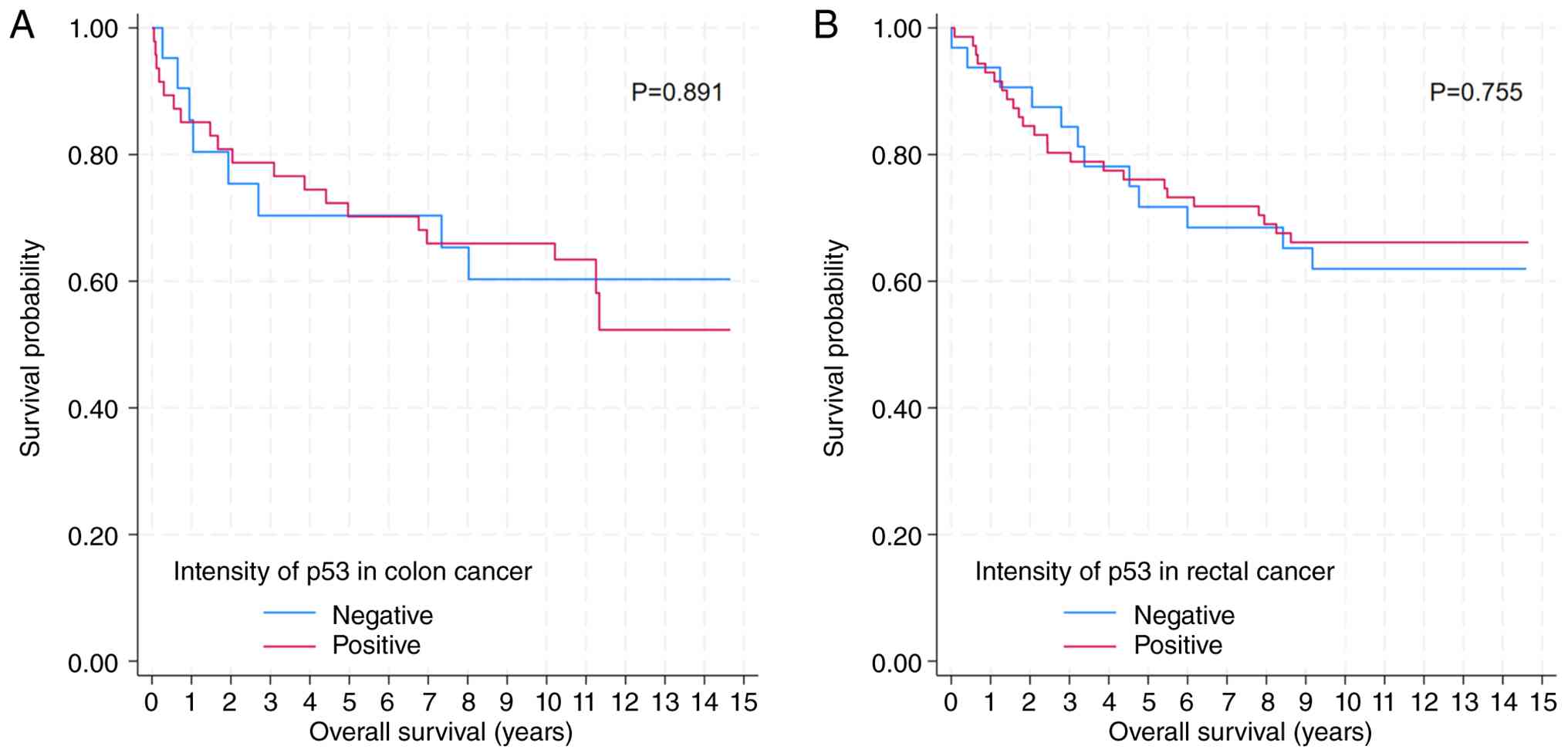
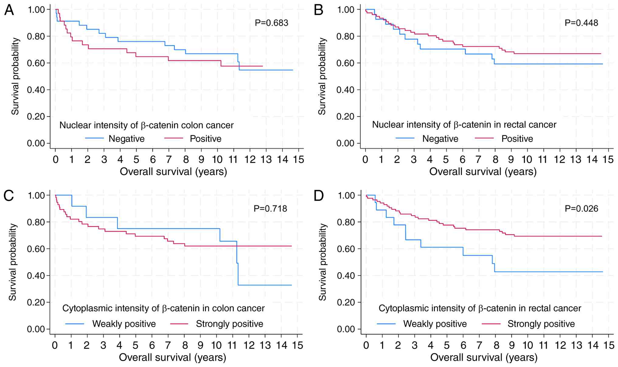
In patients with colon cancer, neither p53
expression nor nuclear β-catenin intensity was significantly
associated with OS (P=0.891 and P=0.683, respectively). Similar
results were observed in rectal cancer patients (P=0.755 and
P=0.448, respectively; Figs. 2, and
3A and B). Cytoplasmic β-catenin
intensity (weak positive vs. strong positive) was not associated
with OS in colon cancer (P=0.718; Fig.
3C). However, among rectal cancer patients (n=103), those with
lower cytoplasmic β-catenin intensity (weak positive) exhibited
significantly shorter OS compared with those with higher intensity
(strong positive) (median OS: 94 months vs. not reached; P=0.026;
Fig. 3D). Subsequent analyses
therefore focused on cytoplasmic β-catenin intensity in the rectal
cancer cohort.
Baseline characteristics according to
cytoplasmic β-catenin intensity in rectal cancer
Among the 103 rectal cancer patients, 18 (17.5%) had
cytoplasmic β-catenin intensity of weak positive and 85 (82.5%)
showed strong positive intensity. Baseline clinicopathological
characteristics, including sex, age, histological differentiation,
TNM stage distribution, and baseline carcinoembryonic antigen (CEA)
levels, did not differ significantly between the two groups
(Table II), indicating
well-balanced baseline characteristics.
| Table II.Baseline characteristics of patients
with rectal cancer. |
Table II.
Baseline characteristics of patients
with rectal cancer.
|
| Cytoplasmic β-catenin
intensity |
|
|---|
|
|
|
|
|---|
| Characteristics | Weakly positive | Strongly
positive | P-value |
|---|
| No. (%) | 18 (17.5) | 85 (82.5) |
|
| Sex, n (%) |
|
| 0.186 |
| Male | 13 (72.2) | 47 (55.3) |
|
|
Female | 5 (27.8) | 38 (44.7) |
|
| Median age, years
(IQR) | 62 (54–70) | 63 (58–70) | 0.487 |
| Histological |
|
| 0.394 |
| differentiation, n
(%) |
|
|
|
|
W/D | 5 (27.8) | 27 (31.8) |
|
|
M/D | 11 (61.1) | 55 (64.7) |
|
| P/D and
others | 2 (11.1) | 3 (3.5) |
|
| TNM stage, n
(%) |
|
| 0.229 |
| I | 1 (5.6) | 22 (25.9) |
|
| II | 7 (38.9) | 33 (38.8) |
|
|
III | 9 (50.0) | 27 (31.8) |
|
| IV | 1 (5.6) | 3 (3.5) |
|
| Mean CEA, ng/ml
(SD) | 3.9 (8.0) | 4.4 (8.2) | 0.137 |
Multivariate prognostic analysis in
rectal cancer
Multivariate Cox proportional hazards regression
analysis demonstrated that lower cytoplasmic β-catenin intensity
(weak positive) was an independent predictor of worse OS compared
with higher intensity (strong positive) (HR 2.271; 95% CI
1.063–4.852; P=0.034), after adjustment for other covariates
(Table III). As expected,
advanced TNM stage (III/IV) was strongly associated with poor OS
(HR 17.856; 95% CI 4.895–65.140; P<0.001). In contrast, p53
expression was not independently associated with OS. A significant
association was observed between cytoplasmic and nuclear β-catenin
intensity (Fisher's exact test; P<0.001; Fig. 4). However, despite this association,
only cytoplasmic β-catenin intensity remained independently
associated with overall survival in rectal cancer, whereas nuclear
β-catenin intensity showed no prognostic significance.
| Table III.Multivariate Cox regression analysis
of overall survival in rectal cancer. |
Table III.
Multivariate Cox regression analysis
of overall survival in rectal cancer.
| Variable | HR (95% CI) | P-value |
|---|
| Sex |
|
|
|
Male | Ref. |
|
|
Female | 0.625
(0.289–1.354) | 0.234 |
| Age, years (per
year) | 1.016
(0.982–1.051) | 0.359 |
| Histological
differentiation |
|
|
|
W/D | Ref. |
|
|
M/D | 0.940
(0.423–2.085) | 0.878 |
| P/D or
others | 3.071
(0.828–11.395) | 0.094 |
| TNM stage |
|
|
| I | Ref. |
|
| II | 1.685
(0.762–3.725) | 0.197 |
|
III/IV | 17.856
(4.895–65.140) | <0.001 |
| p53 |
|
|
|
Negative | Ref. |
|
|
Positive | 1.103
(0.509–2.393) | 0.804 |
| Cytoplasmic
β-catenin intensity |
|
|
| Strong
positive | Ref. |
|
| Weak
positive | 2.271
(1.063–4.852) | 0.034 |
Discussion
The primary objective of this study was to evaluate
the prognostic significance of p53 expression and β-catenin
intensity, considering both nuclear and cytoplasmic localization,
in patients with colorectal cancer. Our findings demonstrate that
neither p53 expression nor nuclear β-catenin intensity is
significantly associated with OS in colorectal cancer. In contrast,
lower cytoplasmic β-catenin intensity is independently associated
with worse OS in rectal cancer, even after adjustment for
established prognostic factors such as TNM stage. As expected,
advanced TNM stage (III/IV) was associated with the highest hazard
ratio (HR=17.856), reflecting its established role in clinical
oncology. Nevertheless, the independent prognostic value of
cytoplasmic β-catenin intensity (HR=2.271, P<0.05) remains
clinically relevant. Multivariate Cox regression confirmed that low
cytoplasmic β-catenin expression serves as an independent
prognostic factor, indicating that it provides incremental risk
stratification beyond TNM staging. This finding may help identify
patients at higher risk of poor outcomes who could benefit from
closer postoperative surveillance.
Previous studies have reported heterogeneous results
regarding the prognostic role of cytoplasmic β-catenin in
gastrointestinal adenocarcinomas (21). Some studies have shown that
increased cytoplasmic β-catenin expression is associated with
improved survival (16). However,
meta-analyses have demonstrated no significant association between
cytoplasmic β-catenin expression and overall survival (22). The prognostic relevance of
membranous β-catenin localization has also been emphasized, with
several studies reporting that reduced membrane β-catenin
expression is associated with poorer survival outcomes (15). Furthermore, the progressive
redistribution of β-catenin from the cell membrane to the nucleus
during the transition from normal colonic epithelium to benign,
premalignant, and malignant lesions suggests that its subcellular
location is closely related to the biological alterations
underlying cancer progression (19).
β-catenin is a multifunctional protein that plays
critical roles in both cellular architecture and signal
transduction. It serves as a key component of adherens junctions,
linking cadherins to the actin cytoskeleton and thereby maintaining
cell-to-cell adhesion (18).
Simultaneously, β-catenin functions as a transcriptional
coactivator in the Wnt signaling pathway (18). In the absence of Wnt signaling,
cytoplasmic β-catenin is targeted for degradation by the
destruction complex. Upon binding of Wnt ligands to their
receptors, this complex is inhibited, leading to cytoplasmic
accumulation of β-catenin (16).
The accumulated β-catenin subsequently translocates to the nucleus,
where it interacts with TCF/LEF transcription factors and activates
the expression of genes involved in cell proliferation,
differentiation, and survival (18). This process is generally recognized
as a key driver of Wnt pathway activation and tumorigenesis
(22). Therefore, the present
finding that lower cytoplasmic β-catenin intensity is associated
with poorer prognosis suggests that the underlying biological
mechanisms may be more complex than previously understood.
One possible explanation for these findings is that,
in rectal cancer, a certain threshold level of cytoplasmic
β-catenin may be necessary to maintain adequate intercellular
adhesion through its interaction with E-cadherin at the cell
membrane (16). Reduced cytoplasmic
β-catenin levels may lead to decreased membrane-associated
β-catenin, resulting in weakened cell-to-cell adhesion, enhanced
cellular motility, and an increased propensity for invasion and
metastasis, ultimately contributing to poorer survival outcomes.
Alternatively, low cytoplasmic β-catenin intensity in rectal cancer
may reflect a distinct pattern of Wnt signaling dysregulation.
Higher cytoplasmic β-catenin levels may be associated with a
relatively less invasive phenotype, whereas the reduced levels
observed in the weak-positive group may impair cellular functions
essential for maintaining epithelial integrity and survival
(23). Furthermore, β-catenin is
known to interact with multiple other signaling pathways, including
EGFR, Hippo, and PI3K/Akt, and the crosstalk among these pathways
may differ between colon and rectal cancers. The molecular
mechanisms linking low cytoplasmic β-catenin expression to poor
prognosis in rectal cancer remain to be fully elucidated, but
several pathways may explain this phenomenon. Cytoplasmic depletion
of β-catenin may reflect accelerated degradation via the
destruction complex or enhanced nuclear translocation, thereby
promoting EMT and tumor invasiveness. In addition, disruption of
the E-cadherin/β-catenin complex may weaken cell-cell adhesion and
contribute to metastatic potential. Although cytoplasmic and
nuclear β-catenin expression are biologically related, only
cytoplasmic β-catenin intensity was independently associated with
prognosis in our cohort. This finding suggests that cytoplasmic
β-catenin expression may have greater clinical relevance than
nuclear localization for risk stratification in rectal cancer.
Nuclear accumulation of β-catenin is generally
regarded as a surrogate marker of Wnt pathway activation and has
traditionally been considered a poor prognostic factor (22–25).
However, previous studies have reported inconsistent prognostic
associations. Consistent with these observations, nuclear β-catenin
intensity was not independently associated with overall survival in
our cohort, suggesting that nuclear localization alone may not
adequately reflect the biologically relevant activity of Wnt
pathway in rectal cancer.
p53 is a well-established tumor suppressor gene, and
aberrant expression is frequently observed in colorectal cancer.
However, the prognostic significance of p53 remains controversial
(26). Some studies have reported
that p53 expression is associated with poorer survival outcomes
(17), whereas others have
demonstrated no significant association or even a favorable
prognostic impact (26). These
discrepancies suggest that the clinical relevance of p53 may depend
on the specific pattern of alteration, including p21 status,
protein overexpression, or complete loss of expression (27). In the present study, the lack of a
significant association between p53 expression and prognosis may be
attributable to the heterogeneity of TP53 mutations within this
cohort, or to attenuation of any early prognostic effect over a
prolonged follow-up period.
This study has several limitations. First, as a
retrospective analysis, it is inherently limited in establishing
causal relationships, and the single-center design restricts the
generalizability of the findings. Second, a notable limitation is
the relatively small sample size of the rectal cancer subgroup with
weak cytoplasmic β-catenin expression (n=18). This small sample
size inherently broadens the confidence intervals, which demands
caution when generalizing the statistical robustness of our
survival analysis. Despite this limitation, the survival deficit
observed in this subgroup was statistically significant;
nevertheless, future large-scale, multi-center prospective studies
are warranted to narrow the confidence intervals and validate these
prognostic trends. Third, the contemporary applicability of these
results may be constrained, as patient enrollment (2000–2007)
preceded the establishment of current standard treatment
strategies. We acknowledge that our study cohort may not fully
reflect recent advancements in rectal cancer management. Over the
past two decades, the widespread implementation of total mesorectal
excision (TME), standardized neoadjuvant chemoradiotherapy (CRT),
and more recently, total neoadjuvant therapy (TNT) have
significantly improved local control and overall survival rates.
These therapeutic shifts could potentially alter the prognostic
impact of cytoplasmic β-catenin expression observed in our
historical cohort. However, our findings remain highly relevant as
they identify a distinct biological subgroup that may exhibit
differential treatment responses, providing a baseline for future
validation in modern cohorts. Finally, although
immunohistochemistry (IHC) is a widely utilized method, it has
inherent limitations in quantitatively and functionally reflecting
protein activity.
In conclusion, lower cytoplasmic β-catenin intensity
is independently associated with poor overall survival in patients
with rectal cancer, whereas p53 expression and nuclear β-catenin
intensity are not prognostically significant. These findings
highlight the importance of β-catenin subcellular localization in
rectal cancer prognosis and warrant further validation in larger,
prospective studies.
Supplementary Material
Supporting Data
Acknowledgements
The authors would like to thank Professor Gyung
Hyuck Ko (Department of Pathology, Gyeongsang National University
Hospital, Jinju, Republic of Korea) for his technical assistance
with immunohistochemical staining.
Funding
The present study was supported by the New Faculty Research
Support Grant from Gyeongsang National University in 2025 (grant
no. GNU-NFRSG-0036), a grant of the National R&D Program for
Cancer Control, Ministry for Health, Welfare and Family Affairs,
Republic of Korea (grant no. 0820050).
Availability of data and materials
The data generated in the present study are not
publicly available due to patient privacy considerations but may be
requested from the corresponding author.
Authors' contributions
MHK designed the study, performed the data analysis
and wrote the manuscript. SIG performed the data analysis. DCK and
DHS performed the immunohistochemical staining. YTJ contributed to
patient selection and to the acquisition and interpretation of
clinical data, including determination of tumor location in cases
with ambiguous primary sites and completion of missing clinical
information. WSL conceived and designed the study and critically
revised the manuscript. MHK and SIG confirm the authenticity of all
the raw data. All authors have read and approved the final version
of the manuscript.
Ethics approval and consent to
participate
The present study was approved by the Institutional
Review Board of Gyeongsang National University Changwon Hospital
(approval no. 2025-06-011; Changwon, Republic of Korea). The
requirement for written informed consent was waived due to the
retrospective nature of the study and the use of anonymized
clinical data.
Patient consent for publication
Not applicable.
Competing interests
The authors declare that they have no competing
interests.
Use of artificial intelligence tools
During the preparation of this work, artificial
intelligence tools were used to generate images, and subsequently,
the authors revised and edited the content produced by the
artificial intelligence tools as necessary, taking full
responsibility for the ultimate content of the present
manuscript.
References
|
1
|
Siegel RL, Miller KD, Fuchs HE and Jemal
A: Cancer statistics, 2021. CA Cancer J Clin. 71:7–33.
2021.PubMed/NCBI
|
|
2
|
Hong S, Won YJ, Park YR, Jung KW, Kong HJ
and Lee ES: Cancer statistics in Korea: Incidence, mortality,
survival and prevalence in 2017. Cancer Res Treat. 52:335–350.
2020. View Article : Google Scholar : PubMed/NCBI
|
|
3
|
Smith G, Carey FA, Beattie J, Wilkie MJ,
Lightfoot TJ, Coxhead J, Garner RC, Steele RJ and Wolf CR:
Mutations in APC, Kirsten-ras and p53-alternative genetic pathways
to colorectal cancer. Proc Natl Acad Sci USA. 99:9433–9438. 2002.
View Article : Google Scholar : PubMed/NCBI
|
|
4
|
Hinton K, Kirk A, Paul P and Persad S:
Regulation of the epithelial to mesenchymal transition in
osteosarcoma. Biomolecules. 13:3982023. View Article : Google Scholar : PubMed/NCBI
|
|
5
|
Ribatti D, Tamma R and Annese T:
Epithelial-mesenchymal transition in cancer: A historical overview.
Transl Oncol. 13:1007732020. View Article : Google Scholar : PubMed/NCBI
|
|
6
|
Takayama T, Miyanishi K, Hayashi T, Sato Y
and Niitsu Y: Colorectal cancer: Genetics of development and
metastasis. J Gastroenterol. 41:185–192. 2006. View Article : Google Scholar : PubMed/NCBI
|
|
7
|
Iacopetta B: TP53 mutation in colorectal
cancer. Hum Mutat. 21:271–276. 2003. View Article : Google Scholar : PubMed/NCBI
|
|
8
|
Liebl MC and Hofmann TG: The role of p53
signaling in colorectal cancer. Cancers (Basel). 13:21252021.
View Article : Google Scholar : PubMed/NCBI
|
|
9
|
Diaz-Lopez A, Moreno-Bueno G and Cano A:
Role of microRNA in epithelial to mesenchymal transition and
metastasis and clinical perspectives. Cancer Manag Res. 6:205–216.
2014.PubMed/NCBI
|
|
10
|
Tang Q, Su Z, Gu W and Rustgi AK: Mutant
p53 on the path to metastasis. Trends Cancer. 6:62–73. 2020.
View Article : Google Scholar : PubMed/NCBI
|
|
11
|
Valenta T, Hausmann G and Basler K: The
many faces and functions of β-catenin. EMBO J. 31:2714–2736. 2012.
View Article : Google Scholar : PubMed/NCBI
|
|
12
|
Kim WK, Kwon Y, Jang M, Park M, Kim J, Cho
S, Jang DG, Lee WB, Jung SH, Choi HJ, et al: β-catenin activation
down-regulates cell-cell junction-related genes and induces
epithelial-to-mesenchymal transition in colorectal cancers. Sci
Rep. 9:184402019. View Article : Google Scholar : PubMed/NCBI
|
|
13
|
Salon C, Lantuejoul S, Eymin B, Gazzeri S,
Brambilla C and Brambilla E: The e-cadherin-beta-catenin complex
and its implication in lung cancer progression and prognosis.
Future Oncol. 1:649–660. 2005. View Article : Google Scholar : PubMed/NCBI
|
|
14
|
Li LF, Wei ZJ, Sun H and Jiang B: Abnormal
beta-catenin immunohistochemical expression as a prognostic factor
in gastric cancer: A meta-analysis. World J Gastroenterol.
20:12313–12321. 2014. View Article : Google Scholar : PubMed/NCBI
|
|
15
|
Matly A, Quinn JA, Mcmillan DC, Park JH
and Edwards J: The Relationship between beta-catenin and patient
survival in colorectal cancer systematic review and meta-analysis.
Crit Rev Oncol Hematol. 163:1033372021. View Article : Google Scholar : PubMed/NCBI
|
|
16
|
Bruun J, Kolberg M, Nesland JM, Svindland
A, Nesbakken A and Lothe RA: Prognostic significance of β-Catenin,
E-Cadherin, and SOX9 in colorectal cancer: Results from a large
Population-Representative series. Front Oncol. 4:1182014.
View Article : Google Scholar : PubMed/NCBI
|
|
17
|
Auvinen A, Isola J, Visakorpi T, Koivula
T, Virtanen S and Hakama M: Overexpression of p53 and long-term
survival in colon carcinoma. Br J Cancer. 70:293–296. 1994.
View Article : Google Scholar : PubMed/NCBI
|
|
18
|
Wong NA and Pignatelli M: Beta-catenin-a
linchpin in colorectal carcinogenesis? Am J Pathol. 160:389–401.
2002. View Article : Google Scholar : PubMed/NCBI
|
|
19
|
Bhattacharya I, Barman N, Maiti M and
Sarkar R: Assessment of beta-catenin expression by
immunohistochemistry in colorectal neoplasms and its role as an
additional prognostic marker in colorectal adenocarcinoma. Med
Pharm Rep. 92:246–252. 2019. View Article : Google Scholar : PubMed/NCBI
|
|
20
|
Nazemalhosseini Mojarad E, Kashfi SM,
Mirtalebi H, Almasi S, Chaleshi V, Kishani Farahani R, Tarban P,
Molaei M, Zali MR and J K Kuppen P: Prognostic significance of
nuclear β-catenin expression in patients with colorectal cancer
from Iran. Iran Red Crescent Med J. 17:e223242015. View Article : Google Scholar : PubMed/NCBI
|
|
21
|
Norwood MG, Bailey N, Nanji M, Gillies RS,
Nicholson A, Ubhi S, Darnton JJ, Steyn RS, Womack C, Hughes A, et
al: Cytoplasmic beta-catenin accumulation is a good prognostic
marker in upper and lower gastrointestinal adenocarcinomas.
Histopathology. 57:101–111. 2010. View Article : Google Scholar : PubMed/NCBI
|
|
22
|
Chen Z, He X, Jia M, Liu Y, Qu D, Wu D, Wu
P, Ni C, Zhang Z, Ye J, et al: β-catenin overexpression in the
nucleus predicts progress disease and unfavourable survival in
colorectal cancer: A meta-analysis. PLoS One. 8:e638542013.
View Article : Google Scholar : PubMed/NCBI
|
|
23
|
Zhang Y and Wang X: Targeting the
wnt/β-catenin signaling pathway in cancer. J Hematol Oncol.
13:1652020. View Article : Google Scholar : PubMed/NCBI
|
|
24
|
Song P, Gao Z, Bao Y, Chen L, Huang Y, Liu
Y, Dong Q and Wei X: Wnt/β-catenin signaling pathway in
carcinogenesis and cancer therapy. J Hematol Oncol. 17:462024.
View Article : Google Scholar : PubMed/NCBI
|
|
25
|
Elzagheid A, Buhmeida A, Korkeila E,
Collan Y, Syrjanen K and Pyrhonen S: Nuclear beta-catenin
expression as a prognostic factor in advanced colorectal carcinoma.
World J Gastroenterol. 14:3866–3871. 2008. View Article : Google Scholar : PubMed/NCBI
|
|
26
|
Kruschewski M, Mueller K, Lipka S,
Budczies J, Noske A, Buhr HJ and Elezkurtaj S: The prognostic
impact of p53 expression on sporadic colorectal cancer is dependent
on p21 status. Cancers (Basel). 3:1274–1284. 2011. View Article : Google Scholar : PubMed/NCBI
|
|
27
|
Nagao K, Koshino A, Sugimura-Nagata A,
Nagano A, Komura M, Ueki A, Ebi M, Ogasawara N, Tsuzuki T, Kasai K,
et al: The complete loss of p53 expression uniquely predicts worse
prognosis in colorectal cancer. Int J Mol Sci. 23:32522022.
View Article : Google Scholar : PubMed/NCBI
|