Introduction
Endometrial carcinoma comprises malignant tumors
occurring in the female genital tract and is the third most common
cause of gynecological cancer-related mortalities (1). The incidence and mortality rates have
increased in recent years, with 66,200 new cases and 13,030
mortalities reported in 2023 in America (2). Despite advancements in diagnosis,
targeted therapies and immunotherapies, the average 5-year survival
rate of endometrial carcinoma is as low as 30%, especially for
patients diagnosed in clinical phase III/IV, due to its rapid
development and distant metastasis (3). Therefore, understanding the underlying
molecular mechanism accounting for the carcinogenesis of
endometrial carcinoma is essential for developing accurate targeted
therapies.
Epigenetic modifications are involved in the
carcinogenesis of numerous types of tumors, such as cervical
(4) and hepatocellular (5) cancer. The
N6-methyladenosine (m6A) modification of RNA
is a reversible epigenetic modification, including for DNA
methylation and histone modifications (6–8).
m6A-related proteins, referred to as ‘writer’, ‘eraser’
and ‘reader’ proteins, regulate pre-mRNA splicing, mRNA stability
and the decay, localization, transport and translation of mRNA
(9–12). The m6A ‘readers’, such as
YTH N6-methyladenosine RNA binding protein C (YTHDC)1/2
and insulin-like growth factor 2 mRNA binding protein
(IGF2BP)1/2/3, identify methylated RNA and participate in the
translation and degradation of downstream proteins (13). IGF2BPs are considered to exert a key
role in biological processes such as embryogenesis, cell migration,
proliferation and invasion (14,15).
IGF2BP3 recruits nudix hydrolase 21 to regulate SPTBN1
m6A modifications, which drives ovarian cancer
progression (16).
IGF2BP3-methyltransferase like-14 complex regulates stearoyl-CoA
desaturase mRNA m6A modifications, resulting in cervical
cancer cell proliferation, metastasis and increased lipid
metabolism (17). IGF2BP3-mediated
m6A modification of the early growth response 1/PTEN
axis regulates the malignant phenotypes of endometrial cancer stem
cells (18).
IGF2BP3 enhances the mRNA stability of E2F
transcription factor by interacting with long independently
transcribed non-coding RNA 958 to promote endometrial carcinoma
progression (19). Long non-coding
RNA metastasis associated lung adenocarcinoma transcript 1 and
urothelial cancer associated 1 can be used as biomarkers for the
prognosis of patients with glioblastoma (20). Circular RNAs (circRNAs/circs) are
dysregulated in nearly all types of cancer and drive tumorigenesis
through tumor-suppressive functions (21). IGF2BP2-modified circ-chromodomain
helicase DNA binding protein 7 promotes endometrial cancer
progression by stabilizing platelet-derived growth factor receptor
β and activating the Janus kinase/STAT signaling pathway (22).
CircEPB41L2 can bind PTBP1 and the E3 ubiquitin
ligase TRIP12 to promote the ubiquitination and degradation of
PTBP1, block the proliferation, invasion and metastasis of NSCLC
(23), and act as a miRNA sponge
involved in the tumorigenesis of colorectal cancer (24) and lung cancer (25).
However, the biological role of circEPB41L2 in
gyneco-logical diseases, and whether IGF2BP3 can regulate
circEPB41L2 expression has not been reported. The present study
explored IGF2BP3 expression in endometrial carcinoma and identified
the underlying mechanism and signal pathway, which can be further
explored for targeted treatment in endometrial carcinoma.
Materials and methods
Cell culture and clinical
specimens
A total of 25 endometrial carcinoma and adjacent
paracancerous tissues were collected from the same female patients
with endometrial carcinoma who underwent tumor resection at The
First Affiliated Hospital of Jinan University (Guangzhou, China)
between August 2024 and December 2025. Adjacent normal tissues were
collected at least 1 cm away from the tumor margin. Patients who
had received chemotherapy, radiotherapy or hormonal therapy were
excluded. The age of the patients ranged 42–65 years, and the mean
age was 55.3 years. All patients provided written informed consent.
Clinical stages were classified according to the International
Federation of Gynecology and Obstetrics 2023 guidelines (26). The clinical parameters of the
patients are summarized in Table I.
The experimental procedures were approved by the Ethics Committee
of The First Affiliated Hospital of Jinan University (Guangzhou,
China; approval no. KY-2025-317).
 | Table I.Association of IGF2BP3 expression
with clinicopathological features in 25 patients with endometrial
carcinoma. |
Table I.
Association of IGF2BP3 expression
with clinicopathological features in 25 patients with endometrial
carcinoma.
| Variable | Total, n | IGF2BP3 (+),
n=16 | IGF2BP3 (−),
n=9 | P-value |
|---|
| Differentiation
stage |
|
|
| 0.0029 |
|
Well-moderate | 18 | 15 | 3 |
|
|
Poor | 7 | 1 | 6 |
|
| FIGO stage |
|
|
| 0.147 |
|
I–II | 19 | 13 | 4 |
|
|
III–IV | 6 | 3 | 5 |
|
| Age, years |
|
|
| 0.2203 |
|
<50 | 6 | 9 | 2 |
|
|
≥50 | 19 | 7 | 7 |
|
| Pelvic lymph node
metastasis |
|
|
| 0.734 |
|
Yes | 10 | 6 | 2 |
|
| No | 15 | 10 | 7 |
|
The HEC-1A endometrial carcinoma cell line was
obtained from American Type Culture Collection. HEC-1A cells were
cultured at 37°C with 5% CO2 in DMEM (Gibco; Thermo
Fisher Scientific, Inc.) supplemented 1% penicillin-streptomycin
solution and 10% fetal calf serum (Gibco; Thermo Fisher Scientific,
Inc.).
Functional annotation and pathway
enrichment analysis
Gene set enrichment analysis software (version
4.3.2; http://www.gsea-msigdb.org/gsea/index.jsp) was used to
perform Gene Ontology (GO) functional annotation and Kyoto
Encyclopedia of Genes and Genomes (KEGG) pathway analyses.
P<0.05 was considered to indicate a statistically significant
difference. Volcano plots displayed the differentially expressed
genes or circ RNA, and the top 20 genes were further analyzed.
Differentially expressed genes were screened with the threshold of
|log2FC|>1 and false discovery rate (FDR)<0.05.
Genes with log2FC>1 and FDR<0.05 were defined as
upregulated genes, while genes with log2FC<-1 and
FDR<0.05 were defined as downregulated genes.
Analysis of the prognostic and
diagnostic performance of IGF2BP3
IGF2BP3 gene expression profiles and matched
clinical follow-up data were retrieved from the Uterine Corpus
Endometrial Carcinoma (UCEC) cohort of The Cancer Genome Atlas
(TCGA) database (https://portal.gdc.cancer.gov) Patients were
stratified into high- and low-IGF2BP3 groups according to the
median expression level of IGF2BP3. Kaplan-Meier survival curves
were plotted for overall survival (OS) and disease-free survival
(DFS), and the log-rank test was used to compare survival
differences between groups. Hazard ratios (HRs) with 95% confidence
intervals were calculated. A receiver operating characteristic
curve was constructed and the area under the curve was calculated
to evaluate the diagnostic performance of IGF2BP3. All statistical
analyses were performed in R software (version 4.3.1; http://www.R-project.org/).
Generation of cell lines with stable
knockdown
Endogenous IGF2BP3 (NM_006547.3) was stably knocked
down using lentiviral vectors containing short hairpin RNA
(shRNA/sh) constructs: shIGF2BP3#1, 5′-CCTTGAAAGTAGCCTATAT-3′;
shIGF2BP3#2, 5′-GGTGAAACTTGAAGCTCAT-3′; and shIGF2BP3#3,
5′-CCAGACACCTGATGAGAAT-3′. The sequence of the shRNA control was as
follows: 5′-CCTAAGGTTAAGTCGCCCT-3′. Validated pLKO.1-based shRNA
and scramble control plasmids were purchased from MilliporeSigma
and used at 1 µg/µl. Lentivirus was packaged in 293T cells
(MilliporeSigma) using a third-generation system. In brief, 3 µg of
plasmids (transfer:psPAX2:pMD2.G-=4:3:1) were transfected into
70–80% confluent 293T cells per 6-well plate via Lipofectamine 3000
(Thermo Fisher Scientific, Inc.). Viral supernatants were collected
at 48–72 h, sterile-filtered (0.45 µm) and stored at −80°C. Target
cells were transduced at an MOI of 2 for 24 h with 8 µg/ml
polybrene. After medium renewal, antibiotic selection was performed
at 48 h post-transduction to establish stable knockdown cell lines.
All downstream experiments were conducted at 72 h after
transduction.
Bioinformatic prediction of
IGF2BP3-binding circRNAs
Potential circRNA targets of IGF2BP3 were predicted
using ENCORI (starBase v2.0; http://starbase.sysu.edu.cn/) database, which curates
and integrates published cross-linking immunoprecipitation followed
by high-throughput sequencing (CLIP-seq) datasets for RNA-binding
protein and RNA interaction analysis (27). Briefly, IGF2BP3 binding peaks we
searched for and candidate circRNAs with statistically significant
binding signals were retained. The default significance cut-off
threshold of the ENCORI pipeline was applied, with FDR<0.05.
Cell Counting Kit-8 (CCK-8) assay
HEC-1A cells transfected with sh-negative control
(sh-NC) or sh-IGF2BP3 were seeded in 96-well plates (5,000 cells
per well). After 24, 48, 72 and 96 h 0 µl CCK-8 solution (APExBIO
Technology LLC) was added to each well. The cells were subsequently
incubated for an additional 2 h at 37°C. The absorbance was
measured at a wavelength of 450 nm.
Transwell assay
A Transwell chamber (cat. no. 3422; Corning, Inc.)
was used to detect cell migration. A total of 3×10⁵
cells suspended in serum-free medium were added to the upper
chamber and 700 µl DMEM with 10% FBS was added to the lower
chamber. For migration assays, uncoated upper chambers were
applied. For invasion assays, upper chambers were pre-coated with
Matrigel matrix (Corning, Inc.) and gelled at 37°C. After
incubation at 37°C for 24 h, the upper chambers were rinsed with
PBS and fixed in 100% methanol for 15 min at room temperature.
Next, cells left on the surface of the chamber were carefully wiped
off with cotton swabs. Crystal violet staining was performed for 10
min at room temperature, and then the lower chambers were rinsed
again with PBS. Cells in three random fields were imaged under a
bright-field microscope (magnification, ×40) and the numbers of
cells that had passed through the cell membrane were counted using
ImageJ (version 1.54u8; National Institutes of Health).
Whole-transcriptome sequencing
Total RNA was isolated using TRIzol reagent (cat.
no. 15596026; Thermo Fisher Scientific, Inc.). Ribosomal RNA
depletion was performed using a Ribo-Zero rRNA Removal Kit (cat.
no. MRZH11124; Illumina, Inc.). For quality verification, the RNA
concentration and purity were determined using a Nanodrop 2000
spectrophotometer (Thermo Fisher Scientific, Inc.), and RNA
integrity was assessed using an Agilent 2100 Bioanalyzer (Agilent
Technologies, Inc.). Only samples with an optical density
(OD)260/280 of 1.8–2.2, OD260/230 of 2.0–2.4 and RNA integrity
number ≥7.0 were used for subsequent analysis.
Whole-transcriptome libraries were constructed using
a TruSeq Stranded Total RNA Library Prep Kit (cat. no. 20020596;
Illumina, Inc.). The final library concentration was accurately
quantified with a Qubit 4.0 Fluorometer (Invitrogen; Thermo Fisher
Scientific, Inc.), and libraries were normalized to a loading
concentration of 1.5 nM. Sequencing was performed on the Illumina
NovaSeq 6000 platform (Illumina, Inc.) with the paired-end 150-bp
sequencing mode.
Raw sequencing reads were quality-trimmed and
filtered using Trimmomatic v0.39 (www.usadellab.org/cms/page=trimmomatic). Clean reads
were aligned to the human reference genome using HISAT2 v2.2.1
(https://daehwankimlab.github.io/hisat2/). Transcript
expression levels (transcripts per million) were quantified using
StringTie v2.2.1(https://ccb.jhu.edu/software/stringtie/). Differential
expression analysis was conducted using DESeq2 v1.52.0 (R v4.6.1),
with thresholds of |log2FC|≥1 and adjusted P<0.05.
All software was used with default parameters as documented in the
official developer manuals.
RT-qPCR
Total RNA was extracted from cells or tissue using
TRIzol Reagent (Invitrogen; Thermo Fisher Scientific, Inc.) in
accordance with the manufacturer's protocol. The PrimeScript™ RT
reagent kit (Takara Biotechnology Co., Ltd.) was used to reverse
transcribe RNA into cDNA. The reaction program was set as 42°C for
2 min (gDNA elimination), 37°C for 15 min (reverse transcription)
and 85°C for 5 sec (heat inactivation). The cDNA was preserved at
−20°C. For qPCR, the reaction system was constructed according to
the protocol of the SYBR Premix Ex Taq™ II kit (Takara Bio, Inc.).
The expression levels of RNA were determined using the Bio-Rad
CFX96 real-time PCR system (Bio-Rad Laboratories, Inc.). The human
housekeeping gene GAPDH was used as the internal control. The
sequences of the primers utilized in the present study were as
follows: IGF2BP3 forward, 5′-CCGTATCCAAGCAGAAACCA-3′ and reverse,
5′-AATCGACTTCTCAGCAGCCC-3′; GAPDH forward, 5′-AAGGCTGTGGGCAAGG-3′
and reverse, 5′-TGGAGGAGTGGGTGTCG-3′; circ-EPB41L2 forward,
5′-ACCAAGGAAGTGCAGACCAA-3′ and reverse, 5′-TCACTTCAGACACAGAGCCT-3′;
circ-WDPCP forward, 5′-AACGATGATGCTTGGCCTTG-3′ and reverse,
5′-TCTGAGATGAGCTTCCCCAC-3′; and circ-PH20 forward,
5′-GAACCGACTTCTCCCCTTGT-3′ and reverse, 5′-AGGCCTAGCATTACCCACAA-3′.
The thermocycling conditions were as follows: One cycle at 95°C for
10 min, followed by 40 cycles of 95°C for 15 sec and 60°C for 60
sec. Melting curves were generated to confirm specific
amplification. Relative gene expression was quantified by
2−ΔΔCq method (28),
normalized to GAPDH.
Western blot analysis
After PBS washing, cells were lysed in RIPA buffer
(cat. no. R0010; Beijing Solarbio Science & Technology Co.,
Ltd.) supplemented with 1% PMSF (cat. no. P0100; Beijing Solarbio
Science & Technology Co., Ltd.) on ice for 30 min. Lysates were
centrifuged at 12,000 × g for 15 min at 4°C and protein
concentrations were quantified using a BCA assay kit (cat. no.
MA0082; Dalian Meilun Biotechnology Co., Ltd.). Proteins (30
mg/lane) were separated by SDS-PAGE (cat. no. P0015L; Beyotime
Biotechnology) and transferred to PVDF membranes (Roche
Diagnostics). Membranes were blocked with quick block buffer (cat.
no. P0023B; Beyotime Biotechnology) at room temperature for 30 min
and incubated with primary antibodies IGF2BP1 (1:1,500; cat. no.
ab184305; Abcam) IGF2BP3 (1:2,000; cat. no. ab225697; Abcam) and
GAPDH (1:1,000; cat. no. MA5-15738; Thermo Fisher Scientific, Inc.)
overnight at 4°C. After incubation with horseradish
peroxidase-conjugated secondary antibody IgG (H+L) (1:1,000; cat.
no. A0352; Beyotime Biotechnology) for 1 h at room temperature,
protein bands were visualized using ECL reagent (cat. no. MA0186;
Dalian Meilun Biotechnology Co., Ltd.).
Methylated RNA immunoprecipitation
(MeRIP)
MeRIP analysis was performed according to MeRIP
m6A kit (cat. no. C11051-1; Guangzhou RiboBio Co., Ltd).
Total RNA was extracted and chemically fragmented at 70°C for 5 min
to produce 100–200 nucleotide RNA fragments. For each IP reaction,
100 µg fragmented RNA was incubated with the kit-supplied
anti-m6A antibody and Protein A/G magnetic beads at 4°C
overnight; IgG from the kit served as the negative control. The
bead-RNA-antibody complexes were washed three times with pre-cooled
1X IP buffer. Immunoprecipitated RNA was eluted, purified and
subjected to RT-qPCR to evaluate m6A enrichment.
Statistical analysis
All experiments were performed in triplicate with
consistent and reproducible results. Statistical analysis was
carried out using SPSS 16.0 software (SPSS, Inc.), and data are
presented as the mean ± SD. Comparisons between two groups were
performed using t-tests. Matched samples were analyzed using a
paired-samples t-test. For independent samples, the homogeneity of
variance test was performed first to select the appropriate test.
If the variance was homogeneous (P>0.05), independent-samples
t-test was used for group comparison. One-way ANOVA was performed
to analyze differences among multiple groups, with Tukey's test
adopted for post hoc pairwise comparisons. The associations between
IGF2BP3 expression and clinicopathological features were analyzed
using Fisher's exact test. P<0.05 was considered to indicate a
statistically significant difference.
Results
m6A reader proteins are
involved in endometrial carcinoma
First, the expression levels of the m6A
methylation-related reader proteins in 35 normal and 546
endometrial carcinoma tissues from TCGA database were compared and
analyzed (Fig. 1A). IGF2BP1,
IGF2BP3 and RNA binding motif protein 15 (RBM15) were revealed to
be upregulated, while YTHDC2 was downregulated in endometrial
carcinoma. Upregulated expression of IGF2BP1, IGF2BP3 and RBM15,
but downregulated expression of YTHDC2 was associated with poorer
survival (Fig. 1B). To further
verify the expression of the aforementioned genes, qPCR was
performed for 5 pairs of endometrial carcinoma tissues and
corresponding paracancerous tissues. The results indicated that
only IGF2BP1 and IGF2BP3 were expressed at significantly higher
levels in endometrial cancer tissues (Fig. 1C). Subsequently, 25 clinical samples
were used to further detect the expression levels of IGF2BP1 and
IGF2BP3 by RT-qPCR and western blotting. AlthoughIGF2BP3 and
IGF2BP1 expression was significantly upregulated in endometrial
carcinoma tissues compared with paracancerous tissues, only the
mRNA and protein expression of IGF2BP3 was consistently increased
(Fig. 1D and E). Therefore, IGF2BP3
was selected as the target gene to perform further research.
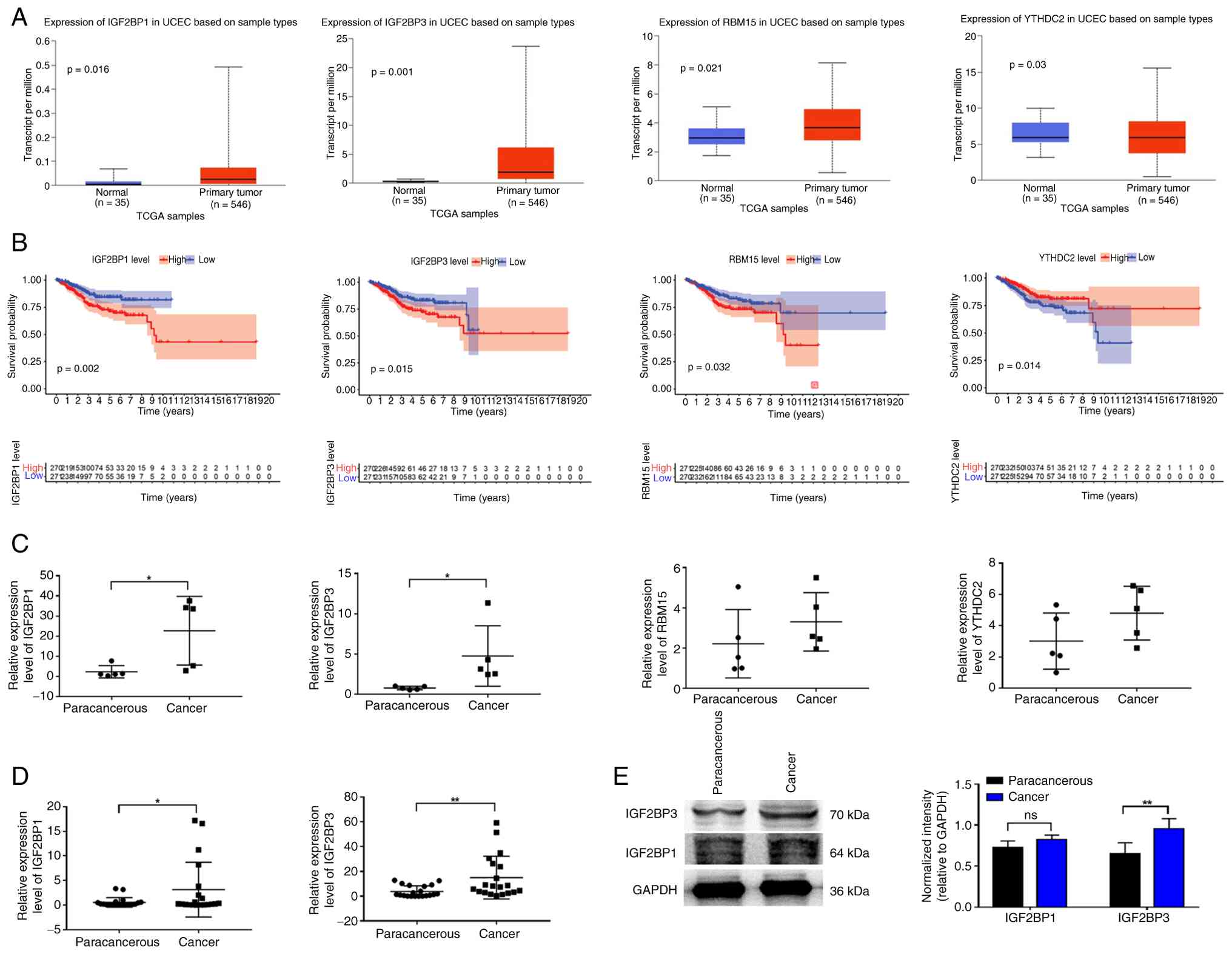 | Figure 1.N6-methyladenosine reader
protein expression in endometrial carcinoma. (A) IGF2BP1, IGF2BP3,
RBM15 and YTHDC2 expression in normal and tumor tissues from TCGA.
(B) Kaplan-Meier analysis of patients with high/low expression of
IGF2BP1, IGF2BP3, RBM15 and YTHDC2. (C) IGF2BP1, IGF2BP3, RBM15 and
YTHDC2 expression was upregulated in cancer compared with
paracancerous tissues from the GEO database. (D) IGF2BP1 and
IGF2BP3 mRNA expression in cancer and paracancerous tissues. (E)
IGF2BP1 and IGF2BP3 protein expression in cancer and paracancerous
tissues. An unpaired-samples t-test was used in the analysis of
normal and tumor tissues from TCGA in (A), while a paired t-test
was used to analyze the data in (C-E). *P<0.05, **P<0.01. ns,
not significant; IGF2BP, insulin-like growth factor 2 mRNA binding
protein; RBM15, RNA binding motif protein 15; YTHDC2, YTH
N6-methyladenosine RNA binding protein C2; TCGA, The
Cancer Genome Atlas; UCEC, uterine corpus endometrial
carcinoma. |
Association of IGF2BP3 with
unfavorable prognosis in endometrial carcinoma
To further explore the association between IGF2BP3
expression and patient prognosis, the overall survival (OS) and The
DFS and OS rates of patients with endometrial carcinoma were
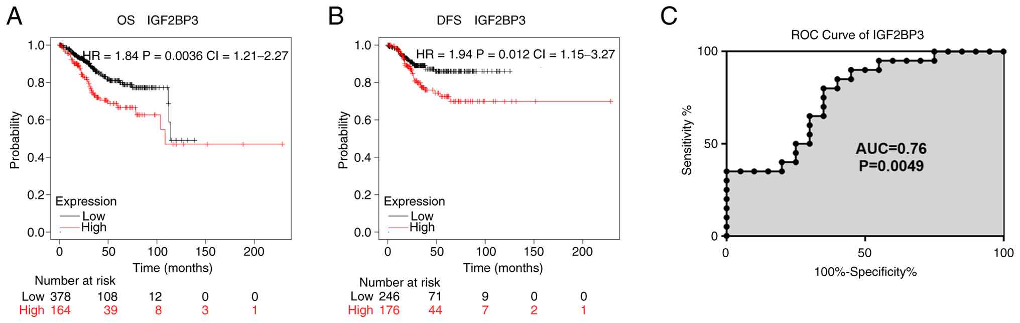
evaluated via Kaplan-Meier survival analysis. Patients with high
IGF2BP3 expression exhibited significantly shorter OS and DFS times
(Fig. 2A and B). Furthermore, the
potential diagnostic value of IGF2BP3 expression in endometrial
carcinoma was evaluated using a receiver operating characteristic
curve. As shown in Fig. 2C, the
area under the curve for IGF2BP3 was 0.76 with a sensitivity of 80%
and a specificity of 65%.
IGF2BP3 facilitates the proliferation
and migration of endometrial carcinoma cells in vitro
To determine the biological role of IGF2BP3 in
vitro, HEC-1A endometrial carcinoma cells were transfected with
sh-IGF2BP3 or sh-NC. The knockdown efficiency of IGF2BP3 was
verified by RT-qPCR (Fig. 3A).
sh-IGF2BP3-1 and sh-IGF2BP3-3 significantly reduced the mRNA level
of IGF2BP3, while sh-IGF2BP3-2 showed no notable knockdown effect
Flow cytometry was used to evaluate lentiviral infection efficiency
by detecting EGFP fluorescence (Fig.
3B). The EGFP-positive rates of all three groups exceeded 93%,
indicating satisfactory lentivirus transduction for subsequent
functional experiments. Western blot analysis demonstrated that
IGF2BP3 protein expression was significantly decreased after
transfection with sh-IGF2BP3-1. (Fig.
3C). Therefore, sh-IGF2BP3-1 and sh-IGF2BP3-3 were used to
detect functional changes in HEC-1A cells. The results indicated
that downregulation of IGF2BP3 significantly decreased cell
viability (Fig. 3D). Transwell
migration and invasion assays demonstrated that knockdown of
IGF2BP3 markedly suppressed cell migration and invasion compared
with the sh-NC group (Fig. 3E).
 | Figure 3.IGF2BP3 regulates the proliferation
and migration of endometrial cancer cells. (A) IGF2BP3 expression
in sh-NC and sh-IGF2BP3 groups was determined by reverse
transcription-quantitative PCR. (B) Transfection efficacy was
determined by flow cytometry. EGFP excited by 488 nm laser emits
green fluorescence at 509 nm, detected via the FITC (B525) channel.
Successfully transfected cells are fluorescence-positive (V1R) and
non-transfected cells are negative (V1L). Transfection efficiency
is the percentage of positive cells. (C) IGF2BP3 expression in the
sh-NC and sh-IGF2BP3 groups was examined by western blotting. (D)
Proliferation of cells in the sh-NC and sh-IGF2BP3 groups was
determined using an CCK-8assay. (E) Migration of cells in the sh-NC
and sh-IGF2BP3 groups was determined by Transwell assay. One-way
ANOVA followed by Tukey's multiple comparison test t were used to
analyze data. **P<0.01. IGF2BP3, insulin-like growth factor 2
mRNA binding protein 3; sh, short hairpin RNA; NC, negative
control; ns, not significant. P1, population 1; V1L, gate V1 left;
V1R, gate V1 right; B525, blue laser; FITC, fluorescein
isothiocyanate. EGFP enhanced green fluorescent protein. |
Identification of the circRNA
expression profile in endometrial carcinoma
CircRNAs are considered to serve a key role in the
clinical diagnosis, treatment and prognosis evaluation of tumors
due to their high stability and tissue specificity (29). RNA-sequencing analysis of
endometrial carcinoma tissue and paracancerous tissue was
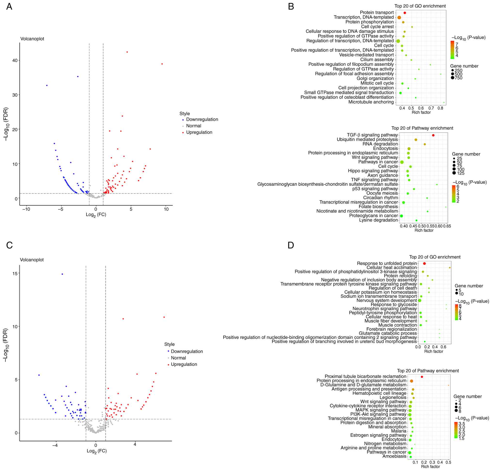
performed. A total of 168 circRNAs were upregulated and 273
circRNAs were downregulated in tumor tissue (Fig. 4A). KEGG and GO analysis indicated
that the significantly altered genes were involved in ‘regulation
of transcription’ and ‘cellular response to DNA damage stimulus’
and associated with the ‘TGF-β signaling pathway’ and the ‘Wnt
signaling pathway’ (Fig. 4B).
Furthermore, the downregulation of IGF2BP3 resulted in dysregulated
gene expression, including upregulation of 178 genes and
downregulation of 232 genes, in transfected endometrial carcinoma
cells (Fig. 4C). The significantly
altered terms and pathways included ‘cellular heat acclimation’,
the ‘transmembrane receptor protein tyrosine kinase signaling
pathway’, the ‘Wnt signaling pathway’ and ‘MAPK, PI3K-Akt pathway’
(Fig. 4D). These results suggested
that circRNAs were abnormally expressed in endometrial carcinoma
and may be regulated by IGF2BP3.
IGF2BP3 regulates circ-EPB41L2 in an
m6A-dependent manner
To determine the specific targeted circRNA which
corresponded to IGF2BP3, screening of candidate circRNAs regulated
by IGF2BP3 was performed using the CLIP-seq database according to
the following criteria: i) The trend of circRNA expression was
consistent with that of IGF2BP3, namely, the expression of circRNA
in endometrial cancer was dysregulated; ii) the expression of
circRNA was downregulated after interference with IGF2BP3
expression; and iii) the circRNA simultaneously had m6A
and IGF2BP3 binding sites. Based on the aforementioned criteria,
three circRNAs (circ-EPB41L2, circ-WD repeat containing planar cell
polarity effector and circ-PH20) were predicted to interact with
IGF2BP3 (Fig. 5A). Pairs supported
by ≥2 independent CLIP-seq experiments (ClipExpNum ≥2) were
considered for subsequent analysis.
 | Figure 5.CLIP-seq database analysis and MeRIP
determining the targets of IGF2BP3. (A) CLIP-seq database analysis
was used to detect potential targets of IGF2BP3. (B) mRNA
expression levels of the three potential circRNAs in cancer and
paracancerous tissues. (C) mRNA expression levels of the three
potential circRNA after downregulation of IGF2BP3 in HEC-1A cells.
(D) MeRIP was used to determine whether IGF2BP3 modulates
circ-EPB41L2 in an m6A-dependent manner.
Independent-samples t-tests were used to analyze the data in (C and
D), whereas a paired-samples t-test was used to analyze the data in
(B). *P<0.05. ns, not significant; NC, negative control; sh,
short hairpin RNA; IGF2BP3, insulin-like growth factor 2 mRNA
binding protein 3; MeRIP, methylated RNA immunoprecipitation;
CLIP-seq, cross-linking immunoprecipitation followed by
high-throughput sequencing; RBP, RNA binding protein; circRNA/circ,
circular RNA; EPB41L2, erythrocyte membrane protein band 4.1
like-2; WDPCP, WD repeat containing planar cell polarity effector;
PH20, PHD finger protein 20; IP, immunoprecipitation;
m6A, N6-methyladenosine. |
Among them, circ-EPB41L2 was prioritized for further
investigation as it exhibited the most consistent IGF2BP3 binding
in CLIP-seq datasets and the most marked upregulation in
endometrial cancer tissues relative to paired paracancerous control
tissues (Fig. 5B). Functional
validation in HEC-1-A cells suggested that only circ-EPB41L2
expression was significantly reduced following IGF2BP3 knockdown
(Fig. 5C), whereas the expression
of the other two candidates was not statistically significantly
altered.
An m6A RIP (Fig. 5D) assay verified that circ-EPB41L2
can be dynamically modulated by m6A modification
supporting a potential regulatory axis involving m6A
modification and IGF2BP3 binding. Collectively, these findings
identified circ-EPB41L2 as a key target for future mechanistic
studies.
Discussion
The reversible m6A modification is
considered to be the most abundant RNA modification, which serves a
key role in regulating RNA splicing, stability and translation via
writer, eraser and reader proteins (30). Inhibition of m6A read
protein YTHDF2 triggers proteotoxic cell death, which affects
breast cancer progression (31).
YTHDF1 associated with high-altitude hypoxia adaptation, modulates
NSCLC progression and cisplatin sensitivity by regulating the
translation of cell cycle genes and the Keap1/Nrf2/AKR1C1 axis
(32). RNA-binding proteins have a
variety of functions, of which the post-transcriptional regulation
of gene expression is a key factor involved in carcinogenesis and
therapeutic responses (33).
Increased IGF2BP3 expression typically implies the malignant
transformation of tissues (34).
IGF2BP3 stabilizes PDPN mRNA to facilitate invadopodia formation
and extracellular matrix degradation, promoting tumorigenesis and
invasion in oral squamous cell carcinoma (35), the role of IGF2BP3 in endometrial
carcinoma has rarely been explored.
In the present study, the expression levels of major
m6A methylation-related ‘reader’ proteins were compared
and analyzed using TCGA, which suggested that IGF2BP1, IGF2BP3 and
RBM15 were upregulated in endometrial carcinoma. High expression
levels of the upregulated proteins were associated with worse
prognosis. Subsequently, the expression levels of the IGF2BP1 and
IGF2BP3 were analyzed by RT-qPCR and western blotting. Lastly,
IGF2BP3 was revealed to be the only gene that exhibited consistent
expression changes at the mRNA and protein level and was related to
the prognosis of patients. Although multivariate survival analysis
was not performed to analyze the association between IGF2BP3
expression and clinicopathological parameters, IGF2BP3 was still
selected as the target gene to perform further research.
IGF2BP3 is associated with the carcinogenesis and
progression of numerous types of cancer. Chen et al
(36) examined TCGA data and
revealed that IGF2BP3 was predominantly upregulated across 18
different types of tumors. Consistent with this study, the present
study revealed that IGF2BP3 was upregulated in patients with
endometrial carcinoma. IGF2BP3-stabilized SIX homeobox 4 promotes
the proliferation, migration, invasion and tube formation of
ovarian cancer cells (37). IGF2BP3
interacts with MYCN and facilitates neuroblastoma cell
proliferation (38). IGF2BP3, as a
reader protein, prevents high mobility group box 1 mRNA decay in
bladder cancer (39). In the
present study, knockdown of IGF2BP3 expression inhibited cell
proliferation and invasion in vitro. These results implied
the role of IGF2BP3 in endometrial carcinoma development.
Therefore, the mechanism of IGF2BP3 in endometrial carcinoma should
be further explored.
circRNAs are involved in proliferation, metastasis,
apoptosis, metabolism and drug resistance in urological cancer
(40). circRNAs can not only
combine with microRNAs to compete with endogenous RNA in colon
cancer but also bind to certain proteins (41). circ-nuclear factor of activated T
cells 3 promotes the proliferation of gastric cancer cells by
binding to IGF2BP3 and restricting its ubiquitination to enhance
cyclin D1 mRNA stability (42).
circ-EPB41L2 serves as a potential diagnostic and prognostic
biomarker in lung adenocarcinoma (43). However, how IGF2BP3 influences the
expression of circ-EPB41L2 has not been explored. In the present
study, a CLIP-seq database was used to predict genes that IGF2BP3
may bind to. circ-EPB41L2 expression was revealed to be positively
associated with IGF2BP3 expression. Although IGF2BP3-RIP-qPCR, RNA
pull-down assays, m6A site mutation experiments,
actinomycin D stability assays and reciprocal rescue experiments
were not performed to demonstrate the direct association between
IGF2BP3 and circ-EPB41L2, MeRIP demonstrated that IGF2BP3
effectively bound to circ-EPB41L2. In the future, Sanger sequencing
and subcellular localization analysis should be performed to
further confirm IGF2BP3 regulation of circ-EPB41L2 in an
m6A-dependent manner. Furthermore, it should be explored
whether circ-EPB41L2 regulates ferroptosis in cancer progression,
similar to long non-coding RNAs (44).
The present study demonstrated that IGF2BP3 was a
tumorigenic RNA-binding protein that may mediate the stabilization
of circ-EPB41L2. However, the detailed mechanisms of IGF2BP3 and
circ-EPB41L2 regulators need to be investigated further in the
future.
The present study has several limitations. First,
the patient sample size needs to be expanded. Second,
m6A methylation site prediction and functional
verification should be performed at multiple levels to confirm the
IGF2BP3-dependent epigenetic regulatory mechanism of circ-EPB41L2.
Finally, additional cell and animal studies should be performed to
further clarify the roles of IGF2BP3 and circ-EPB41L2 in
endometrial carcinoma.
In summary, the present study provided in
vitro evidence demonstrating that IGF2BP3 was a reader protein
that could modify circ-EPB41L2, contributing to the tumorigenesis
of endometrial cancer. IGF2BP3 may therefore be used as a target
for the therapy of endometrial carcinoma.
Acknowledgements
Not applicable.
Funding
The present study was supported by the Science and Technology
Projects in Guangzhou (grant no. 202102010134).
Availability of data and materials
The high-throughput sequencing data generated in the
present study may be found in the Gene Expression Omnibus database
under accession number GSE333959 or at the following URL:
(www.ncbi.nlm.nih.gov/geo/query/acc.cgi?acc=GSE333959).
The other data generated in the present study may be requested from
the corresponding author.
Authors' contributions
LL participated in performing the experimental work
and drafted the manuscript. XF performed the statistical analyses,
prepared the figures and critically read and corrected the
manuscript. YZ collected the specimens and performed the
experimental work. SS participated in conceiving the study and
received the financial support. All authors read and approved the
final version of the manuscript. LL, XF, YZ and SS confirm the
authenticity of all the raw data.
Ethics approval and consent to
participate
Ethics approval was obtained from the Ethics
Committee of The First Affiliated Hospital of Jinan University
(Guangzhou, China; approval no. KY-2025-317). The procedures used
in the present study adhere to the tenets of the Declaration of
Helsinki. Written informed consent was obtained from all
participants.
Patient consent for publication
Not applicable.
Competing interests
The authors declare that they have no competing
interests.
References
|
1
|
Liang B, Tan J, Li J, Wang X, Li G, Li H,
Li T and Gao H: Epidemiology, molecular typing, microbiome-immune
interactions and treatment strategies of endometrial cancer: A
review. Front Immunol. 16:15956382025. View Article : Google Scholar : PubMed/NCBI
|
|
2
|
Siegel RL, Miller KD, Wagle NS and Jemal
A: Cancer statistics, 2023. CA Cancer J Clin. 73:17–48.
2023.PubMed/NCBI
|
|
3
|
Dane C and Bakir S: The effect of
myometrial invasion on prognostic factors and survival analysis in
endometrial carcinoma. Afr Health Sci. 19:3235–3241. 2019.
View Article : Google Scholar : PubMed/NCBI
|
|
4
|
Liu H, Ma H, Li Y and Zhao H: Advances in
epigenetic modifications and cervical cancer research. Biochim
Biophys Acta Rev Cancer. 1878:1888942023. View Article : Google Scholar : PubMed/NCBI
|
|
5
|
Nagaraju GP, Dariya B, Kasa P, Peela S and
El-Rayes BF: Epigenetics in hepatocellular carcinoma. Semin Cancer
Biol. 86:622–632. 2022. View Article : Google Scholar : PubMed/NCBI
|
|
6
|
Koch A, Joosten SC, Feng Z, de Ruijter TC,
Draht MX, Melotte V, Smits KM, Veeck J, Herman JG, Van Neste L, et
al: Analysis of DNA methylation in cancer: Location revisited. Nat
Rev Clin Oncol. 15:459–466. 2018. View Article : Google Scholar : PubMed/NCBI
|
|
7
|
Kraiczy J, Nayak KM, Howell KJ, Ross A,
Forbester J, Salvestrini C, Mustata R, Perkins S, Andersson-Rolf A,
Leenen E, et al: DNA methylation defines regional identity of human
intestinal epithelial organoids and undergoes dynamic changes
during development. Gut. 68:49–61. 2019. View Article : Google Scholar : PubMed/NCBI
|
|
8
|
Roundtree IA, Evans ME, Pan T and He C:
Dynamic RNA modifications in gene expression regulation. Cell.
169:1187–1200. 2017. View Article : Google Scholar : PubMed/NCBI
|
|
9
|
Wang X, Zhao BS, Roundtree IA, Lu Z, Han
D, Ma H, Weng X, Chen K, Shi H and He C: N(6)-methyladenosine
modulates messenger RNA translation efficiency. Cell.
161:1388–1399. 2015. View Article : Google Scholar : PubMed/NCBI
|
|
10
|
Lin S, Choe J, Du P, Triboulet R and
Gregory RI: The m(6)A methyltransferase METTL3 promotes translation
in human cancer cells. Mol Cell. 62:335–345. 2016. View Article : Google Scholar : PubMed/NCBI
|
|
11
|
He L, Li H, Wu A, Peng Y, Shu G and Yin G:
Functions of N6-methyladenosine and its role in cancer. Mol Cancer.
18:1762019. View Article : Google Scholar : PubMed/NCBI
|
|
12
|
Huang H, Weng H and Chen J: The biogenesis
and precise control of RNA m(6)A methylation. Trends Genet.
36:44–52. 2020. View Article : Google Scholar : PubMed/NCBI
|
|
13
|
Jiang X, Liu B, Nie Z, Duan L, Xiong Q,
Jin Z, Yang C and Chen Y: The role of m6A modification in the
biological functions and diseases. Signal Transduct Target Ther.
6:742021. View Article : Google Scholar : PubMed/NCBI
|
|
14
|
Bell JL, Wachter K, Muhleck B, Pazaitis N,
Kohn M, Lederer M and Huttelmaier S: Insulin-like growth factor 2
mRNA-binding proteins (IGF2BPs): Post-transcriptional drivers of
cancer progression? Cell Mol Life Sci. 70:2657–2675. 2013.
View Article : Google Scholar : PubMed/NCBI
|
|
15
|
Degrauwe N, Suva ML, Janiszewska M, Riggi
N and Stamenkovic I: IMPs: An RNA-binding protein family that
provides a link between stem cell maintenance in normal development
and cancer. Genes Dev. 30:2459–2474. 2016. View Article : Google Scholar : PubMed/NCBI
|
|
16
|
Luo X, Xie L, Ma X, Chen N and Xu J, Jiang
X, Liu X, Yang Y, Liu T, Yi P and Xu J: IGF2BP3 recruits NUDT21 to
regulate SPTBN1 alternative polyadenylation and drive ovarian
cancer progression. Commun Biol. 8:6802025. View Article : Google Scholar : PubMed/NCBI
|
|
17
|
Han C, Hu C, Liu T, Sun Y, Hu F, He Y,
Zhang J, Chen J, Ding J, Fan J, et al: IGF2BP3 enhances lipid
metabolism in cervical cancer by upregulating the expression of
SCD. Cell Death Dis. 15:1382024. View Article : Google Scholar : PubMed/NCBI
|
|
18
|
Wang B, Wang Y, Wang W, Wang Z, Zhang Y,
Pan X, Wen X, Leng H, Guo J and Ma XX: WTAP/IGF2BP3 mediated m6A
modification of the EGR1/PTEN axis regulates the malignant
phenotypes of endometrial cancer stem cells. J Exp Clin Cancer Res.
43:2042024. View Article : Google Scholar : PubMed/NCBI
|
|
19
|
Wang C, Kong F, Ma J, Miao J, Su P, Yang
H, Li Q and Ma X: IGF2BP3 enhances the mRNA stability of E2F3 by
interacting with LINC00958 to promote endometrial carcinoma
progression. Cell Death Discov. 8:2792022. View Article : Google Scholar : PubMed/NCBI
|
|
20
|
Hooshmandi S, Jangholi E, Heidarian A,
Dehghani M, Ghassemi A, Heidary A, Rezvanimehr A, Sadeghi E, Ghodsi
SM, Hoseinian M, et al: The role of MALAT1 and UCA1 long
non-codingRNAs on the prognosis of patients with glioblastoma: A
systematic review and meta-analysis. Precis Med Sci. 14:66–74.
2025. View Article : Google Scholar
|
|
21
|
Conn VM, Chinnaiyan AM and Conn SJ:
Circular RNA in cancer. Nat Rev Cancer. 24:597–613. 2024.
View Article : Google Scholar : PubMed/NCBI
|
|
22
|
Shi R, Zhao R, Shen Y, Wei S, Zhang T,
Zhang J, Shu W, Cheng S, Teng H and Wang H: IGF2BP2-modified
circular RNA circCHD7 promotes endometrial cancer progression via
stabilizing PDGFRB and activating JAK/STAT signaling pathway.
Cancer Gene Ther. 31:1221–1236. 2024. View Article : Google Scholar : PubMed/NCBI
|
|
23
|
Wang Y, Wang Y, Wu C, Ji Y, Hou P, Wu X,
Li Z, Li M, Chu S, Ning Q, et al: circEPB41L2 blocks the
progression and metastasis in non-small cell lung cancer by
promoting TRIP12-triggered PTBP1 ubiquitylation. Cell Death Discov.
10:722024. View Article : Google Scholar : PubMed/NCBI
|
|
24
|
Jiang Z, Hou Z, Li L, Liu W, Yu Z and Chen
S: Exosomal circEPB41L2 serves as a sponge for miR-21-5p and
miR-942-5p to suppress colorectal cancer progression by regulating
the PTEN/AKT signalling pathway. Eur J Clin Invest. 51:e135812021.
View Article : Google Scholar : PubMed/NCBI
|
|
25
|
Zhang SJ, Ma J, Wu JC, Hao ZZ, Zhang YN
and Zhang YJ: CircRNA EPB41L2 inhibits tumorigenicity of lung
adenocarcinoma through regulating CDH4 by miR-211-5p. Eur Rev Med
Pharmacol Sci. 25:31502021.PubMed/NCBI
|
|
26
|
Gaffney D, Suneja G, Weil C and Creutzberg
C: International federation of gynecology and obstetrics
endometrial 2023 is better for radiation oncology patients. Pract
Radiat Oncol. 14:574–581. 2024. View Article : Google Scholar : PubMed/NCBI
|
|
27
|
Li JH, Liu S, Zhou H, Qu LH and Yang JH:
starBase v2.0: Decoding miRNA-ceRNA, miRNA-ncRNA and protein-RNA
interaction networks from large-scale CLIP-Seq data. Nucleic Acids
Res. 42:D92–D97. 2014. View Article : Google Scholar : PubMed/NCBI
|
|
28
|
Livak KJ and Schmittgen TD: Analysis of
relative gene expression data using real-time quantitative PCR and
the 2(−Delta Delta C(T)) method. Methods. 25:402–408. 2001.
View Article : Google Scholar : PubMed/NCBI
|
|
29
|
Patop IL, Wüst S and Kadener S: Past,
present, and future of circRNAs. EMBO J. 38:e1008362019. View Article : Google Scholar : PubMed/NCBI
|
|
30
|
Fu Y, Dominissini D, Rechavi G and He C:
Gene expression regulation mediated through reversible m⁶A RNA
methylation. Nat Rev Genet. 15:293–306. 2014. View Article : Google Scholar : PubMed/NCBI
|
|
31
|
Einstein JM, Perelis M, Chaim IA, Meena
JK, Nussbacher JK, Tankka AT, Yee BA, Li H, Madrigal AA, Neill NJ,
et al: Inhibition of YTHDF2 triggers proteotoxic cell death in
MYC-driven breast cancer. Mol Cell. 81:3048–3064. 2021. View Article : Google Scholar : PubMed/NCBI
|
|
32
|
Shi Y, Fan S, Wu M, Zuo Z, Li X, Jiang L,
Shen Q, Xu P, Zeng L, Zhou Y, et al: YTHDF1 links hypoxia
adaptation and non-small cell lung cancer progression. Nat Commun.
10:48922019. View Article : Google Scholar : PubMed/NCBI
|
|
33
|
van Kouwenhove M, Kedde M and Agami R:
MicroRNA regulation by RNA-binding proteins and its implications
for cancer. Nat Rev Cancer. 11:644–656. 2011. View Article : Google Scholar : PubMed/NCBI
|
|
34
|
Mancarella C and Scotlandi K: IGF2BP3 from
physiology to cancer: Novel discoveries, unsolved issues, and
future perspectives. Front Cell Dev Biol. 7:3632019. View Article : Google Scholar : PubMed/NCBI
|
|
35
|
Lin CAOY, Chen ST, Jeng YM, Yeh CC, Chou
HY, Deng YT, Chang CC and Kuo MYP: Insulin-like growth factor II
mRNA-binding protein 3 expression promotes tumor formation and
invasion and predicts poor prognosis in oral squamous cell
carcinoma. J Oral Pathol Med. 40:699–705. 2011. View Article : Google Scholar : PubMed/NCBI
|
|
36
|
Chen P, Xu J, Cui Z, Wu S, Xie T and Zhang
X: Multi-omics analysis of N6-methyladenosine reader IGF2BP3 as a
promising biomarker in pan-cancer. Front Immunol. 14:10716752023.
View Article : Google Scholar : PubMed/NCBI
|
|
37
|
Han J and Hu X: IGF2BP3-stabilized SIX4
promotes the proliferation, migration, invasion and tube formation
of ovarian cancer cells. Mol Med Rep. 26:2322022. View Article : Google Scholar : PubMed/NCBI
|
|
38
|
Zhu K, Gao T, Wang Z, Zhang L, Tan K and
Lv Z: RNA N6-methyladenosine reader IGF2BP3 interacts with MYCN and
facilitates neuroblastoma cell proliferation. Cell Death Discov.
9:1512023. View Article : Google Scholar : PubMed/NCBI
|
|
39
|
Lv L, Wei Q, Zhang J, Dong Y, Shan Z,
Chang N, Zhao Y, Bian P and Yi Q: IGF2BP3 prevent HMGB1 mRNA decay
in bladder cancer and development. Cell Mol Biol Lett. 29:392024.
View Article : Google Scholar : PubMed/NCBI
|
|
40
|
Zhang ZH, Wang Y, Zhang Y, Zheng SF, Feng
T, Tian X, Abudurexiti M, Wang ZD, Zhu WK, Su JQ, et al: The
function and mechanisms of action of circular RNAs in Urologic
Cancer. Mol Cancer. 22:612023. View Article : Google Scholar : PubMed/NCBI
|
|
41
|
Gu Z, Wu W, Lu J and Shen J: Construction
of circular RNA-based competing endogenous RNA network in colon
cancer. Precis Med Sci. 13:52–62. 2024. View Article : Google Scholar
|
|
42
|
Yang F, Ma Q, Huang B, Wang X, Pan X, Yu
T, Ran L, Jiang S, Li H, Chen Y, et al: CircNFATC3 promotes the
proliferation of gastric cancer through binding to IGF2BP3 and
restricting its ubiquitination to enhance CCND1 mRNA stability. J
Transl Med. 21:4022023. View Article : Google Scholar : PubMed/NCBI
|
|
43
|
Li Z, Ma G and Pan Y: Five circRNAs serve
as potential diagnostic and prognostic biomarkers in lung
adenocarcinoma. Clin Lab. 67:10–7754. 2021. View Article : Google Scholar
|
|
44
|
Mo Q, Qiu Y, Liang Y, Wei Q, Fan X and
Pang W: Exploring long Noncoding RNAs and ferroptosis in cancer
progression. Life Conflux. 1:e1282025. View Article : Google Scholar
|