Introduction
N-methyl-N-nitrosourea (MNU)-induced mammary
carcinogenesis in female rats is frequently used as an animal model
for the investigation of breast carcinogenesis and the treatment of
breast cancer in humans (1–12). In our previous studies with
MNU-induced rat mammary carcinogenesis, we observed that the
specific formulation of nutrient supplements containing ascorbic
acid, L-lysine, L-proline, L-arginine, N-acetyl cysteine selenium,
copper and manganese along with green tea extract (GTE) fed to the
animals reduced the incidence, number and weights of the tumors
(12) measured 30 weeks post-MNU.
The combination was designated by the authors as ‘Nutrient Synergy’
(NS) to underscore the synergistic action of various constituents.
The beneficial results obtained in the studies were mainly
attributed to epigallocatechin-3-gallate (EGCG), the principal
anticancer agent present in the GTE in the NS (13,14).
Our previous in vitro studies with cancer cell lines
demonstrated that an increase in the concentration of EGCG in the
cell culture media resulted in increased anticancer activity. These
findings suggested that when the plasma levels of EGCG are
increased, this increase is reflected in the elevated anticancer
activity. Our recent investigation (15) showed that the plasma level of EGCG
in rats was elevated by as much as 25% when a small amount of
quercetin (Q) was administered along with GTE as a nutrient mixture
(E) + nutrient supplement (N). It was therefore hypothesized that
the anticancer activity of E is enhanced when Q is fed in
combination with E+N.
Our previous MNU study (12), based on previous investigations, was
conducted for as extensive a period as 30 weeks following the MNU
injection. Various studies by Thompson and associates (6,8,16)
indicated that the prolonged period of experimentation of such
studies can be considerably reduced when MNU is administered to
21-day-old sexually immature female rats rather than to 50-day-old
female rats. They attributed the decrease in the period needed for
the development of tumors to the rapid induction of pre-malignant
and malignant stages of mammary carcinogenesis by MNU in sexually
immature rats. Therefore, we conducted the proposed study by
injecting MNU into the rats at 21 days of age. In our previous MNU
study, the animals were exposed to treatments for an extensive
period prior to the tumors reaching a palpable size (12). There are reservations in
recommending this approach for human cancer clinical studies as
patients generally seek treatment after the cancer is established.
In view of this consideration, the present study was designed with
the following objectives: a) to accurately mimic the intervention
approach by allowing the tumors to grow in size before starting
different oral treatments; b) to study the anticancer effect of E
administration on well-established tumors; c) to study whether the
addition of N to E improves the anticancer activity and plasma
concentration of EGCG in rats; and d) to study whether further
expansion of the treatment to include Q results in a further
increase in the anticancer effect and plasma concentration of
EGCG.
Materials and methods
Chemicals and other agents used
Green tea was supplied by Vita Tech International
(Tusin, CA, USA). E was made available as Epican Forte™ by Matthias
Rath, Inc. (Santa Clara, CA, USA). Its composition is included in
the product packaging. The 405 mg of N that was used included 202
mg ascorbic acid, 155 mg lysine HCl, 12 mg proline and 36 mg citrus
flavonoids. N was prepared by mixing calculated quantities of the
relevant products supplied by Matthias Rath, Inc. Other reagents
used were of the the highest purity and were obtained from Sigma
Aldrich (Mumbai, India).
Animals
The study was initiated after approval by the
Institutional Animal Ethics Committee. Wistar female rats aged 18
days were obtained from the University Animal House. The rats were
housed in an environmentally controlled animal laboratory,
maintained at 22±2°C with a 12-h light/dark cycle. Diet and water
were provided ad libitum. At 21 days of age the rats were
weighed. Fifty-six rats were given MNU intraperitoneally at a rate
of 50 mg/kg body weight. The solution of MNU (10 mg/ml) was
prepared in sterile acidified normal saline (pH 4.0). The solution
was filter sterilized prior to administration. The 8 rats that were
not given MNU continued in the study as the negative control
(NC).
Commencing 2 weeks after MNU injection, the body
weights of the individual rats and food consumption, as well as
water intake per cage were monitored every week. The mammary glands
of the rats were palpated every week for the presence of tumors,
from the 6th week after the MNU administration. Since no tumors
were palpable until the 14th week post-MNU, a second injection of
MNU (50 mg/kg) was administered on the 100th day after the first
MNU injection. Palpable tumors measuring ≥5 mm in one dimension
were detected in all MNU-administered rats on the 18th week after
the first MNU injection. At this time, the length and width of
palpable tumors were measured using digital calipers. All of the
rats were then divided into seven experimental groups of 8 rats
each on the basis of tumor size so that average tumor size (length
× breadth × 0.5) in the different groups did not differ by >0.15
units. Seven experimental treatments were randomly assigned to the
seven groups (Table I). The
ingredients used in each treatment were suspended in 1 ml normal
saline and were administered individually to the animals. The
material sticking to the syringe was washed down with an additional
0.5 ml of saline. The animals in the PC and NC groups were
administered only 1.5 ml of normal saline. The mammary glands of
the animals were palpated, and tumor size was measured every
week.
 | Table IDetails of the dietary
treatmentsa used. |
Table I
Details of the dietary
treatmentsa used.
| Group | Treatment |
|---|
| NC | Negative control;
only saline (no carcinogen, no treatment given) |
| PC | Positive control; MNU
(carcinogen, no treatment given) |
| GTE | Green tea extract (30
mg) |
| Eb | Epican Forte (130
mg) |
| E+Nc | Epican Forte (130 mg)
+ nutrient mixture (405 mg) |
| E+Q | Epican Forte (130 mg)
+ quercetin (0.3 mg) |
| E+N+Q | Epican Forte (130 mg)
+ nutrient mixture (405 mg) + quercetin (0.3 mg) |
Some rats in the PC group showed ulceration and
necrosis of tumors on the 26th week after the first MNU injection.
The studies were therefore terminated after that week.
At the end of the experiment, the rats were
sacrificed by carbon dioxide asphyxiation and skinned partially to
expose the tumors of the mammary glands. Grossly visible tumors
were excised, and a detailed necropsy was performed on each rat.
Location, number, volume (length × breadth × 0.5) and weight of the
excised mammary tumors were recorded.
Estimation of EGCG in rat plasma
Blood samples from 5 randomly selected rats from
each group were collected for the determination of EGCG. EGCG from
rat plasma was processed and separated as previously described
(17) and estimated by the method
described by Graham (18).
Processing of tumors
Tumors were found in both the abdominal-inguinal
mammary and cervical-thoracic mammary gland chains. The lesions in
the mammary gland chains were processed as described by Thompson
et al (8). In brief, the
tumors along with the mammary gland chains were carefully excised
and fixed in 10% neutral-buffered formalin for 18–24 h, dehydrated
in increasing concentrations of ethanol, and embedded in paraffin.
The specimens were then sectioned (5 μm), and the sections were
stained using the standard hematoxylin and eosin protocol. Tumors
were assayed for their pathological status using the criteria
described by Russo and coworkers (19). Grading of the tumors was carried out
by the modified Bloom-Richardson (B-R) scheme (20–22).
The scoring of the lesions was conducted independently by two
trained oncopathologists. No difference was noted in the grades
assigned to various tumors by the two oncopathologists.
Statistical analysis
The results were analyzed by analysis of variance
and the Student’s t-test using Analyse-It 2.21 for Microsoft Excel
(23).
Results and Discussion
Body weight, food and water intake during
oral administration to rats
The average body weights of the rats of the
different treatment groups did not differ significantly. The food
and water consumption were more or less the same in all of the
groups.
Tumor incidence and size as measured in
live rats
The dietary treatments commenced when every rat in
the study had developed at least one palpable tumor. At the start
of the experiment, each group had 8 rats. During the early course
of the experiment, 2 rats from the PC group and 1 rat from each of
the other groups that were given MNU died. There was no mortality
in the NC group. Necropsy of the dead rats did not indicate that
the mortality was related to the treatments. The number of palpable
tumors in the various dietary groups at the start of the treatments
ranged between 11 and 12. The NC group mice did not develop any
tumors. Total number of palpable tumors in the live animals at
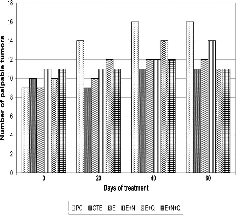
various times during the study increased in the PC group from 9 to
16 (Fig. 1). In the E+N+Q group the
number increased from an initial value of 11 to 12. One of the
tumors regressed by the 60th day. In the E and E+Q groups the tumor
number increased from 9 to 12 and from 10 to 14, respectively. In
the remaining groups the number of tumors increased by 1 or 2.
The average size of the palpable tumors/rat in
different groups did not differ significantly at the beginning of
the study. During the course of the study the tumors in the PC
group grew at a faster rate. By the end of the study (60th day) the
tumor size/rat was 3.38 units. The GTE group reached a tumor size
of 2.17 units. Notably, the tumor size in the E+N+Q group was only
0.574 units. Despite such a large variation in values, the
differences failed to attain significance. When the average sizes
of the palpable tumors/rat for the different groups were compared
with the PC group, the values for the E, E+N+Q and E+Q groups were
found to be lower than that for the PC group (data not shown).
Tumor number, weight and volume after
necropsy
The number of tumors dissected from each group was
higher than the number of tumors palpated in the live rats (data
not shown). Most of the additional tumors detected after
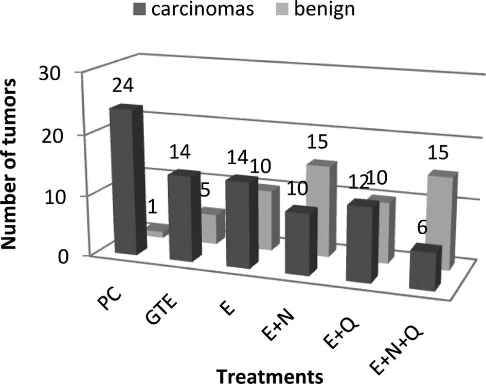
sacrificing the animals were of a smaller size. Histopathological
examinations revealed that some of the tumors were benign (Fig. 2). The PC group had 24 carcinomas,
while the E+N+Q group had only 6 carcinomas. The values for the
other groups ranged between 9 and 14. The rats in the PC group had
≥1 carcinomas per rat. In the E+Q group, 2 rats did not have any
carcinoma. In the remaining groups the number of rats free from any
carcinoma varied from 3 to 4.
The tumor number, volume and weight per rat, as
determined after sacrificing the animals, varied widely among the
groups. Despite such a variation, these values failed to achieve
significance (Table II). The
values for all of the groups were therefore independently compared
with those of the PC using the Student’s t-test using one-tailed
P-values to determine significance (P<0.05). The tumor number,
size and weight per rat for the E+N+Q group were significantly
lower than the corresponding values for the PC group. Percentage
inhibitions with respect to tumor number, size and weight per rat
in the E+N+Q group were 76, 74 and 76%, respectively. Among the
remaining groups, only the E+N and E+Q groups had significantly
smaller numbers of tumors/rat than the value for the PC group.
However, their tumor volume and weight per rat did not differ
significantly from the PC group. The GTE and E groups did not
differ from the PC group in any of the parameters indicated
above.
| Table IIAverage number, size and weight of
malignant tumors per rat as determined after sacrifice of the rats
and histopathological examination of the tumors. |
Table II
Average number, size and weight of
malignant tumors per rat as determined after sacrifice of the rats
and histopathological examination of the tumors.
| Treatment | Number of
tumors/rat | Size of tumors/rat
(cc) | Weight of tumors/rat
(g) |
|---|
| PC | 4.0±0.89 | 10.36±6.00 | 5.71±2.39 |
| GTE | 2.0±1.09 | 7.27±6.32 | 5.33±4.72 |
| E | 2.0±1.05 | 4.13±2.74 | 1.70±1.11 |
| E+N | 1.4±0.81a | 3.39±2.42 | 1.71±1.34 |
| E+Q | 1.4±0.95a | 3.52±2.94 | 1.59±1.25 |
| E+N+Q | 1.0±0.53a | 2.69±1.76a | 1.35±0.93a |
Histopathological analysis of the
tumors
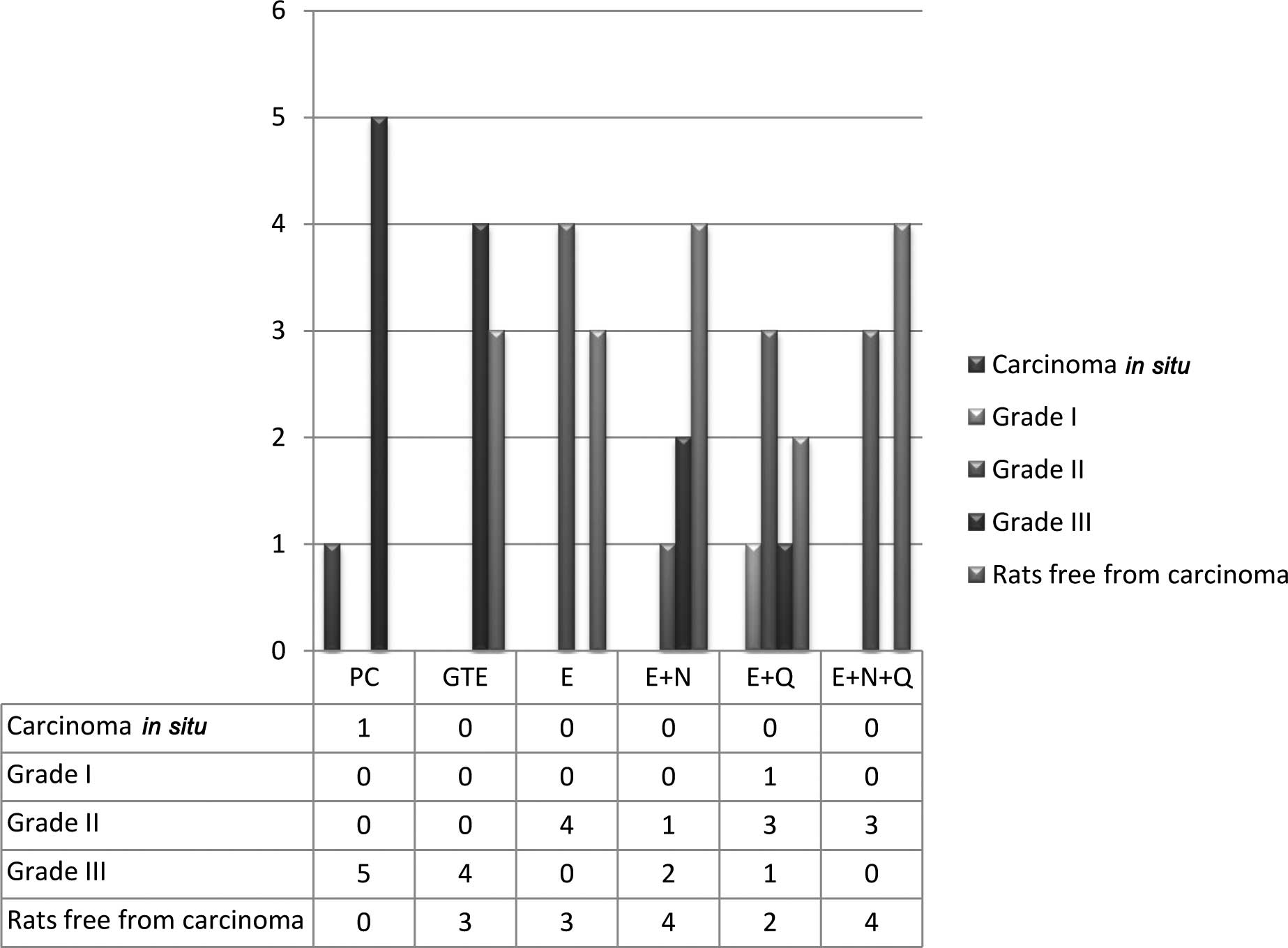
Tumors were assayed for grades of malignancy using
the criteria described by Russo et al (19). The severity of the cancerous tumors
was graded as detailed in the modified Bloom-Richardson (B-R)
scheme (20). The modified B-R
scheme is a semi-quantitative grading method based on three
morphologic features of tumors: degree of tubal tumor formation,
tumor mitotic activity and nuclear pleomorphism of the tumor cells.
In the B-R scheme, seven possible scores are condensed into the
three B-R grades of low, intermediate and high. Out of 6 rats in
the PC group, 5 rats had grade III papillary carcinomas, while 1
rat had carcinoma in situ. In the other groups, 2–4 rats out
of 7 were free from carcinomas (Fig.
3); these rats had consistently shown palpable tumors. In the
GTE group, 3 rats were free from any carcinomas. The remaining 4
rats had grade III carcinomas. In contrast, the E+N+Q group had 4
rats that did not have any carcinomas. The remaining 3 rats in the
group had only grade II carcinomas. The other groups had a
variation of grade II and III carcinomas, except for the E+Q group
which had 1 grade I carcinoma, along with 3 grade II and 1 grade
III carcinomas.
Plasma concentration of EGCG
The plasma concentration of EGCG was very low (<2
ng/ml) in the PC group (Table
III). The relationship between the plasma concentration of EGCG
and the number of carcinomas in the rats of the different treatment
groups is shown in Fig. 4.
| Table IIIAverage plasma concentration of EGCG
(mean ± SE) determined 1 h after the administration of various
dietary treatments. |
Table III
Average plasma concentration of EGCG
(mean ± SE) determined 1 h after the administration of various
dietary treatments.
| Treatment | Plasma concentration
of EGCG (ng/ml) mean ± SE |
|---|
| PC |
1.99±0.81a |
| GTE |
35.94±1.23b |
| E |
42.60±1.26c |
| E+N |
45.81±2.49d |
| E+Q |
61.94±0.83d |
| E+N+Q |
65.94±1.23d |
In conclusion, the results indicate that
intervention of E alone may not be as efficient as it occurs in the
inhibition of carcinogenesis. E must be supplemented with a
nutrient mixture as used in this study along with Q in order to
increase the plasma level of EGCG for significant anticancer
activity. Furthermore, supplementation of E with the nutrient
mixture alone caused a significant decrease in the number of tumors
per rat. Doubling the quantity of E did not offer any advantage
even when it was given with Q and the plasma level was considerably
elevated.
Acknowledgements
The authors are grateful to oncopathologists Dr
Girish Moghe and Dr Sachin Mopkar for the histopathological
evaluation and for the determination of grades for the cancerous
lesions. A.K. and S.G. conducted the experiments, S.K. and S.N.
designed and analyzed the data and drafted the manuscript; W.R.,
V.I., M.R. and A.N. supervised the work and helped in the final
drafting of the manuscript.
Abbreviations:
|
MNU
|
N-methyl-N-nitrosourea
|
|
GTE
|
green tea extract
|
|
EGCG
|
epigallocatechin-3-gallate
|
References
|
1
|
Gullino PM, Pettigrew HM and Grantham FH:
N-Nitrosomethylurea as mammary gland carcinogen in rats. J Natl
Cancer Inst. 54:401–414. 1975.PubMed/NCBI
|
|
2
|
Moon RC, Grubbs CJ, Sporn MB and Goodman
DG: Retinyl acetate inhibits mammary carcinogenesis induced by
N-methyl-N-nitrosourea. Nature. 267:620–621. 1977. View Article : Google Scholar : PubMed/NCBI
|
|
3
|
McCormick DL, Adamoski CB, Fiks A, et al:
Lifetime dose-response relationships for mammary tumor induction by
a single administration of N-methyl-N-nitrosourea. Cancer Res.
41:1690–1694. 1981.PubMed/NCBI
|
|
4
|
Thompson HJ and Meeke LD: Induction of
mammary gland carcinomas by subcutaneous injection of
1-methyl-1-nitrosourea. Cancer Res. 41:1628–1629. 1983.PubMed/NCBI
|
|
5
|
Thomson HJ and Adlakha H: Dose-response
induction of mammary gland carcinomas by intraperitoneal injection
of 1-methyl-1-nitrosourea. Cancer Res. 51:3411–3415.
1991.PubMed/NCBI
|
|
6
|
Thompson HJ, McGinley JN, Rothhammer K, et
al: Rapid induction of mammary intraductal proliferation, ductal
carcinoma in situ and carcinomas by the injection of sexually
immature female rats with 1-methyl-1-nitrosourea. Carcinogenesis.
16:2407–2411. 1995. View Article : Google Scholar
|
|
7
|
Green A, Shilkaitis A and Christov K:
4-(Hydroxyphenyl) retinamide selectively inhibits the development
and progression of ductal hyperplastic lesions and carcinoma in
situ in mammary gland. Carcinogenesis. 20:1535–1540. 1999.
View Article : Google Scholar : PubMed/NCBI
|
|
8
|
Thompson HJ, Singh M and McGinley J:
Classification of premalignant and malignant lesions developing in
the rat mammary gland after injection of sexually immature rats
with 1-methyl-1-nitrosourea. J Mammary Gland Biol Neoplasia.
5:201–210. 2000. View Article : Google Scholar : PubMed/NCBI
|
|
9
|
Kotsopoulos J, Sohn KJ, Martin R, et al:
Dietary folate deficiency suppresses N-methyl-N-nitrosourea-induced
mammary tumorigenesis in rats. Carcinogenesis. 24:937–944. 2003.
View Article : Google Scholar : PubMed/NCBI
|
|
10
|
Kotsopoulos J, Medline A, Renlund R, et
al: Effects of dietary folate on the development and progression of
mammary tumors in rats. Carcinogenesis. 20:1603–1612. 2005.
View Article : Google Scholar : PubMed/NCBI
|
|
11
|
Melancon K, Cheng Q, Kiefer TL, et al:
Regression of MNU-induced mammary tumors with combination of
melatonin and 9-cis-retinoic acid. Cancer Lett. 227:39–48. 2005.
View Article : Google Scholar : PubMed/NCBI
|
|
12
|
Roomi MW, Roomi NW, Ivanov V, et al:
Modulation of N-methyl-N-nitrosourea induced mammary tumors in
Sprague-Dawley rats by combination of lysine, proline, arginine,
ascorbic acid and green tea extract. Br Cancer Res. 7:R291–R295.
2005. View
Article : Google Scholar : PubMed/NCBI
|
|
13
|
Roomi MW, Ivanov V, Kalinovsky T, et al:
In vivo and in vitro antitumiorigenic activity of a mixture of
lysine, proline and green tea extract on human breast cancer cell
lines MDA-MB-231 and MCF-7. Med Oncol. 22:129–138. 2005. View Article : Google Scholar : PubMed/NCBI
|
|
14
|
Ahmad N, Feyes AL, Nieminen R, et al:
Green tea constituent epigallocatechin-3-gallate and induction of
apoptosis and cell cycle arrest in human carcinoma cells. J Natl
Cancer Inst. 89:1881–1886. 1997. View Article : Google Scholar : PubMed/NCBI
|
|
15
|
Kale A, Gawande S, Kotwal S, et al:
Studies on the effects of oral administration of nutrient mixture,
quercetin and red onions on the bioavailability of epigallocatechin
gallet from green tea extract. Phytother Res. July 7–2009.(Epub
ahead of print).
|
|
16
|
Thompson HJ, McGinley JN, Wolfe P, et al:
Temporal sequence of mammary intraductal proliferation, ductal
carcinomas in situ and adenocarcinomas induced by
1-methyl-1-nitrosourea in rats. Carcinogenesis. 19:2181–2185. 1998.
View Article : Google Scholar : PubMed/NCBI
|
|
17
|
Gawande S, Kale A and Kotwal S: Effect of
nutrient mixture and black grapes on the pharmacokinetics of orally
administered (−) epigallocatechin-3-gallate from green tea extract:
a human study. Phytotherapy Res. 22:802–806. 2008.PubMed/NCBI
|
|
18
|
Graham HD: Stabilization of prussian blue
color in the determination of polyphenols. J Agric Food Chem.
40:801–805. 1992. View Article : Google Scholar
|
|
19
|
Russo J, Gusterson BA, Rogers AE, et al:
Comparative study of human and rat tumorigenesis. Lab Invest.
62:244–278. 1990.PubMed/NCBI
|
|
20
|
Bloom HJG and Richardson WW: Histological
grading and prognosis in breast cancer. Br J Cancer. 11:359–377.
1957. View Article : Google Scholar : PubMed/NCBI
|
|
21
|
Dalton LW, Dage DL and Dupont WD:
Histologic grading of breast carcinoma: a reproducibility study.
Cancer. 73:2765–2770. 1994. View Article : Google Scholar : PubMed/NCBI
|
|
22
|
Elston CW: Grading of invasive carcinoma
of the breast. Diagnostic Histopathology of the Breast. Page DL and
Anderson TJ: Edinburgh Churchill Livingston; New York: pp. 300–311.
1987
|
|
23
|
Analyse-it for Microsoft Excel (version
2.20). Analyse-it Software Ltd. http://www.analyse-it.com/.
2009
|
|
24
|
Ahlers I, Solar P, Buresova A, et al: Very
low sensitivity of Wistar:Han female rats to chemocarcinogens in
mammary carcinogenesis induction. Neoplasma. 45:373–376.
1998.PubMed/NCBI
|
|
25
|
Bojkova B, Ahlers I, Kubatka P, et al:
Repeated administration of carcinogen in critical developmental
periods increases the susceptibility of female Wistar:Han rats to
mammary carcinogenesis induction. Neoplasma. 47:230–233. 2000.
|