Introduction
Primary lung cancer is the leading cause of
cancer-related mortality in Japan and in Western countries, with
non-small cell lung cancer (NSCLC) accounting for approximately 80%
of primary lung cancers (1,2). Surgical resection is the only
potentially curative treatment in patients with early disease.
However, 5-year survival after surgical resection remains
unsatisfactory, ranging from 73% in stage IA disease to 25% in
stage IIIA disease. Improvement in the poor survival of patients
with NSCLC requires better clinical predictors of outcomes and of
response to specific therapeutic interventions.
Cell-to-cell adhesion is generally reduced in
various human cancer types. The dissociation of cancer nests is a
crucial step in metastasis. The suppression of cell-to-cell
adhesion may trigger the release of cancer cells from primary
cancer nests, increasing tumor invasion (3). Tight junctions are cell-cell junctions
located at the apical end of the lateral membrane surface of
epithelial or endothelial cells. Tight junctions consist of
membrane and peripheral proteins. Occludin and claudins are
membrane proteins, and zonula occludens (ZO)-1, ZO-2 and ZO-3 are
peripheral proteins. Claudin-7 is a member of the 24-claudin
multigene family, associated with cancer. Loss of claudin-7
expression has been confirmed in ductal carcinoma of the breast and
squamous cell carcinoma of the head and neck (4,5). Usami
et al reported that a reduced claudin-7 expression
correlates with tumor invasion and metastasis in squamous cell
carcinoma of the esophagus (6).
Whether the expression of claudin-7 is associated with the
malignant potential of NSCLC remains to be clarified. The present
study evaluated the clinical significance of claudin-7, especially
its relation to outcome, in resected NSCLC.
Materials and methods
We retrospectively reviewed a total of 76
consecutive patients with pathological (p)-stage I to III NSCLC.
The patients underwent complete tumor resection and nodal
dissection without any pre-operative therapy at the Respiratory
Center, Yokohama City University Medical Center between January
1st, 2000 and November 30th, 2003. One patient succumbed to
operation-related causes and was excluded from the study. The study
group thus comprised 75 patients: 45 with adenocarcinomas, 25 with
squamous cell carcinomas and 5 patients with large cell carcinomas.
The mean follow-up was 1,466 days (range 106–3,328). Informed
consent was obtained from each patient. This study was approved by
Ethics Committee of the Yokohama City Medical Center.
Immunohistochemistry
The primary antibody used to detect claudin-7 was a
mouse monoclonal antibody (Zymed, San Francisco, CA, USA).
Formalin-fixed, paraffin-embedded tissue specimens were cut into
4-μm sections and mounted on slides. The sections were
deparaffinized and rehydrated. The slides were then heated in a
microwave oven three times for 5 min each in a 10-μmol/l citrate
buffer solution at pH 6.0 and cooled at room temperature for 20
min. After quenching the endogenous peroxidase activity with 3%
H2O2 for 5 min, the sections were incubated
for 60 min at room temperature, with the primary antibody diluted
at 1:100. Endogenous biotin was blocked by the Dako Biotin blocking
system (Dako, Glostrup, Denmark), according to the manufacturer’s
specifications. After rinsing, specific staining was visualized
with the use of an LSAB+ system-HRP system (Dako). Color was
produced by the application of 3,3′-diaminobenzidine for 10 min.
The sections were counterstained with Meyer’s hematoxylin (Muto
Pure Chemicals, Tokyo, Japan).
Samples were scored semiquantitatively and
qualitatively for every section, without knowledge of the clinical
data. The distribution of stained cells was scored as follows: 1,
0–30%; 2, 31–60% and 3, 61–100%. The intensity of claudin-7
expression was scored from 1 to 3 as follows: grade 1, weak
staining; grade 2, moderate staining and grade 3, strong staining.
Strong staining of ≥61% cells (i.e., both distribution score and
intensity score = 3) was classified as claudin-7-high. The
remaining staining patterns were classified as claudin-7-low.
Statistical analysis
Counts were compared by the χ2 test.
Continuous data were compared using Student’s t-test. The
postoperative survival rate was analyzed by the Kaplan-Meier method
and differences in survival rates were assessed using the log-rank
test. Death from any cause was included in the calculation of
postoperative survival. Differences were considered significant
when P<0.05. Statistical manipulations were performed using the
SPSS for Windows software system (SPSS, Inc., Chicago, IL,
USA).
Results
The subjects included were 52 men and 23 women with
a mean age of 65.2 years (range 32–82; median 65). The most common
histological type of tumor was adenocarcinoma (60.0%; 45 cases),
followed by squamous cell carcinoma (33.3%; 25 cases) and large
cell carcinoma (6.7%; 5 cases). Tumor size was T1 in 34 patients
(45.3%), T2 in 28 (37.3%), T3 in 9 (12.0%) and T4 in 4 (5.3%).
Thirty-six patients (48.0%) had no metastasis to regional lymph
nodes (N0), whereas 11 (14.7%) had metastatic involvement of the
hilar lymph nodes (N1) and 28 (37.3%) had metastases to the
mediastinal nodes (N2, N3). Thirty tumors (40.0%) were classified
as stage I, 15 (20.0%) were stage II and 30 (40.0%) were stage III.
At the end of follow-up, 34 patients (45.3%) were alive and 41
(54.7%) had succumbed to the disease.
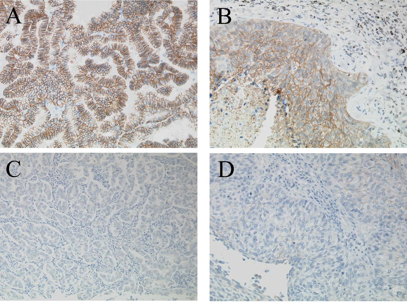
The immunohistochemical expression of claudin-7 was
detected in the epithelial cell membranes of the normal bronchial
mucosa. Claudin-7 expression was also observed in lung cancer cells
(Fig. 1). The immunohistochemical
distribution and intensity of claudin-7 expression are summarized
in Table I. Among the 75 carcinomas
studied, 25 (33.3%) strongly expressed claudin-7. In the 75
carcinomas, ≥61% of the tumor cells stained positively. Therefore,
25 carcinomas were classified as claudin-7-high and 50 (66.7%) were
classified as claudin-7-low.
 | Table IImmunohistochemical results for
claudin-7 in NSCLC. |
Table I
Immunohistochemical results for
claudin-7 in NSCLC.
| Distribution (%) | Intensity (%) |
|---|
|
|
|---|
| 1 | 2 | 3 |
|---|
| 1 (0–30) | 28 (37.3) | 1 (1.3) | 0 (0.0) |
| 2 (31–60) | 8 (10.6) | 3 (4.0) | 0 (0.0) |
| 3 (61–100) | 3 (4.0) | 7 (9.3) | 25 (33.3) |
Claudin-7 expression status did not significantly
correlate with any of the patient characteristics (Table II). The 5-year survival rate was
41.4% in patients with claudin-7-low tumors, compared to 61.3% in
those with claudin-7-high tumors. Survival was significantly poorer
in patients with claudin-7-low tumors than in those with
claudin-7-high tumors (P=0.024) (Table III). Subsequently, we conducted
subset analyses to investigate the prognostic significance of the
claudin-7 status. Among patients with squamous cell carcinoma, the
5-year survival rate was significantly lower in claudin-7-low
(26.3%) than in claudin-7-high patients (80.0%, P=0.011). For
patients with adenocarcinoma, survival was similar in
claudin-7-high and claudin-7-low patients (5-year survival rate,
53.5 and 54.4%, respectively; P=0.664). Claudin-7-high was
associated with a better 5-year survival than claudin-7-low in
patients with stage I disease, male patients and smokers (P=0.023,
0.035 and 0.010, respectively) (Table
III).
| Table IIPatient characteristics and the
claudin-7 expression status. |
Table II
Patient characteristics and the
claudin-7 expression status.
| No. of patients
(%) | Claudin-7
expression | P-value |
|---|
| |
| |
|---|
| | High (%) | Low (%) | |
|---|
| All patients | 75 (100) | 25 (33.3) | 50 (66.7) | |
| Age (mean ± SD) | 65.2±9.49 | 65.8±6.98 | 64.9±10.5 | 0.702 |
| Gender |
| Male | 52 (69.3) | 15 (28.8) | 37 (71.2) | 0.250 |
| Female | 23 (30.7) | 10 (43.5) | 13 (56.5) | |
| Histological
type |
| Adenocarcinoma | 45 (60.0) | 16 (35.6) | 29 (64.4) | 0.262 |
| Squamous cell
carcinoma | 25 (33.3) | 6 (24.0) | 19 (76.0) | |
| Large cell
carcinoma | 5 (6.7) | 3 (60.0) | 2 (40.0) | |
| P-stage |
| I | 30 (40.0) | 12 (40.0) | 18 (60.0) | 0.178 |
| II | 15 (20.0) | 2 (13.3) | 13 (86.7) | |
| III | 30 (40.0) | 11 (36.7) | 19 (63.3) | |
| Smoking |
| Non-smoker | 20 (26.7) | 7 (35.0) | 13 (65.0) | 0.854 |
| Smoker | 55 (73.3) | 18 (32.7) | 37 (67.3) | |
| T-factor |
| T1 | 34 (45.3) | 12 (35.3) | 22 (64.7) | 0.452 |
| T2 | 28 (37.3) | 10 (35.7) | 18 (64.3) | |
| T3 | 9 (12.0) | 1 (11.1) | 8 (88.9) | |
| T4 | 4 (5.3) | 2 (50.0) | 2 (50.0) | |
| N-factor |
| N0 | 36 (48.0) | 13 (36.1) | 23 (63.9) | 0.353 |
| N1 | 11 (14.7) | 2 (18.2) | 9 (81.8) | |
| N2 | 27 (36.0) | 9 (33.3) | 18 (66.7) | |
| N3 | 1 (1.3) | 1 (100) | 0 (0.0) | |
| Table IIIClaudin-7 expression and postoperative
survival in NSCLC. |
Table III
Claudin-7 expression and postoperative
survival in NSCLC.
| 5-year survival rate
(%) | |
|---|
|
| |
|---|
| Claudin-7 high
patients | Claudin-7 low
patients | P-value |
|---|
| All patients | 61.3 | 41.4 | 0.024 |
| Gender |
| Male | 57.7 | 32.4 | 0.035 |
| Female | 66.6 | 68.3 | 0.855 |
| Histological
type |
| Adenocarcinoma | 53.5 | 54.4 | 0.664 |
| Squamous cell
carcinoma | 80.0 | 26.3 | 0.011 |
| P-stage |
| I | 91.6 | 50.0 | 0.023 |
| II, III | 38.5 | 36.4 | 0.365 |
| Smoking |
| Non-smoker | 50.0 | 61.5 | 0.890 |
| Smoker | 65.1 | 34.3 | 0.010 |
Discussion
Claudin-7 expression was reported in various
malignant neoplasms. Kominsky et al showed that loss of the
claudin-7 expression is associated with nodal metastasis in primary
breast carcinomas (5). Sauer et
al found that a reduced claudin-7 expression correlates with
metastatic disease in breast carcinoma (7), while Usami et al reported that
this correlates with tumor invasion and metastasis in squamous cell
carcinoma of the esophagus (6). A
reduced claudin-7 expression correlates with a high tumor grade in
prostatic adenocarcinoma (8).
Oshima et al reported that a reduced claudin-7 expression
correlates with venous invasion and liver metastasis in colorectal
cancer (9). In contrast, an
increased claudin-7 expression was reported in various cancer
types, such as intestinal-type gastric adenocarcinomas (10), ovarian cancer (11) and cervical carcinoma (12). On the other hand, Bello et al
reported that a high and low immunoreactivity of claudin-7 is
associated with a slightly poorer survival than median
immunoreactivity in squamous cell carcinoma of the tongue (13). However, claudin-7 expression in
NSCLC remains largely uninvestigated.
The present study assessed the immunohistochemical
expression of claudin-7 in NSCLC. Two previous studies examined the
immunohistochemical expression of claudin-7 in NSCLC. Moldvay et
al reported a very strong expression of claudin-7 in almost all
types of NSCLC, except for neuroendocrine tumors (14). Soini also reported a strong
expression of claudin-7 in almost all NSCLCs studied (15). However, neither of these studies
assessed the clinical significance of claudin-7 expression. We
found that 66.7% of the NSCLCs showed a low claudin-7 expression,
and two large cell neuroendocrine carcinomas showed a high
claudin-7 expression. The discrepancies noted in the findings of
previous studies may be related to differences in staining
procedures, such as the antibody used.
To the best of our knowledge, this is the first
study to examine the clinical significance of claudin-7 expression
in NSCLC. We found that a reduced expression of claudin-7 was a
significant predictor of poor outcome, especially in squamous cell
carcinoma. In contrast to patients with squamous cell carcinoma,
claudin-7 expression status was unrelated to the postoperative
survival in patients with adenocarcinoma.
Among patients with stage I disease, survival was
significantly poorer in those with claudin-7-low than in those with
claudin-7-high tumors. This finding suggests that claudin-7 is a
prognostic marker for early stage cancer. Although in our study
claudin-7 expression was unrelated to patient characteristics, such
as p-stage and nodal status, further experimental and clinical
studies should be conducted to define the exact role of claudin-7
in the progression of NSCLC.
In conclusion, a reduced claudin-7 expression was
associated with poor outcome in patients with NSCLC, particularly
in squamous cell carcinoma, stage I disease, male gender and
smokers. These results suggest that claudin-7 expression is a
useful biomarker and a potential therapeutic target in patients
with NSCLC.
References
|
1
|
Jemal A, Siegel R, Ward E, Murray T, Xu J
and Thun MJ: Cancer statistics. CA Cancer J Clin. 57:43–66.
2007.
|
|
2
|
Cersosimo RJ: Lung cancer: a review. Am J
Health Syst Pharm. 59:611–642. 2002.PubMed/NCBI
|
|
3
|
Hirohashi S: Inactivation of the
E-cadherin-mediated cell adhesion system in human cancers. Am J
Pathol. 153:333–339. 1998. View Article : Google Scholar : PubMed/NCBI
|
|
4
|
Al Moustafa AE, Alaoui-Jamali MA, Batist
G, et al: Identification of genes associated with head and neck
carcinogenesis by cDNA microarray comparison between matched
primary normal epithelial and squamous carcinoma cells. Oncogene.
21:2634–2640. 2002.PubMed/NCBI
|
|
5
|
Kominsky SL, Argani P, Korz D, et al: Loss
of the tight junction protein claudin-7 correlates with
histological grade in both ductal carcinoma in situ and invasive
ductal carcinoma of the breast. Oncogene. 22:2021–2033. 2003.
View Article : Google Scholar : PubMed/NCBI
|
|
6
|
Usami Y, Chiba H, Nakayama F, et al:
Reduced expression of claudin-7 correlates with invasion and
metastasis in squamous cell carcinoma of the esophagus. Hum Pathol.
37:569–577. 2006. View Article : Google Scholar : PubMed/NCBI
|
|
7
|
Sauer T, Pedersen MK, Ebeltoft K and Naess
O: Reduced expression of Claudin-7 in fine needle aspirates from
breast carcinomas correlate with grading and metastatic disease.
Cytopathology. 16:193–198. 2005. View Article : Google Scholar : PubMed/NCBI
|
|
8
|
Sheehan GM, Kallakury BV, Sheehan CE,
Fisher HA, Kaufman RP Jr and Ross JS: Loss of claudins-1 and −7 and
expression of claudins-3 and −4 correlate with prognostic variables
in prostatic adenocarcinomas. Hum Pathol. 38:564–569. 2007.
|
|
9
|
Oshima T, Kunisaki C, Yoshihara K, et al:
Reduced expression of the claudin-7 gene correlates with venous
invasion and liver metastasis in colorectal cancer. Oncol Rep.
19:953–959. 2008.PubMed/NCBI
|
|
10
|
Johnson AH, Frierson HF, Zaika A, et al:
Expression of tight-junction protein claudin-7 is an early event in
gastric tumorigenesis. Am J Pathol. 167:577–584. 2005. View Article : Google Scholar : PubMed/NCBI
|
|
11
|
Tassi RA, Bignotti E, Falchetti M, et al:
Claudin-7 expression in human epithelial ovarian cancer. Int J
Gynecol Cancer. 18:1262–1271. 2008. View Article : Google Scholar : PubMed/NCBI
|
|
12
|
Sobel G, Paska C, Szabo I, Kiss A, Kadar A
and Schaff Z: Increased expression of claudins in cervical squamous
intraepithelial neoplasia and invasive carcinoma. Hum Pathol.
36:162–169. 2005. View Article : Google Scholar : PubMed/NCBI
|
|
13
|
Bello IO, Vilen ST, Niinimaa A, Kantola S,
Soini Y and Salo T: Expression of claudins 1, 4, 5 and 7 and
occludin, and relationship with prognosis in squamous cell
carcinoma of the tongue. Hum Pathol. 39:1212–1220. 2008. View Article : Google Scholar : PubMed/NCBI
|
|
14
|
Moldvay J, Jackel M, Paska C, Soltesz I,
Schaff Z and Kiss A: Distinct claudin expression profile in
histologic subtypes of lung cancer. Lung Cancer. 57:159–167. 2007.
View Article : Google Scholar : PubMed/NCBI
|
|
15
|
Soini Y: Expression of claudins 1, 2, 3,
4, 5 and 7 in various types of tumours. Histopathology. 46:551–560.
2005. View Article : Google Scholar : PubMed/NCBI
|