Spandidos Publications Logo
  • About
    • About Spandidos
    • Aims and Scopes
    • Abstracting and Indexing
    • Editorial Policies
    • Reprints and Permissions
    • Job Opportunities
    • Terms and Conditions
    • Contact
  • Journals
    • All Journals
    • Oncology Letters
      • Oncology Letters
      • Information for Authors
      • Editorial Policies
      • Editorial Board
      • Aims and Scope
      • Abstracting and Indexing
      • Bibliographic Information
      • Archive
    • International Journal of Oncology
      • International Journal of Oncology
      • Information for Authors
      • Editorial Policies
      • Editorial Board
      • Aims and Scope
      • Abstracting and Indexing
      • Bibliographic Information
      • Archive
    • Molecular and Clinical Oncology
      • Molecular and Clinical Oncology
      • Information for Authors
      • Editorial Policies
      • Editorial Board
      • Aims and Scope
      • Abstracting and Indexing
      • Bibliographic Information
      • Archive
    • Experimental and Therapeutic Medicine
      • Experimental and Therapeutic Medicine
      • Information for Authors
      • Editorial Policies
      • Editorial Board
      • Aims and Scope
      • Abstracting and Indexing
      • Bibliographic Information
      • Archive
    • International Journal of Molecular Medicine
      • International Journal of Molecular Medicine
      • Information for Authors
      • Editorial Policies
      • Editorial Board
      • Aims and Scope
      • Abstracting and Indexing
      • Bibliographic Information
      • Archive
    • Biomedical Reports
      • Biomedical Reports
      • Information for Authors
      • Editorial Policies
      • Editorial Board
      • Aims and Scope
      • Abstracting and Indexing
      • Bibliographic Information
      • Archive
    • Oncology Reports
      • Oncology Reports
      • Information for Authors
      • Editorial Policies
      • Editorial Board
      • Aims and Scope
      • Abstracting and Indexing
      • Bibliographic Information
      • Archive
    • Molecular Medicine Reports
      • Molecular Medicine Reports
      • Information for Authors
      • Editorial Policies
      • Editorial Board
      • Aims and Scope
      • Abstracting and Indexing
      • Bibliographic Information
      • Archive
    • World Academy of Sciences Journal
      • World Academy of Sciences Journal
      • Information for Authors
      • Editorial Policies
      • Editorial Board
      • Aims and Scope
      • Abstracting and Indexing
      • Bibliographic Information
      • Archive
    • International Journal of Functional Nutrition
      • International Journal of Functional Nutrition
      • Information for Authors
      • Editorial Policies
      • Editorial Board
      • Aims and Scope
      • Abstracting and Indexing
      • Bibliographic Information
      • Archive
    • International Journal of Epigenetics
      • International Journal of Epigenetics
      • Information for Authors
      • Editorial Policies
      • Editorial Board
      • Aims and Scope
      • Abstracting and Indexing
      • Bibliographic Information
      • Archive
    • Medicine International
      • Medicine International
      • Information for Authors
      • Editorial Policies
      • Editorial Board
      • Aims and Scope
      • Abstracting and Indexing
      • Bibliographic Information
      • Archive
  • Articles
  • Information
    • Information for Authors
    • Information for Reviewers
    • Information for Librarians
    • Information for Advertisers
    • Conferences
  • Language Editing
Spandidos Publications Logo
  • About
    • About Spandidos
    • Aims and Scopes
    • Abstracting and Indexing
    • Editorial Policies
    • Reprints and Permissions
    • Job Opportunities
    • Terms and Conditions
    • Contact
  • Journals
    • All Journals
    • Biomedical Reports
      • Information for Authors
      • Editorial Policies
      • Editorial Board
      • Aims and Scope
      • Abstracting and Indexing
      • Bibliographic Information
      • Archive
    • Experimental and Therapeutic Medicine
      • Information for Authors
      • Editorial Policies
      • Editorial Board
      • Aims and Scope
      • Abstracting and Indexing
      • Bibliographic Information
      • Archive
    • International Journal of Epigenetics
      • Information for Authors
      • Editorial Policies
      • Editorial Board
      • Aims and Scope
      • Abstracting and Indexing
      • Bibliographic Information
      • Archive
    • International Journal of Functional Nutrition
      • Information for Authors
      • Editorial Policies
      • Editorial Board
      • Aims and Scope
      • Abstracting and Indexing
      • Bibliographic Information
      • Archive
    • International Journal of Molecular Medicine
      • Information for Authors
      • Editorial Policies
      • Editorial Board
      • Aims and Scope
      • Abstracting and Indexing
      • Bibliographic Information
      • Archive
    • International Journal of Oncology
      • Information for Authors
      • Editorial Policies
      • Editorial Board
      • Aims and Scope
      • Abstracting and Indexing
      • Bibliographic Information
      • Archive
    • Medicine International
      • Information for Authors
      • Editorial Policies
      • Editorial Board
      • Aims and Scope
      • Abstracting and Indexing
      • Bibliographic Information
      • Archive
    • Molecular and Clinical Oncology
      • Information for Authors
      • Editorial Policies
      • Editorial Board
      • Aims and Scope
      • Abstracting and Indexing
      • Bibliographic Information
      • Archive
    • Molecular Medicine Reports
      • Information for Authors
      • Editorial Policies
      • Editorial Board
      • Aims and Scope
      • Abstracting and Indexing
      • Bibliographic Information
      • Archive
    • Oncology Letters
      • Information for Authors
      • Editorial Policies
      • Editorial Board
      • Aims and Scope
      • Abstracting and Indexing
      • Bibliographic Information
      • Archive
    • Oncology Reports
      • Information for Authors
      • Editorial Policies
      • Editorial Board
      • Aims and Scope
      • Abstracting and Indexing
      • Bibliographic Information
      • Archive
    • World Academy of Sciences Journal
      • Information for Authors
      • Editorial Policies
      • Editorial Board
      • Aims and Scope
      • Abstracting and Indexing
      • Bibliographic Information
      • Archive
  • Articles
  • Information
    • For Authors
    • For Reviewers
    • For Librarians
    • For Advertisers
    • Conferences
  • Language Editing
Login Register Submit
  • This site uses cookies
  • You can change your cookie settings at any time by following the instructions in our Cookie Policy. To find out more, you may read our Privacy Policy.

    I agree
Search articles by DOI, keyword, author or affiliation
Search
Advanced Search
presentation
Oncology Letters
Join Editorial Board Propose a Special Issue
Print ISSN: 1792-1074 Online ISSN: 1792-1082
Journal Cover
July-August 2010 Volume 1 Issue 4

Full Size Image

Sign up for eToc alerts
Recommend to Library

Journals

International Journal of Molecular Medicine

International Journal of Molecular Medicine

International Journal of Molecular Medicine is an international journal devoted to molecular mechanisms of human disease.

International Journal of Oncology

International Journal of Oncology

International Journal of Oncology is an international journal devoted to oncology research and cancer treatment.

Molecular Medicine Reports

Molecular Medicine Reports

Covers molecular medicine topics such as pharmacology, pathology, genetics, neuroscience, infectious diseases, molecular cardiology, and molecular surgery.

Oncology Reports

Oncology Reports

Oncology Reports is an international journal devoted to fundamental and applied research in Oncology.

Experimental and Therapeutic Medicine

Experimental and Therapeutic Medicine

Experimental and Therapeutic Medicine is an international journal devoted to laboratory and clinical medicine.

Oncology Letters

Oncology Letters

Oncology Letters is an international journal devoted to Experimental and Clinical Oncology.

Biomedical Reports

Biomedical Reports

Explores a wide range of biological and medical fields, including pharmacology, genetics, microbiology, neuroscience, and molecular cardiology.

Molecular and Clinical Oncology

Molecular and Clinical Oncology

International journal addressing all aspects of oncology research, from tumorigenesis and oncogenes to chemotherapy and metastasis.

World Academy of Sciences Journal

World Academy of Sciences Journal

Multidisciplinary open-access journal spanning biochemistry, genetics, neuroscience, environmental health, and synthetic biology.

International Journal of Functional Nutrition

International Journal of Functional Nutrition

Open-access journal combining biochemistry, pharmacology, immunology, and genetics to advance health through functional nutrition.

International Journal of Epigenetics

International Journal of Epigenetics

Publishes open-access research on using epigenetics to advance understanding and treatment of human disease.

Medicine International

Medicine International

An International Open Access Journal Devoted to General Medicine.

Journal Cover
July-August 2010 Volume 1 Issue 4

Full Size Image

Sign up for eToc alerts
Recommend to Library

  • Article
  • Citations
    • Cite This Article
    • Download Citation
    • Create Citation Alert
    • Remove Citation Alert
    • Cited By
  • Similar Articles
    • Related Articles (in Spandidos Publications)
    • Similar Articles (Google Scholar)
    • Similar Articles (PubMed)
  • Download PDF
  • Download XML
  • View XML
Case Report

Unique intense uptake demonstrated by 18F-FDG positron emission tomography/computed tomography in primary pancreatic lymphoma: A case report

  • Authors:
    • Yoshiyuki Abe
    • Katsumi Tamura
    • Ikuko Sakata
    • Jiro Ishida
    • Masaya Mukai
    • Makoto Ohtaki
    • Masato Nakamura
    • Kikuo Machida
  • View Affiliations / Copyright

    Affiliations: Tokorozawa PET Diagnostic Imaging Clinic, Saitama 359-1124, Japan. abe@toko-pet.or.jp
  • Pages: 605-607
    |
    Published online on: July 1, 2010
       https://doi.org/10.3892/ol_00000107
  • Expand metrics +
Metrics: Total Views: 0 (Spandidos Publications: | PMC Statistics: )
Metrics: Total PDF Downloads: 0 (Spandidos Publications: | PMC Statistics: )
Cited By (CrossRef): 0 citations Loading Articles...

This article is mentioned in:


Abstract

Patients with primary pancreatic lymphoma (PPL), which is rare, require a different therapeutic approach and have a better prognosis than those with pancreatic cancer. However, conventional imaging modalities alone are not able to differentiate between pancreatic cancer and other rare tumors such as PPL, although the accurate diagnosis of PPL is crucial. The development of new modalities such as F-18 2'-deoxy-2fluoro-D-glucose (FDG) positron emission tomography combined with computed tomography (PET/CT) contributes to the evaluation of lymphoma staging. However, few reports are currently available regarding PET/CT findings in PPL. In this study, a 56-year old man with PPL was examined using FDG PET/CT imaging, which showed the unique intense uptake of FDG in the pancreas with atypical findings of malignancy in the CT scan and magnetic resonance images.

Introduction

Both Hodgkin’s and non-Hodgkin’s types of lymphoma exist. Non-Hodgkin’s lymphomas often invade extra-lymphatic organs, while Hodgkin’s lymphomas rarely disseminate to extra-lymphatic organs. More than 25% of non-Hodgkin’s lymphomas originate from extra-lymphatic organs, approximately 30% of which may involve the pancreas (1). Primary pancreatic lymphomas (PPL) are usually non-Hodgkin’s lymphomas and isolated PPL is quite rare (2). Only three cases (1.5%) of pancreatic lymphoma were found in a review of 207 cases of malignant pancreatic tumors (3). Clinically, PPL is likely to be misdiagnosed as pancreatic cancer. However, patients with PPL require a different therapeutic approach and have a better prognosis than those with pancreatic adenocarcinoma. Conventional imaging modalities alone are not able to differentiate between pancreatic adenocarcinoma and PPL, although the accurate diagnosis of PPL is crucial.

The development of a new modality such as F-18 2′-deoxy-2fluoro-D-glucose (FDG) positron emission tomography combined with computed tomography (PET/CT) has contributed to the evaluation of human cancers and the usefulness of PET/CT has been well established for lymphoma staging (4,5). However, few reports are currently available that pertain to PET/CT diagnosing PPL. In this study, a 56-year-old man with PPL was examined using PET/CT imaging. Results showed the unique intense uptake of FDG in the pancreas with atypical findings of malignancy in the CT scan and magnetic resonance images (MRI).

Patient and methods

A 56-year-old, asymptomatic man was admitted to Tokai University Hachioji Hospital for further examination following an abdominal ultrasound study that showed a mass shadow in the pancreas. A physical examination did not reveal any abnormal findings or systemic lymphadenopathy. Blood analysis showed a slight elevation of the serum interluekin-2 receptor (604 ng/ml) without other abnormalities, including tumor markers such as carcinoembryonic antigen and CA19-9. An abdominal CT scan showed a 5-cm tumor located in the head of pancreas, while an enhanced CT scan showed a slight increase in the tumor without encasement of arteries or veins. The MRI showed a mass with homogeneously high signal intensity on T2-weighted images and low signal intensity on T1-weighted images with gadolinium enhancement (Fig. 1). The CT and MRI findings described above suggested not pancreatic cancer but massive pancreatitis. Endoscopic retrograde cholangiopancreatography did not demonstrate either stenosis or obstruction of either the main pancreatic or common bile duct. Informed patient consent was obtained, as well as approval for the study by an ethics committee of our institute.

Figure 1

An MRI showed a mass with (A) homogeneously high signal intensity on T2-weighted images and (B) low signal intensity on T1-weighted images with gadolinium enhancement (C, 30sec; D, 60 sec and E, 180 sec).

The patient underwent 18F-FDG PET/CT scans. PET/CT imaging was performed with Biograph Duo (Siemens CTI). The Biograph Duo allows for the simultaneous collection of 64 slices over a span of 15.8 cm with a slice thickness of 2.5 mm and a transaxial resolution of 6.3 mm. All data were reconstructed with OSEM image. One hour (early scan) after the intravenous administration of approximately 3.7 Mbq/kg of 18F-FDG, a transmission scan using CT for attenuation correction and anatomical imaging was acquired for 90 sec with a delayed scan after 2 h. To determine the semi-quantitative FDG uptake, regions of interest (ROIs) were placed over the lesion, including the highest uptake area (circular ROI, 1 cm in diameter), and the 
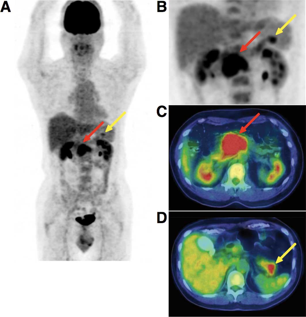
standardized uptake value (SUV) was calculated. The early PET/CT showed an intense abnormal FDG uptake (SUVmax, 8.67) in the head and body of the pancreas, and a moderately increased uptake in the tail of the pancreas (Fig. 2). The delayed scan demonstrated a more intense FDG uptake (SUVmax, 10.3) in the pancreas. No other areas showed an abnormal uptake of FDG apart from the pancreas. The PET/CT findings strongly suggested malignancy of the pancreas without lymphatic or systemic metastasis.

Figure 2

(A) Early PET/CT imaging showed intense abnormal FDG uptake (SUVmax, 8.67) in the head and body of the pancreas (red arrow), and a moderately increased uptake in the tail of the pancreas (yellow arrow). (B–D) A delayed scan demonstrated a more intense FDG uptake (SUVmax, 10.3) in the pancreas.

Results

Histopathologic study

A laparoscopic examination was performed and biopsy 
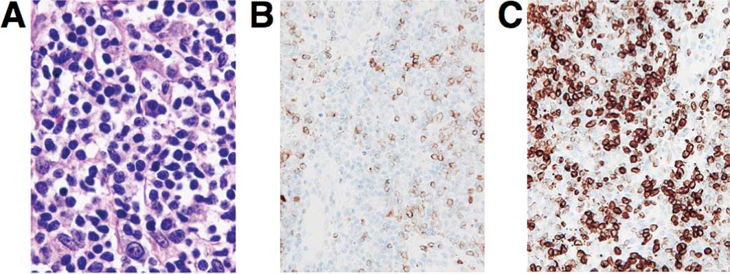
specimens were obtained. Microscopic examination showed diffuse infiltrative growth of T-lymphoid cells (Fig. 3). Multiple foci of B-lymphoid cells with small nodular or aggregate features were noted. The B cells showed a medium to relatively large morphology. The cells were embedded in the fibrous or hyalinizing stroma. The immunohistochemical analysis showed that the neoplastic cells were CD79a(+), CD20(+), BCL-2(+), CD3(−) and CD10(−). The MIB1 (Ki67) index was ~70–80%. A histopathological examination confirmed the diagnosis of a low grade B-cell pancreatic lymphoma. The patient underwent an aspiration biopsy of the bone marrow; however, no abnormality was noted. He received chemotherapy after surgery.

Figure 3

(A) Microscopic examination showed diffuse infiltrative growth of T-lymphoid cells. (B and C) Immunohistochemical analysis showed that the neoplastic cells were positive for CD79a (B) and BCL-2 (C). Original magnification, ×100.

Discussion

PPL is an extremely rare disease which occurs in the pancreas, with or without the involvement of peripancreatic lymph nodes. The clinical manifestation and imaging result of PPL resembles other pancreatic occupying lesions such as pancreatic carcinoma. However, unlike carcinomas, PPLs are potentially treatable even if not found at the early stage. PPL accounts for fewer than 2% of extra-nodal malignant lymphomas and 0.5% of cases of pancreatic masses (6,7). Fewer than 150 cases of PPL had been reported in the literature until 2006 (8).

Imaging results play a key role in the diagnosis of PPL. Percutaneous ultrasound (US), CT scan and MRI are well-established procedures used in the evaluation of pancreatic masses (9,10). CT is the most common imaging technique used in the detection and characterization of pancreatic tumors. The CT image of PPL resembles that of pancreatic carcinoma, including enlargement of the pancreatic head and density changes. However, fewer signs of invasion of large vessels and metastasis to the liver and spleen are observed. Pancreatic duct dilation is less common in PPL compared to pancreatic cancer. Merkle et al reported that the combination of a bulky localized tumor in the pancreas without significant dilation of the main pancreatic duct supports a diagnosis of pancreatic lymphoma as opposed to adenocarcinoma (11).

MRI findings in PPL usually show homogeneously high signal intensity on T2-weighted images and low signal intensity on T1-weighted images (12). Arcari et al stated that conventional imaging techniques suggest the suspicion of PPL but are unable to distinguish PPL from pancreatic adenocarcinoma (13). PPL is a rare disease with non-specific symptoms, laboratory tests and imaging examination results (14). Therefore, the final diagnosis of PPL should depend on a histopathological examination. The first choice for PPL treatment should be a combination of chemotherapy and radiotherapy as opposed to surgery. The development of FDG-PET/CT contributes to the evaluation of human cancer staging and the usefulness of PET/CT has been well established for lymphoma staging (5), whereas only one report regarding PET findings in PPL was previously published (15). Yoon et al reported that FDG-PET imaging, and not PET/CT, showed round intense FDG uptake in the center of the midabdomen in a patient with PPL. However, results of the PET/CT imaging for a 56-year-old man examined in our study showed a unique intense uptake of FDG in the pancreas as well as atypical findings of malignancy from a CT scan and MRI. The results therefore show that further accumulation and analysis of PET/CT images of PPL may provide additional and novel information to the data currently available from conventional imaging such as US, CT and MRI for the evaluation of pancreatic tumors.

Acknowledgements

We thank Mr. Kenji Kawai for his technical assistance.

References

1 

Behrns KE, Sarr MG and Strickler JG: Pancreatic lymphoma: is it a surgical disease? Pancreas. 9:662–667. 1994. View Article : Google Scholar : PubMed/NCBI

2 

Freeman C, Berg JW and Cutler SJ: Occurrence and prognosis of extranodal lymphomas. Cancer. 29:252–260. 1972. View Article : Google Scholar : PubMed/NCBI

3 

Reed K, Vose PC and Jarstfer BS: Pancreatic cancer: 30 year review (1947–1977). Am J Surg. 138:929–933. 1979.PubMed/NCBI

4 

Weiler-Sagie M, Bushelev O, Epelbaum R, Dann EJ, Haim N, Avivi I, Ben-Barak A, Ben-Arie Y, Bar-Shalom R and Israel O: (18)F-FDG avidity in lymphoma readdressed: a study of 766 patients. J Nucl Med. 51:25–30. 2010. View Article : Google Scholar : PubMed/NCBI

5 

Cronin CG, Swords R, Truong MT, Viswanathan C, Rohren E, Giles FJ, O’Dwyer M and Bruzzi JF: Clinical utility of PET/CT in lymphoma. AJR Am J Roentgenol. 194:W91–W103. 2010. View Article : Google Scholar : PubMed/NCBI

6 

Zucca E, Roggero E, Bertoni F and Cavalli F: Primary extranodal non-Hodgkin’s lymphomas. Part 1: gastrointestinal, cutaneous and genitourinary lymphomas. Ann Oncol. 8:727–737. 1997.

7 

Boni L, Benevento A, Dionigi G, Cabrini L and Dionigi R: Primary pancreatic lymphoma. Surg Endosc. 16:1107–1108. 2002. View Article : Google Scholar

8 

Saif MW: Primary pancreatic lymphomas. J Pancreas. 7:262–273. 2006.

9 

McNulty NJ, Francis IR, Platt JF, Cohan RH, Korobkin M and Gebremariam A: Multi-detector row helical CT of the pancreas: effect of contrast-enhanced multiphasic imaging on enhancement of the pancreas, peripancreatic vasculature, and pancreatic adenocarcinoma. Radiology. 220:97–102. 2001. View Article : Google Scholar

10 

Kelekis NL and Semelka RC: MRI of pancreatic tumors. Eur Radiol. 7:875–886. 1997. View Article : Google Scholar : PubMed/NCBI

11 

Merkle EM, Bender GN and Brambs HJ: Imaging findings in pancreatic lymphoma: differential aspects. AJR Am J Roentgenol. 174:671–675. 2000. View Article : Google Scholar : PubMed/NCBI

12 

Masui T, Katayama M, Kobayashi S and Shimizu S: MR imaging of primary malignant lymphoma of the pancreas. Radiat Med. 23:213–215. 2005.PubMed/NCBI

13 

Arcari A, Anselmi E, Bernuzzi P, Berte R, Lazzaro A, Moroniz CF, Trabacchi E, Vallisa D, Vercelli A and Cavanna L: Primary pancreatic lymphoma. Report of five cases. Haematologica. 90:ECR092005.

14 

Lin H, Li SD, Hu XG and Li ZS: Primary pancreatic lymphoma: report of six cases. World J Gastroenterol. 12:5064–5067. 2006.PubMed/NCBI

15 

Yoon SN, Lee MH and Yoon JK: F-18 FDG positron emission tomography findings in primary pancreatic lymphoma. Clin Nucl Med. 29:574–575. 2004. View Article : Google Scholar : PubMed/NCBI

Related Articles

  • Abstract
  • View
  • Download
  • Twitter
Copy and paste a formatted citation
Spandidos Publications style
Abe Y, Tamura K, Sakata I, Ishida J, Mukai M, Ohtaki M, Nakamura M and Machida K: Unique intense uptake demonstrated by 18F-FDG positron emission tomography/computed tomography in primary pancreatic lymphoma: A case report . Oncol Lett 1: 605-607, 2010.
APA
Abe, Y., Tamura, K., Sakata, I., Ishida, J., Mukai, M., Ohtaki, M. ... Machida, K. (2010). Unique intense uptake demonstrated by 18F-FDG positron emission tomography/computed tomography in primary pancreatic lymphoma: A case report . Oncology Letters, 1, 605-607. https://doi.org/10.3892/ol_00000107
MLA
Abe, Y., Tamura, K., Sakata, I., Ishida, J., Mukai, M., Ohtaki, M., Nakamura, M., Machida, K."Unique intense uptake demonstrated by 18F-FDG positron emission tomography/computed tomography in primary pancreatic lymphoma: A case report ". Oncology Letters 1.4 (2010): 605-607.
Chicago
Abe, Y., Tamura, K., Sakata, I., Ishida, J., Mukai, M., Ohtaki, M., Nakamura, M., Machida, K."Unique intense uptake demonstrated by 18F-FDG positron emission tomography/computed tomography in primary pancreatic lymphoma: A case report ". Oncology Letters 1, no. 4 (2010): 605-607. https://doi.org/10.3892/ol_00000107
Copy and paste a formatted citation
x
Spandidos Publications style
Abe Y, Tamura K, Sakata I, Ishida J, Mukai M, Ohtaki M, Nakamura M and Machida K: Unique intense uptake demonstrated by 18F-FDG positron emission tomography/computed tomography in primary pancreatic lymphoma: A case report . Oncol Lett 1: 605-607, 2010.
APA
Abe, Y., Tamura, K., Sakata, I., Ishida, J., Mukai, M., Ohtaki, M. ... Machida, K. (2010). Unique intense uptake demonstrated by 18F-FDG positron emission tomography/computed tomography in primary pancreatic lymphoma: A case report . Oncology Letters, 1, 605-607. https://doi.org/10.3892/ol_00000107
MLA
Abe, Y., Tamura, K., Sakata, I., Ishida, J., Mukai, M., Ohtaki, M., Nakamura, M., Machida, K."Unique intense uptake demonstrated by 18F-FDG positron emission tomography/computed tomography in primary pancreatic lymphoma: A case report ". Oncology Letters 1.4 (2010): 605-607.
Chicago
Abe, Y., Tamura, K., Sakata, I., Ishida, J., Mukai, M., Ohtaki, M., Nakamura, M., Machida, K."Unique intense uptake demonstrated by 18F-FDG positron emission tomography/computed tomography in primary pancreatic lymphoma: A case report ". Oncology Letters 1, no. 4 (2010): 605-607. https://doi.org/10.3892/ol_00000107
Follow us
  • Twitter
  • LinkedIn
  • Facebook
About
  • Spandidos Publications
  • Careers
  • Cookie Policy
  • Privacy Policy
How can we help?
  • Help
  • Live Chat
  • Contact
  • Email to our Support Team