Genetic diagnosis of patients with esophageal cancer using FISH

  • Authors:
    • Idiris Awut
    • Madiniyet Niyaz
    • Xie Huizhong
    • Hadeti Biekemitoufu
    • Zhang Hong Yan
    • Zhang Zhu
    • Ilyar Sheyhedin
    • Zhang Changmin
    • Zhangli Wei
    • Wen Hao
  • View Affiliations

  • Published online on: September 1, 2010     https://doi.org/10.3892/ol_00000142
  • Pages: 809-813
Metrics: Total Views: 0 (Spandidos Publications: | PMC Statistics: )
Total PDF Downloads: 0 (Spandidos Publications: | PMC Statistics: )


Abstract

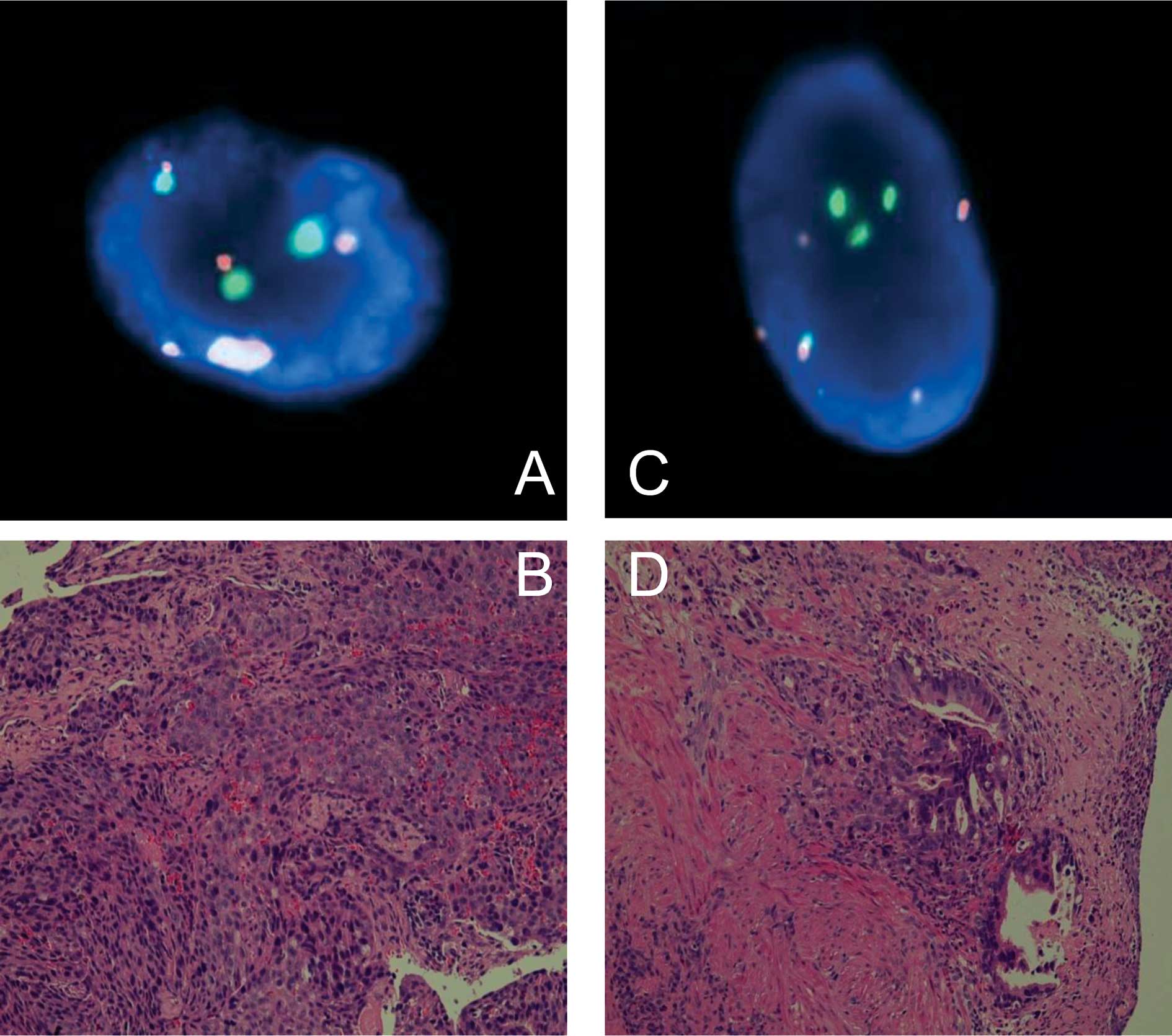
This study aimed to the clarify the diagnostic efficacy of fluorescence in situ hybridization (FISH) in Kazakh patients with esophageal cancer (EC). FISH was compared with the pathological examination of biopsy specimens with DNA probes. We enrolled 20 patients, of which 15 were males and 5 females, with an average age of 58.3 years, who had abnormal esophaguses on barium radiological digital imaging. Touch preparations were performed on biopsy specimens from all of the patients and were examined using FISH for chromosomal abnormalities. We compared the FISH results with the pathology slides stained with hematoxylin and eosin. Classification, according to pathology, identified 2 cases of class II, 3 cases of IIIa, 1 case of IIIb, 2 cases of IV, 12 cases of class V and no cases of class I. The cases classified as class IIIb or higher were considered to be positive for cancer. Using histopathology, 10 cases were diagnosed with squamous cell carcinoma and 5 were diagnosed as adenocarcinoma, with one case being false-negative. Thus, the sensitivity of the pathological examination was 93% and the specificity was 100%. Using FISH, 16 cases showed aberrant copy numbers in either chromosome 3 or 17. By comparison, pathology did not reveal any false-positive or false-negative cases with a sensitivity and specificity of 100%. The centromeres of chromosome 3 copy numbers was significantly higher (p=0.035) than the centromeres of chromosome 17. Our study compared FISH to diagnose aneusomic esophageal cancer cells with the pathology of biopsied tissue. Our findings suggest that FISH is a useful and objective assay for the detection of malignant cells of esophageal cancer. In our study, the centromeres of chromosome 3 was the more sensitive probe for the diagnosis of esophageal cancer in Kazakh patients.
View Figures
View References

Related Articles

Journal Cover

September-October 2010
Volume 1 Issue 5

Print ISSN: 1792-1074
Online ISSN:1792-1082

Sign up for eToc alerts

Recommend to Library

Copy and paste a formatted citation
x
Spandidos Publications style
Awut I, Niyaz M, Huizhong X, Biekemitoufu H, Yan ZH, Zhu Z, Sheyhedin I, Changmin Z, Wei Z, Hao W, Hao W, et al: Genetic diagnosis of patients with esophageal cancer using FISH . Oncol Lett 1: 809-813, 2010
APA
Awut, I., Niyaz, M., Huizhong, X., Biekemitoufu, H., Yan, Z.H., Zhu, Z. ... Hao, W. (2010). Genetic diagnosis of patients with esophageal cancer using FISH . Oncology Letters, 1, 809-813. https://doi.org/10.3892/ol_00000142
MLA
Awut, I., Niyaz, M., Huizhong, X., Biekemitoufu, H., Yan, Z. H., Zhu, Z., Sheyhedin, I., Changmin, Z., Wei, Z., Hao, W."Genetic diagnosis of patients with esophageal cancer using FISH ". Oncology Letters 1.5 (2010): 809-813.
Chicago
Awut, I., Niyaz, M., Huizhong, X., Biekemitoufu, H., Yan, Z. H., Zhu, Z., Sheyhedin, I., Changmin, Z., Wei, Z., Hao, W."Genetic diagnosis of patients with esophageal cancer using FISH ". Oncology Letters 1, no. 5 (2010): 809-813. https://doi.org/10.3892/ol_00000142