Introduction
For the detection of lung cancer cells in the
sputum, we developed melanoma associated gene (MAGE) primers
that can amplify MAGE A1-A6 simultaneously (1), and evaluated the positive rates in
induced sputum of patients with lung cancer (2). Meanwhile, we detected MAGE
expression not only in patients with lung cancer but also in some
patients with non-malignant lung diseases, including tuberculosis
and inflammation. Expression of MAGE by de-methylation of
the MAGE promoter region has been demonstrated (3), and its expression was known to be
restricted to germ cells and cancer cells (4,5).
However, Mecklenburg et al reported that MAGE
expression was also detected in patients with inflammatory diseases
(6).
As inflammation is a critical component of tumor
progression, repeated and chronic inflammation can increase cancer
risk (7). Considering the course of
lung carcinogenesis, before which molecular biological change has
occurred, MAGE expression in the sputum of inflammatory
disease can be regarded as a reasonable event. However, applying
this result to clinical diagnosis can increase the probability of
false positive diagnoses and lead to difficulties in patient care.
For the clinical application of MAGE reverse transcription
polymerase chain reaction (RT-PCR), it would be necessary that the
MAGE result be carefully interpreted along with additional
molecular findings and clinical evidence.
This study was designed to evaluate the performance
of MAGE RT-PCR, MAGE A3 methylation-specific PCR
(MSP), and p16 MSP using induced sputum of patients with
lung cancer or non-cancerous inflammatory diseases, and to compare
the results of genetic tests with the patients' clinical findings.
Eventually, we attempted to elucidate the clinical significance of
MAGE expression in the sputum of patients with pulmonary
diseases.
Subjects and methods
Subjects
Twenty-four biopsy specimens were obtained from
patients with lung cancer who underwent bronchoscopy at Yeungnam
University Hospital from 2006 to 2008. Obtained tissues were
immersed immediately in TRI solution (Molecular Research Center,
Cincinnati, OH), and stored in a deep freezer until RNA
extraction.
During the same period, 133 samples of induced
sputum specimens collected from patients with pulmonary problems
who visited Yeungnam University Hospital were added to sputum RNA
extraction solution (iC&G Co., Daegu, Korea), and stored in a
-70°C refrigerator until required for RNA extraction. Induced
sputum was obtained after inhaling Berotec solution (Boehringer
Ingelheim, Ingelheim, Germany) and 16 ml of 3% hypertonic saline.
To determine the clinical diagnosis for the patients, history
taking, physical examinations, bronchoscopy, computed tomography
(CT) scan, and histopathological biopsies were performed.
Random sputa were collected from 30 healthy
volunteers, and treated equally. All stored specimens were blindly
transferred to the Department of Laboratory Medicine at Daegu
Catholic University Medical Center, then MAGE A1-A6 RT-PCR,
MAGE A3 MSP, and p16 MSP were carried out. Tissue and
sputum procurement procedures were approved by the Institutional
Review Board of the Yeungnam University Hospital. Informed consent
was obtained from all patients.
RNA and DNA extraction and RT-PCR
RNA and DNA extraction from sputum specimens was
conducted with an iC&G extraction kit using magnetic beads. RNA
from tissues was extracted according to the TRI Corporation's
instructions, and its DNA was extracted using TRI remnant. RNA was
reverse-transcribed using ImProm-II reverse transcription (RT)
reagents (Promega Corp., Madison, WI), and MAGE A1-A6
expression was amplified using iC&G PCR reagents. cDNA
integrity was confirmed by GAPD amplifications.
Methylation-specific PCR
Unmethylated cytosine was changed to uracil in the
CG nucleotide gene of extracted DNA using Cp genome change reagent
(Chemicon, Temecula, CA). Nested PCR was performed on treated
genomic DNA for amplification of MAGE A3 and p16 MSP.
At first, the target gene was amplified for 30 cycles, regardless
of methylation. Then, MSP was performed for 30 cycles using
methylation-specific primers and Gold Taq enzyme (Perkin-Elmer,
Boston, MA). Cp™ genome universal methylated DNA (Chemicon) was
used as a positive control sample of MAGE A3 and p16
MSP. Primer sets for MAGE A3, p16 MSP, and
GAPD, annealing temperatures and product sizes are shown in
Table I. The amplified products of
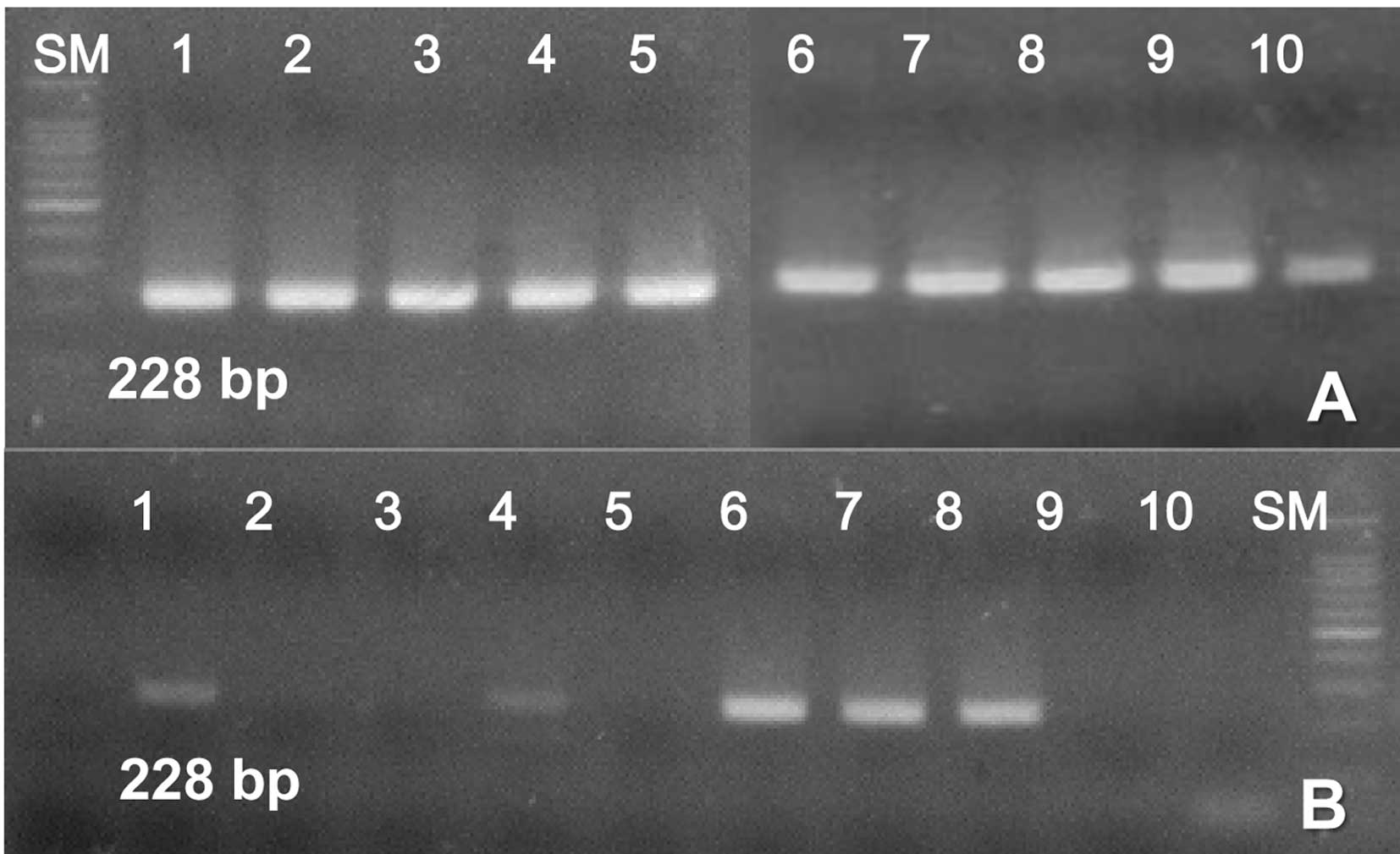
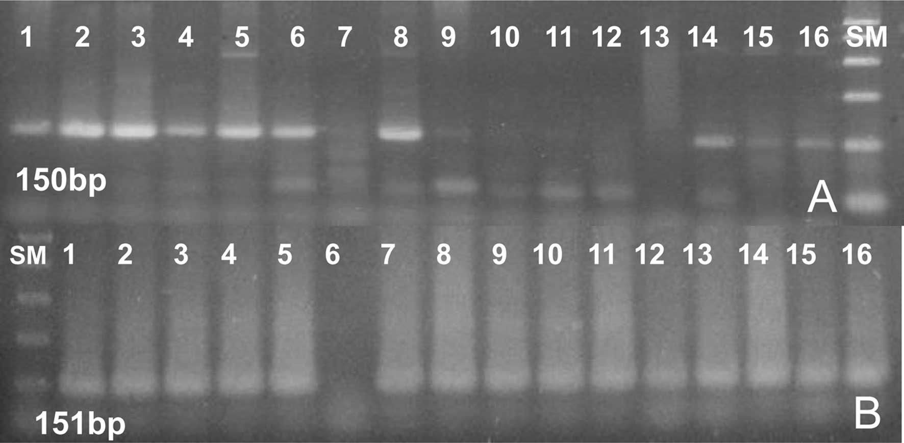
MAGE A3 MSP, and p16 MSP were clearly visualized in
Fig. 1 and Fig. 2, respectively.
 | Table IPrimer sequences used for GAPD
PCR, MAGE A3, and p16 methylation specific PCR. |
Table I
Primer sequences used for GAPD
PCR, MAGE A3, and p16 methylation specific PCR.
| Target | Direction | Sequences
(5′→3′) | Ta (°C) | Size (bp) |
|---|
| GAPD | F | tcg gag tca acg gat
ttg gtc gta | 59 | 320 |
| R | caa atg agc ccc agc
ctt ctc ca | | |
| MAGE A3 | Outer F | tgt tcg gaa ttt agg
gta gta tcg | 56 | 417 |
| Outer R | ttc cct ctc gaa atc
cta acc tta | | |
| IF (UM) | tgt ttt gag taa tga
gtg at | 56 | 228 |
| IR (UM) | act aaa aca aca aaa
atc aac a | | |
| IF (M) | cgt ttt gag taa cga
gcg ac | 56 | 228 |
| IR (M) | act aaa acg acg aaa
atc gac g | | |
| p16 | Outer F | ggt gtt ata ttc gtt
aag tgt tcg | 56 | 482 |
| Outer R | cta cct aat tcc aat
tcc cct aca | | |
| IF (UM) | tta tta gag ggt ggg
gtg gat tgt | 65 | 151 |
| IR (UM) | c aac ccc aaa cca
caa cca taa | | |
| IF (M) | tta tta gag ggt ggg
gcg gat cgc | 65 | 150 |
| IR (M) | gac ccc gaa ccg cga
ccg taa | | |
Statistical analysis
The repeated-measure one factor analysis of the
Cochran test was performed to compare the positive rates of
MAGE A1-A6 RT-PCR, MAGE A3 MSP, and p16 MSP.
The Chi-square test was used for comparison of positive rates among
the patients' groups. Statistical analyses were conducted using
SPSS 14.0 software (SPSS Inc., Chicago, IL). A P-value of <0.05
was considered to indicate statistical significance.
Results
Subjects
The 133 enrolled patients were diagnosed as 65 lung
cancer and 68 benign lung diseases. Patients of benign lung
diseases were classified as follows: 25 no active lung disease, 16
pulmonary tuberculosis, 11 pneumonia, 11 inflammatory diseases, 2
bullae, 2 pleural effusion of unknown cause, and 1 right middle
lobe syndrome.
The mean ages of patients with lung cancer, patients
with benign lung diseases, and healthy volunteers were 66.0±12.7,
59.0±15.6 and 29.5±9.79 years, respectively. The gender
distributions of these groups were 8.29:1, 5.18:1, and 0.58:1,
respectively.
Positive rates for MAGE A1-A6 RT-PCR,
MAGE A3 MSP, and p16 MSP according to the patient group
Positive rates for MAGE A1-A6 RT-PCR,
MAGE A3 MSP, and p16 MSP were as follows. In tissues
of patients with lung cancer, 87.5, 58.3, and 70.8%; in induced
sputa of patients with lung cancer, 50.8, 46.2, and 63.1%; in
induced sputa of patients with benign lung disease, 10.3, 30.9, and
39.7%; in random sputa of healthy people, 3.3, 6.7, and 3.3%. All 3
tests showed statistically significant results using the sputum of
lung cancer, benign diseases, and healthy groups (P<0.05)
(Table II). In the group of lung
cancer, MAGE RT-PCR revealed statistically significant
higher positive rates while in benign lung diseases, significant
lower positive rates than MAGE A3 and p16 MSP results
(P<0.05).
| Table IIPositive rates of MAGE RT-PCR,
MAGE A3, and p16 methylation specific PCR according
to the diagnosis. |
Table II
Positive rates of MAGE RT-PCR,
MAGE A3, and p16 methylation specific PCR according
to the diagnosis.
| Specimen | Diagnosis | n | MAGE
(%) | A3-UM (%) | p16-M
(%) | P-valuea |
|---|
| Tissue | Lung cancer | 24 | 87.5 | 58.3 | 70.8 | 0.058 |
| Induced sputum | Lung cancer | 65 | 50.8 | 46.2 | 63.1 | 0.040 |
| Induced sputum | Benign
diseases | 68 | 10.3 | 30.9 | 39.7 | 0.000 |
| Random sputum | Healthy people | 30 | 3.3 | 6.7 | 3.3 | 0.607 |
| P-valueb | | | 0.00 | 0.00 | 0.00 | |
Positive rates for MAGE RT-PCR, MAGE A3
MSP, and p16 MSP in patients with benign lung diseases
MAGE expression rates were high in patients
with inflammatory diseases and tuberculosis (18.2 and 18.8%,
respectively), while positive rates for MAGE A3
unmethylation and p16 methylation were high in patients with
tuberculosis (56.3 and 62.5%) and in those with pneumonia (45.5 and
54.5%). The average positive rate of MAGE, MAGE A3
unmethylation, and p16 methylation in patients with benign
lung diseases were 10.3, 30.9, and 39.7% (Table III), showing a statistically
significant difference (Table
II).
| Table IIIPositive rates of MAGE RT-PCR,
MAGE A3, and p16 methylation specific PCR in sputum
of patients with benign lung diseases. |
Table III
Positive rates of MAGE RT-PCR,
MAGE A3, and p16 methylation specific PCR in sputum
of patients with benign lung diseases.
| Diagnosis | n | MAGE
(%) | A3-UM (%) | p16-M
(%) |
|---|
| No active lung
disease | 25 | 4.0 | 16.0 | 24.0 |
| Inflammatory
diseases | 11 | 18.2 | 18.2 | 36.4 |
| Tuberculosis | 16 | 18.8 | 56.3 | 62.5 |
| Pneumonia | 11 | 0.0 | 45.5 | 54.5 |
| Othersa | 5 | 20.0 | 20.0 | 20.0 |
| Total | 68 | 10.3 | 30.9 | 39.7 |
| P-value (Chi-square
test) | | 0.306 | 0.047 | 0.094 |
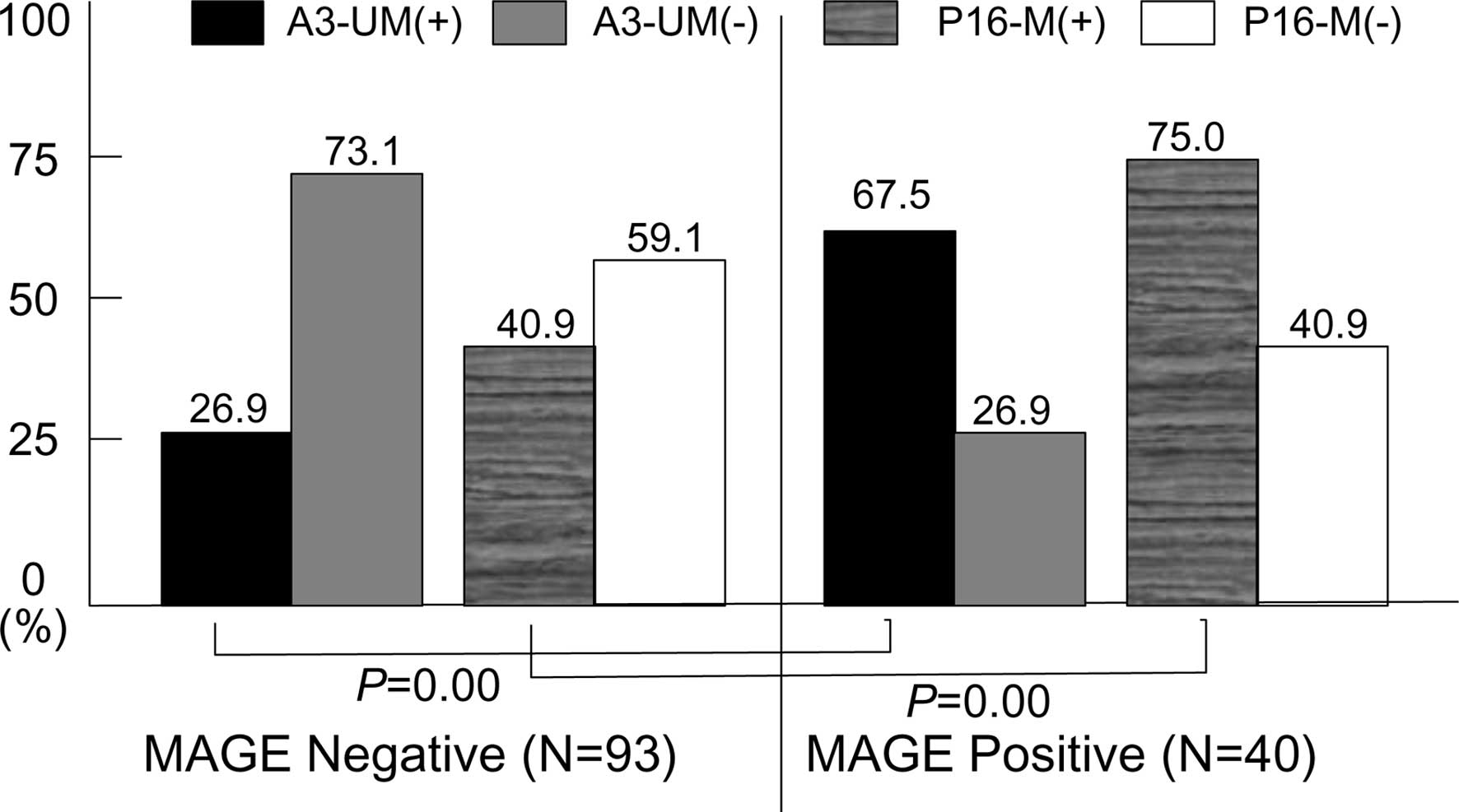
Positive rates for MAGE A3 MSP and p16
MSP in regard to MAGE expression
In the 40 MAGE positive cases of lung cancer
and benign lung diseases, positive rates for MAGE A3
unmethylation and p16 methylation were 67.5 and 75.0%,
respectively. These results showed significant differences compared
with the 26.9% positive rates of MAGE A3 unmethylation and
40.9% of p16 methylation in the 93 MAGE-negative
cases (Fig. 3).
Clinical analysis and methylation
abnormality in the MAGE-positive patients
Of the 40 MAGE-positive cases, 33 were
diagnosed as lung cancer and 7 as benign lung diseases. The
diagnosis of 7 benign lung diseases was as follows: 3 pulmonary
tuberculosis, 2 inflammatory diseases, 1 bullae, and 1 no active
lung disease. From the 7 cases of benign lung diseases, 5 showed
methylation abnormality in both MAGE A3 and p16. One
inflammation showed methylation abnormality in p16 only and
1 bullae showed no methylation abnormality in either gene (Table IV).
| Table IVClinical diagnosis and
methylation-specific PCR results for 7 MAGE-positive cases
of benign lung diseases. |
Table IV
Clinical diagnosis and
methylation-specific PCR results for 7 MAGE-positive cases
of benign lung diseases.
| Case no. | Age | Gender | Diagnosis | A3-UM | p16-M |
|---|
| 1 | 53 | M | No active lung
disease | Positive | Positive |
| 2 | 62 | M | TB pleurisy | Positive | Positive |
| 3 | 77 | M | Pulmonary TB | Positive | Positive |
| 4 | 48 | M | Pulmonary TB | Positive | Positive |
| 5 | 52 | F | Inflammation | Positive | Positive |
| 6 | 74 | M | Inflammation | Negative | Positive |
| 7 | 58 | M | Bullae | Negative | Negative |
Discussion
Mortality of lung cancer is still high; therefore,
early detection of lung cancer is a major issue. The advent of
low-dose spiral chest CT, positron emission tomography, and
autofluorescence bronchoscopy (AFB) have opened a new perspective
for early detection (8), and
specific molecular markers have also been developed (9). Although AFB can detect pre-invasive
lesions and lung cancers in the central airway, the specificity of
AFB is low (10). A diagnostic test
with high sensitivity and high specificity has not yet been
developed.
MAGE is a highly specific tumor marker, and
MAGE A3 expression was observed in 35% of lung cancer
patients through a multi-center study (11). Based on these findings,
MAGE-A3 vaccination has been developed as a promising
treatment modality for lung cancer (12,13).
Atanackovic et al (12)
reported that 14 of 18 lung cancer patients with stage I or stage
II disease had no evidence of disease up to 3 years after
vaccination with MAGE A3-protein. In addition to MAGE
A3, a DNA methylation-based biomarker is considered a rapid and
efficient early detection marker for lung cancer (9,14), and
a previous study reported that DNA methylation marker targeting 4
genes in sputum showed 94% sensitivity and 90% specificity
(14). Anglim et al
demonstrated that aberrant promoter methylation of p16 was
observed 3 years before diagnosis of squamous cell carcinoma in
smokers (9).
In the present study, MAGE gene-positive
rates were 87.5% in lung cancer tissues, 50.8% in induced sputum
specimens of patients with lung cancer, 10.3% in induced sputum
specimens of patients with benign lung diseases, and 3.3% in random
sputum specimens of healthy people. The finding of MAGE
expression in benign lung diseases is consistent with those of an
earlier study, which reported that MAGE A1 or A2 expression
is detected in severe bronchitis and severe actinomycosis with
concomitant tissue regeneration (6). MAGE expression was considered
an early event in lung carcinogenesis (3) and was detected in precancerous
lesions. Therefore, MAGE can be used not only as an early
detection marker for lung cancer but also as a prevention marker
for lung carcinogenesis. In this study, MAGE positive benign
lung diseases were mainly comprised of pulmonary tuberculosis and
inflammatory diseases and severity of inflammation was not
evaluated. Kim et al (15)
reported MAGE expression in tissue samples obtained using a
percutaneous needle aspiration biopsy of tuberculosis patients. We
performed MAGE A1-A6 nested PCR using DNA of
Mycobacterium tuberculosis; however, the MAGE gene
was not amplified. Therefore, MAGE expression of
tuberculosis patients may be not the result of a false-positive
detection by Mycobacterium tuberculosis but the result of
the inflammatory process. Compared with the non-tuberculosis group,
tuberculosis patients showed coexistence of lung cancer and high
incidence rates of lung cancer (16,17).
However, determination of MAGE expression in
the sputum based simply on clinical conditions cannot reflect
molecular biological change at the cellular level. To reflect
molecular biological change, additional molecular biological tests
associated with lung carcinogenesis are necessary. In order to
conduct a proper mutation analysis, a number of tumor cells and a
large number of genetic loci should be investigated. Aberrant
methylation of the p16 promoter is an important mechanism of
lung carcinogenesis (14) and
unmethylation of the MAGE A3 promoter is directly associated
with MAGE expression. Therefore, we performed MAGE A3
and p16 MSP. The MSP is a promising method for detection of
lung cancer because it can detect a small number of cancer cells in
sputum that contains a large number of normal cells (9).
In the present study, unmethylation rates for
MAGE A3 MSP were 46.2% in sputa of patients with lung
cancers, 30.9% in patients with benign lung diseases, and 6.7% in
healthy people. Though unmethylation rates for MAGE A3 MSP
have not yet been reported in the literature review, these findings
are similar to the results of Olaussen et al (18), which reported that positive rates
for MAGE A1 MSP were 50% in cytologically negative sputum
from lung cancers patients, 45% in sputum showing inflammatory
change from smokers, and 6% in cytologically negative sputum from
smokers.
Positive rates for p16 MSP were 63.1% in
sputa of lung cancer patients, 39.7% in patients with benign lung
diseases, and 3.3% in healthy people. Olaussen et al
(18) also reported that positive
rates for p16 MSP were 27% in cytologically negative sputum
from lung cancers patients, 64% in sputum showing cancerous
cytology from smokers, 27% in sputum showing inflammatory change
from smokers, and 47% in sputum showing normal cytology from
smokers. Among studies performed in the Korean population, one
study reported that p16 methylation was detected in 67% of
tumor samples (19). However,
another study reported 22% positive rates in tumor samples, and 1%
in the corresponding non-malignant lung tissues (20). The methylation rate of p16 in
lung cancer tissues was about 80% (21,22).
Liu et al (22) reported
methylation rates of 74.7% in the sputum from lung cancer patients,
and 51.4% from people exposed to coal smoke. Using matched
specimens from lung cancer patients, Hsu et al (23) reported 37% methylation rates in lung
cancer tissues, 33% in sputa, 13% in normal lung tissues, and 14%
in sputa from the control group. Although reported rates for
p16 methylation were variable, p16 MSP was regarded
as a useful molecular marker for use in early detection and
prediction of lung cancer.
Therefore, analysis of molecular abnormality in the
same specimen using MAGE A1-A6 RT-PCR, MAGE A3 MSP,
and p16 MSP simultaneously may be very useful for detection
and prediction of lung cancer at the molecular level. This analysis
can also be applied to non-cancerous groups to understand the
clinical significance of MAGE expression. In this and other
studies, positive rates of MSP remain high in the non-cancerous
group; therefore, abnormality of MSP was just utilized as a
supplemental modality to explain MAGE expression in the
sputum, not as a cancer detection tool.
The MAGE-positive group in sputum showed a
statistically significant higher MAGE A3 unmethylation and
p16 MSP methylation rate than the MAGE negative
group. From the 7 cases of benign lung diseases with MAGE
expression, 5 showed methylation abnormality in both MAGE A3
and p16, 1 case showed methylation abnormality in p16
only, and 1 case showed methylation abnormality in neither
MAGE A3 nor p16. Although the MAGE A3 gene
could not be expressed without unmethylation of MAGE A3,
another gene could be amplified because common primers that can
amplify MAGE A1-A6 together had been used. Moreover,
MAGE A3 MSP is not a quantitative MSP of promoter loci, but
reflects methylation status of primer binding sites. Therefore, the
results of MSP may not be consistent with the results of
RT-PCR.
The question of how to interpret MAGE
expression in patients with benign lung diseases is an important
issue. MAGE proteins form complexes with KAP1, suppress
p53-dependent apoptosis, and contribute to cancer development
(24). We analyzed MAGE
expression acting as a tumor enhancer, and also detected the
methylation status of MAGE A3 and p16 using the same
specimens. Of 40 MAGE positive cases in the sputa, 39 cases
turned out to be lung cancers or benign lung diseases accompanying
methylation abnormality. Therefore, MAGE expression in the
clinical specimen may suggest the presence of cells in the process
of molecular carcinogenesis, even if cancer cells are not
visible.
Thus, the clinical significance of MAGE
expression in the sputum would be i) the presence of lung cancer
cells or ii) pre-cancerous cells. In another study (25), we evaluated MAGE expression
in the peritoneal washes of gastric carcinoma patients, and the
patients were followed up for 5 years in regard to MAGE
expression. As a result, recurrence rates of MAGE-positive
cases (45.5%) were much higher than those of MAGE negative
cases (9.6%). Although gastric carcinogenesis may not be identical
with lung carcinogenesis, MAGE expression in clinical
specimens can be considered a strong indication of tumor
recurrence. However, not all precancerous cells will develop into
cancer, and MAGE expression was regarded as a reversible
change. Thus, MAGE expression in specimens of non-malignant
patients should be interpreted very carefully, and for proper
clinical application, other clinical information, such as 5-year
follow-up results would be studied.
In 2001, Jang et al (3) predicted that the MAGE gene
would be utilized as a tool for lung cancer prevention. Since then,
functions of the MAGE gene as a tumor promoter have been
disclosed, and tumor therapeutic agents targeting the MAGE
gene have been developed and will soon be applied to lung cancer
treatment. The recurrence rate of MAGE-positive cases in
peritoneal washes of gastric carcinoma patients was 45.5%. In the
present study, the MAGE A3 unmethylation or p16
methylation abnormality was demonstrated in MAGE-positive
specimens from non-cancerous patients. Based on these findings,
MAGE expression in the sputum may indicate the presence of
lung cancer cells or pre-cancerous cells. Therefore, a MAGE
positive case in the non-cancer group should be closely followed
up. In conclusion, MAGE could be utilized as a cancer
prediction tool as well as a cancer detection tool. Further studies
including molecular markers, histological examination, and clinical
studies targeting non-cancerous patients with MAGE
expression are inevitable.
References
|
1
|
Park JW, Kwon TK, Kim IH, et al: A new
strategy for the diagnosis of MAGE-expressing cancers. J Immunol
Methods. 266:79–86. 2002. View Article : Google Scholar : PubMed/NCBI
|
|
2
|
Jheon S, Hyun DS, Lee SC, et al: Lung
cancer detection by a RT-nested PCR using MAGE A1-6 common primers.
Lung Cancer. 43:29–37. 2004. View Article : Google Scholar : PubMed/NCBI
|
|
3
|
Jang SJ, Soria JC, Wang L, et al:
Activation of melanoma antigen tumor antigens occurs early in lung
carcinogenesis. Cancer Res. 61:7959–7963. 2001.PubMed/NCBI
|
|
4
|
van der Bruggen P, Traversari C, Chomez P,
et al: A gene encoding an antigen recognized by cytolytic T
lymphocytes on a human melanoma. Science. 254:1643–1647. 1991.
|
|
5
|
De Plaen E, Arden K, Traversari C, et al:
Structure, chromosomal localization, and expression of 12 genes of
the MAGE family. Immunogenetics. 40:360–369. 1994.PubMed/NCBI
|
|
6
|
Mecklenburg I, Stratakis DF, Huber RM, et
al: Detection of melanoma antigen-A expression in sputum and
bronchial lavage fluid of patients with lung cancer. Chest.
125:164S–166S. 2004. View Article : Google Scholar : PubMed/NCBI
|
|
7
|
Chaturvedi AK, Gaydos CA, Agreda P, et al:
Chlamydia pneumoniae infection and risk for lung cancer.
Cancer Epidemiol Biomarkers Prev. 19:1498–1505. 2010. View Article : Google Scholar
|
|
8
|
Pastorino U: Lung cancer screening. Br J
Cancer. 102:1681–1686. 2010. View Article : Google Scholar
|
|
9
|
Anglim PP, Alonzo TA and Laird-Offringa
IA: DNA methylation-based biomarkers for early detection of
non-small cell lung cancer: an update. Mol Cancer. 7:812008.
View Article : Google Scholar : PubMed/NCBI
|
|
10
|
Yasufuku K: Early diagnosis of lung
cancer. Clin Chest Med. 31:39–47. 2010. View Article : Google Scholar
|
|
11
|
Sienel W, Varwerk C, Linder A, et al:
Melanoma associated antigen (MAGE)-A3 expression in stages I and II
non-small cell lung cancer: results of a multi-center study. Eur J
Cardiothorac Surg. 25:131–134. 2004. View Article : Google Scholar : PubMed/NCBI
|
|
12
|
Atanackovic D, Altorki NK, Cao Y, et al:
Booster vaccination of cancer patients with MAGE-A3 protein reveals
long-term immunological memory or tolerance depending on priming.
Proc Natl Acad Sci USA. 105:1650–1655. 2008. View Article : Google Scholar : PubMed/NCBI
|
|
13
|
Tsuji T, Altorki NK, Ritter G, et al:
Characterization of preexisting MAGE-A3-specific CD4+ T
cells in cancer patients and healthy individuals and their
activation by protein vaccination. J Immunol. 183:4800–4808.
2009.PubMed/NCBI
|
|
14
|
Risch A and Plass C: Lung cancer
epigenetics and genetics. Int J Cancer. 123:1–7. 2008. View Article : Google Scholar : PubMed/NCBI
|
|
15
|
Kim H, Kim SJ, Lee SH, et al: Usefulness
of melanoma antigen (MAGE) gene analysis in tissue samples from
percutaneous needle aspiration biopsy of suspected lung cancer
lesions. Lung Cancer. 69:284–288. 2010. View Article : Google Scholar : PubMed/NCBI
|
|
16
|
Zheng W, Blot WJ, Liao ML, et al: Lung
cancer and prior tuberculosis infection in Shanghai. Br J Cancer.
56:501–504. 1987. View Article : Google Scholar : PubMed/NCBI
|
|
17
|
Kurasawa T: The coexistence of pulmonary
tuberculosis and lung cancer. Nihon Rinsho. 56:3167–3170.
1998.PubMed/NCBI
|
|
18
|
Olaussen KA, Soria JC, Park YW, et al:
Assessing abnormal gene promoter methylation in paraffin-embedded
sputum from patients with NSCLC. Eur J Cancer. 41:2112–2119. 2005.
View Article : Google Scholar : PubMed/NCBI
|
|
19
|
Kim YT, Lee SH, Sung SW, et al: Can
aberrant promoter methylation of CpG islands predict the clinical
outcome of non-small cell lung cancer after curative resection? Ann
Thorac Surg. 79:1180–1188. 2005. View Article : Google Scholar : PubMed/NCBI
|
|
20
|
Kim DS, Cha SI, Lee JH, et al: Aberrant
DNA methylation profiles of non-small cell lung cancers in a Korean
population. Lung Cancer. 58:1–6. 2007. View Article : Google Scholar : PubMed/NCBI
|
|
21
|
Ulivi P, Zoli W, Calistri D, et al:
p16INK4A and CDH13 methylation in tumor and serum of non-small cell
lung cancer patients. J Cell Physiol. 206:611–615. 2006. View Article : Google Scholar : PubMed/NCBI
|
|
22
|
Liu Y, An Q, Li L, et al: Methylation of
p16INK4a in Chinese lung cancer patients: biological and clinical
implications. Carcinogenesis. 24:1897–1901. 2003. View Article : Google Scholar : PubMed/NCBI
|
|
23
|
Hsu HS, Chen TP, Wen CK, et al: Multiple
genetic and epigenetic biomarkers for lung cancer detection in
cytologically negative sputum and a nested case-control study for
risk assessment. J Pathol. 213:412–419. 2007. View Article : Google Scholar : PubMed/NCBI
|
|
24
|
Yang B, O'Herrin SM, Wu J, et al: MAGE-A,
mMage-b, and MAGE-C proteins form complexes with KAP1 and suppress
p53-dependent apoptosis in MAGE-positive cell lines. Cancer Res.
67:9954–9962. 2007. View Article : Google Scholar : PubMed/NCBI
|
|
25
|
Jeon CH, Shin IH, Park JB, et al:
Prognostic significance of MAGE in peritoneal washes in gastric
carcinoma patients without peritoneal metastasis: results of a
5-year follow-up study. J Clin Gastroenterol. 44:682–686.
2010.PubMed/NCBI
|