Introduction
Ovarian cancer is the leading cause of
cancer-related deaths among gynecological cancers. Lesions derived
from the surface epithelium of the ovary account for 80–90% of
cases. Unfortunately, ovarian carcinoma is often diagnosed late in
the course of the disease, after the cancer has spread into the
peritoneal cavity, and the 5-year survival rate is only 20–30%.
Invasion and metastasis of tumor cells are the main factors
influencing to prognosis. Thus, defining carcinogenesis and
progression mechanisms plays a vital role in early detection, in
clarifying a diagnosis and antineoplaston therapy for ovarian
cancer.
Glycosyl-antigen, is widely expressed in the cell
membrane and is an important ingredient of glycoprotein and
glycolipid. Lewis y antigen is carried by glycoconjugates
(glycoproteins and glycolipids) at the cell surface. It is an
oligosaccharide with 2 fucoses, belonging to the A, B, H, Lewis
blood group antigens family (1).
Elevated expression of Lewis y has been found in 70–90% of the
human carcinomas of epithelial cell origin, including breast,
ovary, prostate, colon cancers, and its high expression level is
correlated with the tumor’s pathological staging and prognosis
(2). TGF-β (transforming growth
factor β) represents a family of pleiotropic, secreted growth
factors which regulate such diverse processes as embryonic
development, wound healing, organ development, and
immunoregulation. Researchers have found that TGF-β plays an
important role in occurrence, progression and metastasis of ovarian
cancer. TGF-β may cause cell cycle arrest, terminal
differentiation, or apoptosis in most normal ovarian epithelial
cells, whereas most malignant ovarian cell lines are resistant to
TGF-β (3,4). In addition, TGF-β production may
represent a significant tumor escape mechanism from host
immunosurveillance, may increase angiogenesis and enhance the
interaction between cancer cells and extracellular matrix. This
negative control mechanism finally promotes growth and development
of advanced tumor cells. In advanced ovarian cancer, increased
expression of TGF-β or changes in signal transduction pathways may
promote tumor recurrence and resistance to chemotherapy (5).
In previous studies, α1,2-fucosyltransferase
(α1,2-FT) was transfected into the ovarian cancer cell line RMG-I,
and the RMG-I-H cell line was thus developed which highly expressed
the Lewis y antigen. This research showed that the post-tranfected
cell line had increased abilities of proliferation, adhesion,
invasion, metastasis and drug resistance than the pre-transfected
cell line. It illustrated that Lewis y played an important role in
the canceration, development and metastasis in ovarian cancer
(6–8). Moreover, we used microarray analysis
to distinguish between the expression profiles of cancer-related
genes before and after α1,2-FT transfection into ovarian cancer
cells. The microarray results revealed that the expression of the
TGF-β1 gene is up-regulated after transfection (9). Therefore, we hypothesize that the
expression level of TGF-β1 may correlate with Lewis y antigen.
Based on the results of previous studies, this
experiment investigated the expression and correlation of Lewis y
antigen and TGF-β1 in ovarian epithelial carcinoma tissue specimen
using an immunohistochemical method. The immunofluorescence double
labeling method was also used to elucidate the correlation of Lewis
y antigen and TGF-β1. This study will aid in the development of the
theoretical basis of ovarian carcinogenesis, its mechanisms of
development and in the identification of potential biological
treatments.
Materials and methods
Patients and tissue samples
A total of 110 paraffin samples were obtained from
operations performed between 2000 and 2009 in the department of
Gynecology and Obstetrics of our hospital. All the tissue sections
were diagnosed by specialists. There were 60 cases of primary
ovarian epithelial malignant tumors (including 30 mucous
cystadenocarcinoma, 30 serous cystadenocarcinoma), 20 cases of
borderline ovarian tumor, 20 cases of benign ovarian tumor and 10
cases of normal ovarian tissues (obtained from normal ovarian
tissue that was excised during the cervical cancer operations). The
average age of these patients was 47.89 years (15–73 years). The
age range of the ovarian cancer group was 36–73 years, the median
age was 53.5 years; the age range of borderline ovarian tumor group
was 22–55 years, the median age was 35 years; the age ranges of the
benign ovarian tumor and normal tissue were 15–72 and 37–52 years
and the median ages were 44 and 42 years, respectively. There was
not a statistically significant difference between the age ranges
of these groups (P>0.05). According to the pathological grading,
the ovarian cancer group contained 21 cases of high
differentiation, 21 cases of middle differentiation and 18 cases of
low differentiation; this group included 39 cases of I–II stage and
21 cases of III–VI stage according to the International Federation
of Gynecology and Obstetrics (FIGO) standard; 12 cases had
metastases in the pelvic cavity nodes. All the cases were primary
tumors, and the information obtained was complete. Furthermore,
chemical treatment had not been received by any of the patients
before operations.
Chief reagent and methods
The Lewis y monoclonal antibody (clone A70-C/C8) was
purchased from Abcam; the TGF-β1 polyclonal antibody was obtained
from the Boster Company; the goat monoclonal anti-rabbit
immunoglobulin G fluorescein isothiocyanate and the goat monoclonal
anti-mouse immunoglobulin G tetramethylrhodamine isothiocyanate
(TRITC) were purchased from the Mai Xin Company;
4,6-diamidino-2-phenylindole (DAPI) was obtained from the Bao Xin
Company. Other reagents were supplied by our laboratory.
Methods
Histological sections
Five micrometer serial sections were obtained from
each group of ovarian tissues.
Immunohistochemistry
The expression of Lewis y and TGF-β1 in ovarian
carcinoma tissues were analyzed by immunohistochemical SP staining.
Positive and negative immunohistochemistry controls are routinely
used. The study concentration of primary antibodies against Lewis y
and TGF-β1 were all 1:200. The empirical procedure was performed
according to the kit’s instructions.
Immunofluorescence double labeling
method
The tissue sections that showed strong positive
expression by immunohistochemistry, were chosen for
immunofluorescence double labeling. The sections were
simultaneously incubated with primary antibodies against Lewis y
(1:100) and TGF-β1. Negative control sections were incubated with
PBS instead of the primary antibody. The study concentrations of
fluorescein isothiocyanate and TRITC were 1:100. Nuclei were
counterstained with DAPI. The empirical procedure was performed
based on the kit’s instructions.
Assessment standard
Immunohistochemistry
The staining results were considered positive if
there buffy granules were present in the cell membrane and the
cytoplasm. According to the chromatosis intensity: no pigmentation,
light yellow, buffy and brown were scored as 0, 1, 2 and 3,
respectively. We chose 5 high power fields in a series from each
slice, then scored them and took the average percentage of stained
cells: when stained cells were <5% the score was 1; when the
stained cells accounted for 5–25%, the score was 1; when for
26–50%, the score was 2; for 51–75% the score was 3, and when for
>75% the score was 4. These 2 numbers were multiplied, and 0–2
was considered (−), 3–4 was (+), 5–8 was (++), and 9–12 was (+++).
Two observers read the sections to control for error. At the same
time, we used the NIS-Elements BR 2.10 picture analysis software of
the Japanese Nikon company to measure the mean optical density
(MOD).
Immunofluorescence double
labeling
The red fluorescence represented the Lewis y antigen
that was labeled by TRITC and the green fluorescence was labeled by
TGF-β1; the blue fluorescence was nuclear counterstaining by DAPI.
After the pictures were captured, we used the picture analysis
software to build up the 3 fluorescence passages; yellow
fluorescence illustrates that Lewis y and TGF-β1 are located in the
same position.
Statistical analysis
The software of SPSS version 13.0 was used for
statistical analysis. Continuous variables were expressed as mean ±
SD. The χ2-test, Fisher’s exact test, variance analysis
and t-test were used. The correlation between Lewis y and TGF-β1
expression was assessed by the Pearson correlation coefficient C or
by linear regression correlation analysis in ovarian tumor.
P<0.05 was considered statistically significant.
Results
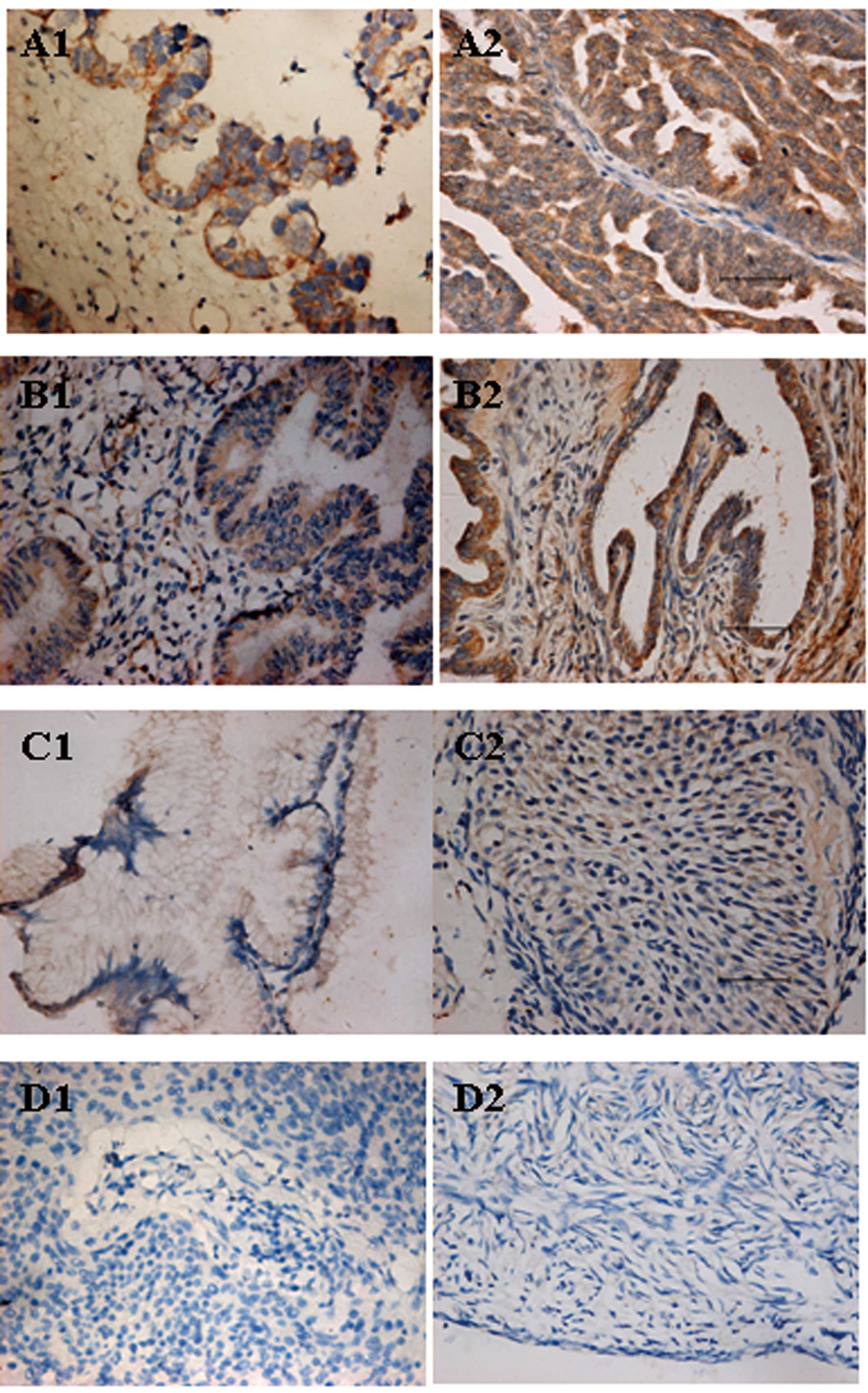
Expression patterns of Lewis y and TGF-β1
in the groups of ovarian tissues
Lewis y was mainly present in the cell membrane and
rarely in the cytoplasm. Its appearance was well-distributed and
granular. The expression of Lewis y in ovarian malignant tumors was
generally enhanced. The positive rate reached 88.33%, significantly
higher than the borderline tumor group (60.00%, P<0.05) and the
benign tumor group (35.00%, P<0.01). No significant difference
was noticed between the borderline tumor group and benign tumor
group (P>0.05). Lewis y was not expressed in any of the 10 cases
of normal ovarian tissue (Table
I).
 | Table IExpression of Lewis y in different
ovarian tissues. |
Table I
Expression of Lewis y in different
ovarian tissues.
| | Lewis y antigen | | |
|---|
| |
| | |
|---|
| Groups | n | − | + | ++ | +++ | Positive cases
(n) | Positive rate
(%) |
|---|
| Malignant tumor | 60 | 7 | 15 | 20 | 18 | 53 | 88.33a |
| Borderline tumor | 20 | 8 | 4 | 7 | 1 | 12 | 60.00b |
| Benign tumor | 20 | 13 | 4 | 3 | 0 | 7 | 35.00 |
| Normal | 10 | 10 | 0 | 0 | 0 | 0 | 0.00 |
TGF-β1 was mainly expressed in the cell membrane and
cytoplasm. The positive expression rates of TGF-β1 in ovarian
malignant, the borderline, benign group and the normal tissue were
78.33, 75.00, 65.00 and 40.00% respectively. TGF-β1 in the
malignant group was expressed more highly than in the benign group
(P<0.05) and normal tissue (P<0.05), but compared with the
borderline group, no significant difference was noticed
(P>0.05). Comparing the 2 groups among the borderline and benign
group with normal tissue, the differences were not significant
(P>0.05) (Table II, Fig. 1).
| Table IIThe expression of TGF-β1 in different
ovarian tissues. |
Table II
The expression of TGF-β1 in different
ovarian tissues.
| | TGF-β1 | | |
|---|
| |
| | |
|---|
| Groups | n | − | + | ++ | +++ | Positive cases
(n) | Positive rate
(%) |
|---|
| Malignant tumor | 60 | 13 | 13 | 26 | 8 | 47 | 78.33a |
| Borderline tumor | 20 | 5 | 6 | 7 | 2 | 15 | 75.00b |
| Benign tumor | 20 | 7 | 6 | 5 | 2 | 13 | 65.00 |
| Normal | 10 | 6 | 2 | 2 | 0 | 4 | 40.00 |
Correlation of Lewis y antigen and TGF-β1
expression with the clinical features of ovarian cancer
In the ovarian serous cystadenocarcinoma, the
positive expression rate of Lewis y was 90.00%. No significant
difference was noticed compared with the mucous cystadenocarcinoma
group (86.67%, P>0.05). The Lewis y was present in 95.24% of
III–IV stage ovarian cancer specimens. It was higher than in I–II
stage (84.62%), but the difference did not reach statistical
significance (P>0.05). The expression rates of Lewis y in the
high, middle and low differentiation group were 80.95, 85.71 and
100.00% respectively, the expression was higher as the
differentiation level descended. Comparison of the 3 groups,
revealed that the differences were not significant (P>0.05). It
has been demonstrated that the expression of Lewis y in ovarian
cancer was not associated with lymphatic metastasis
(P>0.05).
The positive expression rates of TGF-β1 were 73.33
and 83.33% in the ovarian serous and mucous cystadenocarcinoma
groups. The difference beween them was not significant (P>0.05).
TGF-β1 was detected in 18 cases of III–IV stage ovarian cancer
(85.71%), it was obviously higher than in I–II stage tumors
(74.36%), but the disparity beween them was not significant
(P>0.05). The expression rates of TGF-β1 in the high, middle and
low differentiation group were 80.95, 76.19 and 77.78%
respectively; the expression increased as the differentiation level
descended. Comparison of the 3 groups, did not reveal a
statisticaly significant difference (P>0.05). The expression of
TGF-β1 in ovarian cancer was not associated with lymphatic
metastasis (P>0.05) (Table
III).
| Table IIIAssociation between Lewis y and TGF-β1
expression and pathological features. |
Table III
Association between Lewis y and TGF-β1
expression and pathological features.
| | Lewis y antigen | TGF-β1 |
|---|
| |
|
|
|---|
| Features | n | Positive cases
(n) | Rate (%) | P-value | Positive cases
(n) | Rate (%) | P-value |
|---|
| Pathological
type |
| Mucous | 30 | 26 | 86.67 | P>0.05 | 25 | 83.33 | P>0.05 |
| Serous | 30 | 27 | 90.00 | | 22 | 73.33 | |
| FIGO stage |
| I–II | 39 | 33 | 84.62 | P>0.05 | 29 | 74.36 | P>0.05 |
| III–IV | 21 | 20 | 95.24 | | 18 | 85.71 | |
| Differentiation
level |
| High | 21 | 17 | 80.95 | P>0.05 | 17 | 80.95 | P>0.05 |
| Middle | 21 | 18 | 85.71 | | 16 | 76.19 | |
| Low | 18 | 18 | 100.00 | | 14 | 77.78 | |
| Lymphatic
metastasis |
| No | 48 | 41 | 85.42 | P>0.05 | 36 | 75.00 | P>0.05 |
| Yes | 12 | 12 | 100.00 | | 11 | 91.67 | |
Correlation of Lewis y antigen and TGF-β1
expression intensity with the clinical features of ovarian
cancer
We detected and analyzed the optical density value
of the ovarian cancer sections that showed positive expression in
immunohistochemistry. In III-IV stage of ovarian cancer, the MOD of
Lewis y was 0.505±0.072 and its intensity was obviously stronger
than the I–II stage group (0.455±0.065, P<0.05). The MOD of
Lewis y in the low differentiation ovarian cancer group was
0.498±0.084, obviously higher than that of the high differentiation
group (P<0.05). A comparison of the low with the middle
differentiation group, and of the middle with the high
differentiation group, revealed that the disparity beween them was
not significant (P>0.05). The expression intensity of Lewis y in
ovarian cancer was not associated with histological type and
lymphatic metastasis (P>0.05).
The MOD of TGF-β1 in the III–IV stage group of
ovarian cancer was 0.440±0.064 and its intensity was stronger than
in the I–II stage group (0.426±0.055), but the difference did not
reach statistical significance (P>0.05). The MOD of TGF-β1 in
the low differentiation ovarian cancer group was 0.441±0.029,
obviously higher than that of middle (0.402±0.044) and high
differentiation group (0.451±0.065) (P<0.05). A comparison of
the middle with the high differentiation groups revealed that the
disparity was not significant (P>0.05). The expression intensity
of TGF-β1 in ovarian cancer was not associated with histological
type and lymphatic metastasis (P>0.05) (Table IV).
| Table IVAssociation between Lewis y and
TGF-β1 expression intensity and pathological features. |
Table IV
Association between Lewis y and
TGF-β1 expression intensity and pathological features.
| Lewis y
antigen | TGF-β1 |
|---|
|
|
|
|---|
| Features | n | MOD | P-value | n | MOD | P-value |
|---|
| Pathological
type |
| Mucous | 26 | 0.463±0.068 | P>0.05 | 25 | 0.437±0.064 | P>0.05 |
| Serous | 27 | 0.477±0.016 | | 22 | 0.426±0.055 | |
| FIGO stage |
| I and IIstage | 33 | 0.455±0.065 | P<0.05 | 29 | 0.426±0.055 | P>0.05 |
| III and IV
stage | 20 | 0.505±0.072 | | 18 | 0.440±0.064 | |
| Differentiation
level |
| High | 17 | 0.448±0.017 | P<0.05a | 17 | 0.451±0.065 | P<0.05a |
| Middle | 18 | 0.461±0.054 | P>0.05b | 16 | 0.402±0.044 | P<0.05b |
| Low | 18 | 0.498±0.084 | P>0.05c | 14 | 0.441±0.029 | P>0.05c |
| Lymphatic
metastasis |
| No | 41 | 0.459±0.078 | P>0.05 | 36 | 0.424±0.056 | P>0.05 |
| Yes | 12 | 0.476±0.057 | | 11 | 0.452±0.068 | |
The relevance of Lewis y and TGF-β1
expression in ovarian cancer
There were 45 cases that expressed Lewis y and
TGF-β1 positively and simultaneously and 5 cases that negatively
and simultaneously expressed the two factors among the 60 cases of
ovarian cancer tissues. Positive significant correlation between
Lewis y and TGF-β1 was observed in ovarian cancer (C=0.441,
P<0.05) (Table V).
| Table VRelevance of Lewis y and TGF-β1
expression in ovarian cancer. |
Table V
Relevance of Lewis y and TGF-β1
expression in ovarian cancer.
| TGF-β1 |
|---|
|
|
|---|
| Lewis y | Positive | Negative | Total |
|---|
| Positive | 45 | 8 | 53 |
| Negative | 2 | 5 | 7 |
| Total | 47 | 13 | 60 |
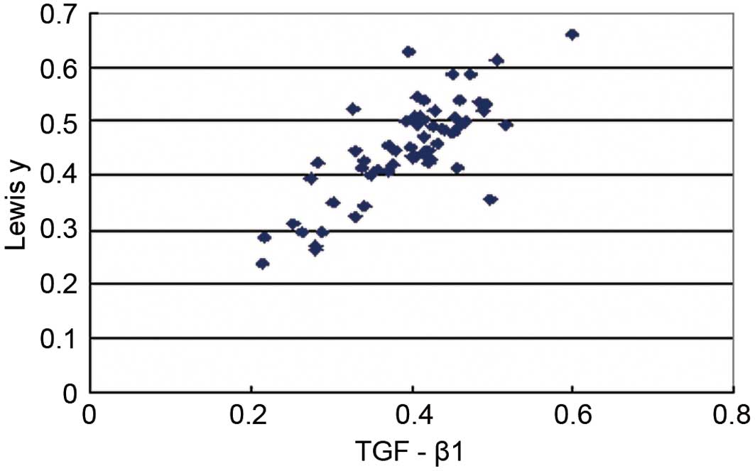
Using linear regression correlation analysis method,
we detected that the expression intensity of Lewis y and TGF-β1
showed linear correlation (r=0.792, P<0.05) (Fig. 2).
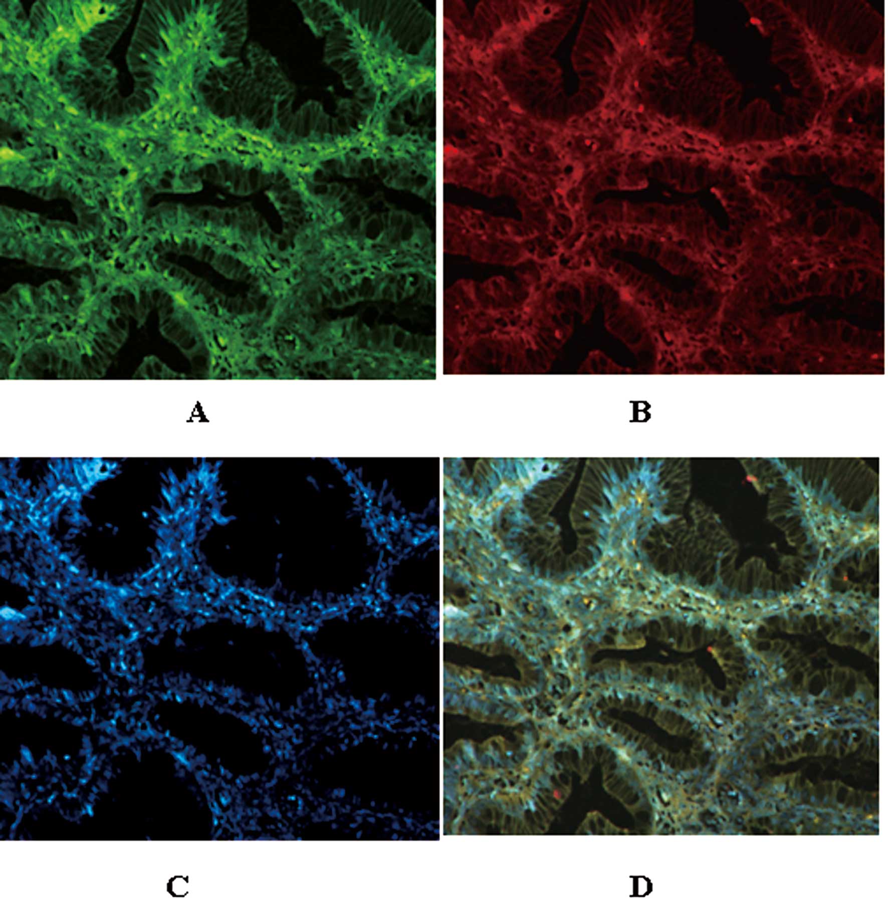
In addition, using the immunofluorescence double
labeling method, we observed that the red fluorescence that labeled
the Lewis y antigen was localized in the membrane; the green
fluorescence that labeled TGF-β1 also appeared in the membrane and
the cytoplasm; the blue fluorescence in the nucleus was the
counterstaining by DAPI. After capturing the pictures, we used the
picture analysis software to build up the 3 fluorescence passages;
yellow fluorescence appeared in the position at which red and green
occurred simultaneously. This fact illustrated that Lewis y and
TGF-β1 were located in the same position (Fig. 3).
Discussion
At present, the etiology and pathogenic mechanisms
of epithelial ovarian cancer are not well understood. Cytokines are
abnormally expressed during the development and progression of
epithelial ovarian cancer, and their potential contributions to
this cancer have become a recent research focus. In fact, it has
been proposed that the dysregulation of cytokines might cause
epithelial cancer. Cytokines act upon tumor cells via autocrine and
paracrine modes, supporting tumor angiogenesis and nutrient
availability and affecting the balance between normal cells and
tumor cells by inducing a state of immunosuppression. These actions
facilitate tumor establishment and progression. Specifically, the
cytokine TGF-β is a primary contributor to the expansion and
metastasis of ovarian tumors. Humans express 3 TGF-β isoforms:
TGF-β1, TGF-β2, and TGF-β3. Among the TGF-β superfamily members,
TGF-β1 exerts the strongest functional action (10), and for this reason, ovarian cancer
research is mainly focused on this isoform.
Initially, TGF-β binds and phosphorylates the TGF-β
receptor (TβR). Phosphorylated TβR activates downstream
intracellular signaling molecules called Smad proteins within the
cell, phosphorylating Smad proteins and interacting with Smads in
heterocomplexes. Phosphorylated Smads can translocate to the
nucleus, bind DNA, and interact with other transcription factors.
The binding of TGF-β and TβR thus indirectly regulates the cell
cycle via Smad signaling to arrest tumor cells in G1, initiate
apoptosis, and inhibit cell proliferation (11). Abnormalities in any component of the
TGF-β signaling pathway can interfere with signal transduction, and
dysfunction of this pathway is closely correlated with tumor
development as well as infiltration and metastasis of tumor cells
(12). Hempel et al
(13) reported that a breakdown in
TGF-β signal transduction significantly increased the infiltration
and movement capabilities of ovarian cancer cells.
TGF-β regulates the growth of normal human ovarian
epithelial cells in vivo by promoting apoptosis, which
inhibits the proliferation of both normal ovarian epithelial cells
and early ovarian cancers. However, the inhibitory action of TGF-β
on cell growth is compromised or even reversed in tumors, where it
promotes their growth and progression. Rodriguez et al
(14) suggested that TGF-β enhances
the infiltration capabilities of most ovarian cancer cell lines by
2–20-fold, but has no effect or an inhibitory effect on the
infiltration capabilities of normal ovarian epithelial cells.
Hirashima et al (15)
reported that TGF-β1 produced by ovarian cancer cells promotes
tumor infiltration by up-regulating plasminogen activator
inhibitor-1 (PAI-1) in peritoneal mesothelial cells. Bristow et
al (16) suggested that,
compared with primary ovarian cancers, recurrent ovarian cancer is
more likely to be associated with changes in TGF-β and its
receptor. An increase in the expression of TGF-β and a concurrent
loss in its receptor promote tumor development and progression, and
facilitate tumor growth, relapse, metastasis, and resistance to
chemotherapeutic drugs. Taken together, these findings support that
TGF-β plays a critical role in the development, progression, and
metastasis of ovarian cancer, especially advanced ovarian
cancer.
The Lewis y antigen is a difucosylated
oligosaccharide; fucose is an end structure of sugar chain
synthesis. The Lewis y antigen is known to be associated with
several cancers. Expression of Lewis y increases when cells undergo
carcinogenesis of the ovary, pancreas, prostate, colon, or the
non-small cell type of the lung (17). We have demonstrated that an increase
in Lewis y promotes the growth, proliferation, and survival
capacity of ovarian cancer cells (6–8). Our
in vitro results indicate an enhancement in cell adhesion,
infiltration, metastasis, and drug resistance.
We used DNA microarrays to obtain genomic expression
profiles of ovarian cancer cells before and after gene transfection
with α1,2-FT. TGF-β1 is up-regulated in human ovarian cancer cells
following transfection (9). We also
determined that Lewis y antigen, an important component of the
transmembrane glycoproteins TβRI and TβRII, can up-regulate
TGF-β1-dependent ERK and PI3K signaling pathways to promote ovarian
cancer cell proliferation (unpublished data). This study provides
histological support of a correlation between the expression of
TGF-β1 and the expression of Lewis y antigen in ovarian epithelial
cancer tissues. The expression of TGF-β1 is significantly higher in
ovarian epithelial cancer cells than in benign tumors (P<0.05)
or in normal ovarian cells (P<0.01). Moreover, the histological
expression intensity of TGF-β1 is elevated with increasing extent
of malignancy (P<0.05) and is correlated with operational stage
(P<0.05). Correlation analyses indicated that the expression
levels of Lewis y antigen and TGF-β1 are positively correlated in
ovarian cancer tissues (C=0.441, P<0.05). Statistical analyses
of the expression intensities of Lewis y antigen and TGF-β1 in
ovarian cancer tissues further support that these molecules are
linearly correlated (r=0.792, P<0.05). Immunofluoresence
double-labeling experiments suggest that Lewis y colocalizes with
TGF-β1 in tissues, lending additional support for a correlation.
This investigation does not prove that Lewis y is colocalized with
TGF-β1; instead, TGF-β1 likely binds its receptor and then
colocalizes with Lewis y.
Growth, infiltration, metastasis, and relapse of
ovarian cancer are associated with refractoriness to treatment and
vascular vessel formation, which are important to the survival of
tumor cells. It has been reported (18) that vascularization is greater in
tissues expressing TGF-β1 and highly expressing vascular
endothelial growth factor (VEGF) compared with tissues negative for
TGF-β1 and only weakly expressing VEGF. The coexpression of TGF-β1
and VEGF promotes angiogenesis in ovarian cancer, thereby
facilitating the growth of ovarian cancer cells. Donovan et
al (19) reported that TGF-β1
enhances tumor development and progression by stimulating cancer
cells to secret VEGF. Our previous experiments indicated that the
expression of Lewis y and VEGF are significantly increased in
ovarian cancer cells following transfection with α1,2-FT,
suggesting that Lewis y may alter the actions of intercellular
messengers, thus directly or indirectly promoting VEGF expression
(20). In addition, Lewis y
facilitates the expression of the VEGF receptor, KDR, by autocrine
and paracrine pathways, which enhance tumor angiogenesis (21). The Lewis y antigen further promotes
tumor cell proliferation by regulating the expression and
phosphorylation status of molecules in the EGFR/PI3K signal
transduction pathways (22). We
suggest that Lewis y increases angiogenesis in ovarian cancer and
facilitates the growth and progression of ovarian cancer cells by
promoting the coexpression of TGF-β1 and VEGF.
TGF-β1 can promote or inhibit metastasis and
infiltration depending on the tumor cell type in which it is
expressed. In advanced ovarian cancer, the inhibitory action of
TGF-β1 is compromised, and this molecule instead enhances malignant
behaviors of tumor cells. TGF-β1 can inhibit the proliferation and
killing activities of various cell types involved in the mediation
of cellular immunity, including cytotoxic T lymphocytes (CTL),
natural killer (NK) and lymphokine-activated killer (LAK) cells
(23). TGF-β1 can inhibit both
macrophage activation and the secretion of corresponding cytokines
in IFN-γ-evoked mice, resulting in T-cell inhibition (24). TGF-β1 is highly expressed in tumor
tissues where it serves as an immunosuppressive factor, forming an
isolating ‘firewall’ around tumor tissues (4). The effect is to shield tumor cells
from host immunosurveillance and allow for infiltration and
metastasis.
Detailed mechanisms regarding the role of Lewis y
and TGF-β1 in the development and progression of ovarian cancer
have not been elucidated. We have demonstrated preliminarily that
both Lewis y and TGF-β1 are related to ovarian cancer. Lewis y
antigen likely evokes abnormal expression of TGF-β1 and/or affects
other signal transduction pathways involved in ovarian cancer,
increasing its malignant extent. Further exploration of the
relationship between Lewis y and cytokines should lend insight into
the development, progression, and metastasis mechanisms of ovarian
cancer and facilitate the clinical diagnosis and evaluation of
ovarian cancer. Interference of the expression or actions of Lewis
y may yield novel approaches for the treatment of ovarian
cancer.
Acknowledgements
This study was supported by the National Natural
Science Foundation of China (30170980, 30571958, 30872757,
81072118); the Liaoning Natural Science Foundation (20052107); the
Scientific and Technical Project of the Educational Department of
Liaoning Province (05L492); the Educational Department Doctor
Projects Fund (20070159023); the Key Laboratory Project of Liaoning
Province Education Office (2008S247); the Free Researchers Plan of
Shengjing Hospital (200807); the Programs of Science and Technology
Commission of Shenyang (F10-14-9-52).
References
|
1
|
Goupille C, Hallouin F, Meflah K and Le
Pendu J: Increase of rat colon carcinoma cells tumorigenicity by
alpha(1–2) fucosyltransferase gene transfection. Glycobiology.
7:221–229. 1997.PubMed/NCBI
|
|
2
|
Hellström I, Garrigues HJ, Garrigues U and
Hellström KE: Highly tumor-reactive, internalizing, mouse
monoclonal antibodies to Le(y)-related cell surface antigens.
Cancer Res. 50:2183–2190. 1990.PubMed/NCBI
|
|
3
|
Markowitz S: TGF-beta receptors and DNA
repair genes, coupled targets in a pathway of human colon
carcinogenesis. Biochim Biophs Acta. 1470:M13–M20. 2000.PubMed/NCBI
|
|
4
|
Shah AH and Lee C: TGF-beta-based
immunotherapy for cancer: breaching the tumor firewall. Prostate.
45:167–172. 2000. View Article : Google Scholar : PubMed/NCBI
|
|
5
|
Ahmed AA, Mills AD, Ibrahim AE, et al: The
extracellular matrix protein TGFBI induces microtubule
stabilization and sensitizes ovarian cancers to paclitaxel. Cancer
Cell. 12:514–527. 2007. View Article : Google Scholar
|
|
6
|
Iwamori M, Tanaka K, Kubushiro K, et al:
Alterations in the glyolipid composition and cellular properties of
ovarian carcinoma-derived RMG-1 cells on transfection of the
α1,2-fucosyltransferase gene. Cancer Sci. 96:26–30. 2005.PubMed/NCBI
|
|
7
|
Zhao Y, Lin B, Hao YY, Yan LM, Liu JJ, Zhu
LC and Zhang SL: The effects of Lewis(y) antigen content on drug
resistance to carboplatin in ovarian cancer line RMG-I. Prog
Biochem Biophys. 35:1175–1182. 2008.
|
|
8
|
Hao YY, Lin B, Zhao Y, et al:
alpha1,2-fucosyltransferase gene transfection influences on
biological behavior of ovarian carcinoma-derived RMG-1 cells. Fen
Zi Xi Bao Sheng Wu Xue Bao. 41:435–442. 2008.(In Chinese).
|
|
9
|
Zhu K, Amin MA, Zha Y, Harlow LA and Koch
AE: Mechanism by which H-2g, a glucose analog of blood group H
antigen, mediates angiogenesis. Blood. 105:2343–2349. 2005.
View Article : Google Scholar : PubMed/NCBI
|
|
10
|
Yasui T, Uemura H, Irahara M and Aono T:
Effects of transforming growth factor-beta on the production of
parathyroid hormone-related peptide in a human ovarian cancer cell
line in vitro. J Obstet Gynaecol Res. 23:231–238. 1997. View Article : Google Scholar : PubMed/NCBI
|
|
11
|
Derynck R and Zhang YE: Smad-dependent and
Smad-independent pathways in TGF-beta family signaling. Nature.
425:577–584. 2003. View Article : Google Scholar : PubMed/NCBI
|
|
12
|
de Caestecker MP, Piek E and Roberts AB:
Role of transforming growth factor-beta signaling in cancer. J Natl
Cancer Inst. 92:1388–1402. 2000.PubMed/NCBI
|
|
13
|
Hempel N, How T, Dong M, Murphy SK, Fields
TA and Blobe GC: Loss of betaglycan expression in ovarian cancer:
role in motility and invasion. Cancer Res. 67:5231–5238. 2007.
View Article : Google Scholar : PubMed/NCBI
|
|
14
|
Rodriguez GC, Haisley C, Hurteau J, Moser
TL, Whitaker R, Bast RC Jr and Stack MS: Regulation of invasion of
epithelial ovarian cancer by transforming growth factor-beta.
Gynecol Oncol. 80:245–253. 2001. View Article : Google Scholar : PubMed/NCBI
|
|
15
|
Hirashima Y, Kobayashi H, Suzuki M, Tanaka
Y, Kanayama N and Terao T: Transforming growth factor-beta1
produced by ovarian cancer cell line HRA stimulates attachment and
invasion through an up-regulation of plasminogen activator
inhibitor type-1 in human peritoneal mesothelial cells. J Biol
Chem. 278:26793–26802. 2003. View Article : Google Scholar
|
|
16
|
Bristow RE, Baldwin RL, Yamada SD, Korc M
and Karlan BY: Altered expression of transforming growth
factor-beta ligands and receptors in primary and recurrent ovarian
carcinoma. Cancer. 85:658–668. 1999. View Article : Google Scholar : PubMed/NCBI
|
|
17
|
Hakomori S: Tumor malignancy defined by
aberrant glycosylation and sphingo(glyco)lipid metabolism. Cancer
Res. 56:5309–5318. 1996.PubMed/NCBI
|
|
18
|
Breier G, Blum S, Peli J, Groot M, Wild C,
Risau W and Reichmann E: Transforming growth factor-beta and Ras
regulate the VEGF/VEGF-receptor system during tumor angiogenesis.
Int J Cancer. 97:142–148. 2002. View
Article : Google Scholar : PubMed/NCBI
|
|
19
|
Donovan D, Harmey JH, Toomey D, Osborne
DH, Redmond HP and Bouchier-Hayes DJ: TGF beta-1 regulation of VEGF
production by breast cancer cells. Ann Surg Oncol. 4:621–627. 1997.
View Article : Google Scholar : PubMed/NCBI
|
|
20
|
Li Y, Lin B, Hao YY, et al: Influence of
alpha1,2-fucosyltransferase gene transfection on vascular
endothelial growth factor in ovarian carcinoma-derived RMG-I cell
xenografts in nude mice. J China Med Univ. 3:284–289. 2008.(In
Chinese).
|
|
21
|
Wang PL, Lin B, Liu Q, Li Y, Li FF, Hao YY
and Zhang SL: Lewis y antigen promotes the expression of vascular
endothelial growth factor receptor in ovarian carcinoma-derived
RMG-I cells. J Modern Oncol. 17:1831–1835. 2009.
|
|
22
|
Liu JJ, Lin B, Hao YY, et al: Lewis(y)
antigen stimulates the growth of ovarian cancer cells via
regulation of the epidermal growth factor receptor pathway. Oncol
Rep. 23:833–841. 2010.PubMed/NCBI
|
|
23
|
Scott S, Kimura T, Ichinohasama R, et al:
Microsatellite mutations of transforming growth factor-beta
receptor type II and caspase-5 occur in human precursor T-cell
lymphoblastic lymphomas/leukemias in vivo but are not associated
with hMSH2 or hMLH1 promoter methylation. Leuk Res. 27:23–34. 2003.
View Article : Google Scholar
|
|
24
|
Langermans JA, Nibbering PH, Van Vuren-Van
Der Hulst ME and Van Furth R: Transforming growth factor-beta
suppresses interferon-gamma-induced toxoplasmastatic activity in
murine macrophages by inhibition of tumour necrosis factor-alpha
production. Parasite Immunol. 23:169–175. 2001. View Article : Google Scholar
|