Introduction
NPC is one of the most common malignant tumors in
southern China and Southeast Asia with incidence rates of 20 to 50
per 100,000 (1). The most effective
treatment for NPC is radiotherapy, which achieves an overall 5-year
survival rate of 65% (2).
Nevertheless, radioresistance remains a serious barrier to
successful treatment in many cases. Radioresistance easily induces
in some NPC patients local recurrence and distant metastases and
the majority of these patients suffer recurrence and metastasis
within 1.5 year after treatment (3,4). To
obtain optimal effect of radiotherapy on NPC patients, it is urgent
to identify subgroup of radioresistant NPC patients and to reveal
the molecular mechanism of NPC radioresistance.
Many studies have been directed toward understanding
the mechanism of radiotherapy response and resistance, with the
ultimate goal of identifying molecular markers that allow the
prediction of radiotherapy response. Several proteins, Epstein-Barr
virus gene BHRF1 (5), antioxidant
enzyme manganese superoxide dismutase (MnSOD2) (6), 14-3-3σ (7), and raf kinase inhibitory protein
(RKIP) (8) have been proved to be
associated with radioresistance in NPC. However, almost all of
these proteins were originally found from RR NPC cells not from RR
NPC tissues with scarcity of clinical specificity. High-throughput
technology is needed which offer the potential ability to find
specific RR proteins in NPC. Proteomics, which aims at identifying
differential proteins associated with differential disease traits,
has been proved to be an effective approach in protein research
(9). To identify the proteins
associated with the radioresistance of NPC, we performed
comparative proteomics on RR and RS NPC tissues in order to find
differential proteins associated with radioresistance.
ERp29 is an endoplasmic reticulum(ER) protein and
has emerged in a variety of physiological and pathological
conditions, such as normal production of dental enamel, antibodies,
and milk, and disorders of the thyroid, spinal cord, and aging eye
(10–13). Many studies indicated that ERp29 was
expressed in a variety of human cancer and it was found to be
highly expressed in primary tumors and cell lines (14–16).
It was also reported that ERp29 was up-regulated in mouse
intestinal epithelia cells when exposure to radiation (17), indicating ERp29 associated with
resistance to radiation stress and may play a potential protective
role against stress. Nevertheless, whether ERp29 is involved in
radioresistance in human cancer has not yet been elucidated.
In this study, based on the results of the
comparative proteomic analysis of RR and RS NPC tissues, we
identified twelve differential proteins and verified ERp29
overexpression in the RR NPC tissues. Then we attempted to
elucidate that ERp29 knockdown attenuated CNE-1 and 6-10B cell
radioresistance and enhanced cell apoptosis.
Materials and methods
Patients and tissues
We selected 88 NPC patients who were treated by
curative-intent radiotherapy (a total dose of 70 Gy) using a
modified linear accelerator in the Xiangya Hospital of Central
South University, China, from January 2003 to June 2007. NPC
patients recruited in this study included 42 RR and 46 RS patients.
RR NPC patients were defined as those with persistent disease
(incomplete regression of tumor) at 6 weeks or more, or those with
recurrent disease at the nasopharynx and/or neck nodes at 2 months
or more after completion of radiotherapy (18). RS NPC patients were defined as those
without local residual lesions at 6 weeks or no recurrence at 2
months after completion of radiotherapy (18). NPC tissue biopsies from these 88
patients were obtained at the time of diagnosis before any therapy
with an informed consent. Ten pairs of RR and RS NPC tissues by
random sampling were frozen in liquid nitrogen for proteomic
analysis, and other tissues were used for immunohistochemical
staining. All cases in this study were histopathologically
diagnosed as poorly differentiated squamous cell carcinomas by
examination of the frozen sections and paraffin imbedded sections
based on the 1978 WHO classification (19). The clinicopathological parameters of
88 archival NPC tissue specimens used in the present study are
shown in Table I. This study was
approved by the ethics committee of Xiangya School of Medicine,
Central South University, China.
 | Table IClinicopathological parameters of the
NPC tissue specimens. |
Table I
Clinicopathological parameters of the
NPC tissue specimens.
| Classification | Number |
|---|
| Gender |
| Male | 63 |
| Female | 25 |
| Age |
| ≥50 | 27 |
| <50 | 61 |
| Histological
type |
| WHO type III | 88 |
| Primary tumor (T)
stage |
| T1 | 7 |
| T2 | 38 |
| T3 | 25 |
| T4 | 18 |
| Lymph node
metastasis |
| Negative | 43 |
| Positive | 45 |
| Distant metastasis
(M) |
| Negative | 82 |
| Positive | 6 |
| Clinical stage |
| II | 29 |
| III | 35 |
| IV | 24 |
| Recurrence |
| Negative | 76 |
| Positive | 12 |
Proteomic analysis
The detailed approach of two-dimensional gel
electrophoresis (2-DE), image analysis and mass spectrometer (MS)
analysis was described by Cheng et al (20). Briefly, 10 pairs of radioresistant
and radiosensitive NPC tissues were dissolved in lysis buffer (7
mol/l urea, 2 mol/l thiourea, 100 mmol/l DTT, 4% CHAPS, 0.5 mmol/l
EDTA, 40 mmol/l Tris, 2% NP40, 1% Triton X-100, 5 mmol/l PMSF, and
2% phamarlyte) at 4˚C for 1 h. Then the supernatant was transferred
after centrifugation at 12,000 rpm for 30 min at 4˚C. After
detection of protein concentration, total proteins were separated
by 2D Quantification kit (Amersham Biosciences) from twenty sets
with each set containing a radioresistant or a radiosensitive NPC
tissue. After Blue Silver staining, the stained 2-DE gels of each
set were scanned by MagicScan software on an Image scanner
(Amersham Biosciences), and analyzed using a PDQuest system
(Bio-Rad Laboratories, Hercules, CA). Proteins were classified as
being differentially expressed between the two types of tissues
when spot intensity showed a 2-fold variation in radioresistant NPC
tissue compared to radiosensitive NPC tissue. All the differential
protein spots were excised from stained gels. After trypsin
digestion, the mixture was analyzed by a Voyager System DE-STR 4307
MALDI-TOF mass spectrometer (MS) (ABI, Foster City, CA, USA) to get
a peptide mass fingerprint (PMF). Mascot Distiller was used to
obtain the monoisotopic peak list from the raw mass spectrometry
files. Peptide matching and protein searches against the Swiss-Prot
database were performed using the Mascot search engine (http://www.matrixscience.com/) with a mass tolerance
of ±50 ppm.
Immunohistochemistry staining
Immunohistochemistry was performed using the
following protocol. Forty-two radioresistant and forty-six
radiosensitive NPC tissues sections were deparaffinized in xylene.
Sections were rehydrated in alcohol, and pretreated with citrate
buffer (10 mmol/l, pH 6.0) for 20 min at 100˚C in a microwave oven.
Endogenous peroxidase activity was blocked with 3% hydrogen
peroxide for 15 min at room temperature, then nonspecific binding
sites were blocked by 10% normal goat serum for 30 min at 37˚C. The
sections were incubated with antibody (rabbit polyclonal anti-ERP29
1:200 dilution, Abcam) overnight at 4˚C. After washing with PBS,
sections were incubated with 1:1000 dilution of biotinylated goat
anti-rabbit IgG (Zhongshan Chemical) for 20 min at 37˚C. Finally,
tissue sections were incubated with 3′,3′-diaminobenzidine (Maixin,
Fuzhou) until a brown color emerged and washed with distilled
water, then counterstained with Harris modified hematoxylin
(Zhongshan Chemical). Primary antibodies were omitted for negative
controls.
Counting and statistical methods
Sections were blindly evaluated by two pathologists
by light microscopy. A semi-quantitative scoring criterion for
immunohistochemistry was used, in which both the intensity and the
percentage of positive cells were evaluated according to the
methods by Hara and Okayasu (21).
More than 10 microscopic fields were chosen randomly with ×400
magnification, and >1000 cells were counted for each section.
The intensity of staining was graded on the following scale: 0, no
stain; 1, mild staining; 2, moderate staining; 3, intense staining.
The number of positive cells was visually evaluated as follows: 0,
≤10% tissue stained positive; 1, 10 to 30% stained positive; 2, 30
to 60% stained positive; 3, >60% stained positive. The minimum
score summed (extension + intensity) was therefore 0 and the
maximum was 6. A combined staining score (extension + intensity) ≤2
was considered to be weak staining; a score 3 or 4 moderate; and 5
or 6 intense staining.
Cell culture
NPC cell lines CNE-1, CNE-2, 5-8F, 6-10B used in
this study were obtained from cancer institute, Central South
University. NPC cells were cultured in RMPI-1640 medium (Gibco, NY,
USA) supplemented with 10% of fetal bovine serum FBS (Gibco) at
37˚C in an incubator at a humidified atmosphere with 5%
CO2 in air.
Western blotting
Briefly, 40 μg of lysates were separated by 10%
SDS-PAGE and transferred to a polyvinylidene difluoride membrane.
Non-specific binding sites were blocked by 5% normal goat serum at
room temperature for 1 h. The membrane was incubated with primary
antibody: anti-ERP29 (Abcam, 1:2000 dilution) overnight at 4˚C.
Then horseradish peroxidase conjugated secondary antibody
(Beyotime, Beijing, China, 1:2000 dilution) for 1 h. The immune
complexes were visualized by enhanced electrochemiluminescence
(ECL) detection. The ECL test kit-based detection was performed
with Chemiluminescence Reagent (Formantas biology) according to the
manufacturer's instructions. β-actin was used for loading control.
The results of Western blot analysis represented the average of
three individual experiments.
Clonogenic survival assay
Cells were plated in triplicate at cell population
of 102, 2×10², 4×10²,
103, 104, 105 per dish, and then
were exposed to a range of radiation doses (0–8 Gy). After
irradiation, the cells were cultured for no less than 12 days and
the number of surviving colonies (defined as a colony with >50
cells) was counted and the data normalized to the appropriate
sham-irradiated control group. Survival parameters D0
and N were fitted according to the linear quadratic equation
[S=1−(1−e−D/Do)N] using SigmaPlot 9.0 software (Systat Software
Inc., USA). Three independent experiments were done.
Stable transfection
The ERp29-targeted shRNA lentiviral particles
(sc-60599-V) and no-targeted shRNA lentiviral particles (sc-108080)
as control, purchased from Santa Cruz Biotechnology (USA), were
transfected into NPC cells according to the manufacturer's
instructions. After 14 days of selection in RIPM-1640 containing
10% FBS and 10 μg/ml puromycin (Santa Cruz Biotechnology),
individual puromycin-resistant colonies were isolated and expanded.
The expression of ERp29 was determined by Western blot analysis as
above described.
Cell viability assay
2-(2-methoxy-4-nitrophenyl)-3-(4-nitrophenyl)-5-(2,4-disulfophenyl)-2H-tetrazolium,
monosodium salt (CCK-8, Beyotime) assay was used to detect cell
viability in response to irradiation. Briefly, cells were seeded in
96-well culture plates at 3×10³ for CNE-1 cells or
2×10³ for 6-10B cells per well. After incubation for 8
h, the cells were exposed to 8 Gy X-ray irradiation. Cell viability
was determined by CCK-8 at various time intervals according to the
manufacturer's instructions. Optical densities were determined on a
microtiter plate reader (Peskin and Winterbourn 2000) at 450 nm.
Three independent experiments were done in triplicate.
Flow cytometry analysis of cell
apoptosis
Cells were seeded in 6-well culture plates at
1×10⁵ for CNE-1 cells or 5×10⁴ for 6-10B
cells per well. Then the cells were harvested at 72 h after
irradiation with 8 Gy X-ray. According to the manufacturer's
instructions of Annexin V-FITC apoptosis detection kit (Beyotime),
cells were stained using Annexin-V-FITC for 10 min at room
temperature and then were stained using propidium iodure (PI) for
10 min in dark. The cells were analyzed immediately on a FSCAN flow
cytometer (BD Biosciences, USA). All samples were assayed in
triplicate.
Statistics
All statistical analyses were carried out using SPSS
for Windows version 13.0 (SPSS). χ2 test was applied to
analyze the relationship between ERp29 expression and
clinicopathologic features. Student's t-test and One-way analysis
of variance (ANOVA) were used to analyze the cell experimental
data. Data are presented as the mean ± standard deviation (SD).
Differences were considered statistically significant for
P<0.05.
Results
Screening for radioresistance-associated
proteins by proteomic analysis
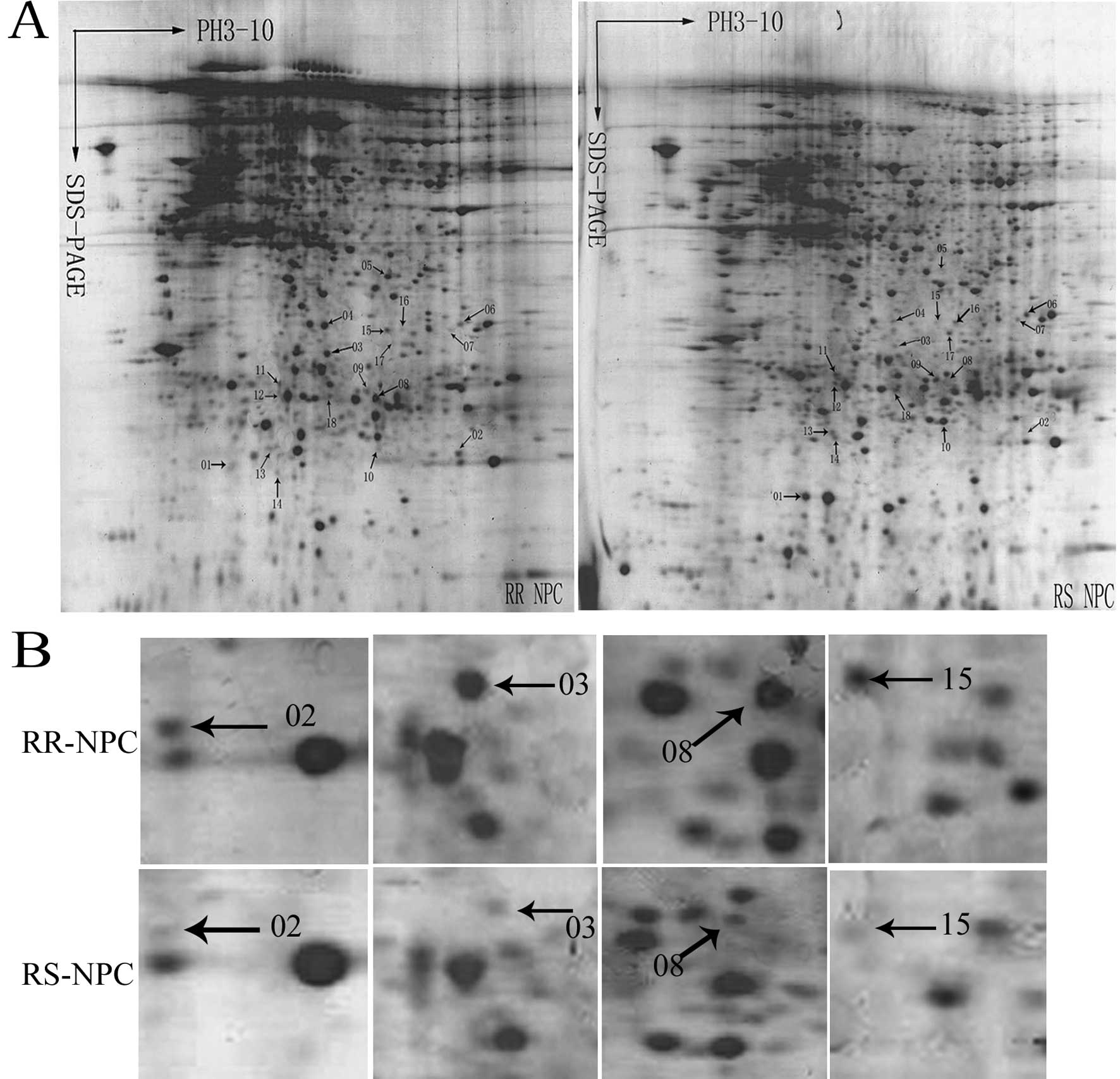
Comparative proteomic study of RR and RS NPC tissues
was performed to identify the proteins associated with
radioresistance. Ten pairs of 2-DE maps from RR NPC tissues and
control RS NPC tissues were constructed with PDQuest image
software. A total of 18 differential protein spots (≥2-fold) in the
two types of tissues were detected, and subjected to the analysis
of MAL-DI-TOF-MS. Two representative 2-DE maps from RR and RS NPC
tissues are shown in Fig. 1A, and
18 differential protein spots are marked with arrows. Images of the
region of the 2-DE showing spot 02 (Mn-SOD, up-regulated 3.1-fold),
spot 03 (ERp29, up-regulated 4.7-fold) and spot 08 (HSP27,
up-regulated 3.9-fold) in the RR NPC tissues compare to the RS NPC
tissues are shown in Fig. 1B. The
18 differential protein spots were analyzed by MAL-DI-TOF-MS and 12
proteins were identified. Among these proteins, Mn-SOD, ERp29,
HSP27 and GST ω1 were up-regulated in the RR NPC tissues; TSP-1,
PKM2, human electron transfer flavoprotein, MRP-14, DJ-1, GMFG,
prohibitin and cytochrome c oxidase were down-regulated in the RR
NPC tissues. The annotation of all the identified proteins is
summarized in Table II.
| Table IIDifferential expression proteins
between RR and RS NPC identified by MALDI-TOF MS. |
Table II
Differential expression proteins
between RR and RS NPC identified by MALDI-TOF MS.
| Spot no. | NCBInr
Identification no. | Protein name | Score | Coverage (%) | Expression in RR/RS
NPC |
|---|
| 1 | gi 20150229 | Multidrug
resistance-associated protein 14 (MRP-14) | 150 | 91 | ↓ |
| 2 | gi 38503339 | Manganese
superoxide dismutase (Mn-SOD) | 73 | 52 | ↑ |
| 3 | gi 5803013 | Endoplasmic
reticulum protein 29 (ERp29) | 77 | 58 | ↑ |
| 6 | gi 4503607 | Human electron
transfer flavoprotein | 169 | 59 | ↓ |
| 7 | gi 88191913 | Thrombospondin-1
(TSP-1) | 68 | 33 | ↓ |
| 8 | gi 662841 | Heat shock protein
27 (Hsp27) | 68 | 47 | ↑ |
| 10 | gi 31543380 | Parkinson protein 7
(DJ-1) | 88 | 52 | ↓ |
| 11 | gi 66910342 | Pyruvate kinase,
muscle (PKM2) | 72 | 39 | ↓ |
| 12 | gi 4505773 | Prohibitin | 252 | 75 | ↓ |
| 14 | gi 34783447 | Glia maturation
factor-γ (GMFG) | 95 | 53 | ↓ |
| 15 | gi 4758484 | Glutathione
S-transferase ω1 (GST ω1) | 109 | 56 | ↑ |
| 18 | gi 18999392 | Cytochrome c
oxidase | 67 | 28 | ↓ |
Expression of ERp29 in clinical specimens
and NPC cell lines
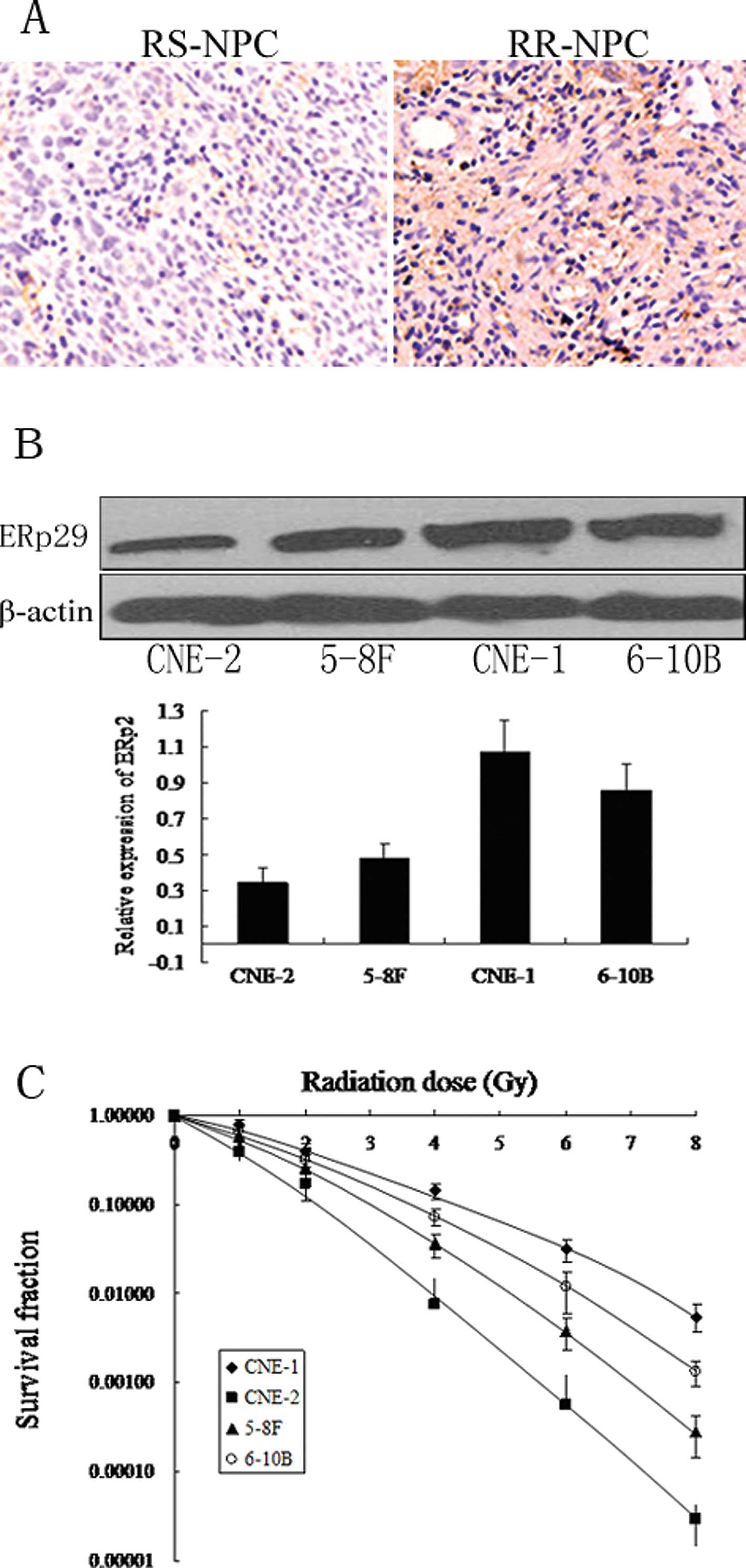
To validate the relevance of ERp29 to NPC
radioresistance, we detected ERp29 expression in 42 radioresistant
and 46 radiosensitive formalin-fixed paraffin-embedded biopsy
samples by immunohistochemistry. ERp29 staining, when present, was
evident in the cytoplasm of tumor cells (Fig. 2A). ERp29 antigen showed intense
staining in the majority of radioresistant NPC tissues (57.1%) and
weak staining in the majority of radiosensitive NPC tissues
(67.4%). ERp29 overexpression was significantly associated with
radioresistant tumors (P<0.001, Table III). To further detect the
association of ERp29 expression with NPC radioresistance, we
detected the ERp29 expression in NPC cell lines CNE-1, CNE-2, 5-8F
and 6-10B with different radioresistant potentials. As shown in
Fig. 2B, the expression of ERp29
descending order is CNE-1, 6-10B, 5-8F and CNE-2. Meanwhile, the
radioresistance of NPC cell lines in descending order is also
CNE-1, 6-10B, 5-8F and CNE-2 (Fig.
2C). This indicates that ERp29 expression positively correlated
with radioresistance of NPC cell lines.
| Table IIIThe difference of ERp29 expression in
the RR and RS of NPC tissues. |
Table III
The difference of ERp29 expression in
the RR and RS of NPC tissues.
| | Score | |
|---|
| |
| |
|---|
| n | Low (0–2) | Moderate (3–4) | High (5–6) | P |
|---|
|
Radioresistance | 42 | 5 (11.9%) | 13 (31.0%) | 24 (57.1%) | <0.001 |
|
Radiosensitivity | 46 | 31 (67.4%) | 13 (28.3%) | 2 (4.3%) | |
ERp29 knockdown attenuated CNE-1 and
6-10B cells radioresistance
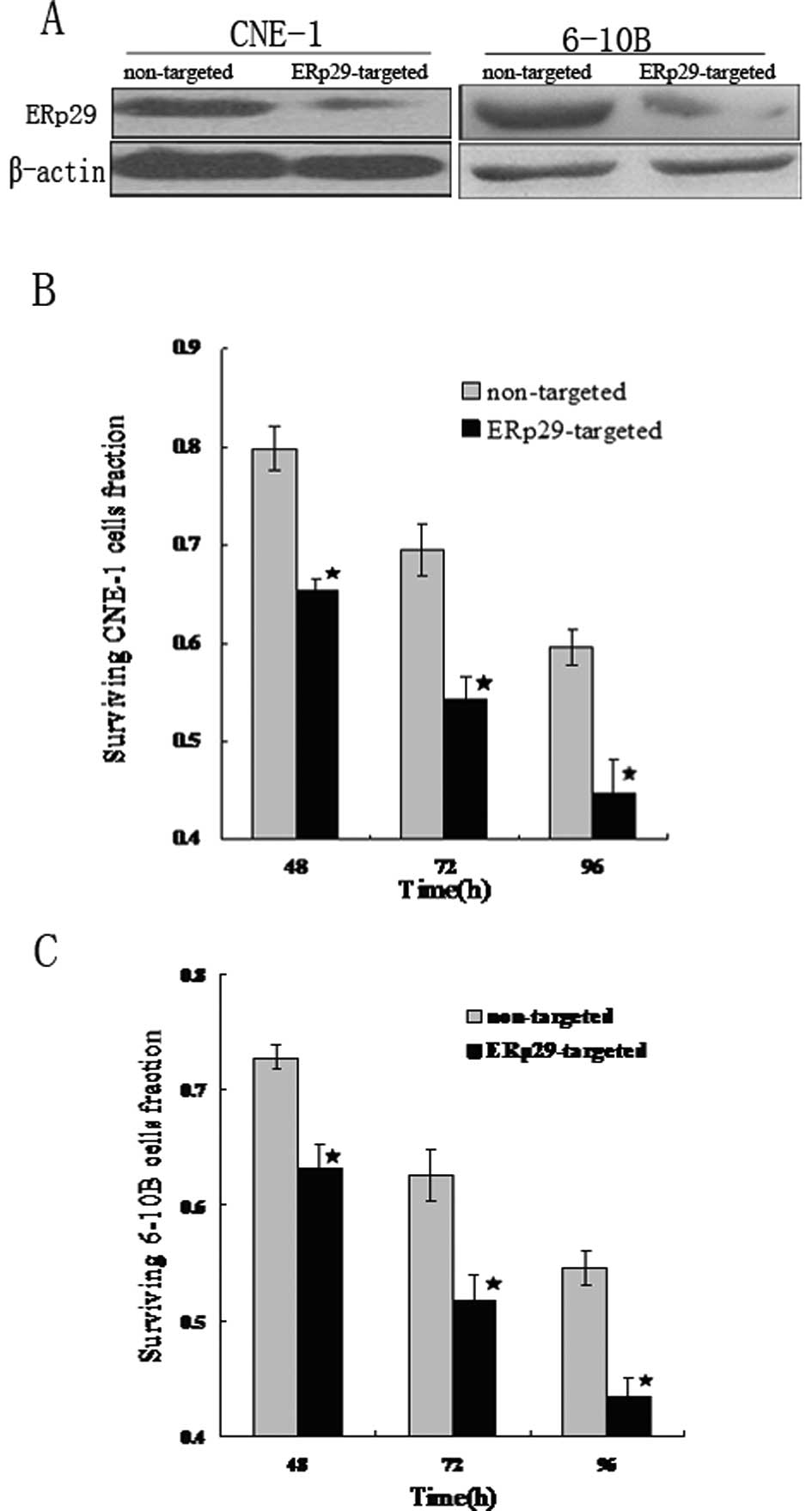
NPC cells CNE-1 and 6-10B with higher ERp29
expression and stronger radioresistance were transfected with
ERp29-targeted shRNA or non-targeted shRNA, and then cell viability
assays and clonogenic survival assays were performed. Western blot
analysis confirmed ERp29 knockdown in CNE-1 and 6-10B cells
(Fig. 3A). Cell viability assays
showed that CNE-1 and 6-10B cells transfected with ERp29-targeted
shRNA resulted in a significant reduction of the cell viability at
48 h (P<0.001), as well as 72 and 96 h (P<0.001) after
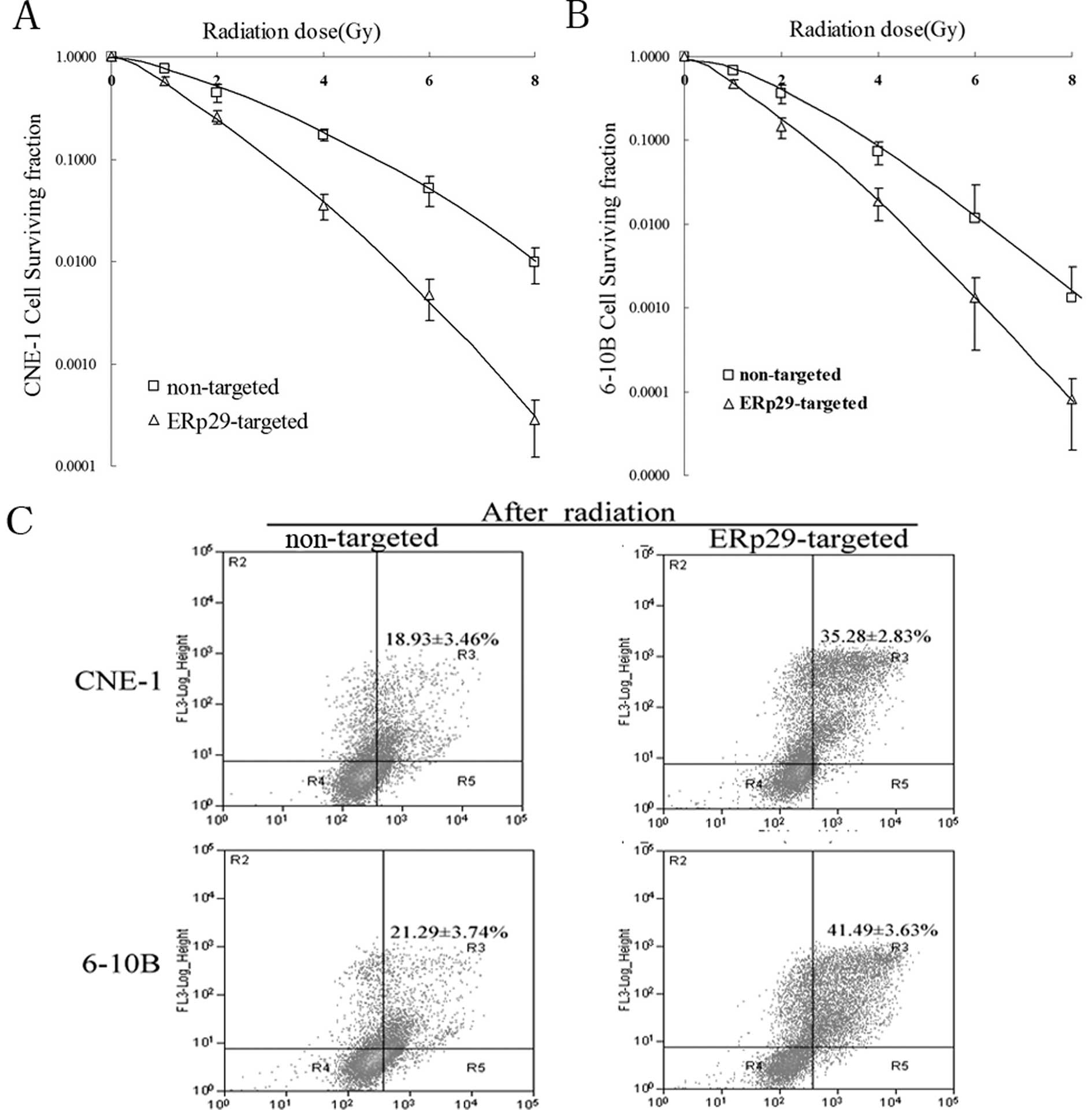
irradiation, comparing with the controls (Fig. 3B and C). In clonogenic survival
assay, after ERp29 knockdown, SF2 and LD50 of CNE-1 cells ranged
from 0.462 to 0.212 and 1.843 to 1.208, respectively, and SF2 and
LD50 of 6-10B cells ranged from 0.324 to 0.144 and 1.142 to 0.875,
respectively. In addition, the curve of ERp29-targeted shRNA CNE-1
cells was much steeper than the control (Fig. 4A). A similar situation was seen in
6-10B cells (Fig. 4B). Flow
cytometric analysis showed transfected with ERp29-targeted shRNA
CNE-1 and 6-10B cells resulted in a higher proportion of apoptotic
cells comparing to transfected with non-targeted shRNA at 72 h
(P<0.001) after irradiation (Fig.
4C). The result indicates that ERp29 up-regulation confers a
significant protection against ionizing radiation and increases the
resistance of NPC cells to X-ray radiation.
Discussion
Proteome analysis of RR and RS tumor tissues allows
the identification of aberrantly expressed proteins in cancer that
might provide key information for identification of biomarkers
predicting cancer radiosensitivity (22). To identify the proteins associated
with the radioresistance of NPC, comparative proteomics was used to
filtrate for differential proteins in the RR and RS NPC tissues. As
a result, twelve differentially expressed proteins were identified.
ERp29, a potential radioresistance-associated protein, was found
significantly up-regulated in RR NPC tissues compared to RS NPC
tissues.
To confirm the association of ERp29 with NPC
radioresistance, immunohistochemistry was performed to detect the
expression of ERp29 in the RR and RS NPC tissues as well as NPC
cell lines with different radiosensitivity, and the correlation of
its expression levels with NPC radioresistance was evaluated. The
result showed that the expression level of ERp29 was positively
related to the radioresistance of NPC tissues and cell lines.
Furthermore, knockdown of ERp29 rendered CNE-1 and 6-10B cells more
sensitive to radiation, which strongly indicated that ERp29
up-regulation plays an important role in the development of NPC
radioresistance. These results demonstrate that ERp29 plays a
protective role against radiation stress and is a factor inducing
radioresistance in NPC.
ERp29 is a characterized resident of the cellular ER
and it is expressed ubiquitously and abundantly in mammalian
tissues (23). In most cases, ERp29
interacts with BiP/GRP78, an abundant ER-resident molecular
chaperone, and this combination has been strengthened under ER
stress (16,24). It was reported that ERp29 was
up-regulated under conditions of homocysteine or dopamine invoked
ER stress (25,26). In addition, when mouse intestinal
epithelial cells were exposed to radiation, ERp29 was highly
expressed and involved in ER stress (17) indicating that ERp29 is associated
with resistance to oxidative and radiation stress and may play a
potential protective role against stress.
Radiation therapy usually activates unfolded protein
response (UPR) and results in the overaccumulation of malfolded,
denatured or aggregated proteins in the cytoplasm (27,28).
In response to UPR, XBP1 (a gene of ER stress sensor) was
alternatively spliced by the activated endonuclease domain. Under
these conditions, ER chaperones, ERp29, protein disulphide
isomerase (PDI)-like proteins and GRP78, are up-regulated and
accomplished by binding to denatured or aggregated cellular
proteins to facilitate their refolding, thereby alleviating cell
stress response (24,29). Besides, other molecules may also
play a critical role in ERp29 mediated stress protection. A recent
study found that ERp29 could potentiate resistance to doxorubicin
by up-regulating Hsp27 in breast cancer cells through sequently
down-regulating the α subunit of the eukaryotic initiation factor 2
(elF2α) (30), which can trigger
apoptotic signals by activation of downstream molecule CHOP
(31,32). While Hsp27 overexpression has been
reported to inhibit apoptosis through direct inhibition of caspase
activation (33–35).
In summary, we used proteomic approach identifying
ERp29 up-regulated in the RR NPC tissues. We further showed that
ERp29 contributes to NPC radioresistance and is a potential
biomarker for predicting NPC response to radiotherapy. The findings
reported here could have clinical value in distinguishing
radiosensitive from radioresistant NPC and in identifying subgroups
of NPC patients that could benefit from personalized therapeutic
strategies.
Acknowledgements
This study was supported by the Scientific Research
Fund of Hunan Provincial Education Department.
References
|
1
|
Yu MC and Yuan JM: Epidemiology of
nasopharyngeal carcinoma. Semin Cancer Biol. 12:421–429. 2002.
View Article : Google Scholar : PubMed/NCBI
|
|
2
|
Chow E, Payne D, O'Sullivan B, Pintilie M,
Liu FF, Waldron J, Warde P and Cummings B: Radiotherapy alone in
patients with advanced nasopharyngeal cancer: comparison with an
intergroup study. Is combined modality treatment really necessary?
Radiother Oncol. 63:269–274. 2002. View Article : Google Scholar
|
|
3
|
Lee N, Xia P, Quivey JM, Sultanem K, Poon
I, Akazawa C, Akazawa P, Weinberg V and Fu KK: Intensity-modulated
radiotherapy in the treatment of nasopharyngeal carcinoma: an
update of the UCSF experience. Int J Radiat Oncol Biol Phys.
53:12–22. 2002. View Article : Google Scholar : PubMed/NCBI
|
|
4
|
Leung SF, Teo PM, Shiu WW, Tsao SY and
Leung TW: Clinical features and management of distant metastases of
nasopharyngeal carcinoma. J Otolaryngol. 20:27–29. 1991.PubMed/NCBI
|
|
5
|
Huang H, Pan XH, Zhou JH, Yu L, Kong XT,
Zhou SM, Li ZJ, Fu Q and Sun XY: The effect of Epstein-Barr virus
gene BHRF1 expression on radioresistance of nasopharyngeal
carcinoma cells. ORL J Otorhinolaryngol Relat Spec. 60:329–333.
1998. View Article : Google Scholar : PubMed/NCBI
|
|
6
|
Qu Y, Zhao S, Hong J and Tang S:
Radiosensitive gene therapy through imRNA expression for silencing
manganese superoxide dismutase. J Cancer Res Clin Oncol.
136:953–959. 2010. View Article : Google Scholar : PubMed/NCBI
|
|
7
|
Feng XP, Yi H, Li MY, Li XH, Yi B, Zhang
PF, Li C, Peng F, Tang CE, Li JL, Chen ZC and Xiao ZQ:
Identification of biomarkers for predicting nasopharyngeal
carcinoma response to radiotherapy by proteomics. Cancer Res.
70:3450–3462. 2010. View Article : Google Scholar : PubMed/NCBI
|
|
8
|
Ruan L, Wang GL, Yi H, Chen Y, Tang CE,
Zhang PF, Li MY, Li C, Peng F, Li JL, Chen ZC and Xiao ZQ: Raf
kinase inhibitor protein correlates with sensitivity of
nasopharyngeal carcinoma to radiotherapy. J Cell Biochem.
110:975–981. 2010. View Article : Google Scholar : PubMed/NCBI
|
|
9
|
Reymond MA and Schlegel W: Proteomics in
cancer. Adv Clin Chem. 44:103–142. 2007. View Article : Google Scholar
|
|
10
|
Hubbard MJ: Functional proteomics: The
goalposts are moving. Proteomics. 2:1069–1078. 2002. View Article : Google Scholar : PubMed/NCBI
|
|
11
|
Van Anken E, Romijn EP, Maggioni C,
Mezghrani A, Sitia R, Braakman I and Heck AJ: Sequential waves of
functionally related proteins are expressed when B cells prepare
for antibody secretion. Immunity. 18:243–253. 2003.PubMed/NCBI
|
|
12
|
Baryshev M, Sargsyan E, Wallin G, Lejnieks
A, Furudate S, Hishinuma A and Mkrtchian S: Unfolded protein
response is involved in the pathology of human congenital
hypothyroid goiter and rat non-goitrous congenital hypothyroidism.
J Mol Endocrinol. 32:903–920. 2004. View Article : Google Scholar
|
|
13
|
Li D, Sun F and Wang K: Caloric
restriction retards age-related changes in rat retina. Biochem
Biophys Res Commun. 318:253–258. 2003.
|
|
14
|
Myung JK, Afjehi-Sadat L,
Felizardo-Cabatic M, Slavc I and Lubec G: Expressional patterns of
chaperones in ten human tumor cell lines. Proteome Sci. 2:82004.
View Article : Google Scholar : PubMed/NCBI
|
|
15
|
Cheretis C, Dietrich F, Chatzistamou I,
Politi K, Angelidou E, Kiaris H, Mkrtchian S and Koutselini H:
Expression of ERp29, an endoplasmic reticulum secretion factor in
basal-cell carcinoma. Am J Dermatopathol. 28:410–412. 2006.
View Article : Google Scholar : PubMed/NCBI
|
|
16
|
Shnyder SD, Mangum JE and Hubbard MJ:
Triplex profiling of functionally distinct chaperones
(ERp29/PDI/BiP) reveals marked heterogeneity of the endoplasmic
reticulum proteome in cancer. J Proteome Res. 7:3364–3372. 2008.
View Article : Google Scholar : PubMed/NCBI
|
|
17
|
Zhang B, Wang M, Yang Y, Wang Y, Pang X,
Su Y, Wang J, Ai G and Zou Z: ERp29 is a radiation-responsive gene
in IEC-6 cell. Radiat Res. 49:587–596. 2008. View Article : Google Scholar : PubMed/NCBI
|
|
18
|
To EW, Chan KC, Leung SF, Chan LY, To KF,
Chan AT, Johnson PJ and Lo YM: Rapid clearance of plasma
Epstein-Barr virus DNA after surgical treatment of nasopharyngeal
carcinoma. Clin Cancer Res. 9:3254–3259. 2003.PubMed/NCBI
|
|
19
|
Shanmugaratnam K and Sobin LH: The World
Health Organization histological classification of tumours of the
upper respiratory tract and ear. A commentary on the second
edition. Cancer. 71:2689–2697. 1993. View Article : Google Scholar
|
|
20
|
Cheng AL, Huang WG, Chen ZC, Zhang PF, Li
MY, Li F, Li JL, Li C, Yi H, Peng F, Duan CJ and Xiao ZQ:
Identificating cathepsin D as a biomarker for differentiation and
prognosis of nasopharyngeal carcinoma by laser capture
microdissection and proteomic analysis. J Proteome Res.
7:2415–2426. 2008. View Article : Google Scholar : PubMed/NCBI
|
|
21
|
Hara A and Okayasu I: Cyclooxygenase-2 and
inducible nitric oxide synthase expression in human astrocytic
gliomas: correlation with angiogenesis and prognostic significance.
Acta Neuropathol. 108:43–48. 2004. View Article : Google Scholar : PubMed/NCBI
|
|
22
|
Latterich M, Abramovitz M and
Leyland-Jones B: Proteomics: new technologies and clinical
applications. Eur J Cancer. 44:2737–2741. 2008. View Article : Google Scholar : PubMed/NCBI
|
|
23
|
Hubbard MJ, McHugh NJ and Carne DL:
Isolation of ERp29, a novel endoplasmic reticulum protein, from rat
enamel cells evidence for a unique role in secretory protein
synthesis. Eur J Biochem. 267:1945–1957. 2000. View Article : Google Scholar : PubMed/NCBI
|
|
24
|
Mkrtchian S, Baryshev M, Matvijenko O,
Sharipo A, Sandalova T, Schneider G and Ingelman-Sundberg M:
Oligomerization properties of ERp29, an endoplasmic reticulum
stress protein. FEBS Lett. 431:322–326. 1998. View Article : Google Scholar : PubMed/NCBI
|
|
25
|
Hung YC, Wang PW, Pan TL, Bazylak G and
Leu YL: Proteomic screening of antioxidant effects exhibited by
radix Salvia miltiorrhiza aqueous extract in cultured rat aortic
smooth muscle cells under homocysteine treatment. J Ethnopharmacol.
124:463–474. 2009. View Article : Google Scholar
|
|
26
|
Dukes AA, Van Laar VS, Cascio M and
Hastings TG: Changes in endoplasmic reticulum stress proteins and
aldolase A in cells exposed to dopamine. J Neurochem. 106:333–346.
2008. View Article : Google Scholar : PubMed/NCBI
|
|
27
|
Harding HP, Calfon M, Urano F, Novoa I and
Ron D: Transcriptional and translational control in the Mammalian
unfolded protein response. Annu Rev Cell Dev Biol. 18:575–599.
2002. View Article : Google Scholar : PubMed/NCBI
|
|
28
|
Sidrauski C, Chapman R and Walter P: The
unfolded protein response: An intracellular signaling pathway with
many surprising features. Trends Cell Biol. 8:245–249. 1998.
View Article : Google Scholar : PubMed/NCBI
|
|
29
|
Rutkowski DT and Kaufman RJ: A trip to the
ER: Coping with stress. Trends Cell Biol. 14:20–28. 2004.
View Article : Google Scholar : PubMed/NCBI
|
|
30
|
Zhang D and Putti TC: Over-expression of
ERp29 attenuates doxorubicin-induced cell apoptosis through
up-regulation of Hsp27 in breast cancer cells. Exp Cell Res.
316:3522–3531. 2010. View Article : Google Scholar : PubMed/NCBI
|
|
31
|
Matsumoto M, Minami M, Takeda K, Sakao Y
and Akira S: Ectopic expression of CHOP (GADD153) induces apoptosis
in M1 myeloblastic leukemia cells. FEBS Lett. 395:143–147. 1996.
View Article : Google Scholar : PubMed/NCBI
|
|
32
|
McCullough KD, Martindale JL, Klotz LO, Aw
T and Holbrook NJ: Gadd153 sensitizes cells to endoplasmic
reticulum stress by down-regulating Bc1-2 and perturbing the
cellular redox state. Mol Cell Biol. 21:1249–1259. 2001. View Article : Google Scholar : PubMed/NCBI
|
|
33
|
Hadchity E, Aloy MT, Paulin C, Armandy E,
Watkin E, Rousson R, Gleave M, Chapet O and Rodriguez-Lafrasse C:
Heat shock protein 27 as a new therapeutic target for radiation
sensitization of head and neck squamous cell carcinoma. Mol Ther.
17:1387–1394. 2009. View Article : Google Scholar : PubMed/NCBI
|
|
34
|
Aloy MT, Hadchity E, Bionda C, Diaz-Latoud
C, Claude L, Rousson R, Arrigo AP and Rodriguez-Lafrasse C:
Protective role of Hsp27 protein against gamma radiation-induced
apoptosis and radiosensitization effects of Hsp27 gene silencing in
different human tumor cells. Int J Radiat Oncol Biol Phys.
70:543–553. 2008. View Article : Google Scholar : PubMed/NCBI
|
|
35
|
Maliutina IaV and Kabakov AE:
Preirradiation heat shock protein induction increases cellular
radioresistance. Radiats Biol Radioecol. 47:273–279. 2007.(In
Russian).
|