Introduction
Lung cancer is a leading cause of cancer-related
deaths in both men and women worldwide (1). Approximately 80% of these patients
represent non-small cell lung cancer (NSCLC). Without treatment,
the median survival of patients with NSCLC is 4–5 months, with a
survival rate at one year of 10% (2). Chemotherapy is recognized as efficient
treatment for patients with advanced NSCLC since it reduces
symptoms and improves the quality of life (3,4).
Importantly, standard regimens of platinum-based chemotherapy
resulted in a median survival of ~10 months (5). However, low response rates to
platinum-based chemotherapy in patients with advanced NSCLC were
observed due to the resistance to platinum compounds (6).
BAG-1 is an anti-apoptotic protein which binds to a
variety of cellular proteins and modulates their intracellular
functions (7–9). Moreover, overexpression of BAG-1 has
been found in many forms of cancers, including breast, lung,
squamous cell carcinomas and glioblastoma (10). Enhanced expression of BAG-1 was also
detected in tumor samples obtained from patients with NSCLC
(11). However, the involvement of
BAG-1 in the tumor development and chemotherapy in NSCLC patients
has not been clarified.
Particular gene polymorphisms may modify the
susceptibility to NSCLC development, which includes the 308 G/A and
238 G/A polymorphisms in the promoter region of TNF-α, the cyclin
D1 (CCND1) A870G gene polymorphism and the matrix metalloproteinase
(MMP) 3 promoter polymorphism (12–14).
DNA sequencing analysis exhibited that C/T transition occurred in
exon 7 of BAG-1 gene codon 324, leading to changes in the encoded
amino acid from Thr to Ile (15).
Such gene polymorphisms may affect the susceptibility to tumor
development. Hence, characterization of a genetic profile unveils
the critical role in contributing to the definition of a better
chemotherapy treatment.
In the present study, elevated BAG-1 expression was
found in the tumor tissues of patients with NSCLC. This study also
advanced NSCLC patients by examining the blood of Bag-1 expression
in various genotypes on the efficacy of chemotherapy regimens
containing different platinum. The genotype at C/C genotype at
Bag-1 condon 324 was closely correlated with the sensitivity
to platinum-based chemotherapeutics in advanced NSCLC patients.
These observations provide a theoretic basis for the design of
individual chemotherapies for NSCLC patients.
Materials and methods
Patients
A total of 120 patients who underwent NSCLC surgery
(I–IIIA stage) were selected from the First Affiliated Hospital of
Liaoning Medical University between May 2004 and March 2006.
Clinicopathological features of these patients are summarized in
Table I. Samples used for
immunohistochemistry and reverse transcription polymerase chain
reaction (RT-PCR) were obtained from these patients. In China,
NSCLC accounts for 80–85% of lung cancers and most patients
(>70%) at the time of diagnosis are at unresectable IIIB or IV
stage. In order to analyze the allelic frequency and gene
polymorphism of Bag-1 at codon 324, 142 patients with newly
diagnosed advanced NSCLC (IIIB–IV stage) assessed via
bronchofiberscope or exfoliative cytological were included
(Table IV). Since small amounts of
pathological tissues obtained by puncture were not enough for the
follow-up study, venous blood samples were collected prior to
chemotherapy. All patients with a Karnofsky performance status
(KPS) score ≥70 had solid tumors, which were confirmed by computed
tomography (CT) or magnetic resonance imaging (MRI). Blood, liver
and renal functions and electrocardiograms were within normal
range. Informed consent was obtained from all participants who met
eligibility criteria. The ethics committee at the First Affiliated
Hospital of Liaoning Medical University approved this study.
 | Table IRelationship between BAG-1 expression
and clinicopathological characteristics of NSCLC patients. |
Table I
Relationship between BAG-1 expression
and clinicopathological characteristics of NSCLC patients.
| | BAG-1 expression | |
|---|
| |
| |
|---|
| n | Positive | Negative | P-value |
|---|
| Subjects |
| Healthy control | 120 | 14 | 106 | <0.05 |
| NSCLC | 120 | 86 | 34 | |
| Benign lung
tumor | 10 | 1 | 9 | |
| Gender |
| Male | 90 | 66 | 24 | >0.05 |
| Female | 28 | 20 | 8 | |
| Age (years) |
| ≤52 | 56 | 42 | 14 | >0.05 |
| >52 | 64 | 44 | 20 | |
| Pathological
types |
| Squamous
carcinoma | 62 | 44 | 18 | >0.05 |
| Adenocarcinoma | 58 | 42 | 16 | |
| Differentiation
stage |
| Moderate/high
differentiation | 68 | 56 | 12 |
<0.05 |
| Poor
differentiation | 52 | 30 | 22 | |
| Clinical stage |
| I + II | 90 | 66 | 24 | >0.05 |
| IIIA | 30 | 20 | 10 | |
| Node
metastasis |
| With lymph node
metastasis | 78 | 50 | 28 | >0.05 |
| Without lymph node
metastasis | 42 | 28 | 14 | |
| Table IVAssociation between
clinicopathological factors and chemotherapeutic efficacy in NSCLC
patients. |
Table IV
Association between
clinicopathological factors and chemotherapeutic efficacy in NSCLC
patients.
| The effect of
chemotherapy | | |
|---|
|
| | |
|---|
| Variables | SD+PD cases | CR+PR cases | χ2 | P-value |
|---|
| Gender |
| Male | 61 | 28 | 0.095 | 0.758 |
| Female | 35 | 18 | | |
| Age (years) |
| ≥52 | 56 | 22 | 0.375 | 0.540 |
| <52 | 40 | 24 | | |
| Smoking |
| Yes | 59 | 20 | 3.377 | 0.066 |
| No | 37 | 26 | | |
| Pathological
type |
| Squamous
carcinoma | 52 | 30 | | |
|
Adenocarcinoma | 44 | 16 | 1.136 | 2.286 |
| Grade |
| High | 22 | 16 | 3.527 | 0.06 |
| Intermidiate | 38 | 20 | | |
| Low | 36 | 10 | | |
| High and
intermediate | 60 | 36 | | |
| Clinical stage |
| IIIB | 61 | 35 | 1.699 | 0.192 |
| IV | 35 | 11 | | |
| PS |
| 70 | 30 | 12 | 1.129 | 0.569 |
| 80 | 35 | 15 | | |
| 90 | 31 | 18 | | |
| Chemotherapy
regimens |
| DDP+NVB | 49 | 17 | | |
| DDP+TAX | 47 | 29 | 1.946 | 0.163 |
Chemotherapy
Sixty-six cases were treated with an intraperitoneal
(IP) injection of cisplatin (DDP) (30 mg/m2) for 2–4
days and vinorelbine (NVB) (25 mg/m2) on Days 1 and 8.
Seventy-six patients were treated with IP injections of DDP (30
mg/m2) for 2–4 days and paclitaxel (TAX) (175
mg/m2) on Day 1. The procedures were repeated every 3 or
5 weeks.
Immunohistochemistry and scoring
methods
BAG-1 expression in tumor tissues from patients was
detected using immunohistochemistry analysis. The 10%
formalin-fixed and paraffin-embedded tissue sections were stained
with anti-BAG-1 antibody at a dilution of 1:150 (Santa Cruz
Biotechnology, Santa Cruz, CA). Immunohistochemical analysis was
performed with a two-step immunohistochemistry detection kit
(PV-6000-G) according to the manufacturer's instructions (Beijing
Zhong Shan-Golden Bridge Biological Technology Co., Ltd., Beijing,
China). PBS was used instead of BAG-1 primary antibody as negative
control. Micrographs were analyzed by microscopy image technology.
BAG-1 mainly distributed in nuclear, showing brown-yellow or dark
brown vesicles. Five fields were randomly selected under microscope
and the immunostaining was scored as previously described (6). The intensity of immunostaining was
scored as: 0, negative staining; 1, light yellow; 2, brown-yellow
and 3, dark brown. The percentage of the immuno-positive cells was
assigned to one of five categories: 0, negative staining; 1, ≤10%;
2, 11–50%; 3, 51–75%; and 4, >75.0%. The weighted score for each
tumor specimen was determined by multiplying the percentage score
by the intensity score. The weighted score ≥3 was recognized as
positive.
RT-PCR
Total RNA was isolated using a kit (Tiangen Biotech
Beijing Co., Ltd.) from 50 mg tissue sample according to the
manufacturer's instructions. cDNA was then transcribed with the
application of Thermo M-MLV reverse transcriptase, Oligo (dT) and
dNTP (Takara). Specific primers for BAG-1 or β-actin were designed
using the Primer 5.0 software and were synthesized by Takara
Biotechnology (Dalian, Co., Ltd.) as follows: Bag-1, forward:
5′-GTTG TCAGCACTTGGAATAC-3′; reverse: 5′-AGATGTTCTG CTCCACTGT-3′;
β-actin, forward: 5′-AAGTACTCCGT GTGGATCGG-3′; reverse:
5′-ATGCTATCACCTCCCC TGTG-3′. PCR reaction was carried out in a
final reaction volume of 25 μl using the following conditions: a
preheating cycle at 94˚C for 5 min, followed by 35 cycles at 94˚C
for 30 sec, 53˚C for 50 sec, and 72˚C for 35 sec and finally
elongated at 72˚C for 10 min. The expected products were separated
on a 1% agarose gel.
PCR-restriction fragment length
polymorphism (PCR-RFLP) analysis
Prior to chemotherapy, 3–5 ml of venous blood was
collected and DNA was extracted using AxyPrep Multisource Genomic
DNA Minprep kit obtained from Axygen Biotechnology Ltd. (Hangzhou,
Zhejiang, China). Specific primers for BAG-1 codon 324 (synthesized
by Takara Biotechnology Dalian Co., Ltd.) were forward:
5′-CAGGTAGTGTAGGAGCGTGGTG-3′; and reverse:
5′-CACCCAGAGGTCCAAACAGC-3′. PCR reactions were carried out in a
final reaction volume of 20 μl using the following conditions: a
preheating cycle at 95˚C for 5 min, followed by 35 cycles at 95˚C
for 30 sec, 63˚C for 45 sec, and 72˚C for 45 sec and finally
elongated at 72˚C for 10 min. The PCR products were digested using
HpyCH4III restriction enzyme (New England BioLabs) and separated on
a 3% agarose gel.
Therapeutic evaluation and survival
analysis
Therapeutic effect was evaluated two or three weeks
after chemotherapy according to the Response Evaluation Criteria In
Solid Tumors (RECIST): complete response (CR), disappearance of all
target lesions, confirmed at ≥4 week; partial response (PR), ≥30%
decrease from baseline, confirmed at 4 weeks; progressive disease
(PD), ≥20% increase over the smallest sum observed, or appearance
of new lesions; stable disease (SD), neither PR nor PD criteria
were met. CR plus PR was recognized as a response, while SD+PD had
no response. The overall survival of individual patients was
defined from the day of surgery up to the last follow-up
examination (July 31th, 2010). Median progression-free survival
(PFS) and overall survival (OS) were plotted.
Statistical analyses
Statistical analyses were performed using SPSS 13.0
software. Data were presented as mean ± standard deviation (SD).
P<0.05 was considered to be statistically significant.
Comparisons between the two groups of subjects were performed using
a χ2 test. Kaplan-Meier analysis was used to estimate
survival. Differences between factors were evaluated using the
log-rank test. Cox regression was used for determining prognostic
factors.
Results
Expression of BAG-1 in lung tissues of
NSCLC patients
RT-PCR and immunohistochemistry were performed to
examine the expression of BAG-1 in lung tissues of NSCLC patients
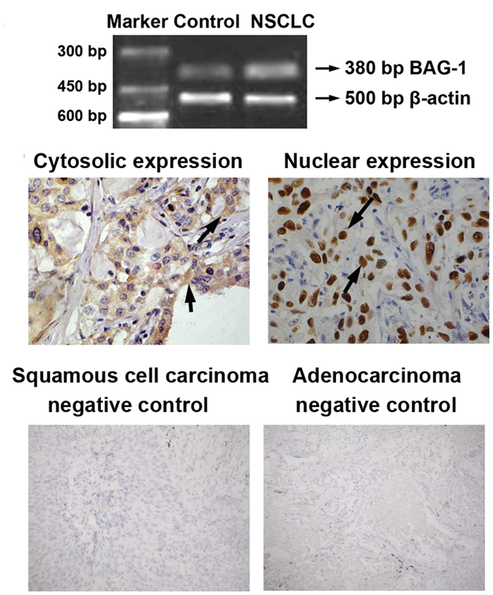
and healthy subjects. As shown in Fig.
1A, enhanced BAG-1 mRNA level was detected in lung tissues of
NSCLC patients, whereas a relatively low level of BAG-1 mRNA was
found in lung tissues obtained from healthy controls. In addition,
immunohistochemical analyses found that most of the BAG-1 protein
was located in the cytoplasm, although nuclear staining of BAG-1
was also observed (Fig. 1B).
Relationship between BAG-1 expression and
clinicopathological characteristics
We next evaluated the potential correlations between
BAG-1 expression and clinicopathological characteristics of NSCLC
patients. As shown in Table I,
BAG-1 expression was closely related to differentiation stage, as
elevated BAG-1 expression was found in patients with moderate/high
differentiation as compared with poor differentiation (P=0.031).
However, no statistically significant difference in BAG-1
expression was found in the various genders, ages, pathological
types, clinical stages or node metastases (P>0.05, Table I). Statistical analyses showed that
11.67% (14 cases) of healthy subjects and 61.67% (74 cases) of
NSCLC patients expressed both mRNA and BAG-1 protein in the lung.
BAG-1 expression was significantly higher in NSCLC patients than in
healthy controls (χ2=5.601, P<0.05).
Survival analysis
Multivariate analyses (Cox regression model) were
used to examine the correlation between prognostic factors and
survival in NSCLC patients. As shown in Table II, differentiation stage, clinical
stage as well as BAG-1 expression were identified as the
independent prognostic factors for survival in patients with NSCLC
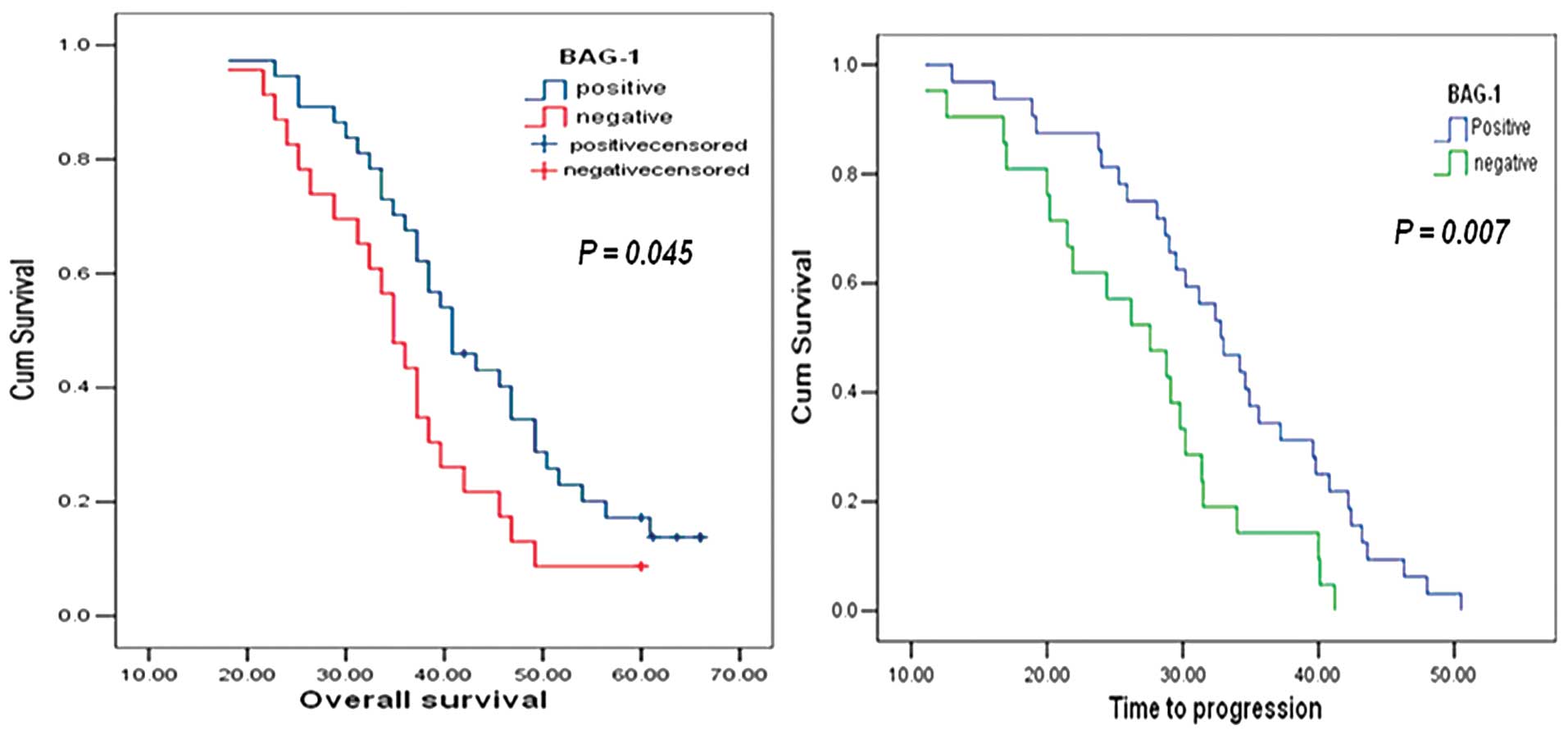
(P<0.05). Overall survival (OS) was counted from the day of
surgery to the last follow-up examination and was longer with BAG-1
positive as compared with BAG-1 negative (log-rank test, P=0.045)
(Fig. 2A). Life-table analysis
showed that the time to progression (TTP) of NSCLC patients with
BAG-1 negative expression (46 cases) was 37.5 months and the 5-year
survival rate was 8.69%. However, both TTP and the survival rate of
NSCLC patients with BAG-1 positive expression (74 cases) were
dramatically increased (TTP, 49.3 months; 5-year survival rate,
16.21%) (χ2=7.243, P=0.007) (Fig. 2B). These results suggested that
BAG-1 expression was positively associated with prolonged survival
of patients with NSCLC.
| Table IISurvival analysis of NSCLC patients
by Cox proportional hazards model. |
Table II
Survival analysis of NSCLC patients
by Cox proportional hazards model.
| F | Hazard ratio | Wald | P-value |
|---|
| Gender | 1 | 1.548 | 1.967 | 0.161 |
| Pathological
types | 1 | 0.748 | 0.801 | 0.371 |
| Differentiation
stage | 1 | 0.505 | 4.518 | 0.034 |
| Clinical stage | 1 | 2.014 | 4.798 | 0.028 |
| Node
metastasis | 1 | 1.094 | 0.088 | 0.766 |
| BAG-1 positive | 1 | 0.513 | 7.613 | 0.006 |
Evaluation of genotype frequencies and
Hardy-Weinberg equilibrium testing
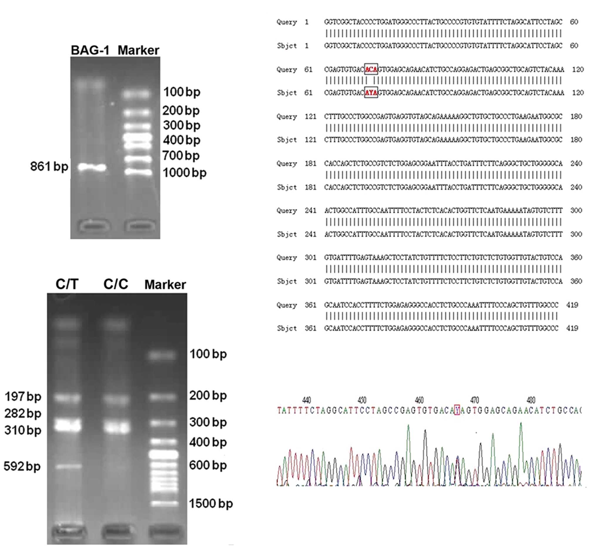
A total of 142 patients with newly diagnosed
advanced NSCLC were included for determination of genotype
frequencies. Venous blood samples were collected and BAG-1 DNA (861
bp in length) was amplified using PCR (Fig. 3A). Two BAG-1 digestion patterns were
identified by digestion: 72, 197, 282 and 310 bp fragments as wild
homozygous CC genotype; 72, 197, 282, 310 and 592 bp fragments as
heterozygous CT genotype (Fig. 3B).
The PCR products were confirmed by DNA sequencing (Fig. 3C and D). In addition, 110 out of 142
cases (77.46%) had the C/C genotype at BAG-1 nucleotide codon 324,
while 32 out of 142 cases (22.54%) had the C/T genotype at codon
324. The T/T genotype was not found in these patients. Each
genotype observed in the NSCLC patients were not significantly
different for the values expected at BAG-1 codon 324 according to
Hardy-Weinberg equilibrium (χ2=3.598, P=0.057). This
indicates that two types of polymorphisms come from the mendelian
population, which is in accordance with genetic equilibrium.
The correlation between gene polymorphism
and the sensitivity to chemotherapy
The association between gene polymorphism (C/C, C/T)
and the effect of chemotherapy were tested. Therapeutic effect was
evaluated two or three weeks following chemotherapy according to
the RECIST as described in Patients and methods. Note that the
Bag-1 polymorphisms might influence clinical outcomes to
chemotherapy as NSCLC patients carrying the C/C genotype exhibited
better responses to chemotherapy (Table III). However, other factors,
including gender, age, smoking, pathological types, differentiation
stage, clinical stage, KPS score and chemotherapy regimens, were
not significantly correlated with chemotherapy sensitivity
(P>0.05, Table IV). These
results demonstrated that the sensitivity to chemotherapy in NSCLC
patients were associated with Bag-1 polymorphisms at codon 324.
| Table IIICorrelation between gene polymorphism
and the effect of chemotherapy. |
Table III
Correlation between gene polymorphism
and the effect of chemotherapy.
| The effect of
chemotherapy | | | | |
|---|
|
| | | | |
|---|
| Genotype | SD+PD cases
(%) | CR+PR cases
(%) | χ2 | P-value | OR | 95% CI |
|---|
| C/C | 64 | 39 | 4.254 | 0.039 | 2.852 | 1.133–7.182 |
| C/T | 32 | 7 | | | | |
Correlation between gene polymorphism and
survival of NSCLC patients
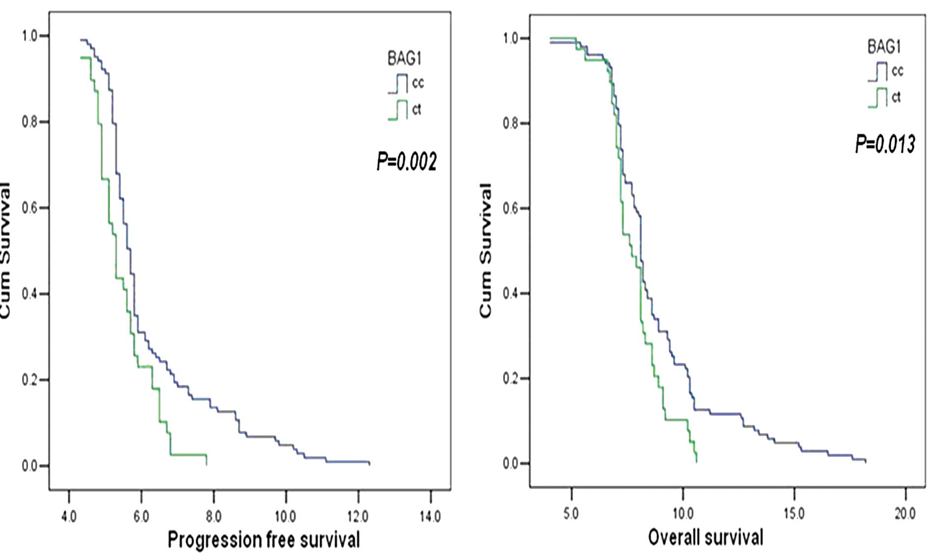
Kaplan Meier analyses showed that statistically
significant differences were observed in both progression free
survival (PFS) and overall survival (OS) of NSCLC patients carrying
the C/C genotype or C/T genotype (PFS, P=0.002; OS, P=0.013)
(Fig. 4). NSCLC patients carrying
the C/C genotype exhibited prolonged PFS and OS as compared with
those carrying the C/T genotype at Bag-1 codon 324. Cox
proportional hazards analyses further revealed that, besides gene
polymorphisms, the PFS was also associated with the clinical stage
and differentiation stage of patients with advanced NSCLC (Table V). Note that the progression risks
of patients carrying the C/C genotype at Bag-1 codon 324
were 1.87 times higher than in patients carrying the C/T genotype
(Table V). Hence, advanced clinical
stage, poor differentiation, C/T genotype at Bag-1 codon 324
could be key factors contributing to the development of NSCLC.
Moreover, we found that gender, age, KPS score were not correlated
with PFS (gender, P=0.831; age, P=0.585; KPS score, P=0.155).
| Table VCorrelation between progression-free
survival and clinical stage, differentiation and gene
polymorphism. |
Table V
Correlation between progression-free
survival and clinical stage, differentiation and gene
polymorphism.
| Regression
coefficient b | Standard error
b | Wald | V | P-value | OR | 95% CI |
|---|
| Clinical
stagea | 0.658 | 0.194 | 11.472 | 1 | 0.001 | 1.930 | 1.319–2.825 |
| Differentiation
stageb | 0.353 | 0.117 | 9.120 | 1 | 0.003 | 1.423 | 1.132–1.789 |
| C324Tc | 0.629 | 0.199 | 10.045 | 1 | 0.002 | 1.870 | 1.272–2.770 |
Discussion
Expression of a variety of anti-apoptotic genes are
involved in the development of NSCLC. Elevated Bcl-2 levels were
linked to increased disease-free and overall survival (16), while decreasing Bcl-2 expression was
related to metastatic NSCLC (17).
Altered expression of the p53 gene was correlated with poor
survival in NSCLC patients (18).
The Bcl-2-binding protein BAG-1 (19), has been recognized as a
multifunctional regulator of cell growth, survival and death
(8,9). Overexpression of BAG-1 inhibits
caspase activation induced by chemotherapeutic agents, radiation
and growth factor deprivation, and therefore suppresses apoptotic
cell death (20,21). However, the mechanism by which BAG-1
mediates cell survival is poorly understood. Importantly,
overexpression of BAG-1 is linked to various human cancers, and may
serve as an independent prognostic factor in the management of
certain cancers (10). For example,
BAG-1 isoforms are potential molecular markers for the pathogenesis
of breast cancer (22,23). However, the correlation between
BAG-1 expression and NSCLC occurrence has not been illustrated.
In the present study, we found that 11.67% (14
cases) of healthy subjects and 61.67% (74 cases) of NSCLC patients
expressed both mRNA and protein of lung BAG-1. These observations
were consistent with a previous report which showed overexpression
of BAG-1 in 73% NSCLC patients (n=85) (11). In addition, immunostaining showed
that BAG-1 protein was mostly located in the cytoplasm, although
the nucleus was also observed (Fig.
1B). Studies have suggested that expression of BAG-1 in the
cytoplasm might be related to a reduced risk of death in patients
with NSCLC (11).
BAG-1 expression was found to be related to the
differentiation of NSCLC, but not other clinicopathological
characteristics, including gender, age, pathological types,
clinical stage or node metastasis (Table I), suggesting BAG-1 may be involved
in the progress of NSCLC and contribute to disease development. Cox
multivariate analysis revealed that tumor differentiation, clinical
stage as well as BAG-1 expression were independent prognostic
factors for survival in patients with NSCLC (Table II). Compared with patients without
BAG-1, TTP and survival rate of NSCLC patients with BAG-1 positive
expression were significantly increased. This, together with
another report (11), demonstrated
that BAG-1 expression was sensitive to chemotherapy and could serve
as an independent prognostic factor in NSCLC. However, our results
demonstrating a correlation between BAG-1 expression and lung
cancer patient survival was contrary to a recent study, which
showed that TNM stage I lung cancer patients with BAG-1
low-expression had better survival compared to patients with high
expression of BAG-1 (24). This
discrepancy may be due to the different types of lung cancer
studied.
Theoretically, patients with high expression of the
anti-apoptosis protein BAG-1 should have poor prognosis. However,
in this present study, we found that BAG-1 expression was
positively associated with prolonged survival of patients with
NSCLC. It is possible since the anti-apoptotic activity of tumor
cells may also benefit patients with NSCLC. Tormanen et al
analyzed 75 cases of NSCLC and revealed that enhanced apoptosis
showed a 1.9-fold risk for a shortened survival in NSCLC patients
(25). Nevertheless, the involved
mechanism needs to be further clarified.
In addition to the anti-apoptotic effect of BAG-1
during cancer development, BAG-1 may also contribute to drug
resistance of chemotherapy. In HeLa cells, down-regulation of BAG-1
conferred resistance to anti-cancer drugs, including actinomycin D,
camptothecin, paclitaxel, staurosporine, thapsigargin and etoposide
(26). Liu et al (24) reported that knock down of BAG-1 by
RNA interference (RNAi) sensitized lung cancer cell lines (A549 and
L9981) to cisplatin-induced apoptosis. In the present study, we
found that BAG-1 expression was closely associated with the
sensitivity to platinum-based chemotherapeutics in NSCLC patients,
suggesting BAG-1 may be a biomarker for evaluating sensitivity to
chemotherapy.
PCR-RFLP technique was applied to screen for DNA
polymorphisms of BAG-1 in codon 324. C/C and C/T genotypes were
detected in a total of 142 patients with newly diagnosed advanced
NSCLC. NSCLC patients carrying the C/C genotype exhibited better
responses to chemotherapy, with prolonged PFS and OS, as compared
with those carrying the C/T genotype (Table III, Fig. 4). A previous study showed that BAG-1
is a novel regulator of nuclear factor-κB (NF-κB) as knock down of
BAG-1 inhibited NF-κB activity (27). Therefore, we reasoned that the C/T
transition at the BAG-1 codon 324 induced changes in encoded amino
acid from Thr to Ile that contributed to alterations in protein
expression and inhibition of apoptotic pathways triggered by
chemotherapeutic drugs. In addition, the T/T genotype was not
observed in this study, which might be due to the low incidence of
two allele mutations (28).
However, we cannot rule out the possibility that this may be due to
the small number of patients in our study. Therefore, BAG-1 codon
324 gene polymorphism serves as a biomarker for predicting the
sensitivity of chemotherapy, and may provide a theoretical basis
for individual treatment of patients with advanced NSCLC.
In summary, our results indicate that BAG-1 is
overexpressed in patients with NSCLC, which is associated with
sensitivity to platinum-based chemotherapeutics. These results
imply that BAG-1 may be a novel biomarker for predicting the
sensitivity to chemotherapy and provide evidence for the
application of individualized therapy in NSCLC. Future studies are
needed to explore the mechanisms by which BAG-1 is involved in
NSCLC progression.
Acknowledgements
This study was supported by the Scientific Research
Fund of Liaoning Provincial Education Department (Grant no.
L2010286). We thank Medjaden Bioscience Limited for assisting in
the preparation of this manuscript.
References
|
1
|
Jemal A, Siegel R, Xu J and Ward E: Cancer
statistics, 2010. CA Cancer J Clin. 60:277–300. 2010. View Article : Google Scholar
|
|
2
|
Rapp E, Pater JL, Willan A, et al:
Chemotherapy can prolong survival in patients with advanced
non-small cell lung cancer - report of a Canadian multicenter
randomized trial. J Clin Oncol. 6:633–641. 1988.PubMed/NCBI
|
|
3
|
Grilli R, Oxman AD and Julian JA:
Chemotherapy for advanced non-small cell lung cancer: how much
benefit is enough? J Clin Oncol. 11:1866–1872. 1993.PubMed/NCBI
|
|
4
|
Cullen MH, Billingham LJ, Woodroffe CM, et
al: Mitomycin, ifosfamide, and cisplatin in unresectable non-small
cell lung cancer: effects on survival and quality of life. J Clin
Oncol. 17:3188–3194. 1999.PubMed/NCBI
|
|
5
|
Schiller JH, Harrington D, Belani CP, et
al: Comparison of four chemotherapy regimens for advanced non-small
cell lung cancer. N Engl J Med. 346:92–98. 2002. View Article : Google Scholar
|
|
6
|
Belani CP and Langer C: First-line
chemotherapy for NSCLC: an overview of relevant trials. Lung
Cancer. 38(Suppl 4): 13–19. 2002. View Article : Google Scholar : PubMed/NCBI
|
|
7
|
Tang SC: BAG-1, an anti-apoptotic tumour
marker. IUBMB Life. 53:99–105. 2002. View Article : Google Scholar : PubMed/NCBI
|
|
8
|
Townsend PA, Cutress RI, Sharp A, Brimmell
M and Packham G: BAG-1: a multifunctional regulator of cell growth
and survival. Biochim Biophys Acta. 1603:83–98. 2003.PubMed/NCBI
|
|
9
|
Liman J, Faida L, Dohm CP, Reed JC, Bahr M
and Kermer P: Subcellular distribution affects BAG1 function. Brain
Res. 1198:21–26. 2008. View Article : Google Scholar : PubMed/NCBI
|
|
10
|
Cutress RI, Townsend PA, Brimmell M,
Bateman AC, Hague A and Packham G: BAG-1 expression and function in
human cancer. Br J Cancer. 87:834–839. 2002. View Article : Google Scholar : PubMed/NCBI
|
|
11
|
Rorke S, Murphy S, Khalifa M, Chernenko G
and Tang SC: Prognostic significance of BAG-1 expression in
non-small cell lung cancer. Int J Cancer. 95:317–322. 2001.
View Article : Google Scholar
|
|
12
|
Gautschi O, Hugli B, Ziegler A, et al:
Cyclin D1 (CCND1) A870G gene polymorphism modulates smoking-induced
lung cancer risk and response to platinum-based chemotherapy in
non-small cell lung cancer (NSCLC) patients. Lung Cancer.
51:303–311. 2006. View Article : Google Scholar : PubMed/NCBI
|
|
13
|
Shih CM, Lee YL, Chiou HL, et al:
Association of TNF-alpha polymorphism with susceptibility to and
severity of non-small cell lung cancer. Lung Cancer. 52:15–20.
2006. View Article : Google Scholar : PubMed/NCBI
|
|
14
|
Fang S, Jin X, Wang R, et al:
Polymorphisms in the MMP1 and MMP3 promoter and non-small cell lung
carcinoma in North China. Carcinogenesis. 26:481–486. 2005.
View Article : Google Scholar : PubMed/NCBI
|
|
15
|
Humphray SJ, Oliver K, Hunt AR, et al: DNA
sequence and analysis of human chromosome 9. Nature. 429:369–374.
2004. View Article : Google Scholar : PubMed/NCBI
|
|
16
|
Silvestrini R, Costa A, Lequaglie C, et
al: Bcl-2 protein and prognosis in patients with potentially
curable non-small cell lung cancer. Virchows Arch. 432:441–444.
1998. View Article : Google Scholar : PubMed/NCBI
|
|
17
|
Dosaka-Akita H, Katabami M, Hommura H,
Fujioka Y, Katoh H and Kawakami Y: Bcl-2 expression in non-small
cell lung cancers: higher frequency of expression in squamous cell
carcinomas with earlier pT status. Oncology. 56:259–264. 1999.
View Article : Google Scholar : PubMed/NCBI
|
|
18
|
Mitsudomi T, Hamajima N, Ogawa M and
Takahashi T: Prognostic significance of p53 alterations in patients
with non-small cell lung cancer: a meta-analysis. Clin Cancer Res.
6:4055–4063. 2000.PubMed/NCBI
|
|
19
|
Takayama S, Sato T, Krajewski S, et al:
Cloning and functional analysis of BAG-1: a novel Bcl-2-binding
protein with anti-cell death activity. Cell. 80:279–284. 1995.
View Article : Google Scholar : PubMed/NCBI
|
|
20
|
Takayama S and Reed JC: Molecular
chaperone targeting and regulation by BAG family proteins. Nat Cell
Biol. 3:E237–E241. 2001. View Article : Google Scholar : PubMed/NCBI
|
|
21
|
Hohfeld J: Regulation of the heat shock
conjugate Hsc70 in the mammalian cell: the characterization of the
anti-apoptotic protein BAG-1 provides novel insights. Biol Chem.
379:269–274. 1998.PubMed/NCBI
|
|
22
|
Millar EK, Anderson LR, McNeil CM, et al:
BAG-1 predicts patient outcome and tamoxifen responsiveness in
ER-positive invasive ductal carcinoma of the breast. Br J Cancer.
100:123–133. 2009. View Article : Google Scholar : PubMed/NCBI
|
|
23
|
Nadler Y, Camp RL, Giltnane JM, et al:
Expression patterns and prognostic value of Bag-1 and Bcl-2 in
breast cancer. Breast Cancer Res. 10:R352008. View Article : Google Scholar : PubMed/NCBI
|
|
24
|
Liu H, Bai Y, Liu B, et al: The expression
of BAG-1 and its clinical significance in human lung cancer.
Zhongguo Fei Ai Za Zhi. 11:489–494. 2008.(In Chinese).
|
|
25
|
Tormanen U, Eerola AK, Rainio P, et al:
Enhanced apoptosis predicts shortened survival in non-small cell
lung carcinoma. Cancer Res. 55:5595–5602. 1995.PubMed/NCBI
|
|
26
|
Takahashi N, Yanagihara M, Ogawa Y,
Yamanoha B and Andoh T: Down-regulation of Bcl-2-interacting
protein BAG-1 confers resistance to anti-cancer drugs. Biochem
Biophys Res Commun. 301:798–803. 2003. View Article : Google Scholar : PubMed/NCBI
|
|
27
|
Clemo NK, Collard TJ, Southern SL, et al:
BAG-1 is up-regulated in colorectal tumour progression and promotes
colorectal tumour cell survival through increased NF-kappaB
activity. Carcinogenesis. 29:849–857. 2008. View Article : Google Scholar : PubMed/NCBI
|
|
28
|
Li J, Wang B and Li SN: Advances in the
research of single nucleotide polymorphism. J Yunan Normal
University (Natural Sciences Edition). 27:40–44. 2007.
|