Introduction
Cervical cancer is the third most commonly diagnosed
cancer and the fourth leading cause of cancer-related death in
females worldwide, accounting for 9% (529,800) of the total new
cancer cases and 8% (275,100) of the total cancer deaths among
females in 2008 (1). Surgery, alone
or combined with radiotherapy has long been the cornerstone in the
treatment of cervical cancer (2,3).
Unfortunately, not all patients respond to such treatment, leading
to an extremely low survival rate for advanced stage. To increase
survival rates, different strategies with neoadjuvant chemotherapy
have been developed (4,5). Additionally, cisplatin-based
chemotherapy has traditionally been reserved as part of treatment
for metastatic or recurrent cervical cancer disease (6). However, resistance of cervical cancer
cells to antineoplastic agents is the major reason why
chemotherapy-based treatment modalities of malignant tumors may
fail (7,8). Therefore, exploring a novel approach
to improve drug sensitivity appeared to be urgently needed.
Accumulating evidence has demonstrated that cervical
cancer cells can exhibit a cross-resistant phenotype against
several unrelated drugs that differ widely with respect to
molecular structure and target specificity (9,10).
This phenomenon has been termed multidrug resistance (MDR). MDR has
most often been linked to overexpression of MDR1/P-gp, which is
overexpressed in many drug-resistant cell lines and in cervical
cancer (11). MDR1/P-gp functions
as a xenobiotics pump transporting a variety of toxic agents
including anticancer drugs from the intracellular milieu to the
outside of the cell (12,13). In view of the vital role of
MDR-1/P-gp in the treatment of cancer, prevention of MDR-1/P-gp
induction in cancer cells may help to avert drug resistance.
Twist1, a basic helix-loop-helix transcription
factor, has been known to contribute to tumor metastasis by
promoting an epithelial-mesenchymal transition (EMT), which is a
process initially observed in embryonic development in which cells
lose epithelial characteristics and gain mesenchymal properties to
increase motility and invasion (14,15).
Recently, a bulk of evidence has demonstrated a close link between
EMT and insensitivity to several growth factors or chemotherapeutic
agents (16,17). A novel function of Twist1 has been
reported in the development of acquired chemoresistance in human
cancer cells. Upregulation of Twist1 was associated with cellular
resistance to microtubule-targeting anticancer drugs in various
types of cancers (18–20). In addition, overexpression of Twist1
confers radioresistance or chemoresistance of cervical cancers,
thereby leading to a poorer prognosis (21). Therefore, targeting Twist1 could be
a novel therapeutic approach for the treatment of cervical cancer
by overcoming drug resistance.
These above observations prompted us to examine
whether Twist1 acts as a potential regulator of MDR1/P-gp.
Therefore, the purpose of this study was to investigate the
relationship of Twist1 and MDR1/P-gp in cervical cancer and to
explore whether Twist1 played an important role in drug resistance
of cervical cancer cells by regulating MDR1/P-gp. To the best of
our knowledge, this is the first report to describe that the
expression of MDR1/P-gp was significantly positively associated
with Twist1 expression in clinical cervical cancer tissues and that
Twist1 silencing not only downregulated MDR1/P-gp expression but
also enhanced chemosensitivity of human cervical cancer HeLa cells
to cisplatin.
Materials and methods
Cell line and tissue specimens
The human cervical cancer HeLa cell line (obtained
from ATCC) was cultured in Dulbecco’s modified Eagle’s medium
(DMEM) supplemented with 10% fetal bovine serum (FBS) at 37°C in a
5% CO2 atmosphere. Forty-two human cervical cancer
(including 26 squamous cell carcinoma and 16 adenocarcinoma) tissue
specimens were obtained from patients who underwent surgical
resection with the approval of the Institutional Review Board (IRB)
of the First Affiliated Hospital of Medical College of Xi’an
Jiaotong University.
Immunohistochemistry (IHC)
IHC was conducted using a Dako Autostainer Plus
system (Dako Corp., Carpinteria, CA) according to the
manufacturer’s protocol. Formalin-fixed, paraffin-embedded cervical
tissues were cut at a thickness of 4 μm, then were deparaffinized,
rehydrated and subjected to 5-min pressure-cooking antigen
retrieval in citrate buffer (10 mM, pH 6.0), 10 min double
endogenous enzyme block, 60 min mouse anti-Twist1 (Abcam, 1:50) or
mouse anti-MDR1/P-gp (Santa Cruz Biotechnology, 1:100) primary
antibody incubation and 30-min incubation with DakoCytomation
EnVision HRP reagent for mouse antibodies. Signals were detected by
adding the substrate hydrogen peroxide using diaminobenzidine (DAB)
as a chromogen followed by a 45-sec hematoxylin counterstaining. As
a negative control, the primary antibody was replaced with normal
IgG at an appropriate dilution. All of the immunostained slides
were analyzed by two histopathologists. A case was diagnosed as
positive when Twist1 staining in the nuclei and cytoplasm and the
MDR1/P-gp staining in the cytoplasm and membrane were observed in
>10% cells.
Cell transfection and clone
selection
Chemically-synthesized pGPU6/GFP/Neo vectors
containing short hairpin RNA (shRNA) against Twist1 (GenBank
accession no. NM_000474) mRNA pGPU6/GFP/Neo-Twist1 (sh-Twist1) (F:
5′-CACCG GTACATCGACTTCCTCTACCTTCAAGAGAGGTAGAGG
AAGTCGATGTACCTTTTTTG-3′; R: 5′-GATCCAAAAAA
GGTACATCGACTTCCTCTACCTCTCTTGAAGGTAGAG GAAGTCGATGTACC-3′; target
sequence: 5′-GGTACATC GACTTCCTCTACC-3′) and pGPU6/GFP/Neo vectors
containing negative control shRNA (sh-NC) were purchased from
Shanghai Genepharma Co. (Shanghai, China). For transient
transfection, HeLa cells were seeded in 6-well plates at the
density of 5×10⁴ cells/well and incubated at 37°C in an
atmosphere with 5% CO2 for 12 h. For each well, 7 μl of
Fugene HD Transfection Reagent (Roche) and 2 μg of sh-Twist1 or
sh-NC mixed together were diluted into 100 μl of DMEM culture
medium without serum and incubated for 20 min. Subsequently, the
mixture was added to the cells. Six hours later, the medium was
replaced with 2 ml of fresh DMEM medium containing 10% FBS, and
subsequent experiments were performed after culturing for another
24 h. For stable transfection, cells were cultured in DMEM culture
medium for 48 h after transfection as above, and then cells were
grown in 10-cm cell plates with medium containing 300 μg/ml G418
(Invitrogen). After 3 weeks of culture, visible colonies were
picked up and expanded. The stably transfected clones of HeLa cells
(HeLa/sh-Twist1 and HeLa/sh-NC) were observed to show green
fluorescence under microscope. Western blotting was used to
identify the positive clone.
Cell viability assay
Cell viability and IC50 values (drug
concentration causing 50% inhibition of cell growth) were analyzed
by the methyl tetrazolium (MTT) assay. For transient transfection,
cells were seeded at 5×10³ cells/well in 96-well plates
one day before transfection, then the MTT assay was performed just
before transfection as well as at 24, 48 and 72 h after
transfection. For stably transfected clones, cisplatin at various
concentrations (from 0 to 50 μM) was added to each 96-well plate
and the MTT assay was performed at 48 and 72 h after intervention.
For the assay, 20 μl of 5 mg/ml MTT (Invitrogen) was added to each
well, then the cells were incubated for 4 h before 180 μl
dimethylsulphoxide (DMSO, Sigma) was added. After the insoluble
crystals were completely dissolved, the absorbance of each well was
measured at 490 nm using a microplate reader (Bio-Rad Laboratories,
Hercules, CA, USA).
Apoptosis assay
HeLa cells were harvested 48 h after sh-Twist1 or
sh-NC transfection and resuspended in binding buffer (100
μl/500,000 cells: 140 mmol/l NaCl, 5 mmol/l CaCl2, and
10 mmol/l HEPES buffer) followed by 3 washes with
phosphate-buffered saline (PBS, pH 7.4). Annexin-V-FITC (5 μl) and
10 μl propidium iodide (PI, 1 μg/ml) were added and then the cell
suspension was incubated in a dark chamber at room temperature for
10 min. After centrifugation, the cell pellet was resuspended in
200 μl binding buffer and subjected to FACS analysis using the
FACSort flow cytometer (BD Biosciences). The percentage of
apoptotic and necrotic cells was determined using FCS express
software (DeNovo Software, Los Angeles, CA).
Reverse transcription-polymerase chain
reaction (RT-PCR)
After shRNA transfection, total RNA was isolated
using the RNAfast200 Total RNA Extract kit (Fastagene, China). The
RNA (2 μg) was reverse transcribed using the RevertAid™ First
Strand cDNA Synthesis kit (MBI Fermentas, Germany) according to the
manufacturer’s instructions. All PCR analyses were subsequently
performed with 1 μg of the cDNA reaction utilizing conditions as
follows: 94°C, 5 min; 32 cycles of 94°C, 30 sec; 58°C, 30 sec;
72°C, 45 sec. Reactions were terminated with 72°C, 7-min extension.
Primers used were F: 5′-CGTTTC CAGGAGGCCTGGCG-3′ and R:
5′-GCGAGACTGGCGA GCTGGAC-3′ for Twist1 and for β-actin F:
5′-GGCGGCACC ACCATGTACCCT-3′ and R: 5′-AGGGGCCGGACTCGT CATACT-3′).
PCR products were analyzed by 2% agarose gel electrophoresis and
visualized using ethidium bromide staining.
Western blotting
After the transfection, cells were lysed in ice-cold
RIPA lysis buffer (1% NP-40, 0.1% SDS, 0.5% sodium deoxycholate,
150 mmol/l NaCl and 10 mmol/l Tris-HCl) containing a protease
inhibitor cocktail. A total of 30 μg of protein was separated by
10% SDS-PAGE and transferred to nitrocellulose membranes. After
being blocked in Tris-buffered saline (TBS) containing 5% skim milk
at room temperature for 1 h, the membranes were incubated with
mouse monoclonal Twist1 antibody (Abcam, 1:200) or mouse monoclonal
MDR1 antibody (Santa Cruz Biotechnology, 1:250) at 4°C for 12 h,
and then with horseradish peroxidase (HRP)-conjugated anti-mouse
antibody (Zhongshan, China) at a dilution of 1:3,000 at room
temperature for 1 h. Signals were detected on X-ray film using the
ECL detection system (Pierce, Rockford, IL, USA). Loading
differences were normalized using a monoclonal GAPDH antibody.
Immunofluorescence staining
HeLa/sh-NC and HeLa/sh-Twist1 cells were cultured on
glass coverslips for 24 h. The cells were washed with PBS and fixed
with 4% paraformaldehyde for 20 min, permeabilized with 0.1% Triton
X-100 and blocked with 3% bovin serum albumin (BSA) for 1 h.
Coverslips were incubated with Twist1 or MDR1/P-gp primary
antibodies described above (dilution rate for both antibodies used
were 1:100) at 4°C overnight and then with TRITC or FITC labeled
secondary IgG conjugates (Zhongshan, China). Atfer
4,6-diamidino-2-phenylindole (DAPI, 1 μg/ml, Sigma) counterstaining
for nuclei, fluorescence was visualized by fluorescence microscopy
(Olympus Optical Co., Ltd., Tokyo, Japan).
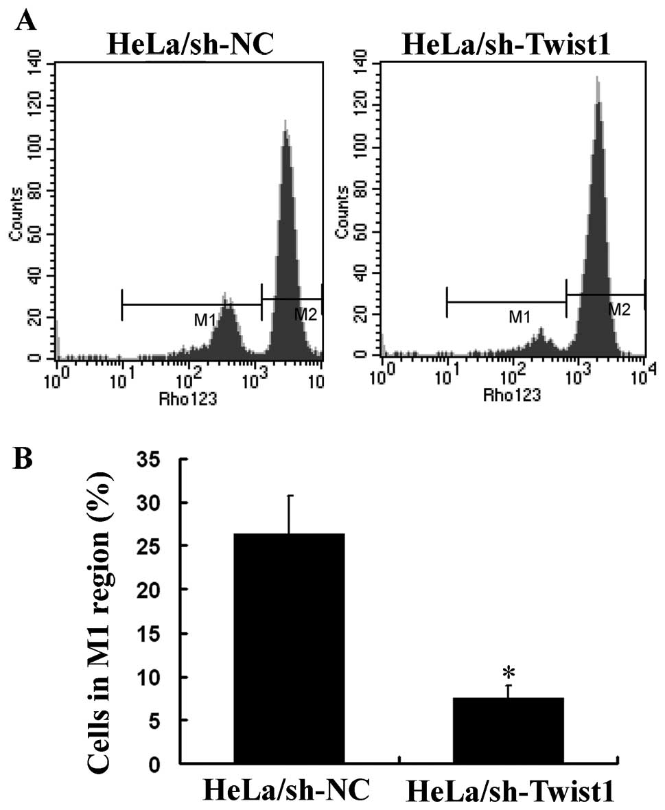
Detection of MDR1/P-gp function
Efflux of rhodamine 123 (Rh123) was chosen to detect
the MDR1/P-gp function of cells. HeLa/sh-NC and HeLa/sh-Twist1
cells were cultured in 6-well plates, and when the cells reached
70–80% confluence, Rh123 (Sigma) was added to the cells at a final
concentration of 0.25 μg/ml and incubated at 37°C for 1 h. Cells
were washed 3 times with 4°C PBS and resuspended at
5–10×105 cells/ml in 4°C PBS. Rh123 fluorescence was
analyzed with a FACStar flow cytometer (BD Biosciences) equipped
with an argon laser. The blast population was gated by forward and
side scatter characteristics. Rh123 fluorescence of 10,000 cells
was measured logarithmically through a 530 nm bandpass filter at an
excitation wavelength of 488 nm. HeLa cells without incubation with
Rh123 served as a negative control. Rh123 efflux was measured by
counting cells in the M1 region of the plot and calculated as the
percentage of cells in the M1 region of the plot. The higher the
percentage of cells in the M1 region the greater the cellular Rh123
efflux and also the greater the MDR1/P-gp function.
Statistical analysis
All statistical analyses were performed using SPSS
16 (SPSS Inc., Chicago, IL). Statistical significance of
differences among the control and various treatment groups were
compared by one-way ANOVA, followed by the Dunnett’s t-test for
separate comparisons. The expression of Twist1 and MDR1/P-gp were
compared using the Pearson’s test. P<0.05 was considered
statistically significant.
Results
Expression of Twist1 and MDR1/P-gp in
clinical cervical carcinoma tissues
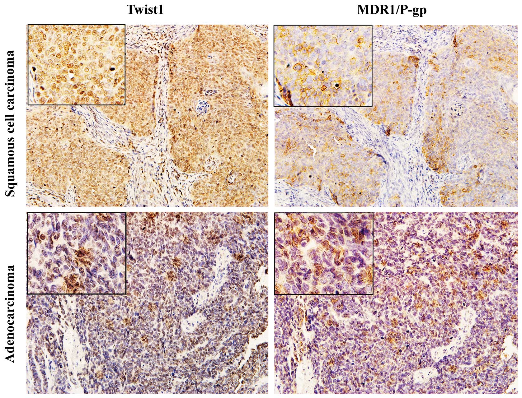
Immunohistochemical staining showed strong
cytoplasmic and/or nuclear staining for Twist1, cytoplasmic and/or
membrane staining for MDR1/P-gp in both cervical squamous cell
carcinoma and adenocarcinoma tissues (Fig. 1). According to the total 42 tissue
specimens, the positive expression of Twist1 and MDR1/P-gp was
31/42 (73.8%) and 27/42 (64.3%), respectively. Of the 31
Twist1-positive specimens, 24 (77.4%) showed positive staining of
MDR1/P-gp. Additionally, of the 11 Twist1-negative specimens, 8
(72.7%) were MDR1/P-gp negative. The Spearman analysis showed that
the expression level of MDR1/P-gp was significantly positively
associated with Twist1 expression (R=0.460, P=0.008; Table I).
 | Table ICorrelation analysis between MDR1/P-gp
and Twist1 expression in human cervical carcinoma tissues. |
Table I
Correlation analysis between MDR1/P-gp
and Twist1 expression in human cervical carcinoma tissues.
| MDR1/P-gp | |
|---|
|
| |
|---|
| Twist1 | Positive | Negative | Total |
|---|
| Positive | 24 | 7 | 31 |
| Negative | 3 | 8 | 11 |
| Total | 27 | 15 | 42 |
shRNA silencing of Twist1 at the mRNA and
protein levels
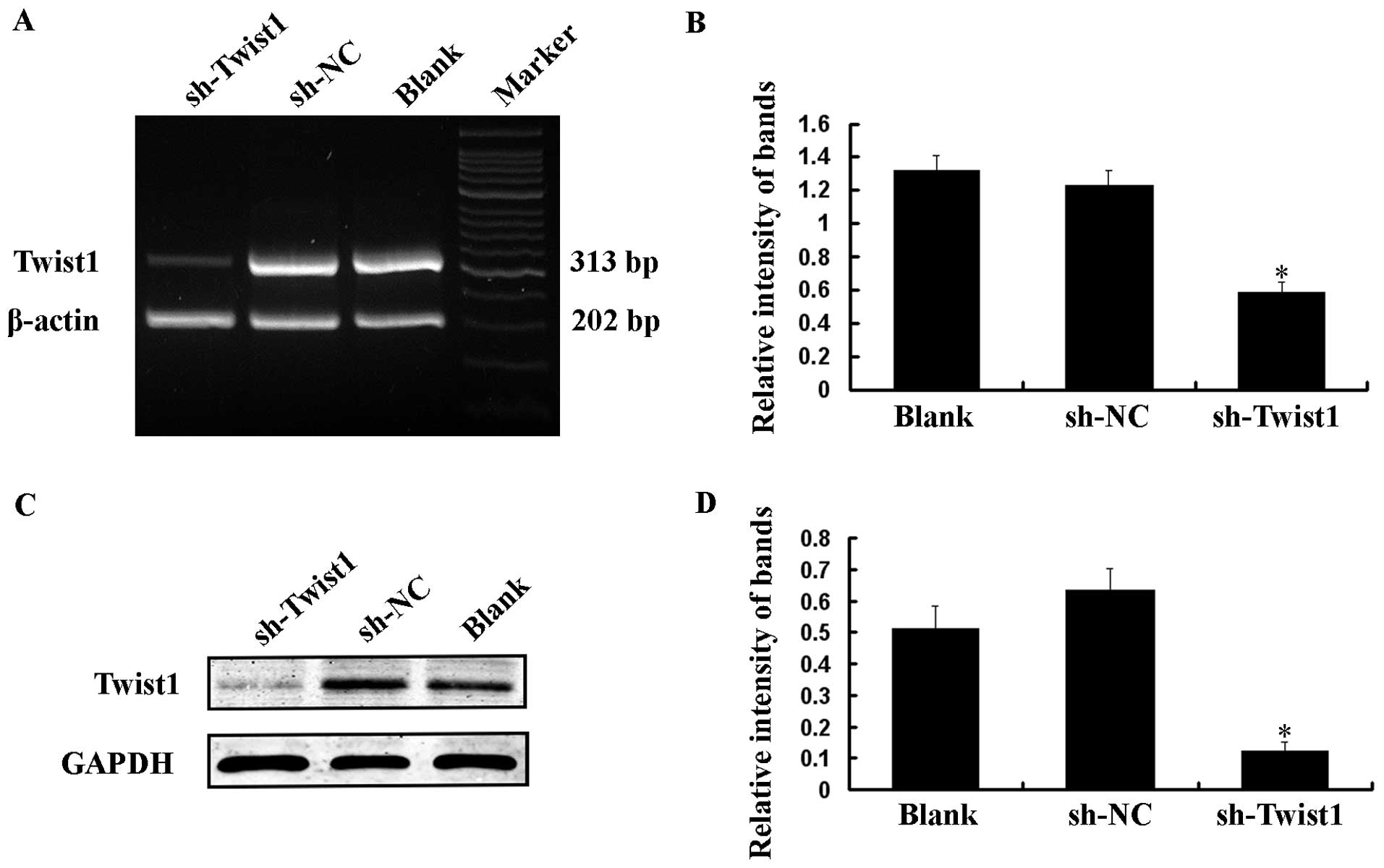
HeLa cells, which have a high level of Twist1
expression, were transiently transfected with 1 μg/ml sh-Twist1 or
sh-NC. Total RNA and protein were isolated and analyzed by RT-PCR
and western blotting at 48 h after transfection. Compared with the
Blank (no shRNA) and sh-NC transfected cells, the expression of
Twist1 was obviously suppressed in cells transfected with sh-Twist1
at both the mRNA and protein levels (Fig. 2).
Downregulation of Twist1 inhibits growth
and promotes apoptosis of HeLa cells
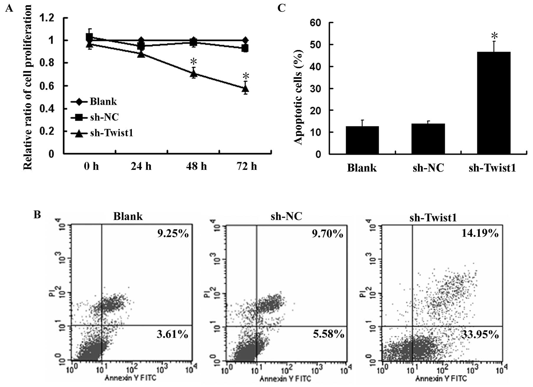
The role of Twist1 in the growth of HeLa cells was
determined by MTT assay. As shown in Fig. 3A, downregulation of Twist1 by
transient transfection of sh-Twist1 in HeLa cells caused
significant inhibition of cell proliferation compared with the
Blank and sh-NC transfected cells (P<0.05). The proliferation
rate of HeLa cells transfected with 1 μg/ml sh-Twist1 was
0.88±0.032, 0.71±0.046 and 0.58±0.039 for 24, 48 and 72 h,
respectively, which indicated a time-dependent growth inhibition
effect.
Since we observed the inhibitory effects of Twist1
silencing on the growth of HeLa cells, we next examined the effects
of Twist1 silencing on the apoptosis of HeLa cells using Annexin-V
and PI double staining. The effects of Twist1 silencing on the
apoptosis of HeLa cells, as measured by flow cytometry, are shown
in Fig. 3B and C. sh-Twist1
resulted in 48.14% of apoptotic cells, while the baseline of the
control cells (Blank) was 12.86% (P<0.05). These results
indicate that downregulation of Twist1 inhibited growth and
promoted apoptosis in HeLa cells.
Silencing of Twist1 represses MDR1/P-gp
expression
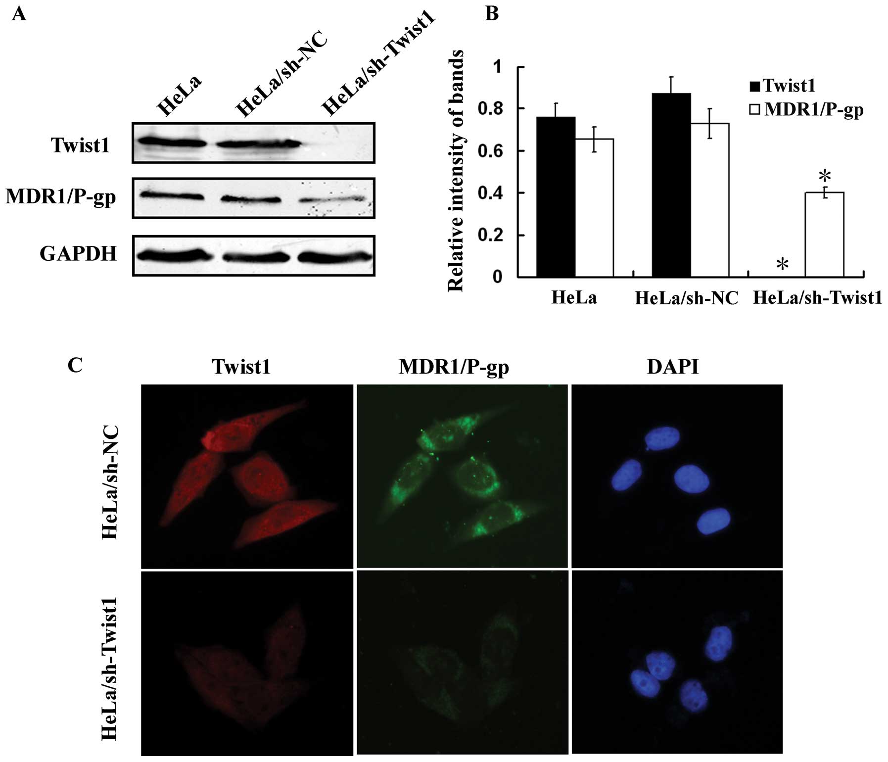
To further investigate the relationship between
Twist1 and MDR1/P-gp in cervical cancer, stable Twist1 knockdown
(HeLa/sh-Twist1) and control (HeLa/sh-NC) HeLa cells were
generated. The protein expression of Twist1 and MDR1/P-gp were then
investigated by western blotting. As shown in Fig. 4A and B, Twist1 ablation in
HeLa/sh-Twist1 cells accompanied with a significant repression on
MDR1/P-gp expression was observed. Immunofluorescence staining
confirmed these findings (Fig.
4C).
Silencing Twist1 abates MDR1/P-gp
function
Since MDR1/P-gp was downregulated by Twist1
ablation, we next examined the effects of silencing Twist1 on
MDR1/P-gp function. Rh123 efflux is sensitive and specific for
indicating the transport function of MDR1/P-gp. MDR1/P-gp-mediated
transport indicated by intracellular decrease of Rh123 fluorescence
was studied using flow cytometry. Compared with HeLa/sh-NC cells, a
significant increase of intracellular Rh123 was observed in
HeLa/sh-Twist1 cells, the mean percentage of Rh123 efflux cells
(cells in M1 region) was 26.41% and 7.53% in HeLa/sh-NC cells and
HeLa/sh-Twist1 cells, respectively (P<0.05) (Fig. 5). These results suggest that the
repression of MDR1/P-gp expression by silencing Twist1 in
HeLa/sh-Twist1 cells results in a great lost of MDR1/P-gp function
in drug efflux.
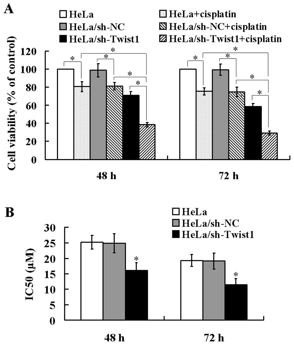
Abated MDR1/P-gp function increases cell
sensitivity to cisplatin
To evaluate the biological significance of Twist1 on
the cell sensitivity to cisplatin, MTT assay was then performed.
Compared with HeLa and HeLa/sh-NC cells, HeLa/sh-Twist1 cells
showed a significant decrease in cell viability over 48 and 72 h.
Cisplatin (15 μM) induced only a moderate (but significant)
decrease in cell viability of HeLa cells and HeLa/sh-NC cells, but
this decrease was much more significant in HeLa/sh-Twist1 cells
(Fig. 6A). The IC50
value to cisplatin for HeLa cells, HeLa/sh-NC cells and
HeLa/sh-Twist1 cells was 25.2±2.79, 24.9±1.84, 16.1±1.98 μM and
19.3±2.31, 18.7±1.92, 11.4±2.01 μM for 48 and 72 h, respectively,
suggesting the reduced IC50 value to cisplatin by Twist1
silencing (Fig. 6B).
Discussion
Recently, a novel function of Twist1 has been
reported in the development of acquired chemoresistance in human
cancer cells (18–20). However, the reason that Twist1
contributes to drug resistance in the treatment of cervical cancer
has not yet been established. In this study, we firstly analyzed
the relationship between Twist1 and MDR1/P-gp expression in
cervical cancer specimens and demonstrated a positive correlation
between Twist1 and MDR1/P-gp expression in the same patient.
Additionally, we provided the first evidence that silencing of
Twist1 downregulated MDR1/P-gp expression, inhibited its efflux
activity, and sensitized cervical cancer cells to cisplatin
treatment.
Twist1 is a highly conserved transcription factor
that belongs to the family of basic helix-loop-helix proteins,
which play a central role in cell type determination and cell
differentiation (14,15). Recently, accumulating studies have
shown that Twist1 was overexpressed in a variety of solid cancers
including breast, prostate and gastric carcinomas rendering Twist1
as a potential oncogene in tumorigenesis (22–24).
Twist1 has been found to function as an antiapoptotic factor
through both p53-dependent and p53-independent pathways (22). Downregulation of Twist1 through
small interfering RNA promotes apoptosis in human breast cancer and
melanoma cell lines (25).
Consistent with these findings, we also observed that inactivation
of Twist1 by RNA interference induced cell apoptosis in cervical
cancer HeLa cells though the mechanisms have not been studied.
EMT is a complex process which disaggregates
structured epithelial units to enable cell motility and
morphogenesis in embryonic development and is critically linked
with up-regulated invasion, metastasis and angiogenesis in cancer
progression (14,15). In addition, the EMT of cancer cells
not only causes increased metastasis, but also contributes to drug
resistance (19). Furthermore, it
has been reported that drug-resistant MCF-7 cells exhibit EMT gene
expression patterns and paclitaxel-resistant epithelial ovarian
carcinoma cells lose their epithelial features, acquire mesenchymal
characteristics, and increase their capacity for migration and
invasion, which are events characteristic of the EMT (16). Twist1 is a major regulator of EMT
and it has also been identified as capable of promoting carcinoma
metastasis. The elevated Twist1 expression is found to be
positively correlated with aggressiveness of cancer and poor
survival rate (22,24). Recently, Li et al
demonstrated that cells undergoing EMT displayed up-regulation of
MDR1/P-gp, MDR to chemotherapeutic agents as well as increased
in vitro invasiveness potential (19). Twist1 RNAi largely inhibited EMT
induction and partially reversed MDR phenotype (19). However, the relationship between EMT
and drug resistance seems complicated. Whether the expression of
MDR1/P-gp is regulated by Twist1 remains to be elucidated.
The expression of MDR1/P-gp is regulated at the
transcriptional level by multiple signaling pathways (26), including those mediated by
hypoxia-inducible factor-1α (HIF-1α), p53, and even chromosomal
rearrangement (26–28). MDR1/P-gp expression is also
regulated by epigenetic mechanisms, such as methylation and
acetylation (29).
Post-transcriptional regulation of MDR1/P-gp expression by microRNA
has also been reported (30,31).
Recently, a potential transcriptional regulatory role of Twist1 has
been identified in chemotherapy drug resistance. During generation
of acquired resistance to paclitaxel, nasopharyngeal carcinoma
cells showed upregulation of Twist1 at both the mRNA and protein
levels and were also cross-resistant to vincristine (18). In the present study, our findings
indicate a novel role of Twist1 in maintaining the
cisplatin-resistant phenotype of cervical cancer HeLa cells through
regulating MDR1/P-gp expression. However, further research is
needed to define the molecular regulatory mechanisms.
Hypoxia is well known to induce resistance to drugs
and radiation in solid tumors and a number of studies have
indicated a direct regulation of Twist1 by HIF-1α (32). Ding et al found that there
was a positive correlation between HIF-1α and MDR1/P-gp expression
in colon carcinoma (33).
Additionally, HNE1-T3 cells that acquired paclitaxel-resistant were
reported to show a high amplification of the Twist1 gene,
decreased p53 and p21Waf1, but only a moderate
alteration in MDR1/P-gp, which indicated that the Twist1-induced
drug resistance results from interference with p53-related pathways
(18). Furthermore, Cheng et
al demonstrated that AKT2 is a downstream target of Twist1 and
AKT2 is responsible for at least in part the Twist1-mediated
paclitaxel resistance of breast cancer MCF-7 cells (20). Thus, the involvement of HIF-1α, p53
and AKT2 in the Twist1/MDR1-mediated cisplatin-resistant phenotype
of cervical cancer will be investigated in our further studies.
In conclusion, this study provides the first
evidence that Twist1 expression was significantly positively
associated with MDR1/P-gp expression in human cervical cancer and
that Twist1-mediated modulation of MDR1/P-gp expression plays an
important role in the sensitization of cervical cancer cells to
cisplatin treatment. Our results indicate a novel therapeutic
strategy to overcome drug-resistance through inactivation of Twist1
expression in cervical cancer.
References
|
1
|
Jemal A, Bray F, Center MM, Ferlay J, Ward
E and Forman D: Global cancer statistics. CA Cancer J Clin.
61:69–90. 2011. View Article : Google Scholar
|
|
2
|
Greven K, Petereit D, Vermorken JB and
Lanciano R: Current developments in the treatment of newly
diagnosed cervical cancer. Hematol Oncol Clin North Am. 13:275–303.
1999. View Article : Google Scholar : PubMed/NCBI
|
|
3
|
Cornelio DB, Roesler R and Schwartsmann G:
Emerging therapeutic agents for cervical cancer. Recent Pat
Anticancer Drug Discov. 4:196–206. 2009. View Article : Google Scholar : PubMed/NCBI
|
|
4
|
Sardi JE, Boixadera MA and Sardi JJ:
Neoadjuvant chemotherapy in cervical cancer: a new trend. Curr Opin
Obstet Gynecol. 17:43–47. 2005. View Article : Google Scholar : PubMed/NCBI
|
|
5
|
Moore DH: Neoadjuvant chemotherapy for
cervical cancer. Expert Opin Pharmacother. 4:859–867. 2003.
View Article : Google Scholar : PubMed/NCBI
|
|
6
|
Tao X, Hu W, Ramirez PT and Kavanagh JJ:
Chemotherapy for recurrent and metastatic cervical cancer. Gynecol
Oncol. 110:S67–S71. 2008. View Article : Google Scholar : PubMed/NCBI
|
|
7
|
Brabec V and Kasparkova J: Molecular
aspects of resistance to antitumor platinum drugs. Drug Resist
Updat. 5:147–161. 2002. View Article : Google Scholar : PubMed/NCBI
|
|
8
|
Djeu JY and Wei S: Clusterin and
chemoresistance. Adv Cancer Res. 105:77–92. 2009.PubMed/NCBI
|
|
9
|
Konishi I, Nanbu K, Mandai M, et al: Tumor
response to neoadjuvant chemotherapy correlates with the expression
of P-glycoprotein and PCNA but not GST-pi in the tumor cells of
cervical carcinoma. Gynecol Oncol. 70:365–371. 1998. View Article : Google Scholar : PubMed/NCBI
|
|
10
|
Britten RA, Liu D, Tessier A, Hutchison MJ
and Murray D: ERCC1 expression as a molecular marker of cisplatin
resistance in human cervical tumor cells. Int J Cancer. 89:453–457.
2000.PubMed/NCBI
|
|
11
|
Takara K, Sakaeda T and Okumura K: An
update on overcoming MDR1-mediated multidrug resistance in cancer
chemotherapy. Curr Pharm Des. 12:273–286. 2006. View Article : Google Scholar : PubMed/NCBI
|
|
12
|
Lage H: MDR1/P-glycoprotein (ABCB1) as
target for RNA interference-mediated reversal of multidrug
resistance. Curr Drug Targets. 7:813–821. 2006.PubMed/NCBI
|
|
13
|
Xia Z, Zhu Z, Zhang L, et al: Specific
reversal of MDR1/P-gp-dependent multidrug resistance by RNA
interference in colon cancer cells. Oncol Rep. 20:1433–1439.
2008.PubMed/NCBI
|
|
14
|
Peinado H, Olmeda D and Cano A: Snail, Zeb
and bHLH factors in tumour progression: an alliance against the
epithelial phenotype? Nat Rev Cancer. 7:415–428. 2007.PubMed/NCBI
|
|
15
|
Yang J, Mani SA, Donaher JL, et al: Twist,
a master regulator of morphogenesis, plays an essential role in
tumor metastasis. Cell. 117:927–939. 2004.PubMed/NCBI
|
|
16
|
Iseri OD, Kars MD, Arpaci F, Atalay C, Pak
I and Gunduz U: Drug resistant MCF-7 cells exhibit
epithelial-mesenchymal transition gene expression pattern. Biomed
Pharmacother. 65:40–45. 2011. View Article : Google Scholar : PubMed/NCBI
|
|
17
|
Yang AD, Fan F, Camp ER, et al: Chronic
oxaliplatin resistance induces epithelial-to-mesenchymal transition
in colorectal cancer cell lines. Clin Cancer Res. 12:4147–4153.
2006. View Article : Google Scholar : PubMed/NCBI
|
|
18
|
Wang X, Ling MT, Guan XY, et al:
Identification of a novel function of TWIST, a bHLH protein, in the
development of acquired taxol resistance in human cancer cells.
Oncogene. 23:474–482. 2004. View Article : Google Scholar : PubMed/NCBI
|
|
19
|
Li QQ, Xu JD, Wang WJ, et al:
Twist1-mediated adriamycin-induced epithelial-mesenchymal
transition relates to multidrug resistance and invasive potential
in breast cancer cells. Clin Cancer Res. 15:2657–2665. 2009.
View Article : Google Scholar
|
|
20
|
Cheng GZ, Chan J, Wang Q, Zhang W, Sun CD
and Wang LH: Twist transcriptionally up-regulates AKT2 in breast
cancer cells leading to increased migration, invasion, and
resistance to paclitaxel. Cancer Res. 67:1979–1987. 2007.
View Article : Google Scholar : PubMed/NCBI
|
|
21
|
Shibata K, Kajiyama H, Ino K, et al: Twist
expression in patients with cervical cancer is associated with poor
disease outcome. Ann Oncol. 19:81–85. 2008. View Article : Google Scholar : PubMed/NCBI
|
|
22
|
Kwok WK, Ling MT, Lee TW, et al:
Up-regulation of TWIST in prostate cancer and its implication as a
therapeutic target. Cancer Res. 65:5153–5162. 2005. View Article : Google Scholar : PubMed/NCBI
|
|
23
|
Martin TA, Goyal A, Watkins G and Jiang
WG: Expression of the transcription factors snail, slug, and twist
and their clinical significance in human breast cancer. Ann Surg
Oncol. 12:488–496. 2005. View Article : Google Scholar : PubMed/NCBI
|
|
24
|
Yan-Qi Z, Xue-Yan G, Shuang H, et al:
Expression and significance of TWIST basic helix-loop-helix protein
over-expression in gastric cancer. Pathology. 39:470–475. 2007.
View Article : Google Scholar : PubMed/NCBI
|
|
25
|
Puisieux A, Valsesia-Wittmann S and
Ansieau S: A twist for survival and cancer progression. Br J
Cancer. 94:13–17. 2006. View Article : Google Scholar : PubMed/NCBI
|
|
26
|
Chen T: Overcoming drug resistance by
regulating nuclear receptors. Adv Drug Deliv Rev. 62:1257–1264.
2010. View Article : Google Scholar : PubMed/NCBI
|
|
27
|
Comerford KM, Wallace TJ, Karhausen J,
Louis NA, Montalto MC and Colgan SP: Hypoxia-inducible
factor-1-dependent regulation of the multidrug resistance (MDR1)
gene. Cancer Res. 62:3387–3394. 2002.PubMed/NCBI
|
|
28
|
Sampath J, Sun D, Kidd VJ, et al: Mutant
p53 cooperates with ETS and selectively up-regulates human MDR1 not
MRP1. J Biol Chem. 276:39359–39367. 2001. View Article : Google Scholar : PubMed/NCBI
|
|
29
|
El-Osta A, Kantharidis P, Zalcberg JR and
Wolffe AP: Precipitous release of methyl-CpG binding protein 2 and
histone deacetylase 1 from the methylated human multidrug
resistance gene (MDR1) on activation. Mol Cell Biol. 22:1844–1857.
2002. View Article : Google Scholar : PubMed/NCBI
|
|
30
|
Gomez-Martinez A, Garcia-Morales P,
Carrato A, et al: Post-transcriptional regulation of P-glycoprotein
expression in cancer cell lines. Mol Cancer Res. 5:641–653. 2007.
View Article : Google Scholar : PubMed/NCBI
|
|
31
|
Chen Y, Tang Y, Wang MT, Zeng S and Nie D:
Human pregnane X receptor and resistance to chemotherapy in
prostate cancer. Cancer Res. 67:10361–10367. 2007. View Article : Google Scholar : PubMed/NCBI
|
|
32
|
Yang MH, Wu MZ, Chiou SH, et al: Direct
regulation of TWIST by HIF-1alpha promotes metastasis. Nat Cell
Biol. 10:295–305. 2008. View
Article : Google Scholar : PubMed/NCBI
|
|
33
|
Ding Z, Yang L, Xie X, et al: Expression
and significance of hypoxia-inducible factor-1 alpha and
MDR1/P-glycoprotein in human colon carcinoma tissue and cells. J
Cancer Res Clin Oncol. 136:1697–1707. 2010. View Article : Google Scholar : PubMed/NCBI
|