Introduction
Colorectal cancer (CRC) is one of the most common
internal malignancies and is one of the leading causes of
cancer-related morbidity and mortality in the world. Therefore,
increasing efforts are being focused on developing more effective
screening and prevention measures for CRC. Several epidemiological
studies have shown that regular use of aspirin or other
non-steroidal anti-inflammatory drugs (NSAIDs) results in 40–50%
reduced risk of colorectal cancer (1–3).
Furthermore, NSAIDs cause regression of pre-existing adenomas in
patients with familial adenomatous polyposis (4) and significantly inhibit tumor growth
in animal models of CRC (1,5). The mechanism by which aspirin and
NSAIDs inhibit cyclooxygenase (COX) activity was first described by
Vane (6). This observation led to
the hypothesis that both the toxicity and efficacy of NSAIDs are
mediated through the inhibition of COX-mediated prostaglandin (PG)
synthesis (7). In the early 1990s,
several groups reported the discovery of two COX isoforms, COX-1
and COX-2. COX-1 is constitutively expressed in normal intestine
and appears to function as a physiological regulatory enzyme in
most tissues, including the gastric mucosa, the kidney and
platelets. However, the COX-1 level does not change in intestinal
tumors. COX-2 is nearly undetectable in most tissues under normal
physiological conditions (7) and is
a strongly inducible form that is involved in growth proliferation
and the inflammatory response (8,9). COX-2
protein expression is enhanced in human CRC (10). COX-2-derived PG was found to play a
key role in colorectal tumorigenesis in ApcΔ16 knockout mice
(11). The invasiveness of colon
cancer cells was increased by COX-2 transfection in association
with PG production and was reversed by sulindac sulfide, a COX
inhibitor (12). COX-2 and
COX-2-derived PGs mediated tumor growth and metastasis in animal
models by inducing the formation of blood vessels (13). Specific COX-2 inhibition, either by
pharmacological intervention or by targeted knockout of the COX-2
gene, has been shown to effectively reduce the growth of xenograft
tumor in mice (3,13).
Celecoxib is a drug that was designed to treat the
signs and symptoms of adult arthritis (14). Celecoxib inhibits the inflammatory
COX-2 enzyme at therapeutic doses in humans that do not lower
gastrointestinal prostaglandin levels associated with mucosal
protection (15). Celecoxib blocked
angiogenesis and inhibited lung and colon cancer xenograft growth
in mice (13). Several recent
reports have suggested that COX-2 expression plays an important
role in haematogenous metastasis of CRC to the liver (16,17).
These previous reports demonstrated that celecoxib has
anti-metastatic effects when cancer cells were pretreated with
celecoxib. We previously developed an animal model of orthotopic
transplanted CRC associated with spontaneous lymph node and
hematological metastases by intra-rectal injection of tumor cells
in nude mouse rectum, consistent with the progression of CRC in
humans (18).
The aim of the present study was to investigate
whether oral administration of COX-2 inhibitor celecoxib prevents
tumorigenesis and spontaneous lymph node and lung metastases of CRC
by using a rectal xenograft model identical to human CRC.
Materials and methods
Animals
All animal experiments were performed according to
Kanazawa University’s standard guidelines. Nude mice
(BALB/cAnNCrj-nu/nu) aged 5 weeks were purchased from Charles River
Japan, Inc. (Kanagawa, Japan). After at least 1 week of
observation, the mice were used for this study at the age of 6–7
weeks. All mice were housed in the Laboratory for Animal
Experiments, Research Institute, Kanazawa University School of
Medicine, under laminar airflow conditions. Housing was
temperature-controlled, with a 12-h/12-h light/dark cycle.
Cell line
The human colon cancer cell line HT-29 was used.
Cells were cultured in RPMI-1640 medium (RPMI) (Nissui, Tokyo,
Japan) supplemented with 10% heat-inactivated fetal bovine serum
(JRH Biosciences, Lenexa, KS) and antibiotics (100 U/ml penicillin
and 100 μg/ml streptomycin), at 37°C in a humidified atmosphere of
95% air/5%CO2. The cells were passaged when
confluent.
Human cancer xenograft models
Log-phase HT-29 human colon carcinoma cells were
harvested with Trypsin EDTA, washed three times with RPMI and
resuspended at a cell density of 1×10⁷/ml in RPMI.
Animals were anaesthetized with ether and placed in a supine
position. To prevent colonic obstruction resulting from rectal
tumor progression, a 7-mm cut of the anterior end of the ano-rectal
wall in the ano-rectal region was performed. Tumor cells
(1×10⁶/0.1 ml/mouse) in RPMI were slowly injected
submucosally into the posterior wall with a 27-gauge needle.
Preparation of celecoxib (SC-58635)
The preferential COX-2 inhibitor, celecoxib, was
purchased from Pfizer Japan Inc. (Tokyo, Japan). We prepared chows
containing 150, 750 or 1500 ppm celecoxib. The mice received
celecoxib chow for 4 weeks starting at 2 weeks after tumor cells
inoculation. Control mice received normal chow.
Antitumor efficacy
To evaluate the antitumor and anti-metastatic
effects of celecoxib, the mice were sacrificed 6 weeks after tumor
cells inoculation. The antitumor effect of celecoxib in the
xenograft was evaluated by measuring the weight of the
peri-ano-rectal tumor. To evaluate metastasis, the lymph nodes
around the abdominal aorta and lung were excised. DNA from each
specimen was extracted by the standard proteinase K
digestion-phenol chloroform extraction method as described
previously (19). The human
β-globin-related sequence in the extracted DNA was amplified by
quantitative PCR by using the 5′ nuclease assay and an ABI PRISM
7700 Sequence Detector (TaqMan) (PE Biosystems Japan, Tokyo,
Japan). One microgram of each DNA sample was amplified in a 50-μl
reaction mixture consisting of 200 nM of forward and reverse
primers, 100 nM of probe and TaqMan Universal Master Mix (PE
Biosystems Japan), which contained Ampli-Taq Gold DNA polymerase,
reaction buffer, dNTP, dUTP and AmpErase uracil-N-glycosylase. The
PCR reaction was carried out on an ABI 7700 with the following
thermo-cycler conditions: 50°C for 2 min, 95°C for 5 min, followed
by 45 cycles of 95°C for 15 sec and 60°C for 1 min. Data were
analyzed by using Sequence Detection Software (PE Biosystems
Japan). The number of metastasized tumor cells in the excised whole
organ was calculated from the standard curve of a serial dilution
series of HT-29 DNA after quantitative PCR as described previously
(18). Primer and probe sequences
for human β-globin gene amplification were as follows: forward,
CACT GACTCTCTCTGCTATTGGTC; reverse, AGGAGTGGAC AGATCCCCAAA; TaqMan
probe, 6FAM5′-CTACCCTTG GACCCAGAGGTTCTTTGAGTC-3′ TAMRA.
Quantification of human COX-2 mRNA
expression
We evaluated the human COX-2 mRNA level in the
rectal xenograft. The primary xenograft, para-aortic lymph nodes
and lung tissue were obtained from 5 mice bearing rectal tumors 6
weeks after tumor cell inoculation. RNA was extracted by using
Isogen systems (Nippon Gene Co., Ltd., Tokyo, Japan). After heat
denaturation at 68°C for 15 min with 500 pmol of the oligo(dT)
primer, 10 μg of RNA was reverse-transcribed at 42°C for 60 min
into first-strand cDNA in reverse-transcription solution [400 units
of Moloney murine leukemia virus reverse transcriptase (Invitrogen
Japan K.K., Tokyo, Japan), 50 mM Tris-HCl (pH 8.3), 75 mM KCl, 3 mM
MgCl2, 0.01 M DTT, 0.5 mM of each dNTP and 16 units of
RNasin (Promega, Madison, WI)] with a total volume of 100 μl. A
reverse-transcribed cDNA solution corresponding to 100 ng of total
RNA was amplified quantitatively with primers and a probe specific
for the targeted cDNA with an ABI PRISM 7700 Sequence Detector
(TaqMan). The final concentrations of reaction components, except
for template cDNA, and thermo-cycler conditions were the same as
those for the human β-globin-related sequence amplification
described above. Internal standard gene expression was examined by
using TaqMan GAPDH control reagents and TaqMan rodent GAPDH control
reagents VIC (PE Biosystems Japan). After standardization by
internal standard gene expression, human COX-2 gene expression was
calculated relative to the in vitro COX-2 expression level
of HT-29 cells. The primer and probe sequences for COX-2 mRNA
amplification were as follows: forward, GGT TGCTGGTGGTAGGAATGTT;
reverse, CATAAAGCGTT TGCGGTACTCA; TaqMan probe, 6FAM5′-CCGCAGTAC
AGAAAGTATCACAGGCTTCCA-3′ TAMRA.
PGE2 measurement
The concentration of PGE2 in the rectal
xenograft was determined by quantitative enzyme-linked
immunosorbent assay (ELISA) (Cayman Chemical, Ann Arbor, MI),
according to the manufacturer’s instructions.
Quantification of microvessel density
(MVD)
The MVD in the rectal xenograft was determined using
immunohistochemistry with an anti-mouse CD31 antibody. The tumor
xenografts were sliced to 5-μm sections at the maximum diameter.
The sliced specimens were fixed in acetone at 4°C overnight. After
rinsing with PBS, they were immersed in 30% sucrose-PBS overnight
for at least 4 h until they sunk. They were embedded in Cryomold
(Sakura Finetek Japan, Tokyo, Japan) by Tissue-Tek O.C.T. compound
(Sakura Finetek Japan). They were frozen on dry-ice acetone. They
were sectioned with a cryothome at a thickness of 7 μm and mounted
on precoated slides. These slides were incubated at 4°C overnight
with rat anti-mouse CD31 antibody (BD Biosciences Pharmingen, San
Diego, CA). Then, the slides were incubated in goat anti-rat IgG
HRP (Santa Cruz Biotechnology, Santa Cruz, CA) as the secondary
antibody. Vascularity was measured by the average number of vessels
per field counted in 8 random areas at ×20 magnification. Only
areas of viable tumor tissues were imaged; necrotic regions and
overlying subdermal regions were excluded. MVD was calculated as
the average number of vessels per mm2.
Western blot analysis
We examined the effect of celecoxib treatment on the
expression of mouse rectal xenograft proteins involved in
angiogenesis. Rectal xenograft samples from 1500 ppm
celecoxib-treated and control groups were assessed for the
expression of vascular endothelial growth factor (VEGF) protein by
specific immunoblot analyses. The protein concentration in each
sample was measured using a Bio-Rad protein assay kit II (Bio-Rad
Laboratory, Richmond, CA). For SDS-PAGE, 10 μg of protein from each
sample was loaded on 12% polyacrylamide gels. Proteins were
transferred to a polyvinylidene difluoride membrane with a tank
transfer system (Bio-Rad Laboratory), and then blocked with a
buffer containing 5% low fat skim milk and 0.1% Tween-20 in
tris-buffered saline (TBST) at room temperature for 1 h. We used
rabbit anti-human VEGF which cross-reacted with VEGF of mouse as
the primary antibody (Santa Cruz Biotechnology). The primary
antibody was diluted in TBST containing 5% skim milk at a dilution
of 1:2000. The membrane was incubated with the primary antibody
overnight at 4°C. After washing three times with TBST, the membrane
was incubated with a horseradish peroxidase-conjugated secondary
antibody (0.02 μg/ml in TBST) at room temperature for 1 h. We used
goat anti-rabbit IgG HRP (1:5000 dilution; Chemicon International,
Temecula, CA) as the secondary antibody. Detection of
chemiluminescence was performed with ECL western blot detection
kits (Amersham, Little Chalfont, UK), according to the
manufacturer’s instructions.
Measurement of apoptosis
We examined the effect of celecoxib treatment on the
degree of apoptosis of mouse rectal xenograft tumor cells by
terminal deoxynucleotidyl transferase-mediated nick end labeling
(TUNEL) assay. Rectal xenografts were embedded in paraffin and
sliced to 5-μm sections. The sections were dewaxed and rehydrated
using routine procedures. Apoptosis in situ detection kit
was purchased from Wako (Osaka, Japan). Apoptosis was assessed by
the average number of apoptotic cells per field counted in 8 random
areas at ×40 magnification. The average number of tumor cells per
field was also determined at ×40 magnification. The apoptotic index
was calculated as follows: apoptotic index (%) = (no. of apoptotic
cells/no. of total tumor cells × 100).
Statistical analysis
The effect of celecoxib was assessed for
associations with number of metastasized tumor cells,
PGE2 production, VEGF production, MVD counting and
apoptosis index by using the Mann-Whitney’s U test. P<0.05 was
considered to be statistically significant.
Results
Rectal xenograft model
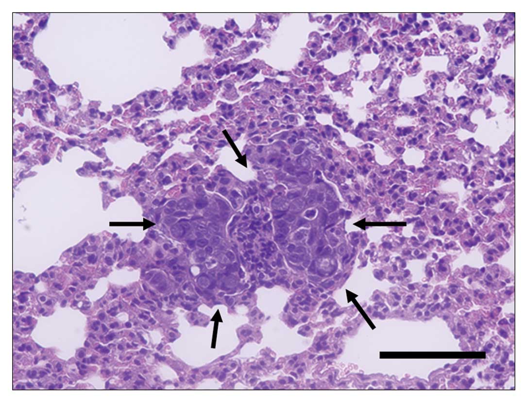
Macroscopically, a rectal xenograft developed into a
rectal tumor within 1 week after tumor cell inoculation. The HT-29
xenograft produced a locally aggressive rectal tumor and
subsequently showed lymph node metastasis around the abdominal
aorta 6 weeks after tumor cell inoculation. No apparent metastatic
nodules were observed macroscopically in the lung, liver, spleen,
kidneys, or peritoneum. Histologically, multiple tiny metastatic
foci in the lungs were observed 6 weeks after tumor cell
inoculation (Fig. 1).
Quantification of human COX-2 mRNA
expression
Expression levels of human COX-2 mRNA from tumor
cells in the primary xenograft tissue, lymph nodes and lungs from
mice bearing rectal xenografts were analyzed (Table I). COX-2 mRNA expression was
increased 3.63-, 3.32- and 37.6-fold in the rectal xenograft
tissue, lymph nodes, and lungs to that in the in vitro HT-29
cells, respectively.
 | Table IQuantification of human COX-2 mRNA
expression in rectal xenograft models. |
Table I
Quantification of human COX-2 mRNA
expression in rectal xenograft models.
| Tissue | Relative expression
ratio of human COX-2a |
|---|
| Rectal
xenograft | 3.63
(2.46–4.23) |
| Lymph node | 3.32
(1.51–106) |
| Lung | 1.6 (0–56.8) |
Antitumor efficacy of celecoxib in
xenografts
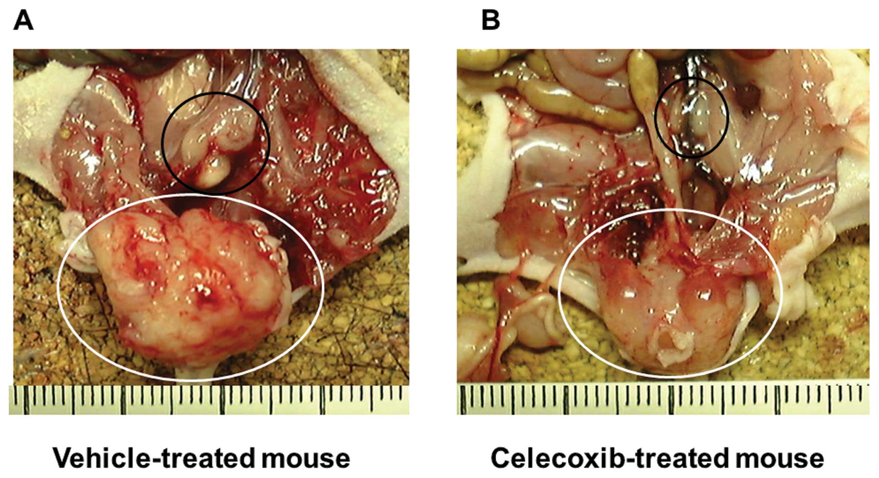
Celecoxib macroscopically inhibited xenograft growth
and abdominal lymph node metastasis (Fig. 2). The antitumor activity of
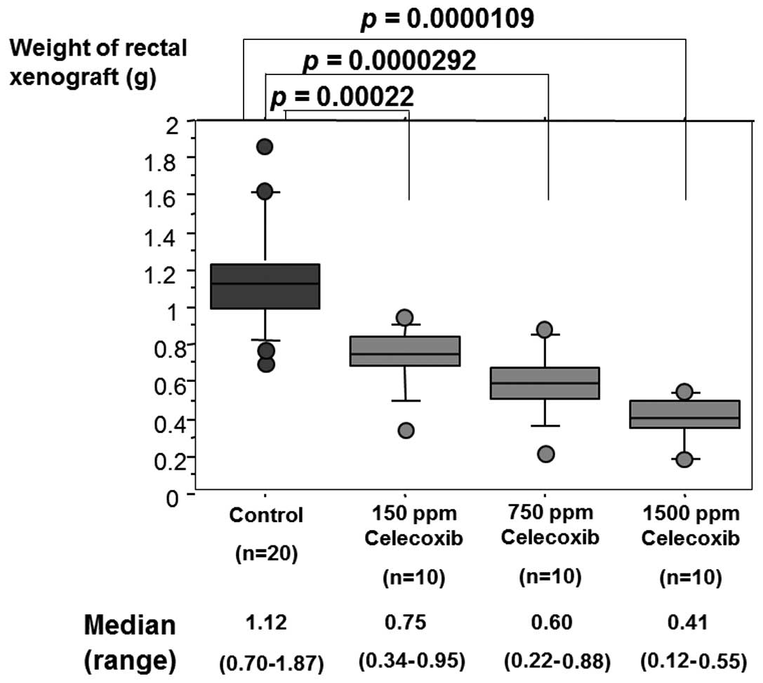
celecoxib in rectal xenograft was examined by comparing the wet
weight of each xenograft. Celecoxib suppressed growth of rectal
xenograft in a dose-dependent manner. Celecoxib (150, 750 and 1500
ppm) significantly inhibited HT-29 xenograft growth in comparison
with normal chows by 33.0% (p=0.00022), 46.4% (p=0.0000292) and
63.4% (p=0.0000109), respectively (Table II, Fig.
3).
| Table IIEffect of celecoxib on rectal
xenograft growth and lymph node and hematological metastases. |
Table II
Effect of celecoxib on rectal
xenograft growth and lymph node and hematological metastases.
| Inhibition rate
(p-value) |
|---|
|
|
|---|
| | Metastatic
organ |
|---|
| |
|
|---|
| Celecoxib
(ppm) | Rectal
xenograft | Lymph node | Lung |
|---|
| 150 | 33.0 (0.00022) | 86.7 (0.0263) | − (0.263) |
| 750 | 46.4
(0.0000292) | 90.3 (0.00638) | 53.3 (0.0107) |
| 1500 | 64.3
(0.000109) | 96.0
(0.000894) | 78.3 (0.00022) |
Anti-metastatic efficacy of
celecoxib
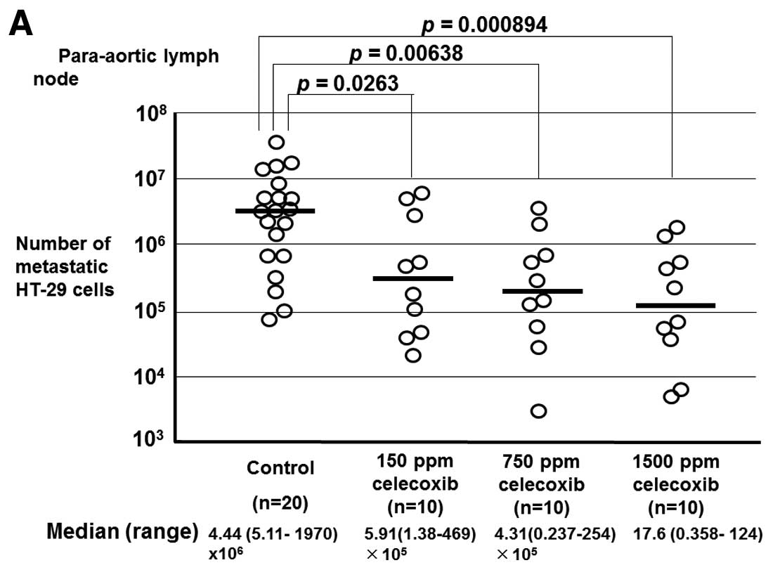
The anti-metastatic activity of celecoxib was
examined by amplifying the human β-globin-related sequence in the
lymph nodes and the lung of rectal xenograft mice. Quantification
of cancer metastasis by calculating metastasized tumor cells from
the quantitatively amplified β-globin gene revealed that 150, 750
or 1500 ppm celecoxib significantly inhibited lymph node metastasis
and 750 or 1500 ppm celecoxib significantly inhibited lung
metastasis of HT-29 cells in rectal xenograft (Fig. 4). Assessment of metastasis by the
median level of quantified metastatic cells revealed that the
inhibition ratios of lymph node metastasis by 150, 750 and 1500 ppm
celecoxib were 86.7% (p=0.0263), 90.3% (p=0.00638) and 96.0%
(p=0.000894), respectively. Inhibition ratios of lung metastasis by
750 and 1500 ppm celecoxib were 53.3% (p=0.00107) and 78.3%
(p=0.00022), respectively (Table
II). The results also demonstrated that celecoxib suppressed
lymph node and lung metastases in a dose-dependent manner.
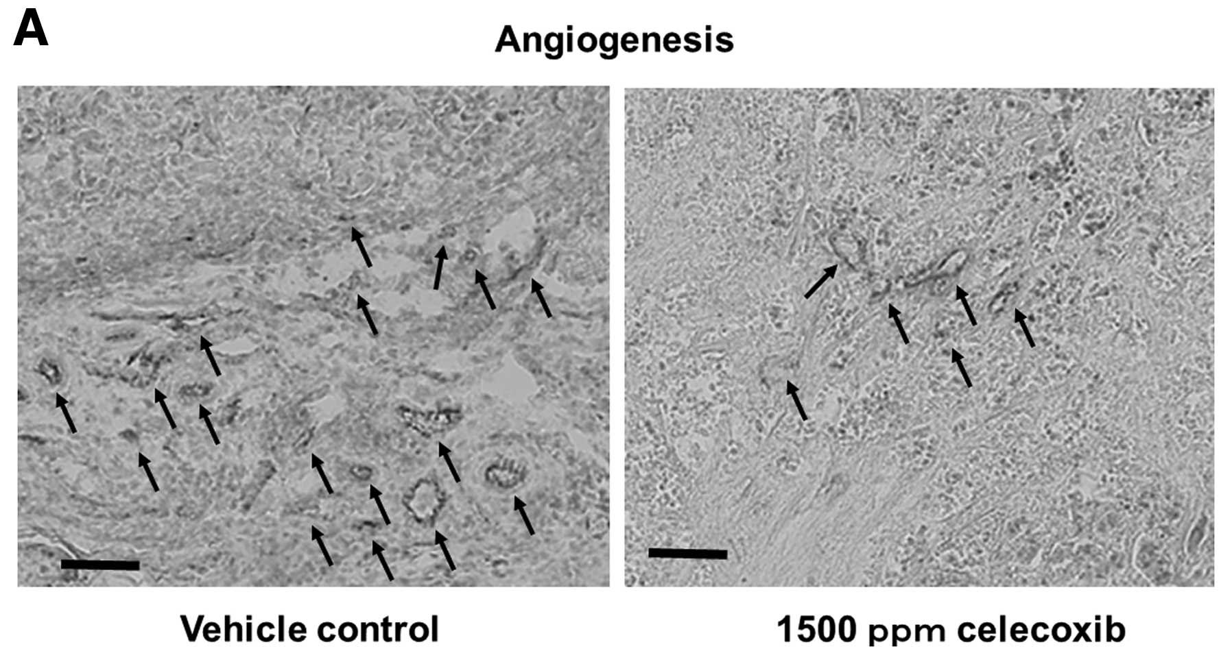
Anti-angiogenesis efficacy of
celecoxib
The anti-angiogenetic activity of celecoxib was
examined by MVD count in rectal xenografts. Celecoxib (1500 ppm)
significantly reduced MVD (Fig. 5).
Assessment of angiogenesis by comparison of the median level of MVD
in normal chow-fed mice with 1500 ppm celecoxib chows-treated mice
revealed that celecoxib inhibited angiogenesis in the rectal
xenograft by 48.2% (p=1.3×10⁻¹²) (Table III).
| Table IIIEffect of celecoxib on
PGE2 production, angiogenesis and apoptosis induction in
human colon cancer xenograft. |
Table III
Effect of celecoxib on
PGE2 production, angiogenesis and apoptosis induction in
human colon cancer xenograft.
| Median (range) | | |
|---|
|
| | |
|---|
| Vehicle (n=10) | 1500 ppm celecoxib
(n=10) | P-value | Relative
valuea |
|---|
| PGE2
production (ng/ml) | 1647
(1125–13030) | 100.3
(8.14–509) | 0.000157 | 0.684 |
| MVD counting
(/mm2) | 56 (32–80) | 27 (9–42) |
1.33×10⁻¹² | 0.482 |
| Apoptosis index
(%) | 1.64
(0.82–8.2) | 4.1
(1.64–11.5) |
3.00×10⁻¹⁴ | 2.5 |
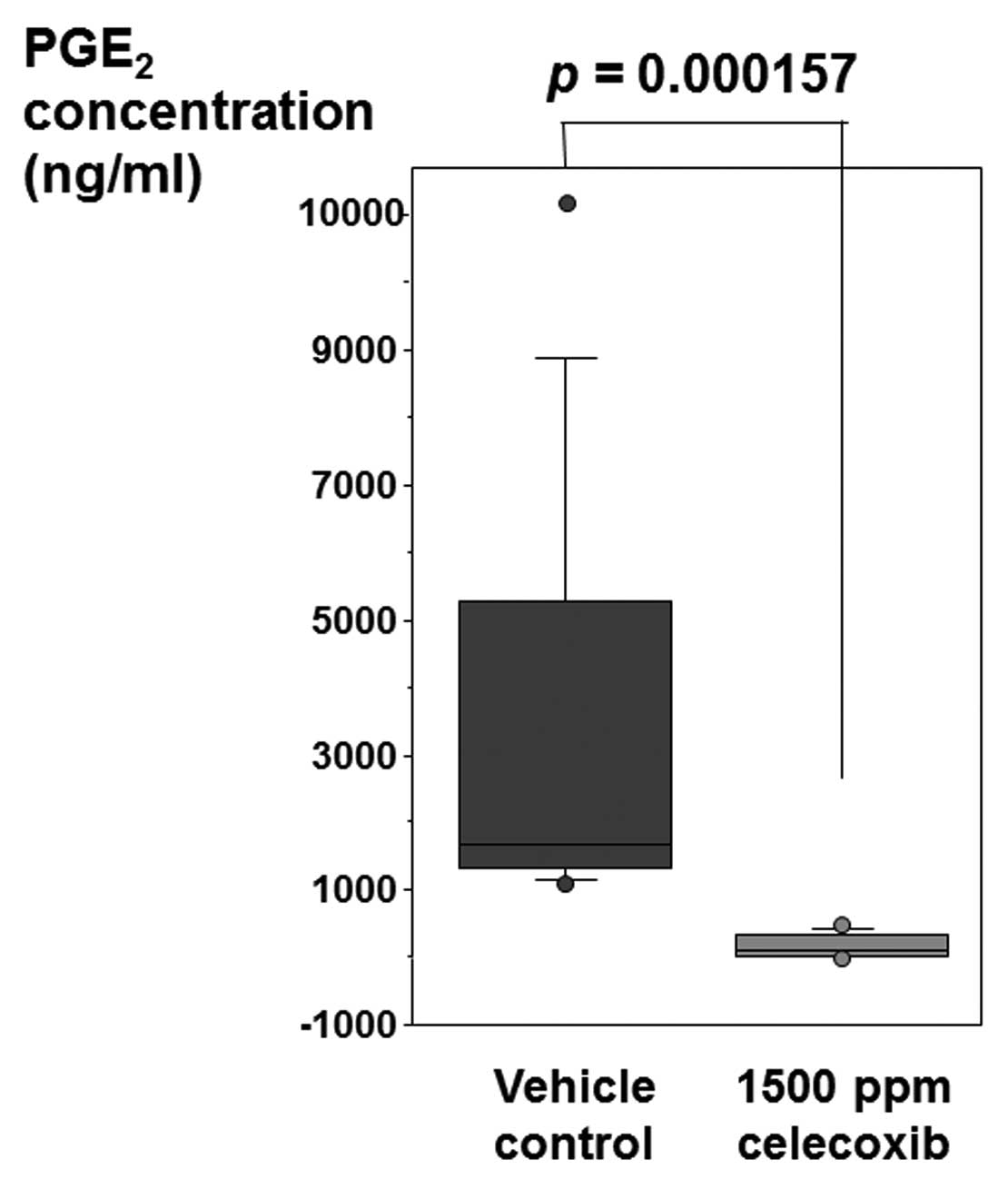
Efficacy of celecoxib treatment on
PGE2 production
Rectal xenograft samples from celecoxib-treated and
control mice were assessed for PGE2 concentration by
quantitative ELISA. The concentration of PGE2 was
reduced in the rectal xenograft of mice treated with celecoxib in
comparison with control animals (Fig.
6). Celecoxib (1500 ppm) inhibited PGE2 production
in the rectal xenograft by 68.4% (Table III). Celecoxib significantly
suppressed PGE2 production in the xenograft
(p=0.000157).
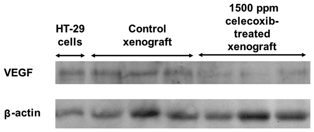
Efficacy of celecoxib treatment on VEGF
expression
We examined the effect of celecoxib treatment on the
expression of VEGF which is involved in angiogenesis in rectal
xenograft by western blotting. HT-29 human colon cancer cells and
control xenograft showed a stronger signal of VEGF protein than
1500 ppm celecoxib-treated xenograft (Fig. 7).
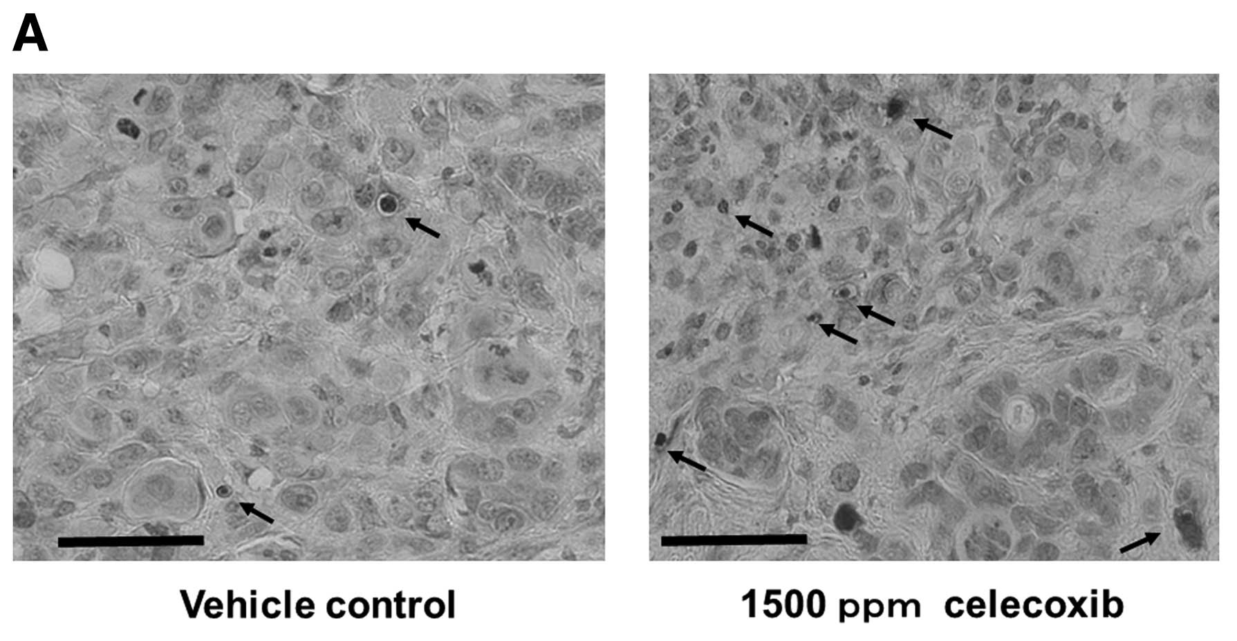
Efficacy of celecoxib treatment on
apoptosis
The TUNEL assay revealed that celecoxib
significantly induced apoptosis of HT-29 cells in rectal xenograft
(p=3.0×10⁻¹⁴) (Fig. 8).
Celecoxib induced tumor cell apoptosis consisting of a 2.5-fold
increase in tumor cell apoptotic index from 1.64% (0.820–8.20) in
vehicle to 4.1% (1.64–11.5) with 1500 ppm celecoxib treatment
(Table III).
Discussion
In the present study, the selective COX-2 inhibitor
celecoxib inhibited tumor growth and prevented spontaneous lymph
node and lung metastases of CRC in a dose-dependent manner in a
mouse rectal xenograft model. Celecoxib inhibited angiogenesis,
VEGF expression and PGE2 production, and induced tumor
cell apoptosis in the rectal xenograft.
In this study, we examined the antitumor efficacy of
celecoxib in rectal xenograft tumor. Celecoxib suppressed growth of
rectal xenograft in a dose-dependent manner. Because the location
of the xenograft in this model is intrinsically near the incident
position of rectal carcinoma, we thought that this would be a good
model to assess the efficacy of antitumor agents such as celecoxib.
Previous reports suggested that COX-2 expression plays an important
role in hematogenous metastasis of colorectal carcinomas to the
liver (16). Oral administration of
COX-2 inhibitors, rofecoxib and JTE-522, reduced the metastatic
potential of colon cancer cells injected in the spleen (3,20).
Celecoxib suppressed tumor growth and lung metastasis of a rodent
mammary cancer and human breast cancer xenograft (21–23).
Pretreatment with celecoxib inhibited liver metastasis of colon
cancer cells including HT-29 that express a high level of COX-2 in
the enforced metastasis model by cancer cell splenic injection
(17). These previous reports
proved the anti-metastatic effect of celecoxib by pre-treatment of
cancer cells with celecoxib or by the non-physiological enforced
metastasis model. We previously showed the usefulness of orthotopic
transplanted mouse rectal xenograft model of CRC presenting
spontaneous lymph node and lung metastases, identical to human
rectal cancer (18). This mouse
model presented microscopic metastatic foci in the lungs.
Therefore, a special technique is needed for detection and
quantification of metastatic tumor cells. To detect and quantify
the small amount of the metastatic cancer cells, we amplified human
β-globin related sequence by TaqMan PCR. Endo et al(24) first reported the usefulness of
amplifying a human-specific DNA fragment by PCR to detect
metastatic human cancer cells in chick embryo. DNA PCR can assess
all of the tumor-related signals in the whole tissue, regardless of
the size, density and number of tumor colonies. Quantification by
DNA amplification is more reliable than reverse transcription-PCR
assessment of enzymes originating from the human cancer cells,
because the expression levels of enzymes may vary under various
tumor conditions. This animal model is useful to assess the
anti-metastatic efficacy of novel anti-cancer agents (18). The human colon cancer cell line
HT-29 expressed COX-2 enzyme in vitro. However, in the model
with subcutaneously injected cancer cell xenograft including HT-29
cells xenograft, COX-2 expression was found only in the stromal
component without cancer cells, unlike human cancer (25,26).
The present study showed that the transplanted xenograft and the
metastatic cancer cells in the lymph nodes and lung presented
enhanced COX-2 mRNA like human CRC (Table I). Therefore, the mouse rectal
xenograft is an optimal model to assess the effect of anti-COX-2
agents. Our present study demonstrated that celecoxib inhibited
lymph node and lung metastases of rectal xenograft in a
dose-dependent manner.
In this study, we also showed that celecoxib reduced
PGE2 production and VFGF expression, had an
anti-angiogenetic effect, and induced apoptosis in CRC rectal
xenograft tumor. These results are consistent with previous reports
using various cancer xenografts. Inhibition of COX-2 by celecoxib
resulted in loss of intra-tumor PGE2 levels and reduced
tumor growth with increased apoptosis of both tumor and stromal
cells (inflammatory and neovascular) in head and neck xenograft
tumors (27). Celecoxib inhibited
VEGF expression and reduced angiogenesis and metastasis of human
pancreatic cancer that was orthotopically transplanted in nude mice
(28). Celecoxib inhibited
angiogenesis and VEGF-A expression, and induced the mitochondrial
pathway of apoptosis in a murine mammary cancer model (21). Recent studies have confirmed the
hypothesis that tumor growth is dependent on angiogenesis. Any
significant increase in tumor mass must be preceded by an increase
in the vascular supply to deliver nutrients and oxygen to the
tumor. COX-2 inhibition leads to reduced conversion of arachidonic
acids to PGs, and inhibition of PGE2 synthesis is
thought to be one of the antitumor mechanisms (29). PGE1 and PGE2
have the ability to induce angiogenesis in the rat cornea (30). COX-2 and COX-2-derived
prostaglandins may play a major role in the development of cancer
through numerous biochemical mechanisms, including stimulation of
tumor cell growth and neovascularization (13). The antitumor activity of celecoxib
may be attributable, at least in part, to a direct effect on host
stromal elements, such as the angiogenic vasculature (26). We also showed inhibition of lymph
node metastasis in the present study. Although we did not evaluate
the effect of celecoxib on lymphangiogenesis, celecoxib might block
lymphangiogenesis via downregulation of VEGF-C as reported in lung
adenocarcinoma xenograft (31).
Experiments on the kinetics of metastasis using
rectal xenograft showed that the initial lymph node and lung
metastasis occurred 2 and 4 weeks after tumor cell inoculation,
respectively (18). Celecoxib
inhibited angiogenesis in the primary tumor. Inhibition of tumor
cell dissociation from the primary tumor as well as reduced
establishment of metastases by circulating tumor cells might be
attributable to the anti-metastatic effect of celecoxib in rectal
xenograft model (32).
The ability of celecoxib to block angiogenesis and
suppress tumor growth and metastasis suggests a novel application
of this anti-inflammatory drug in the treatment of human cancer.
The tumor-suppressive action of celecoxib was not associated with
noticeable side effects on late wound healing and on the
gastrointestinal tract (33).
Celecoxib prevents morphine-induced stimulation of COX-2,
PGE2, angiogenesis, tumor growth, metastasis and
mortality without compromising analgesia (34). Therefore, prophylactic use of the
drug can be advocated in many clinical situations of cancer
treatment, such as residual tumors or contamination of surgical
fields by tumor cells. The combination of celecoxib and morphine
might also be optimal for analgesia in cancer patients with chronic
and severe pain. To prove the usefulness of celecoxib in cancer
treatment, clinical trials for various situations in cancer
treatment are warranted.
References
|
1
|
Smalley WE and DuBois RN: Colorectal
cancer and non-steroidal anti-inflammatory drugs. Adv Pharmacol.
39:1–20. 1997. View Article : Google Scholar
|
|
2
|
Tsujii M, Kawano S, Tsuji S, Sawaoka H,
Hori M and DuBois RN: Cyclooxygenase regulates angiogenesis induced
by colon cancer cells. Cell. 93:705–716. 1998. View Article : Google Scholar : PubMed/NCBI
|
|
3
|
Yao M, Kargman S, Lam EC, et al:
Inhibition of cyclooxygenase-2 by rofecoxib attenuates the growth
and metastatic potential of colorectal carcinoma in mice. Cancer
Res. 63:586–592. 2003.PubMed/NCBI
|
|
4
|
Steinbach G, Lynch PM, Phillips RK, et al:
The effect of celecoxib, a cyclooxygenase-2 inhibitor, in familial
adenomatous polyposis. N Engl J Med. 342:1946–1952. 2000.
View Article : Google Scholar : PubMed/NCBI
|
|
5
|
Tomozawa S, Nagawa H, Tsuno N, et al:
Inhibition of haematogenous metastasis of colon cancer in mice by a
selective COX-2 inhibitor, JTE-522. Br J Cancer. 81:1274–1279.
1999. View Article : Google Scholar : PubMed/NCBI
|
|
6
|
Vane JR: Inhibition of prostaglandin
synthesis as a mechanism of action for aspirin-like drugs. Nat New
Biol. 231:232–235. 1971. View Article : Google Scholar : PubMed/NCBI
|
|
7
|
Wolfe MM, Lichtenstein DR and Singh G:
Gastrointestinal toxicity of non-steroidal antiinflammatory drugs.
N Engl J Med. 340:1888–1899. 1999. View Article : Google Scholar
|
|
8
|
Masferrer JL, Seibert K, Zweifel B and
Needleman P: Endogenous glucocorticoids regulate an inducible
cyclooxygenase enzyme. Proc Natl Acad Sci USA. 89:3917–3921. 1992.
View Article : Google Scholar : PubMed/NCBI
|
|
9
|
Kujubu DA, Fletcher BS, Varnum BC, Lim RW
and Herschman HR: TIS10, a phorbol ester tumor promoter-inducible
mRNA from Swiss 3T3 cells, encodes a novel prostaglandin
synthase/cyclooxygenase homologue. J Biol Chem. 266:12866–12872.
1991.
|
|
10
|
Kargman SL, O’Neill GP, Vickers PJ, Evans
JF, Mancini JA and Jothy S: Expression of prostaglandin G/H
synthase-1 and -2 protein in human colon cancer. Cancer Res.
55:2556–2559. 1995.PubMed/NCBI
|
|
11
|
Oshima M, Dinchuk JE, Kargman SL, et al:
Suppression of intestinal polyposis in ApcΔ716
knockout mice by inhibition of cyclooxygenase 2 (COX-2). Cell.
87:803–809. 1996.
|
|
12
|
Tsujii M, Kawano S and DuBois RN:
Cyclooxygenase-2 expression in human colon cancer cells increases
metastatic potential. Proc Natl Acad Sci USA. 94:3336–3340. 1997.
View Article : Google Scholar : PubMed/NCBI
|
|
13
|
Masferrer JL, Leahy KM, Koki AT, et al:
Antiangiogenic and antitumor activities of cyclooxygenase-2
inhibitors. Cancer Res. 60:1306–1311. 2000.PubMed/NCBI
|
|
14
|
Penning TD, Talley JJ, Bertenshaw SR, et
al: Synthesis and biological evaluation of the 1,5-diarylpyrazole
class of cyclooxygenase-2 inhibitors: identification of
4-[5-(4-methyl-phenyl)-3-(trifluoromethyl)-1H-pyrazol-1-yl]benze
nesulfonamide (SC-58635, celecoxib). J Med Chem. 40:1347–1365.
1997.PubMed/NCBI
|
|
15
|
Silverstein FE, Faich G, Goldstein JL, et
al: Gastrointestinal toxicity with celecoxib vs. non-steroidal
anti-inflammatory drugs for osteoarthritis and rheumatoid
arthritis: the CLASS study: a randomized controlled trial.
Celecoxib long-term arthritis safety study. JAMA. 284:1247–1255.
2000. View Article : Google Scholar
|
|
16
|
Chen WS, Wei SJ, Liu JM, Hsiao M, Kou-Lin
J and Yang WK: Tumor invasiveness and liver metastasis of colon
cancer cells correlated with cyclooxygenase-2 (COX-2) expression
and inhibited by a COX-2-selective inhibitor, etodolac. Int J
Cancer. 91:894–899. 2001. View Article : Google Scholar : PubMed/NCBI
|
|
17
|
Kakiuchi Y, Tsuji S, Tsujii M, et al:
Cyclooxygenase-2 activity altered the cell-surface carbohydrate
antigens on colon cancer cells and enhanced liver metastasis.
Cancer Res. 62:1567–1572. 2002.PubMed/NCBI
|
|
18
|
Ninomiya I, Terada I, Yoshizumi T, et al:
Anti-metastatic effect of capecitabine on human colon cancer
xenografts in nude mouse rectum. Int J Cancer. 112:135–142. 2004.
View Article : Google Scholar : PubMed/NCBI
|
|
19
|
Blin N and Stafford DW: A general method
for isolation of high molecular weight DNA from eukaryotes. Nucleic
Acids Res. 3:2303–2308. 1976. View Article : Google Scholar : PubMed/NCBI
|
|
20
|
Nagatsuka I, Yamada N, Shimizu S, et al:
Inhibitory effect of a selective cyclooxygenase-2 inhibitor on
liver metastasis of colon cancer. Int J Cancer. 100:515–519. 2002.
View Article : Google Scholar : PubMed/NCBI
|
|
21
|
Yoshinaka R, Shibata MA, Morimoto J,
Tanigawa N and Otsuki Y: COX-2 inhibitor celecoxib suppresses tumor
growth and lung metastasis of a murine mammary cancer. Anticancer
Res. 26:4245–4254. 2006.PubMed/NCBI
|
|
22
|
Evans DM and Sloan Stakleff KD: Control of
pulmonary metastases of rat mammary cancer by inhibition of uPA and
COX-2, singly and in combination. Clin Exp Metastasis. 21:339–346.
2004. View Article : Google Scholar : PubMed/NCBI
|
|
23
|
Zhang S, Lawson KA, Simmons-Menchaca M,
Sun L, Sanders BG and Kline K: Vitamin E analog α-TEA and celecoxib
alone and together reduce human MDA-MB-435-FL-GFP breast cancer
burden and metastasis in nude mice. Breast Cancer Res Treat.
87:111–121. 2004.
|
|
24
|
Endo Y, Sasaki T, Harada F and Noguchi M:
Specific detection of metastasized human tumor cells in embryonic
chicks by the polymerase chain reaction. Jpn J Cancer Res.
81:723–726. 1990. View Article : Google Scholar
|
|
25
|
Williams CS, Tsujii M, Reese J, Dey SK and
DuBois RN: Host cyclooxygenase-2 modulates carcinoma growth. J Clin
Invest. 105:1589–1594. 2000. View
Article : Google Scholar : PubMed/NCBI
|
|
26
|
Leahy KM, Ornberg RL, Wang Y, Zweifel BS,
Koki AT and Masferrer JL: Cyclooxygenase-2 inhibition by celecoxib
reduces proliferation and induces apoptosis in angiogenic
endothelial cells in vivo. Cancer Res. 62:625–631. 2002.PubMed/NCBI
|
|
27
|
Zweifel BS, Davis TW, Ornberg RL and
Masferrer JL: Direct evidence for a role of cyclooxygenase
2-derived prostaglandin E2 in human head and neck xenograft tumors.
Cancer Res. 62:6706–6711. 2002.PubMed/NCBI
|
|
28
|
Wei D, Wang L, He Y, Xiong HQ, Abbruzzese
JL and Xie K: Celecoxib inhibits vascular endothelial growth factor
expression in and reduces angiogenesis and metastasis of human
pancreatic cancer via suppression of Sp1 transcription factor
activity. Cancer Res. 64:2030–2038. 2004. View Article : Google Scholar
|
|
29
|
Honn KV, Bockman RS and Marnett LJ:
Prostaglandins and cancer: a review of tumor initiation through
tumor metastasis. Prostaglandins. 21:833–864. 1981. View Article : Google Scholar : PubMed/NCBI
|
|
30
|
Rochels R: Pathobiochemical aspects of
corneal neovascularization. Fortschr Med. 102:101–102. 1984.(In
German).
|
|
31
|
Liu H, Yang Y, Xiao J, et al: Inhibition
of cyclooxygenase-2 suppresses lymph node metastasis via VEGF-C.
Anat Rec. 292:1577–1583. 2009. View
Article : Google Scholar : PubMed/NCBI
|
|
32
|
Backhus LM, Sievers E, Lin GY, et al:
Perioperative cyclooxygenase 2 inhibition to reduce tumor cell
adhesion and metastatic potential of circulating tumor cells in
non-small cell lung cancer. J Thorac Cardiovasc Surg. 132:297–303.
2006. View Article : Google Scholar : PubMed/NCBI
|
|
33
|
Roh JL, Sung MW, Park SW, Heo DS, Lee DW
and Kim KH: Celecoxib can prevent tumor growth and distant
metastasis in postoperative setting. Cancer Res. 64:3230–3235.
2004. View Article : Google Scholar : PubMed/NCBI
|
|
34
|
Farooqui M, Li Y, Rogers T, et al: COX-2
inhibitor celecoxib prevents chronic morphine-induced promotion of
angiogenesis, tumour growth, metastasis and mortality, without
compromising analgesia. Br J Cancer. 97:1523–1531. 2007. View Article : Google Scholar
|