Introduction
Wilms’ tumor (nephroblastoma) arises from embryonal
blastema cells, which should arrest in cell cycle and
differentiation. It is one of the most common pediatric abdominal
tumors and usually occurs in 1:10,000 children. The most cases of
Wilms’ tumors are sporadic, however, 5% of incidences arise with
three congenital syndromes WAGR, Denys-Drash, Beckwith-Wiedemann
(1). Despite of high cure rate of
patients with Wilms’ tumor (90% of cases), it is necessary to
search for new prognostic factors, which will help better
understand the biology of this tumor and to create better target
therapy. Moreover, in majority of Wilms’ cases the primary genetic
causes are still unknown.
The development of genetic and epigenetic events in
multiple loci is characteristic of Wilms’ tumors. Gene expression
studies revealed the influence of C/EBPB, H4FG up-regulation
and p21, CF542255 down-regulation on this tumor relapse
(2). High hTERT expression also
correlated with Wilms’ recurrence. However, alterations in
chromosomal regions appear to play crucial role in nephroblastoma
development. Molecular biology analysis revealed imbalances in
chromosomal regions spanning 1p, 7p, 17q, 16q, 11q13 (WT1
gene), 11p15 (WT2 gene).
The most interesting of the affected chromosomes
appears to be 16q. LOH at that region has been connected with
Wilms’ development, poor prognosis and increased risk of relapse
and death (3–5). Moreover, at 16q the common fragile
site FRA16D is located, and also at 16q23.3–24.1 tumor suppressor
gene WWOX is spanning approximately one million base
pairs.
WWOX encodes protein containing 414 amino
acids (46 kDa) that possesses two N-terminal WW domains of
interaction with protein and C-terminal short-chain dehydrogenase
domain (SDR) (6). WWOX protein
through its first WW domain interacts with many partners such as
ERBB4 (7,8), TP73 (9), transcription factor AP2γ (10,11),
YAP (7), Jun (12). The role of WWOX protein is not well
determined, however, it probably participates in hormonal
metabolism. The relative high expression of WWOX was
detected in reproductive, endocrine and exocrine organs (for
example mammary epithelium, ovaries, endometrium, prostate gland,
testes, liver, stomach, thyroid, parathyroid, adenohypofisis, and
brain cells). In vivo studies indicated that
WWOX-/- mice display abnormalities in growth, survival
and bone metabolism (13).
Simultaneously, mice with phenotype WWOX+/- more
frequently develop tumors (14). In
many types of cancers alterations in WWOX expression were
observed due to loss of heterozygosity (15–20)
and its promoter methylation (15,21,22).
Inactivation of one allele of tumor suppressor WWOX gene is
sufficient for tumorigenesis initiation (mechanism of
haploinsufficiency) (14).
The aim of our study was to evaluate the role of
WWOX gene in Wilms’ tumors. In our experiment we analyzed
correlation between expression level of WWOX gene and genes
involved with proliferation (Ki67), apoptosis (BCL2,
BAX), transduction signal (ERBB4, ERBB2, EGFR), cell
cycle (CCNE1, CCND1), cell adhesion (CDH1) and transcription
(TP73). Moreover, we evaluated loss of heterozygosity and
methylation status of WWOX promoter and examined correlation
between those events and gene expression.
Patients and methods
Patients
This study was performed on 23 Wilms’ tumor samples
obtained from children at the age between 0.25 and 8.92 years (mean
3.41 years).
The children were treated in the Department of
Paediatric Oncology and Hematology, Medical University of Lodz and
in the Department of Bone Marrow Transplantation, Pediatric
Oncology, and Hematology, Medical University, Wroclaw. The
experimental group consisted of 10 males and 13 females. According
to International Classification of renal tumors in childchood, 7
tumors were of high risk, 1 low and the remaining group were of
intermediate risk (23). Tumor
division according to stage was as follows: stage I, 5 cases; stage
II, 6 cases; stage III, 3 cases; stage IV, 3 cases; stage V, 1
case. Samples of 4 cases had no stage characterisation. According
to clinical records 7 out of 23 tumors were metastatic. Disease
recurrence was observed in the group of 5 patients and the
mortality was at 17% (4 cases). Before tumor resection, children
were subjected to chemotherapy according to SIOP scheme therapy.
This study was conducted after receiving patients’ family
consent.
Isolation of RNA, DNA and cDNA
synthesis
Resected tumor samples were stored in −80°C in
RNAlater (Ambion), RNA was isolated using TRIzol reagent
(Invitrogen, USA). cDNA synthesis was performed from 10 μg
of total RNA at the total volume of 100 μl with ImProm RT-II
reverse transcriptase (Promega). Reverse transcription was
performed under following conditions: 5-min incubation at
25°C and 60 min at 42°C, heating at 70°C for
15 min. Then, synthesized cDNA was diluted with sterile deionized
water to 150 and 2 μl of cDNA was used in PCR reaction.
Organic remains of TRIzol after RNA isolation were used for DNA
isolation, according to the manufacturer’s instructions.
Real-time RT-PCR analysis
Real-time RT-PCR was performed with LightCycler 480
II (Roche). PCR products were detected with SYBR Green I and qPCR
Core kit for SYBR Green I (Eurogentec). Reactions were performed in
duplicate. We analyzed relative expression of 11 genes (BAX,
BCL2, EGFR, Ki67, WWOX, ERBB4 (isoforms JM-a and JM-b),
CCNE1,CCND1,CDH1, TP73). The expression level of studied genes
was normalized to two reference genes (BMG2, RPS17).
WWOX mRNA level was relatively low, therefore
we performed a semi-nested RT-PCR for assessing WWOX
expression level. Following primers in the first PCR reaction were
used: 5′-TGCAACATCCTCTTCTCCAACGAGCTGCAC-3′ and
5′-TCCCTGTTGCATGGACTTGGTGAAAGGC-3′ in 50 μl volume. Next, after
200-fold PCR product dilution (171 bp), 2 μl was a template
for semi-nested PCR. The cycling protocol was as follows: 2 min at
94°C, 30 sec denaturation at 94°C, 30 sec annealing
at 63°C, 1 min extension at 72°C, repeated for 77
cycles, additional extension for 7 min at the same temperature.
Primer sequences, the PCR reaction conditions and the length of
received products are available upon request.
Roche algorithm was used for relative expression
level calculation. The Universal Human Reference RNA (composed of
10 cell lines) was used as a calibrator for each reaction. All
primers were designed to be intron spanning in order to exclude
genomic DNA amplification. Detection temperature was designated
above non-specific/primer-dimer melting temperature.
LOH analysis
Loss of heterozygosity was analyzed with high
resolution melting (HRM) of LightCycler 4800 (Roche). We used two
microsatellites markers D16S3096 and D16S504 located on chromosome
16; on intron 8 and intron 1 of WWOX gene, respectively. The
primer sequences were obtained from the Genome database. PCR
conditions were as follows: initial denaturation 95°C for 10 min;
35 cycles of repeated denaturation at 94°C for 30 sec, annealing at
56°C (for D16S3096) or 55°C (for D16S518) for 30 sec, elongation at
72°C for 60 sec.
Analysis of WWOX methylation status
We performed MethylScreen assay, which is based on a
set of methylation specific restriction digestions; subsequently
associated with real-time PCR (24). DNA of samples (2 μl) were
divided into four parts and treated with different digestions:
MSRE, two methylation-sensitive enzymes HhaI and
HpaII, which are cutting only unmethylated DNA; MDRE, one
methylation-dependent restriction enzyme McrBC, which is cutting
only methylated DNA; MOCK, positive control without enzymes; DD,
negative control of reaction with both MSRE and MDRE enzymes.
The digestions reactions contained 1X NEB buffer 2
(10 mM Tris-HCl, 55 mM NaCl, 10 mM MgCl2, 1 mM DTT), 1
μg/ml bovine serum albumin (BSA), 2 mM guanosine-50-triphosphate
(GTP), appropriate enzymes. Next, this mixture was incubated for 4
h at 37°C and then 20 min at 65°C to restrain enzyme activity.
Afterwards, samples were analyzed with real-time PCR using the
RotorGene 3000™ system. We analyzed two fragments containing the
promoter and first exon of the WWOX gene. The PCR for the
first region of WWOX gene (−508 to −174 bp) was performed
using the following primers: the forward primer sequence was
5′-ACAGAAGCCCAGGACAACAGCATGG-3′, and the reverse primer sequence
was 5′-ACCACGAAGCTGAA ATCCAGTCTCCG-3′. For the analysis of the
second region (from −171 to +239 bp) covering the 3′ end of the
promoter and part of exon 1 following primers were used: forward
primer was 5′-AGACTGGATTTCAGCTTCGTGGTCG-3′, and the reverse primer
sequence was 5′-AAGCTCCTTAACAGTTACT TTCACTTTGCAC-3′.
For the first analyzed promoter fragment of
WWOX gene, the PCR mix included 2.5 μl of SYBR Green I, qPCR
Core kit for SYBR Green I reagents (Eurogentec), 10 nM of each
primer, 4 μl of digested DNA template. Real-time PCR was conducted
at the following conditions: 95°C for 5 min, followed by 50 cycles
at 94°C for 30 sec, 55°C for 30 sec, and 72°C for 90 sec, and 77°C
for 15 sec (additional temperature for reading only specific
amplification product size 384 bp). The PCR mix for the second
WWOX promoter fragment consisted of 4 μl of digested
DNA template, 10 nM of each primer, 2.5 μl of SYBR Green I,
qPCR Core kit for SYBR Green I reagents, 70 mM of betaine.
Real-time PCR was performed in the following conditions: 1 cycle of
95°C for 5 min, followed by 50 cycles at 94°C for 30 sec, 50°C for
30 sec, and 72°C for 90 sec, and 83°C for 15 sec (additional
temperature for reading only specific amplification product size
413 bp). All reactions were performed in duplicate.
To analyze methylation status of WWOX gene,
we made standard curve for each patient by setting the MOCK
positive control as 100 standard and DD negative control as 0.001
standard. If the differences between Mock and MSRE/MDRE digestions
Ct was >1 this DNA samples, it was defined as methylated.
Statistical analysis
For the analysis of correlation between expression
of WWOX and other genes we used non-parametric Spearman rank
correlation test. Estimation of differences between WWOX
expression in relation to LOH, methylation status and clinical
factors were performed with Mann-Whitney t-test. All results were
assumed as statistically significant at the confidence level
>95% (p<0.05).
Real-time RT-PCR validation
In order to validate real-time RT-PCR we performed
western blot analysis. We have chosen representative tumor samples
with low and high relative WWOX gene expression. The results
are presented on Fig. 1.
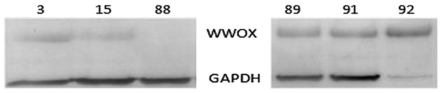 | Figure 1Western blot analysis demonstrating
WWOX protein expression in tumor samples with low and high relative
gene expression. Samples 3, 15 and 88 represent tumors with low
WWOX relative gene expression with values 1,480, 0,214 and
0,957, respectively. Samples 89, 91 and 92 represent tumors with
high WWOX expression and relative expression values: 58,304,
36,491 and 49,213 respectively. |
Protein isolation and immunoblotting
assay
Total protein was extracted from frozen tissues
using RIPA protein extraction buffer consisting of 50 mM Tris-HCl
(pH 8.0), 150 mM NaCl, 0.5% Na Doc, 0.1% NP-40, 0.1% SDS, 2 mM
EDTA, supplemented with protease, phosphatase inhibitor coctail and
1 mM PMSF (Sigma-Aldrich, Germany). The Bradford method (Bio-Rad
Laboratories) for determination of protein concentration was used,
according to the manufacturer’s instructions. The amount of 30
μg of sample protein were run on 10% SDS-PAGE gel
electrophoresis and transferred to PVDF membrane (Sigma-Aldrich).
Membranes were blocked in 5% non-fat milk in TBST (20 mM Tris-HCl,
500 mM NaCl, Tween-20, pH 7.5) for 1 h at room temperature. Then,
the membranes were incubated for 19 h at 4°C in primary
antibodies (Santa Cruz Biotechnology Inc., USA) in 1:200
concentration. Then, 3 times wash with TBST was performed and one
hour incubation in secondary antibodies conjugated with alkaline
phosphatase (Sigma-Aldrich). Post-incubation washes were repeated
in the same conditions as previously. Novex® AP
Chromogenic Substrate (Invitrogen) was used for the induction of
colour reaction. Band visualization was performed on membranes.
Glyceraldehydes-3-phosphate dehyrogenase (GAPDH) (Santa Cruz
Biotechnology) was used as the reference protein.
Results
LOH analysis
In all 23 samples we were able to examine loss of
heterozygosity for locus D16S3096 and in 30.4% cases LOH was
observed. The level of noticed hemizygosity in intron 1 of
WWOX gene (marker D16S518) was lower then homozygosity
observed in the population (according to Genome databases).
Detailed information on LOH analysis is presented in Table I. Analysis of correlation between
LOH for microsatelite locus D16S3096 and expression level of
WWOX gene did not reveal statistically significant reduction
of this gene transcription.
 | Table ILOH analysis in Wilms’ tumor
samples. |
Table I
LOH analysis in Wilms’ tumor
samples.
| D16S3096 (%) | D16S518 (%) |
|---|
| Observed
hemizygosity in Wilms’ tumors | 30.4 | 6.7 |
| Population
homozygosity | 26 | 17 |
| Predicted loss of
heterozygosity | 4.4 | 0 |
Analysis of WWOX methylation status
The methylation analysis of WWOX gene in
Wilms’ tumor samples revealed that the first region from −508 to
−174 bp was methylated in 13.64% cases and the second region in
8.7% cases. We noted statistically significant reduction of
WWOX gene expression level in samples with methylated region
covering the 3′ end of the promoter and part of exon 1. The mean
was 15.94 for unmethylated and 4.01 for methylated samples
(p=0.036). Also in the second promoter region of WWOX gene
its expression decreased after methylation (means were 14.92 vs
0.83; p=0.0037, for unmethylated vs. methylated samples,
respectively).
Expression
Our experiment revealed correlations between
WWOX expression level and expression levels of examined
genes. Spearman rank correlation analysis showed that WWOX
expression level positively correlated with anti-apoptotic
BCL2 gene (Rs=0.8676; p<0.0001) and
BCL2/BAX expression ratio (Rs=0.8368;
p<0.0001). Correlation between WWOX and proapoptotic
BAX was not statistically significant. Positive correlation
was also found between examined suppressor gene and ERBB4
isoform JM-a (Rs=0.5720, p=0.0054), TP73
(Rs= 0.9229; p<0.0001), EGFR
(Rs=0.4812; p=0.0201). On the other hand, WWOX
gene expression was inversely associated with both cyclins
CCND1, CCNE1 (Rs=−0.6594, p=0.0006,
Rs=−0.6594, p=0.0006, respectively). More detailed
information on received correlation is shown in Table II. There were no statistically
significant correlations between WWOX gene expression and
clinical factors such as age, stage or risk.
| Table IICorrelation between expression of the
WWOX gene and other analyzed genes. |
Table II
Correlation between expression of the
WWOX gene and other analyzed genes.
| Gene | Correlation
coefficient Rs | p-value |
|---|
| BAX | −0.2219 | 0.3089(NS) |
|
BCL2 | 0.8676 | <0.0001 |
|
BCL2/BAX | 0.8368 | <0.0001 |
|
CCND1 | −0.6594 | 0.0006 |
|
CCNE1 | −0.6594 | 0.0006 |
|
EGFR | 0.4812 | 0.0201 |
| ERBB4
JM-a | 0.5720 | 0.0054 |
| ERBB4
JM-b | 0.3521 | 0.0994 (NS) |
| TP73 | 0.9229 | <0.0001 |
| CDH1 | −0.1127 | 0.6087 (NS) |
| Ki67 | −0.1887 | 0.3884 (NS) |
Discussion
Reduced expression level of WWOX gene was
demonstrated in many studies conducted on different types of tumors
(breast, gastric, esophageal, pancreatic, lung cancer). One of the
explanatory mechanism of this decrease is loss of heterozygosity
(15–20). This is also a frequent event in
Wilms’ tumors comprising examined region of chromosome. The LOH on
chromosome 16 in this embryonal tumor was associated with higher
risk of recurrence and death (4,5,25). In
our experiment we observed loss of heterozygosity only at intron 8
of WWOX in 30.4% samples. In the second analyzed
microsatellite marker located at intron 1, the incidence of
hemizygosity was at the general population level. However, LOH did
not influence the WWOX expression level.
WWOX promoter methylation in nephroblastoma
specimens was also examined. According to previous findings in
Wilms’ tumors, epigenetic mechanism appears frequently and
regulates expression of IGF2, H19 and CDKNIC
(p57KIP−2) genes (26).
As for WWOX, this event has also been observed and
methylation of its promoter correlated with reduction of its
expression in breast, lung, bladder, gastric and hematopoietic
cancers (15,20,21).
On this basis we analyzed Wilms’ samples in terms of WWOX
promoter methylation and we observed this epigenetic change of −508
to −174 bp region in 13.64% cases and 8.7% for −171 bp to +239 bp
region.
Contrary to LOH, methylation of region covering 3′
end of the promoter and part of exon 1 was associated with
statistically significant reduction of WWOX gene expression
level. Taking into consideration these results, we may assume that
LOH in Wilms’ tumors does not play as vital role as methylation
status in lowering expression of this tumor suppressor gene.
In this study we also focused on examining the
WWOX expression correlation with other genes with definite
function in proliferation, apoptosis, signal transduction, cell
cycle, cell adhesion and transcription. First of all, the conducted
analysis on nephroblastoma samples revealed negative correlation
between WWOX mRNA and the expression level of the cyclins
CCNE1 and CCND1.
Cyclin E is a marker of cell cycle progression from
G1 into S phase and controls RB protein phosphorylation by
cyclin-dependent kinase 2 (Cdk2). Loeb et al identified in
the promoter of CCNE1 sites binding of WT1 gene (Wilms tumor
supressor gene) (27). Moreover,
high protein level of cyclin E was associated with aggressiveness
in this type of tumor (28).
Whereas, overexpression of cyclin D1 was observed in 28% of cases,
but authors indicated a role of cyclin D2, which was overexpressed
in 86% of Wilms’ tumors but did not correlate with
clinicopathological factors and recurrence (29). Moreover, we revealed a positive
correlation between expression of WWOX and TP73,
which is a member of p53 family and WWOX partner at the same
time.
In Wilms’ tumors an interaction between TP73 and WT1
was demonstrated and it was suggested as a possible influence on
the biological function of TP73 gene (30). The balance between apoptosis and
proliferation is essential for tumor cells growth and response to
drugs. In this study a positive correlation between WWOX
expression level and anti-apoptotic BCL2 and ratio
BCL2/BAX was found. The role of apoptosis in progression of
Wilms’ tumors is ambiguous. Ghanem et al, demonstrated
significant relationship between blastemal BCL2 expression,
BCL2/BAX ratio and clinical progression, with no relevance to
stage. Patients with low BCL2/BAX ratio
(BCL2-/BAX+) appeared to have better
prognosis in comparison with three groups of patients with ratio
BCL2-/BAX-, BCL2+/BAX-,
BCL2+/BAX+ (31). Other authors noted significant
difference in the apoptotic index between stages I and IV and there
was lower apoptotic process in patients with unfavorable histology.
However, they did not observe correlation between apoptotic index
and BCL2 expression suggesting the role of other regulator genes in
apoptosis (32).
Recently conducted studies revealed interaction of
WWOX protein by the first WW domain with two PPxY motifs of JM-a
isoform ERBB4 protein. WWOX protein prevents cleaving membranous
ERBB4 through tumor necrosis factor α and γ-secretase enzymes to
intracellular domain (ICD) fragment which in nucleus regulates gene
transcription (7). In Wilms’ tumors
we have also observed a positive relation of ERBB4 JM-a
expression level with WWOX and such coexpression with
membranous ERBB4 was found in breast cancer patients with
favourable prognosis (8).
In Wilms’ tumors overexpression of HER family
members i.e., EGFR (HER1) and HER2 was reported with indication on
more frequent EGFR expression in intermediate- and low-risk tumors
(33). Another study did not reveal
any association between EGFR overexpression and gene amplification,
stage, histopathology nor prognosis (34). In the present study we obtained
positive correlation for WWOX and EGFR mRNA
level.
In conclusion, the presented study conducted on
Wilms’ tumors samples revealed that expression of WWOX is
positively associated with process of apoptosis, signal
transduction through the ErbB4 pathway and EGFR and negatively with
regulation of cell cycle (by cyclin E1 and D1). Moreover, our
analysis demonstrated statistically significant correlation between
WWOX gene promoter methylation and its expression.
Our data indicate that epigenetic mechanism can
regulate WWOX gene transcription in Wilms’ tumors.
Furthermore, all obtained results are similar to those revealed for
this gene in other cancer types and are of importance in Wilms’
tumor and seems to influence the process of its development.
However, further studies ought to be conducted.
Acknowledgements
The authors wish to thank Ewa Latkowska for her
excellent technical support. This study was supported by the Polish
Ministry of Science and Higher Education 2670/B/P01/2008/34 and
0757/B/P01/2009/37. The funders had no role in study design, data
collection and analysis, decision to publish, or preparation of the
manuscript.
Abbreviations:
|
WWOX
|
WW domain containing
oxidoreductase
|
|
LOH
|
loss of heterozygosity
|
|
MSRE
|
methylation-sensitive restriction
enzyme
|
|
MDRE
|
methylation-dependent restriction
enzyme
|
|
DD
|
double-digest
|
|
ICD
|
intracellular domain
|
References
|
1
|
Green DM, D’Angio GJ, Beckwith JB, Breslow
NE, Grundy PE, Ritchey ML and Thomas PR: Wilms tumor. CA Cancer J
Clin. 461:46–63. 1996. View Article : Google Scholar
|
|
2
|
Li W, Kessler P, Yeger H, Alami J, Reeve
AE, Heathcott R, Skeen J and Williams BR: A gene expression
signature for relapse of primary wilms tumors. Cancer Res.
65:2592–2601. 2005. View Article : Google Scholar : PubMed/NCBI
|
|
3
|
Grundy PE, Telzerow PE, Breslow N,
Moksness J, Huff V and Paterson MC: Loss of heterozygosity for
chromosomes 16q and 1p in wilms’ tumors predicts an adverse
outcome. Cancer Res. 54:2331–2333. 1994.
|
|
4
|
Grundy PE, Breslow NE, Li S, Perlman E,
Beckwith JB, Ritchey ML, Shamberger RC, et al: Loss of
heterozygosity for chromosomes 1p and 16q is an adverse prognostic
factor in favorable-histology wilms tumor: a report from the
national wilms tumor study group. J Clin Oncol. 23:7312–7321. 2005.
View Article : Google Scholar
|
|
5
|
Messahel B, Williams R, Ridolfi A, A’hern
R, Warren W, Tinworth L, Hobson R, et al: Allele loss at 16q
defines poorer prognosis wilms tumour irrespective of treatment
approach in the UKW1–3 clinical trials: a children’s cancer and
leukaemia group (CCLG) study. Eur J Cancer. 45:819–826.
2009.PubMed/NCBI
|
|
6
|
Bednarek AK, Laflin KJ, Daniel RL, Liao Q,
Hawkins KA and Aldaz CM: WWOX, a novel WW domain-containing protein
mapping to human chromosome 16q23.3–24.1, a region frequently
affected in breast cancer. Cancer Res. 60:2140–2145.
2000.PubMed/NCBI
|
|
7
|
Aqeilan RI, Donati V, Palamarchuk A,
Trapasso F, Kaou M, Pekarsky Y, Sudol M and Croce CM: WW
domain-containing proteins, WWOX and YAP, compete for interaction
with ErbB-4 and modulate its transcriptional function. Cancer Res.
65:6764–6772. 2005. View Article : Google Scholar : PubMed/NCBI
|
|
8
|
Aqeilan RI, Donati V, Gaudio E, Nicoloso
MS, Sundvall M, Korhonen A, Lundin J, et al: Association of Wwox
with ErbB4 in breast cancer. Cancer Res. 67:9330–9336. 2007.
View Article : Google Scholar : PubMed/NCBI
|
|
9
|
Aqeilan RI, Pekarsky Y, Herrero JJ,
Palamarchuk A, Letofsky J, Druck T, Trapasso F, et al: Functional
association between Wwox tumor suppressor protein and P73, a P53
homolog. Proc Natl Acad Sci USA. 101:4401–4406. 2004. View Article : Google Scholar : PubMed/NCBI
|
|
10
|
Aqeilan RI, Palamarchuk A, Weigel RJ,
Herrero JJ, Pekarsky Y and Croce CM: Physical and functional
interactions between the Wwox tumor suppressor protein and the
AP-2gamma transcription factor. Cancer Res. 64:8256–8261. 2004.
View Article : Google Scholar : PubMed/NCBI
|
|
11
|
Guler G, Huebner K, Himmetoglu C, Jimenez
RE, Costinean S, Volinia S, Pilarski RT, et al: Fragile histidine
triadprotein, WW domain-containing oxidoreductase protein Wwox, and
activator protein 2gamma expression levels correlate with basal
phenotype in breast cancer. Cancer. 115:899–908. 2009. View Article : Google Scholar
|
|
12
|
Gaudio E, Palamarchuk A, Palumbo T,
Trapasso F, Pekarsky Y, Croce CM and Aqeilan RI: Physical
association with WWOX suppresses C-Jun transcriptional activity.
Cancer Res. 66:11585–11589. 2006. View Article : Google Scholar : PubMed/NCBI
|
|
13
|
Aqeilan RI, Hassan MQ, de Bruin A, Hagan
JP, Volinia S, Palumbo T, Hussain S, et al: The WWOX tumor
suppressor is essential for postnatal survival and normal bone
metabolism. J Biol Chem. 283:21629–21639. 2008. View Article : Google Scholar : PubMed/NCBI
|
|
14
|
Aqeilan RI, Trapasso F, Hussain S,
Costinean S, Marshall D, Pekarsky Y, Hagan JP, et al: Targeted
deletion of Wwox reveals a tumor suppressor function. Proc Natl
Acad Sci USA. 104:3949–3954. 2007. View Article : Google Scholar : PubMed/NCBI
|
|
15
|
Maeda N, Semba S, Nakayama S, Yanagihara K
and Yokozaki H: Loss of WW domain-containing oxidoreductase
expression in the progression and development of gastric carcinoma:
clinical and histopathologic correlations. Virchows Arch.
457:423–432. 2010. View Article : Google Scholar
|
|
16
|
Aqeilan RI, Kuroki T, Pekarsky Y, Albagha
O, Trapass F, Baffa R, Huebner K, et al: Loss of WWOX expression in
gastric carcinoma. Clin Cancer Res. 10:3053–3058. 2004. View Article : Google Scholar : PubMed/NCBI
|
|
17
|
Kuroki T, Trapasso F, Shiraishi T, Alder
H, Mimori K, Mori M and Croce CM: Genetic alterations of the tumor
suppressor gene WWOX in esophageal squamous cell carcinoma. Cancer
Res. 62:2258–2260. 2002.PubMed/NCBI
|
|
18
|
Kuroki T, Yendamuri S, Trapasso F,
Matsuyama A, Aqeilan RI, Alder H, Rattan S, et al: The tumor
suppressor gene WWOX at FRA16D is involved in pancreatic
carcinogenesis. Clin Cancer Res. 10:2459–2465. 2004. View Article : Google Scholar : PubMed/NCBI
|
|
19
|
Yendamuri S, Kuroki T, Trapasso F, Henry
AC, Dumon KR, Huebner K, Williams NN, et al: WW domain containing
oxidoreductase gene expression is altered in non-small cell lung
cancer. Cancer Res. 63:878–881. 2003.PubMed/NCBI
|
|
20
|
Chen T, Sahin A and Aldaz CM: Deletion map
of chromosome 16q inductalcarcinoma in situ of the breast: refining
a putative tumor suppressor gene region. Cancer Res. 56:5605–5609.
1996.PubMed/NCBI
|
|
21
|
Iliopoulos D, Guler G, Han SY, Johnston D,
Druck T, McCorkell KA, Palazzo J, et al: Fragile genes as
biomarkers: epigenetic control of WWOX and FHIT in lung, breast and
bladder cancer. Oncogene. 24:1625–1633. 2005. View Article : Google Scholar : PubMed/NCBI
|
|
22
|
Ishii H, Vecchione A, Furukawa Y,
Sutheesophon K, Han SY, Druck T, Kuroki T, et al: Expression of
FRA16D/WWOX and FRA3B/FHIT genes in hematopoietic malignancies. Mol
Cancer Res. 1:940–947. 2003.PubMed/NCBI
|
|
23
|
Vujanic GM, Sandstedt B, Harms D, Kelsey
A, Leuschner I and de Kraker J: Revised international society of
paediatric oncology (SIOP) working classification of renal tumors
of childhood. Med Pediatr Oncol. 38:79–82. 2002. View Article : Google Scholar : PubMed/NCBI
|
|
24
|
Holemon H, Korshunova Y, Ordway JM, Bedell
JA, Citek RW, Lakey N, Leon J, et al: MethylScreen: DNA methylation
density monitoringusing quantitative PCR. Biotechniques.
43:683–693. 2007. View Article : Google Scholar : PubMed/NCBI
|
|
25
|
Skotnicka-Klonowicz G, Rieske P,
Bartkowiak J, Szymik-Kantorowicz S, Daszkiewicz P and
Debiec-Rychter M: 16q heterozygosity loss in Wilms’ tumour in
children and its clinical importance. Eur J Surg Oncol. 26:61–66.
2000.
|
|
26
|
Feinberg AP: Imprinting of a genomic
domain of 11p15 and loss of imprinting in cancer: an introduction.
Cancer Res. 59:S1743–S1746. 1999.PubMed/NCBI
|
|
27
|
Loeb DM, Korz D, Katsnelson M, Burwell EA,
Friedman AD and Sukumar S: Cyclin E is a target of WT1
transcriptional repression. J Biol Chem. 277:19627–19632. 2002.
View Article : Google Scholar : PubMed/NCBI
|
|
28
|
Berrebi D, Leclerc J, Schleiermacher G,
Zaccaria I, Boccon-Gibod L, Fabre M, Jaubert F, et al: High cyclin
E staining index in blastemal, stromal or epithelial cells is
correlated with tumor aggressiveness in patients with
nephroblastoma. PLoS One. 3:e22162008. View Article : Google Scholar : PubMed/NCBI
|
|
29
|
Faussillon M, Monnier L, Junien C and
Jeanpierre C: Frequent overexpression of cyclin D2/cyclin-dependent
kinase 4 in Wilms’ tumor. Cancer Lett. 221:67–75. 2005.PubMed/NCBI
|
|
30
|
Scharnhorst V, Dekker P, van der Eb AJ and
Jochemsen AG: Physical interaction between wilms tumor 1 and P73
proteins modulates their functions. J Biol Chem. 275:10202–10211.
2000. View Article : Google Scholar : PubMed/NCBI
|
|
31
|
Ghanem MA, van der Kwast TH, Den Hollander
JC, Sudaryo MK, van den Heuvel MM, Noordzij MA, Nijman RJ, et al:
The prognostic significance of apoptosis-associated proteins BCL-2,
BAX and BCL-X in clinical nephroblastoma. Br J Cancer.
85:1557–1563. 2001. View Article : Google Scholar : PubMed/NCBI
|
|
32
|
Tanaka K, Granata C, Wang Y, O’Briain DS
and Puri P: Apoptosis and Bcl-2 oncogene expression in wilms’
tumor. Pediatr Surg Int. 15:243–247. 1999.
|
|
33
|
Wetli SC, Leuschner I, Harms D, Rufle A,
Foerster A, Bihl M, Graf N, et al: KIT, PDGFRalpha and EGFR
analysis in nephroblastoma. Virchows Arch. 452:637–650. 2008.
View Article : Google Scholar : PubMed/NCBI
|
|
34
|
Vasei M, Modjtahedi H, Ale-Booyeh O,
Mosallaei A, Kajbafzadeh AM, Shahriari M, Ghaderi AA, et al:
Amplification and expression of EGFR and ERBB2 in Wilms’ tumor.
Cancer Genet Cytogenet. 194:88–95. 2009.
|


