Introduction
At present, a major issue related to treating
gastrointestinal cancers is dealing with metastasis, as this may
determine patient prognosis. Among the different types of cancer,
colon cancer is characterized by susceptibility to hematogenous
metastasis, particularly hepatic metastasis. Successful management
of hematogenous metastasis is considered vital for an improvement
in the survival rate of these patients (1,2).
Colon cancer metastasis occurs possibly through the
following mechanism: after detaching from the primary lesion, the
cancer cells invade capillary vessels, pass through the portal
system and greater circulatory system, spread across the whole
body, adhere to vascular endothelial cells of a target organ,
transmigrate and invade outside the blood vessels and grow in the
metastatic lesion. Cancer cells have been clinically shown to
undergo such invasive processes, and those cancer cells that have
high invasive ability are considered likely to cause metastasis and
poor prognosis (3). The endocrine
gland-derived vascular endothelial growth factor (EG-VEGF) gene
that was investigated in this study was identified by LeCouter
et al in 2001 as an angiogenic growth factor for organs of
the endocrine system. EG-VEGF consists of 305 amino acids with a
molecular weight of 8.6 kDa. A mature EG-VEGF protein has 86 amino
acids containing 10 cysteines, with 80% homology to a non-toxic
protein purified from the venom of the black mamba snake, whereas
homology to the vascular endothelial growth factor (VEGF) is weak
(4). Recently, prokineticin
receptor (PK-R) 1 and 2 were identified as EG-VEGF receptors and
these receptors were shown to mediate physiological changes
(5–7).
This study demonstrates our findings of a new
mechanism by which signal transmission to matrix metalloproteinase
(MMP) via PK-R2 occurs after stimulation with the EG-VEGF protein,
thereby accelerating cell invasion in colon cancer cells.
Materials and methods
Cell culture
The human colon cancer cell lines, DLD-1 and HCT116,
were cultured at 37°C in 5% CO2 in RPMI-1640 medium
containing 10% fetal bovine serum (8).
Antibody
The following antibodies were used: anti-human PK-R1
and PK-R2 Ab (Novus Biochemicals, Littleton, CO, USA).
Immunohistostaining
The cells were plated in 96-well plates at
1×10⁴ and incubated for 12 h. The cells were analyzed
for protein expression using the streptavidin-biotin peroxidase
method (9,10).
Chemicals
EG-VEGF proteins were dissolved in distilled water
according to the manufacturer’s instructions (Shenandoah
Biotechnology, Inc., Warwick, PA, USA).
Antibody treatment
Cancer cells were plated in 10-cm dishes at
5×10⁵ and incubated for 12 h. The cells were treated
with antibody at 10 μg/ml for 3 h.
Tumor cell invasion assay
Transwells (Biocoat Matrigel 6-well invasion
chamber) with filters coated with an extracellular matrix
(Matrigel) on the upper surface were purchased from BD Biosciences
(San Jose, CA, USA). A complete medium was added to the bottom
chamber to induce the invasion of the cells through the Matrigel. A
serum-free medium with or without EG-VEGF protein was added to the
cells (2×10⁵), and seeded to the top chamber. The
Matrigel invasion chamber was incubated for 48 h at 37°C with 5%
CO2. Non-invading cells were removed from the top of the
Matrigel with a cotton-tipped swab. The number of invasive cells
was determined by counting the stained cells. Cell numbers were
counted with a hemocytometer (11).
RNA extraction and RT-PCR analysis
Total RNA was extracted from cells using Isogen
(Wako, Osaka, Japan). The single-strand cDNA prepared from 3 μg of
total RNA using PrimeScript RT reagent kit (Takara, Japan) was used
as the template for the polymerase chain reaction (PCR) (12). The primers for PCR to amplify MMP-2
gene-coding regions were as follows: 5′ primer MMP-2-AX,
5′-ACCCATTTACACCTACACCAAG-3′; 3′ primer MMP-2-BX,
5′-GTATACCGCATCAATCTTTTCCG. The primers for PCR to amplify MMP-7
gene-coding regions were as follows: 5′ primer MMP-7-AX,
5′-TCTTTGGCCTACCTATAACTGG-3′; 3′ primer MMP-7-BX,
5′-CTAGACTGCTACCATCCGTCA-3′. The primers for PCR to amplify MMP-9
gene-coding regions were as follows: 5′ primer MMP-9-AX,
5′-TGGGCTACGTGACCTATGACAT-3′; 3′ primer MMP-9-BX,
5′-GCCCAGCCCACCTCCACTCCTC-3′. GAPDH amplification was used as an
internal PCR control with 5′-GGGGAGCCAAAAGGGTCATCATCT-3′ as the
sense primer and 5′-GACGCCTGCTTCACCACCTTCTTG-3′ as the antisense
primer. Thirty cycles of denaturation (94°C, 1 min), annealing
(50°C, 1.5 min) and extension (72°C, 2 min) were carried out in a
thermal cycler (PTC-100, Programmable Thermal Controller; MJ
Research Inc., MA, USA). PCR products (10 μl) were resolved by
electrophoresis in 1.2% agarose gel. The sequencing was performed
on PCR products showing the bands in RT-PCR analysis. Ethidium
bromide staining of the gels identified a band of the MMP-2, -7 and
-9 mRNA. To ensure reproducibility, all PCR amplifications were
performed in triplicate (8,12).
Statistical considerations
Characteristics of the two treatment arms were
compared using the Chi-square test. Values of P<0.05 were
considered to indicate statistically significant results.
Results
Expression levels of PK-R1 and PK-R2 in
colon cancer cells
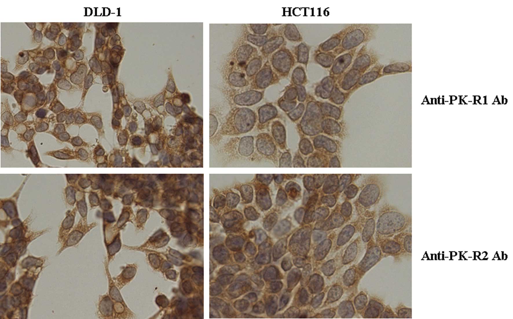
Fig. 1 shows the
stained image of colon cancer cells HCT116 and DLD-1 incubated with
anti-PK-R1 and anti-PK-R2 antibodies. The expression levels of
PK-R1 and PK-R2 in these cancer cell lines were confirmed.
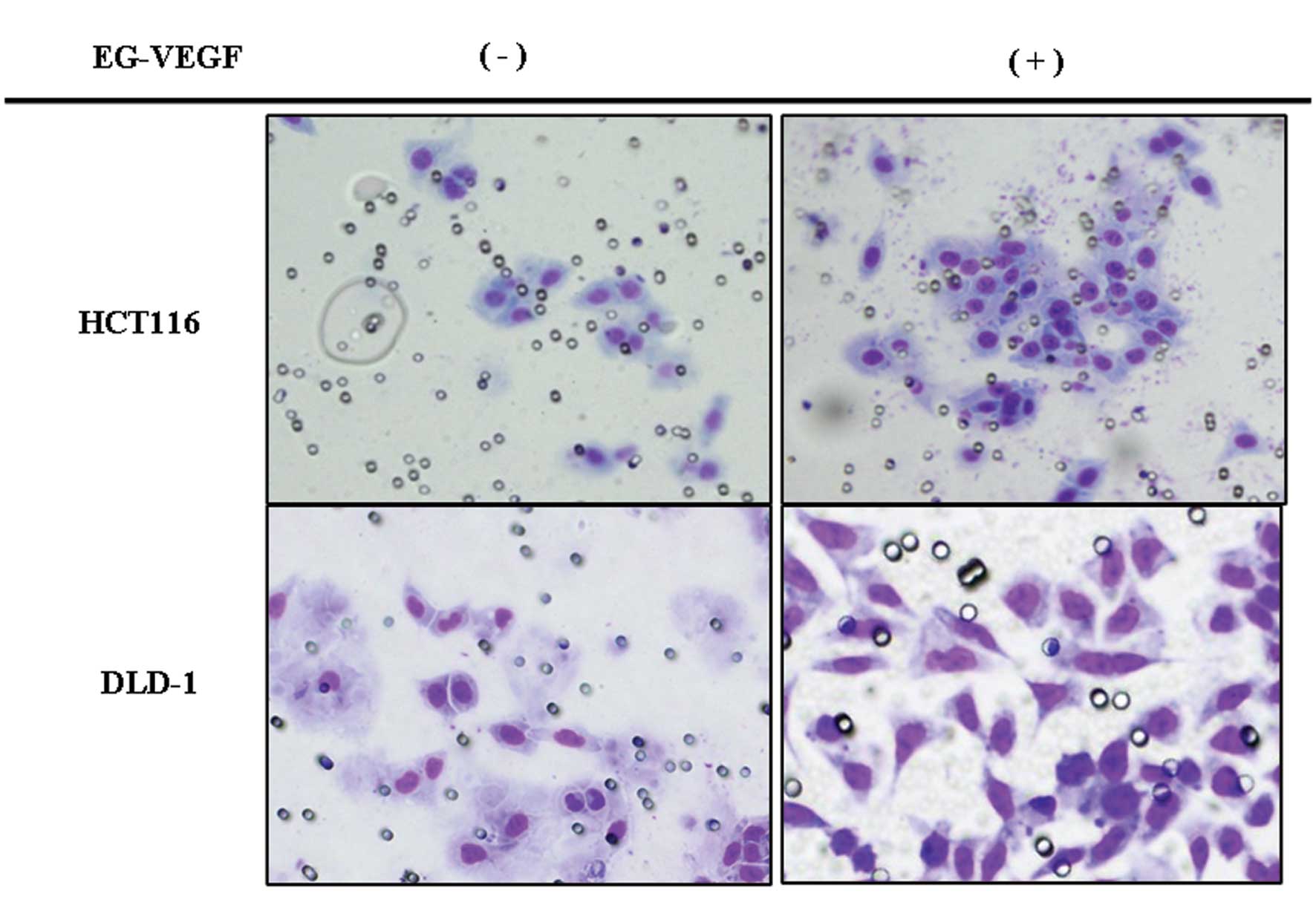
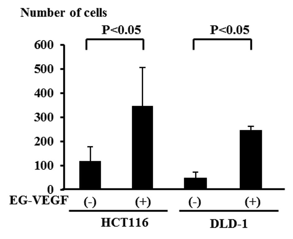
Invasive ability of the colon cancer cell
lines
Figs. 2 and 3 demonstrate the cell invasion after
stimulation of colon cancer cells with the EG-VEGF protein. While
the number of invasive HCT116 colon cancer cells was 117 on
average, the number of invasive HCT116 cells after stimulation with
EG-VEGF was 346 on average. Similarly, while the mean number of
invasive DLD-1 colon cancer cells was 49, the mean number of
invasive DLD-1 cells following stimulation with EG-VEGF was
significantly higher at 288.
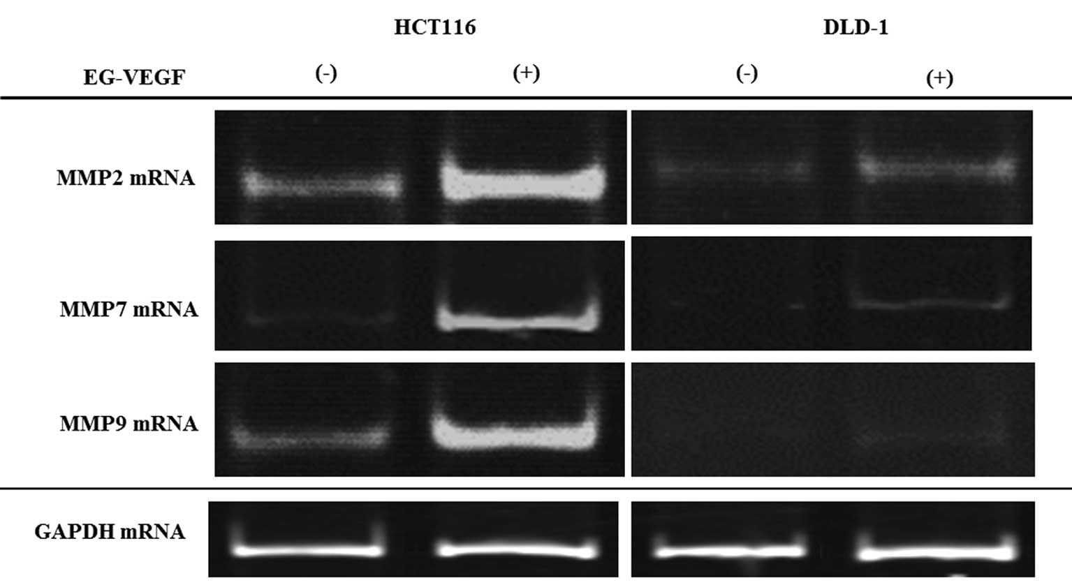
Expression of MMP-2, -7 and -9 mRNAs in
colon cancer cell lines
Results of the expression of MMP-2, -7 and -9 mRNAs
in the colon cancer cell lines stimulated with the EG-VEGF protein
are shown in Fig. 4. Compared with
the cell lines that were not stimulated with EG-VEGF protein,
stimulation with EG-VEGF protein increased the expression of MMP-2,
-7, and -9 mRNAs in both the HCT116 and DLD-1 cell lines, although
the amount of increase was not the same for both cell lines.
Suppression of the invasive ability of
colon cancer cell lines by anti-PK-R1 antibody
While 346 HCT116 colon cancer cells were invasive
after EG-VEGF stimulation, 316 HCT116 cells were invasive following
exposure to the anti-PK-R1 receptor antibody following EG-VEGF
stimulation. While 331 DLD-1 cells were invasive following EG-VEGF
stimulation, 275 DLD-1 cells were invasive after exposure to the
anti-PK-R1 receptor antibody following EG-VEGF stimulation
(Fig. 5). Thus, cell invasion was
not significantly suppressed.
Suppression of the invasive ability of
colon cancer cell lines by anti-PK-R2 antibody
While 346 HCT116 colon cancer cells were invasive
after EG-VEGF stimulation, 119 HCT116 cells were invasive following
exposure to anti-PK-R2 antibody followed by EG-VEGF stimulation.
While 331 DLD-1 cells were invasive following EG-VEGF stimulation,
93 DLD-1 cells were invasive after exposure to the anti-PK-R2
receptor antibody followed by EG-VEGF stimulation (Fig. 5). Thus, cell invasion was
significantly suppressed.
Discussion
The most serious life-threatening condition
associated with malignant tumors is metastasis. Possible
countermeasures include i) preventing cancer cells from leaving the
primary lesion and reaching a target organ; and ii) suppressing the
growth of lesions in the metastatic organs. An important phenomenon
in the course of metastasis is angiogenesis. Angiogenesis
intricately involves the platelet-derived growth factor (PDGF),
VEGF and other growth factors as well as their binding status to
receptors on an endothelial surface (13–16).
Many reports have been published regarding the relationships
between these factors and the development of malignant tumors. With
the recent advancement of molecular biological techniques, the
mechanism of angiogenesis has been elucidated, and various types of
angiogenesis inhibitors have shown efficacy in clinical
applications (17–22).
EG-VEGF investigated in this study is weakly
homologous to and different from VEGF, a known angiogenic factor.
According to previous studies, the expression of EG-VEGF in normal
human tissue is limited to hormone-producing cells including ovary,
testis and placenta, and does not occur in normal gastrointestinal
membranes such as the stomach and colon (4). In connection with our investigations
on primary lesions in colon cancer, we report the following. The
prognosis of patients with positive expression of EG-VEGF mRNA is
significantly worse than for patients with negative expression and
intensification of EG-VEGF expression is related to angiogenesis
and hepatic metastasis (23,24).
EG-VEGF expression is also considered highly significant in other
malignant tumors and is associated with metastasis of prostate
cancer/neuroblastoma and the intensification of malignancy in
pancreatic duct cancer (25–28).
Furthermore, our study showed that EG-VEGF is related to cell
invasion ability, which is an indicator of cell malignancy and an
important element for the development of hematogenous, lymph node
and peritoneal metastasis. The importance of EG-VEGF in malignant
cells was thus indicated.
Although not many studies have been conducted on
EG-VEGF in colon cancer, regarding the transmission system of
EG-VEGF, it is known that information enters the cell via PK-R1 and
2 on the cell membrane, eventually inducing physiological phenomena
and other various important events (5–7). To
the best of our knowledge, our study showed for the first time that
invasive ability of colon cancer cells increases with the
stimulation of the EG-VEGF protein and that information of the
EG-VEGF protein is possibly transmitted via the PK-R2, which we
proved by inhibition of invasion by the anti-PK-R2 antibody.
Furthermore, we examined the information transmission system in
detail by focusing on the MMP family, which plays an important role
in the degradation of the extracellular matrix at the time of
migration from the primary lesion to the interstitium and to the
inside of vascular channels (29,30).
Among the MMP family, MMP-2, -7, and -9, are involved in the
invasive ability of colon cancer cells (31,32).
We examined MMP family members and found that stimulation with the
EG-VEGF protein augmented the expression of MMP-2, -7 and -9 genes,
suggesting the importance of the MMP family genes.
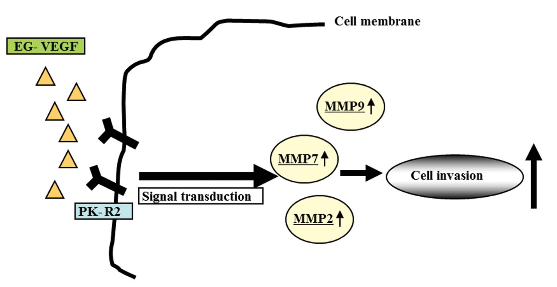
We showed that the EG-VEGF protein acts on the MMP
family genes via the PK-R2 receptor on the cellular membrane,
ultimately intensifying the cell invasion ability in colon cancer
(Fig. 6).
Abbreviations:
|
PK-R1
|
prokineticin receptor 1
|
|
PK-R2
|
prokineticin receptor 2
|
References
|
1
|
Muto T, Kotake K and Koyama Y: Colorectal
cancer statistics in Japan: data from JSCCR registration,
1974–1993. Int J Clin Oncol. 6:171–176. 2001.PubMed/NCBI
|
|
2
|
Koyama Y and Kotake K: Overview of
colorectal cancer in Japan: report from the Registry of the
Japanese Society for Cancer of the Colon and Rectum. Dis Colon
Rectum. 40:2–9. 1997. View Article : Google Scholar
|
|
3
|
Fidler IJ and Ellis LM: The implications
of angiogenesis for the biology and therapy of cancer metastasis.
Cell. 79:185–188. 1994. View Article : Google Scholar : PubMed/NCBI
|
|
4
|
LeCouter J, Kowalski J, Foster J, Hass P,
Zhang Z, Dillard-Telm L, Frantz G, Rangell L, DeGuzman L, Keller
GA, et al: Identification of an angiogenic mitogen selective for
endocrine gland endothelium. Nature. 412:877–884. 2001. View Article : Google Scholar : PubMed/NCBI
|
|
5
|
Soga T, Matsumoto S, Oda T, Saito T,
Hijama H, Takasaki J, Kamohara M, Ohishi T, Matsushime H and
Furuichi K: Molecular cloning and characterization of prokineticin
receptors. Biochim Biophys Acta. 1579:173–179. 2002. View Article : Google Scholar : PubMed/NCBI
|
|
6
|
Lin DC, Bullock CM, Eheler FJ, Chen JL,
Tian H and Zhou QY: Identification and molecular characterization
of two closely related G protein-coupled receptors activated by
prokineticins/endocrine gland vascular endothelial growth factor. J
Biol Chem. 277:19276–19280. 2002. View Article : Google Scholar
|
|
7
|
Masuda Y, Takatsu Y, Terao Y, Kumano S,
Ishibashi Y, Suenaga M, Abe M, Fukusumi S, Watanabe T, Shintani Y,
et al: Isolation and identification of EG-VEGF/prokineticins as
cognate ligands for two orphan G-protein-coupled receptors. Biochem
Biophys Res Commun. 293:396–402. 2002. View Article : Google Scholar : PubMed/NCBI
|
|
8
|
Goi T, Yamaguchi A, Nakagawara G, Urano T,
Shiku H and Furukawa K: Reduced expression of deleted colorectal
carcinoma (DCC) protein in established colon cancers. Br J Cancer.
77:466–471. 1998. View Article : Google Scholar : PubMed/NCBI
|
|
9
|
Yamaguchi A, Urano T, Goi T, Saito M,
Takeuchi K, Hirose K, Nakagawara G, Shiku H and Furukawa K:
Expression of a CD44 variant containing exons 8 to 10 is a useful
independent factor for the prediction of prognosis in colorectal
cancer patients. J Clin Oncol. 14:1122–1127. 1996.PubMed/NCBI
|
|
10
|
Fujishima Y, Goi T, Kimura Y, Hirono Y,
Katayama K and Yamaguchi A: MUC2 protein expression status is
useful in assessing the effects of hyperthermic intraperitoneal
chemotherapy for peritoneal dissemination of colon cancer. Int J
Oncol. 40:960–964. 2012.
|
|
11
|
Sawai K, Goi T, Hirono Y, Katayama K and
Yamaguchi A: Survivin-3B gene decreases the invasion-inhibitory
effect of colon cancer cells with 5-fluorouracil. Oncol Res.
18:541–547. 2010. View Article : Google Scholar : PubMed/NCBI
|
|
12
|
Goi T, Yamaguchi A, Takeuchi K, Nakagawa
G, Yamashiro S, Furukawa K, Urano T and Shiku H: CD44 with variant
exons 8-10 in colorectal tumors: expression analysis by a variant
exon 9-specific monoclonal antibody. Int J Oncol. 8:657–662.
1996.PubMed/NCBI
|
|
13
|
Ferrara N, Gerber HP and LeCouter J: The
biology of VEGF and its receptors. Nat Med. 9:669–676. 2003.
View Article : Google Scholar : PubMed/NCBI
|
|
14
|
Sakurai T and Kudo M: Signaling pathways
governing tumor angiogenesis. Oncology. 81:24–29. 2011. View Article : Google Scholar
|
|
15
|
Ferrara N and Davis-Smyth T: The biology
of vascular endothelial growth factor. Endocr Rev. 18:4–25. 1997.
View Article : Google Scholar
|
|
16
|
Andrae J, Gallini R and Betsholtz C: Role
of platelet-derived growth factors in physiology and medicine.
Genes Dev. 22:1276–1312. 2008. View Article : Google Scholar : PubMed/NCBI
|
|
17
|
Hurwitz H, Fehrenbacher L, Novotny W,
Cartwright T, Hainsworth J, Heim W, Berlin J, Baron A, Griffing S,
Holmgren E, et al: Bevacizumab plus irinotecan, fluorouracil, and
leucovorin for metastatic colorectal cancer. N Engl J Med.
350:2335–2342. 2004. View Article : Google Scholar : PubMed/NCBI
|
|
18
|
Giantonio BJ, Catalano PJ, Meropol NJ,
O’Dwyer PJ, Mitchell EP, Alberts SR, Schwartz MA and Benson AB III:
Bevacizumab in combination with oxaliplatin, fluorouracil, and
leucovorin (FOLFOX4) for previously treated metastatic colorectal
cancer: results from the Eastern Cooperative Oncology Group Study
E3200. J Clin Oncol. 25:1539–1544. 2007. View Article : Google Scholar
|
|
19
|
Jonker DJ, O’Callaghan CJ, Karapetis CS,
Zalcberg JR, Tu D, Au HJ, Berry SR, Krahn M, Price T, Simes RJ, et
al: Cetuximab for the treatment of colorectal cancer. N Engl J Med.
357:2040–2048. 2007. View Article : Google Scholar : PubMed/NCBI
|
|
20
|
Cunningham D, Humblet Y, Siena S, Khayat
D, Bleiberg H, Santoro A, Bets D, Mueser M, Harstrick A, Verslype
C, et al: Cetuximab monotherapy and cetuximab plus irinotecan in
irinotecan-refractory metastatic colorectal cancer. N Engl J Med.
351:337–345. 2004. View Article : Google Scholar : PubMed/NCBI
|
|
21
|
Van Cutsem E, Nowacki M, Lang I, Cascinu
S, Shchepotin I, Maurel J, Rougier P, Cunningham D, Nippgen J and
Kohne C: Randomized phase III study of irinotecan and 5-FU/LV with
or without cetuximab in the first line treatment of patients with
metastatic colorectal cancer. J Clin Oncol. 25:40002007.
|
|
22
|
Van Cutsem E, Peeters M, Siena S, Humblet
Y, Hendlisz A, Neyns B, Canon JL, Van Laethem JL, Maurel J,
Richardson G, et al: Open-label phase III trial of panitumumab plus
best supportive care compared with best supportive care alone in
patients with chemotherapy-refractory metastatic colorectal cancer.
J Clin Oncol. 25:1658–1664. 2007.PubMed/NCBI
|
|
23
|
Nagano H, Goi T, Koneri K, Hirono Y,
Katayama K and Yamaguchi A: Endocrine gland-derived vascular
endothelial growth factor (EG-VEGF) expression in colorectal
cancer. J Surg Oncol. 96:605–610. 2007. View Article : Google Scholar : PubMed/NCBI
|
|
24
|
Goi T, Fujioka M, Satoh Y, Tabata S,
Koneri K, Nagano H, Hirono Y, Katayama K, Hirose K and Yamaguchi A:
Angiogenesis and tumor proliferation/metastasis of human colorectal
cancer cell line SW620 transfected with endocrine
gland-derived-vascular endothelial growth factor, as a new
angiogenic factor. Cancer Res. 64:1906–1910. 2004. View Article : Google Scholar
|
|
25
|
Ngan ES, Sit FY, Lee K, Miao X, Yuan Z,
Wang W, Nicholls JM, Wong KK, Garcia-Barcelo M, Lui VC and Tam PK:
Implications of endocrine gland-derived vascular endothelial growth
factor/prokineticin-1 signaling in human neuroblastoma progression.
Clin Cancer Res. 13:868–875. 2007. View Article : Google Scholar
|
|
26
|
Pasquali D, Rossi V, Staibano S, De Rosa
G, Chieffi P, Prezioso D, Mirone V, Mascolo M, Tramontano D,
Bellastella A and Sinisi AA: The endocrine-gland-derived vascular
endothelial growth factor (EG-VEGF)/prokineticin 1 and 2 and
receptor expression in human prostate: up-regulation of
EG-VEGF/prokineticin 1 with malignancy. Endocrinology.
147:4245–4251. 2006. View Article : Google Scholar : PubMed/NCBI
|
|
27
|
Morales A, Vilchis F, Chávez B, Chan C,
Robles-Díaz G and Díaz-Sánchez V: Expression and localization of
endocrine gland-derived vascular endothelial growth factor
(EG-VEGF) in human pancreas and pancreatic adenocarcinoma. J
Steroid Biochem Mol Biol. 107:37–41. 2007. View Article : Google Scholar
|
|
28
|
Jiang X, Abiatari I, Kong B, Erkan M, De
Oliveira T, Giese NA, Michalski CW, Friess H and Kleeff J:
Pancreatic islet and stellate cells are the main sources of
endocrine gland-derived vascular endothelial growth
factor/prokineticin-1 in pancreatic cancer. Pancreatology.
9:165–172. 2009.
|
|
29
|
Chakraborti S, Mandal M, Das S, Mandal A
and Chakraborti T: Regulation of matrix metalloproteinases: an
overview. Mol Cell Biochem. 253:269–285. 2003. View Article : Google Scholar : PubMed/NCBI
|
|
30
|
Rydlova M, Holubec L Jr, Ludvikova M Jr,
Kalfert D, Franekova J, Povysil C and Ludvikova M: Biological
activity and clinical implications of the matrix
metalloproteinases. Anticancer Res. 28:1389–1397. 2008.PubMed/NCBI
|
|
31
|
Zucker S and Vacirca J: Role of matrix
metalloproteinases (MMPs) in colorectal cancer. Cancer Metastasis
Rev. 23:101–117. 2004. View Article : Google Scholar : PubMed/NCBI
|
|
32
|
Tomita T and Iwata K: Matrix
metaIloproteinases and tissue inhibitors of metalloproteinases in
colonic adenomas-adeno-carcinomas. Dis Colon Rectum. 39:1255–1264.
1996. View Article : Google Scholar : PubMed/NCBI
|