Introduction
Hepatocellular carcinoma (HCC) is one of the most
prevalent and lethal human malignancies, and it causes
approximately half a million deaths each year worldwide (1). Although curative treatments such as
hepatic resection, percutaneous regional treatments, and even liver
transplantation are available for HCC, it still has a high
frequency of postoperative recurrence and poor prognosis (2,3). To
date, there is no proven effective systemic chemotherapy for HCC
due to the resistance of tumor cells to cytotoxic drugs (4,5). The
efficacy of local chemotherapy, using agents such as cisplatin by
means of transarterial chemoembolization, is limited due to the
poor response rate of tumor cells to treatment and tumor re-growth
after treatment (6). These reasons
exemplify the need to fully understand the mechanism underlying
chemoresistance in HCC to allow the design of more effective
therapeutic strategies.
Survivin has recently been identified as the
smallest member of the inhibitor of apoptosis protein (IAP) family,
which counteracts apoptosis and regulates cell division (7). Survivin is expressed in embryonic
tissues as well as in the majority of types of human cancers, but
is undetectable in most terminally differentiated normal adult
tissues (8,9). High levels of survivin expression are
associated with cancer progression, decreased survival time and
poor prognosis, which make survivin a promising target for
anticancer therapies (10). It has
been reported that survivin promotes cell proliferation in human
HCC both in vitro and in vivo(11). Survivin is also reported to be
involved in cancer cell resistance to radiation therapy, as well as
to chemotherapy (12,13). Previous studies have demonstrated
that the inhibition of survivin expression by RNAi inhibits the
tumor growth of HCC cells in vitro and in
vivo(14). However, whether the
suppression of survivin enhances the chemosensitivity of HCC cells
to cisplatin has not been investigated until now.
In the present study, we constructed the siRNA
plasmid expression vector targeting survivin and stably transfected
it into HepG2 and SMMC-7721 HCC cells. Then changes in survivin
expression and the role of siRNA in inducing cancer cell apoptosis
and enhancing chemosensitivity to cisplatin were investigated. The
present research provides a foundation for further study on
survivin siRNA gene therapy for human hepatocellular carcinoma.
Materials and methods
Cell lines and cell culture
Human hepatocellular carcinoma cell lines HepG2 and
SMMC-7721 were provided by Culture Central of Wuhan University
(Wuhan, China). They were maintained in DMEM culture medium,
supplemented with 10% fetal bovine serum at 37°C in a humidified
atmosphere containing 5% CO2, fed with fresh medium
every 3 days and subcultured when confluency was reached.
Construction and transfection of the
siRNA plasmid expression vector
Based on the human survivin gene sequence (GenBank
no. NM_001168), we designed the survivin siRNA targeting against
the sequence, 5′-ACCGCATCTCTACATTC AAGA-3′, while the negative
control siRNA targeted the sequence, 5′-GACCTACCACTCACGATTAAT-3′.
Short hairpin primers were designed around these sequences and
annealed before their ligation into the BbsI sites of the
psiRNA-hH1neo plasmid (Invivogen). The survivin-targeted and the
negative control nucleotide sequences were both confirmed against
the Genbank database to prevent any improper interaction on other
mRNA transcripts. The recombinant plasmids were designated as
psiRNA-sur and psiRNA-con and confirmed by enzyme cutting and
sequencing.
When cells were 70–80% confluent, transfection was
performed following the manufacturer’s protocol. In brief, HepG2
and SMMC-7721 cells were co-transfected with 1 μg of plasmid and 3
μl of Lipofectamine (Invitrogen), respectively. The medium was
replaced with fresh complete medium 6 h after transfection. Then
cells were selected with 500 μg/ml of G418 for 2 weeks. The
resulting resistant clones were isolated and maintained with 200
μg/ml of G418.
Semi-quantitative RT-PCR
The total RNA of the untransfected HCC cells and the
psiRNA-sur or psiRNA-con stably transfected cells was extracted
with TRIzol reagent (Invitrogen) according to the manufacturer’s
instructions. The RNA of each group (1 μg) was used to synthesize
cDNA, and the cDNA was added to a final volume of 25 μl, with 1 μl
of each primer. The primers for human survivin were
5′-CTCAAGGACCACC GCATCT-3′ (forward) and 5′-AGCGCAACCGGACGAAT-3′
(reverse). β-actin was used as an internal control, and its primers
were 5′-CGGGAAATCGTGCGTGAC-3′ (forward) and
5′-GATCTTCATTGTGCTGGGTG-3′ (reverse). The RT-PCR products were
electrophoresed on a 1.5% ethidium bromide-stained agarose gel, and
densitometric analysis was performed using Scion Image
software.
Western blot analysis
The total cellular protein extract for each cell
group was obtained by lysing the cancer cells in lysis buffer.
Western blot analysis was then performed following conventional
protocols. An equal amounts of each protein sample (20 μg) was
electrophoresed on 12% sodium dodecyl sulfate-polyacrylamide gel
(SDS-PAGE), and then transferred onto a PVDF membrane (Millipore).
The PVDF membrane was blocked in PBS containing 5% skimmed milk and
incubated overnight with anti-survivin (1:1,000) and anti-actin
(1:2,000; Santa Cruz Biotechnology), and then incubated with
secondary horseradish peroxidase-conjugated antibodies for 1 h at
room temperature. After extensive rinsing, the blots were developed
with a Luminol chemiluminescence detection kit (Santa Cruz
Biotechnology). Band densities of the blots from photographic film
were quantified using digital image analysis.
Detection of cell proliferation
For cell proliferation analysis, the stably
transfected cells and untransfected HepG2 and SMMC-7721 cells were
seeded in 6-well plates and cultured in the presence of 10% FBS for
8 days. Cells were harvested by trypsinization at various times and
stained with trypan blue. Viable cells were counted using a
hematocytometer and an inverted microscope. The cell numbers were
averaged over 3 independent experiments.
Analysis of the cell cycle by flow
cytometry
Stably transfected HepG2 and SMMC-7721 cells and
untransfected cells were seeded in 6-well plates. When the cells
were ~80% confluent, cells were collected and washed with cold PBS,
and then fixed in 2 ml of 70% ethanol and preserved at 4°C. The
fixed cells were washed three times, and resuspended in PBS
containing 50 mg/ml of RNase A for 30 min, and then incubated with
10 mg/ml propidium iodide (PI) for 20 min in the dark, and the cell
cycle distribution was analyzed by flow cytometry (BD
Biosciences).
Caspase-3 activity assay
The CaspACE™ Assay system kit (Promega) was used to
measure the activity of caspase-3, according to the manufacturer’s
instructions (15). The groups of
HCC cells were collected and washed with cold PBS, and the cell
lysates were prepared. Assays were performed on 96-well plates. We
added 2 μl of the DEVD-pNA substrate (10 mM stock) to 20 μl of the
cell lysate (50 μg total protein) in each well, followed by
incubation at 37°C for 4 h. Absorbance was measured at 405 nm with
an ELISA reader.
Analysis of apoptosis
We identified the effect of different concentrations
of cisplatin on HepG2 cels proliferation by MTT assay, and selected
a concentration of 10 μM as the optimal low dose of cisplatin
(16). Cells of each group were
seeded into 6-well plate. When the cells were ~70% confluent, 10 μM
of cisplatin was added to the cells (Sigma). After 48 h, the cells
were harvested and washed by PBS twice. Annexin V and PI staining
were carried out using the Annexin V-FITC/PI kit (BD Biosciences),
following the manufacturer’s protocol. After incubation for 20 min
at room temperature avoiding light, cell apoptosis was immediately
detected by flow cytometry. All of the samples were assayed in
triplicate.
Chemosensitivity assessment with MTT
assays
Cells were plated in quadruplicate at a density of
5,000 cells/well in 96-well plates, and were allowed to adhere
overnight. The cells were then treated with cisplatin at a
concentration of 10 μM. After 48 h cells were washed with PBS, and
MTT was added to each well at 50 μg/well for 4 h and the produced
formazan was solubilized for 15 min by DMSO. Absorbance was
measured at 570 nm with a microplate reader. Cells treated with
equivalent amounts of DMSO were used as controls.
Statistical analysis
SPSS 10.0 software was used to analyze the data,
which were expressed as the means ± SD. Statistical comparisons of
numerical data within groups were carried out using one-way ANOVA
test. A P-value of <0.05 was considered to indicate a
statistically significant result.
Results
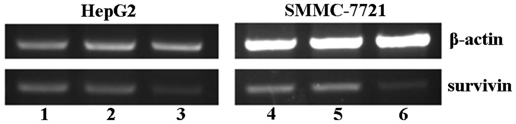
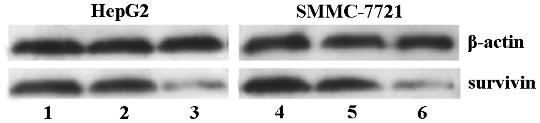
Suppression of survivin expression in HCC
cells by RNAi
psiRNA-sur was transfected into HepG2 and SMMC-7721
cells and stable transfectants were obtained by G418 selection. To
confirm that siRNA transfection downregulates survivin expression
in each cell line, RT-PCR assay and western blotting were
performed. Survivin mRNA and protein were both strongly expressed
in HepG2 and SMMC-7721 cells as reflected by semi-quantitative
RT-PCR and western blotting. Our data showed that after stable
transfection with survivin siRNA, the inhibition ratios of survivin
mRNA and protein expression were 68.38±3.94 and 75.35±4.08% in
HepG2 cells, respectively, and 70.43±3.53 and 76.59±4.61% in
SMMC-7721 cells, respectively (Figs.
1 and 2) (P<0.05).
Transfection with psiRNA-con did not alter survivin expression
levels, indicating that the inhibitory effect of survivin siRNA was
specific. In addition, siRNA did not cause non-specific
downregulation of gene expression, as determined by the β-actin
control. Together, these results proved that the vector based RNAi
effectively and stably suppressed survivin expression in
hepatocellular carcinoma cells.
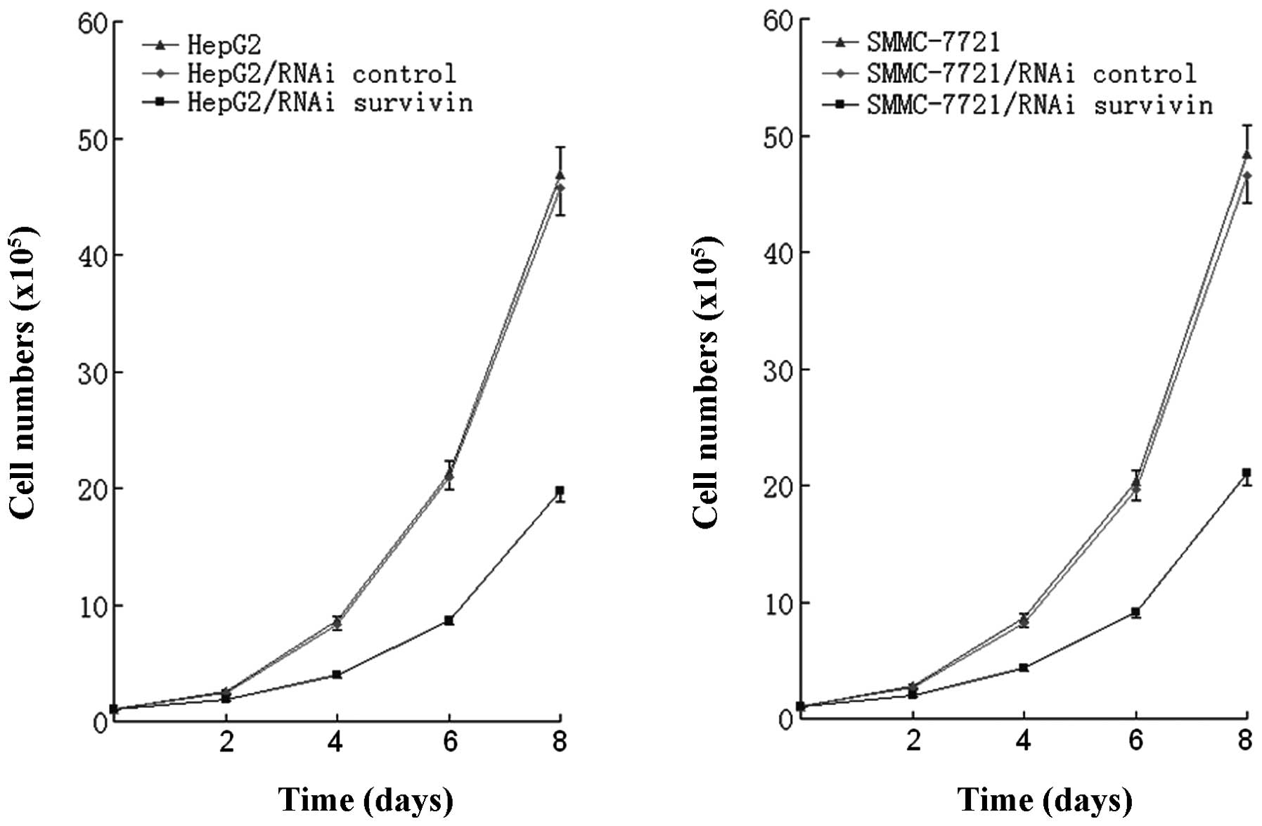
Survivin siRNA significantly inhibits HCC
cell growth
Overexpression of survivin has been reported in the
majority of human cancers including HCC, but not in normal tissue.
Thus, increased levels of survivin may play an important role in
the growth advantage of HCC. Our data showed that siRNA targeting
against survivin significantly decreased the growth rate of cancer
cells in a time-dependent manner, and the highest inhibition rates
of cell proliferation were 57.78±2.63 and 56.40±3.02% for HepG2 and
SMMC-7721 cells, respectively, on Day 8 (P<0.05) (Fig. 3). However, transfection with
psiRNA-con did not alter the growth rate of cancer cells.
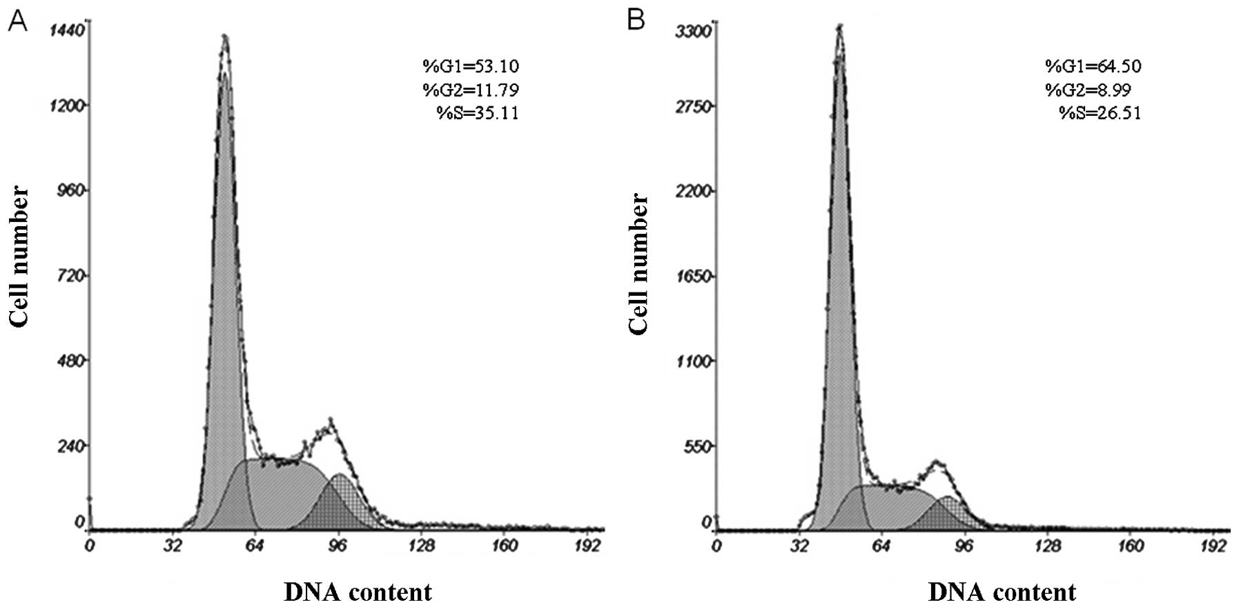
Alteration in the cell cycle by survivin
siRNA
The cell cycle distribution of the HepG2 and
SMMC-7721 cells was analyzed by flow cytometry after stable
transfection by survivin siRNA. Our results showed that, compared
with the untransfected HepG2 cells, psiRNA-survivin-transfected
cells were arrested in the G0/G1 phase (64.52±3.08%) (P<0.05)
and the number of cells was reduced in the G2/M phase (8.98±0.52%)
(P<0.05). psiRNA-survivin-transfected SMMC-7721 cells were also
arrested in the G0/G1 phase (61.23±3.15%) (P<0.05) and the
number of cells was reduced in the G2/M phase (12.36±0.87%)
(P<0.05) (Fig. 4, Table I).
 | Table IComparison of the cell cycle
distribution following transfection of the cell lines with survivin
siRNA (means ± SD). |
Table I
Comparison of the cell cycle
distribution following transfection of the cell lines with survivin
siRNA (means ± SD).
| Cells | G0/G1 (%) | S (%) | G2/M (%) |
|---|
| HepG2 | 53.10±2.92 | 35.11±1.83 | 11.79±0.84 |
| HepG2/RNAi | 64.52±3.08 | 26.50±1.55 | 8.98±0.52 |
| SMMC-7721 | 50.85±2.81 | 29.95±1.89 | 19.20±1.76 |
| SMMC-7721/RNAi | 61.23±3.15 | 26.41±1.72 | 12.36±0.87 |
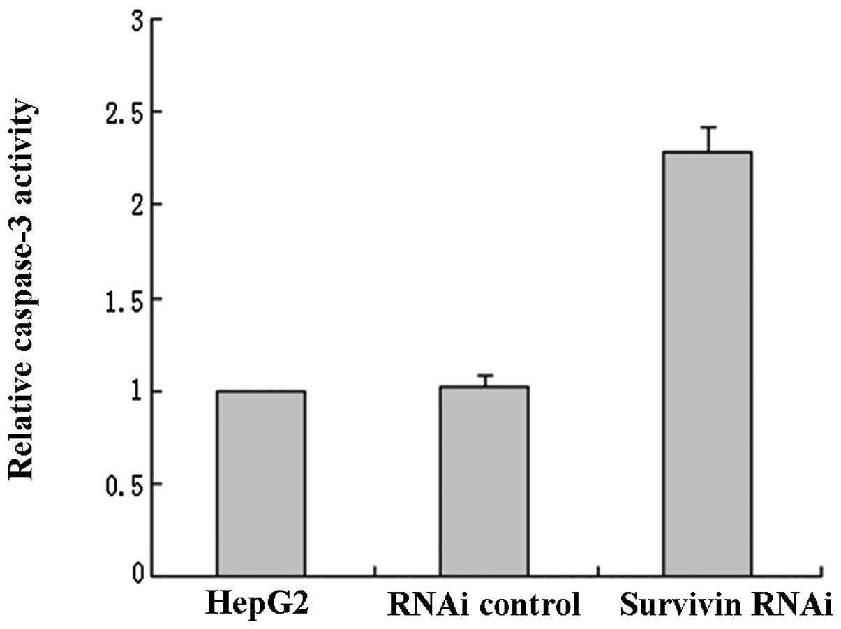
Effect of survivin siRNA on the activity
of caspase-3
We measured the enzymatic activity of caspase-3 to
investigate whether inhibition of survivin expression induces
apoptosis in HCC cells. The results showed that activity of
caspase-3 in HepG2 cells was markedly increased by survivin-siRNA
(2.28±0.19-fold, P<0.05), while there was no difference between
the blank control and the negative control cells (Fig. 5). The suppression of survivin
expression significantly increased the caspase-3 activity in HepG2
cells, leading to cell apoptosis.
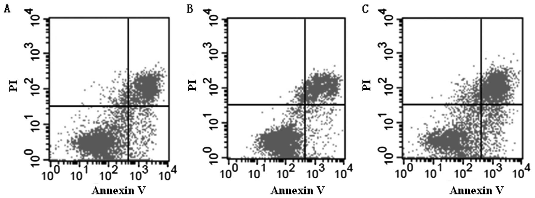
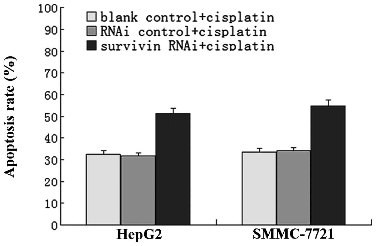
Suppression of survivin expression
enhances cisplatin-induced cytotoxicity
When treated with cisplatin for 48 h, the apoptosis
rates in the untransfected HepG2 cells, negative control group, and
the positive experimental group were 32.54±2.97, 31.82±2.86 and
51.25±3.53%, respectively. The results were similar to that in the
SMMC-7721 cells, which were 33.65±2.74, 34.13±2.27 and 54.58±3.81%,
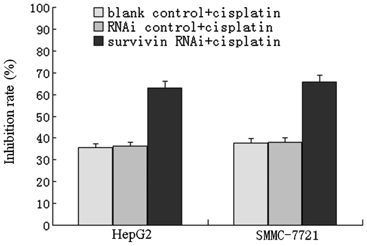
respectively (Figs. 6 and 7). Additionally, following cisplatin
chemotherapy for 48 h, the MTT test showed that the inhibition
rates of cell growth in the untransfected HepG2 cells, negative
control group and positive experimental group were 35.53±2.79,
36.27±2.95 and 62.88±3.84%, respectively, in HepG2 cells, and
37.76±2.65, 38.21±2.58 and 65.74±3.42%, respectively, in the
SMMC-7721 cells (Fig. 8). These
results demonstrated that transfection with survivin siRNA combined
with cisplatin treatment markedly enhanced cell apoptosis and
cytotoxicity, suggesting that inhibition of survivin expression
significantly enhances the chemosensitivity of hepatocellular
carcinoma cells.
Discussion
Previous reports have demonstrated that survivin
plays an important role in cancer proliferation and progression, by
inhibiting apoptosis and facilitating mitosis, giving tumor cells a
survival and growth advantage (17,18).
Survivin expression is also involved in tumor cell resistance to
chemotherapy and radiotherapy (19). Since survivin is specifically
overexpressed in most malignant tumors but not in most normal adult
tissues, the inhibition of survivin has been pursued as a
compelling strategy for cancer therapy (20).
Various strategies have been employed in order to
knock down the overexpression of survivin in cancer cells,
including dominant-negative mutation (21), antisense oligonucleotides (22) and anticancer vaccine (23). The recent emergence of siRNA
technology as a powerful tool to specifically silence gene
expression offers a new modality for anticancer therapeutics
(24). RNAi is mediated by short
interfering RNAs (siRNAs) that are produced from exogenous or
endogenous long dsRNAs by Dicer endonuclease. The resulting siRNAs
are then incorporated into a nuclease complex, which then cleaves
corresponding mRNA in a sequence-specific manner (25,26).
RNAi has become a new technique for silencing gene expression, and
plays an important role in gene function studies and in gene
therapy for many diseases. In the present study, we successfully
constructed recombinant siRNA plasmid expression vector psiRNA-sur
and stably transfected the plasmid into hepatocellular carcinoma
HepG2 and SMMC-7721 cells. Semi-quantitative RT-PCR and western
blotting demonstrated that survivin mRNA and protein expression
levels were significantly reduced by over 60% when transfected with
the siRNA targeting survivin in each cell line.
Survivin is the smallest member of the inhibitors of
the apoptosis protein (IAP) family, and is the most powerful
apoptosis inhibitory factor as far as we know. Survivin is a
bifunctional protein, which has been involved in both control of
apoptosis and regulation of cell division. It was demonstrated that
survivin can directly bind to terminal effector caspase-3 and
caspase-7, and suppresses apoptosis by the inhibition of caspase
activities (27). Survivin inhibits
the apoptosis pathway differently from Bcl-2, which blocks
mitochondrial cytochrome c release into the cytosol, resulting in
the inhibition of the mitochondrial apoptotic pathway.
Overexpression of survivin may also overcome the G2/M phase
checkpoint to enforce progression of cells through mitosis
(28). Survivin expression in
hepatocellular carcinoma is reported to be corelated with
proliferation, metastasis and prognosis (29,30).
In the present study, we found that the growth rates of the HepG2
and SMMC-7721 cell lines were inhibited by survivin siRNA. In
addition, survivin siRNA induced a specific G0/G1 arrest. Moreover,
we found that the caspase-3 activity in both cell lines was
significantly increased by survivin siRNA, which demonstrated that
inhibition of survivin expression induced cell apoptosis. These
results are important since they revealed the role of survivin in
the development of hepatocellular carcinoma. Chen and Deng
(31) also reported that siRNA
targeting of survivin effectively inhibited the growth of gastric
cancer MGC-803 cells.
To further investigate the role of survivin in the
chemoresistance of hepatocellular carcinoma, we compared the
changes in the chemosensitivity to cisplatin in the survivin
siRNA-transfected and untransfected HCC cells. Our data showed that
following cisplatin chemotherapy, survivin siRNA significantly
enhanced cell apoptosis and the cytotoxicity of both HepG2 and
SMMC-7721 cells in vitro, indicating that inhibition of
survivin expression by siRNA significantly enhances the
chemosensitivity of hepatocellular carcinoma cells. Our results are
consistent with another report that inhibition of survivin
expression enhances cisplatin sensitivity in squamous cell
carcinoma of the tongue (32).
In summary, hepatocellular carcinoma HepG2 and
SMMC-7721 cells transfected with survivin siRNA showed decreased
proliferation, increased apoptosis and caspase-3 activity, and
increased chemosensitivity to cisplatin. Our data provide strong
evidence that suppession of survivin expression by siRNA attenuates
the malignant phenotype of HCC, and may provide a novel approach
for anticancer gene therapy.
Acknowledgements
This study was supported by the National Natural
Science Foundation of China (81201676), and the Young Talent
Program of Science and Technology from the Changzhou Municipal
Health Bureau (QN201103).
References
|
1
|
Mann CD, Neal CP, Garcea G, Manson MM,
Dennison AR and Berry DP: Prognostic molecular markers in
hepatocellular carcinoma: a systematic review. Eur J Cancer.
43:979–992. 2007. View Article : Google Scholar : PubMed/NCBI
|
|
2
|
Sakae M, Kubo S, Takemura S, Sakata C,
Uenishi T, Kodai S, Shinkawa H, Urata Y, Ohata K, Kaneda K,
Nishioka T, Nozawa A and Suehiro S: Effect of interferon therapy on
first and second recurrence after resection of hepatitis c
virus-related hepatocellular carcinoma. Hepatol Res. 42:564–573.
2012. View Article : Google Scholar : PubMed/NCBI
|
|
3
|
Fuks D, Dokmak S, Paradis V, Diouf M,
Durand F and Belghiti J: Benefit of initial resection of
hepatocellular carcinoma followed by transplantation in case of
recurrence: an intention-to-treat analysis. Hepatology. 55:132–140.
2012. View Article : Google Scholar : PubMed/NCBI
|
|
4
|
Barbare JC, Bouche O, Bonnetain F, Raoul
JL, Rougier P, Abergel A, Boige V, Denis B, Blanchi A, Pariente A,
Milan C and Bedenne L: Randomized controlled trial of tamoxifen in
advanced hepatocellular carcinoma. J Clin Oncol. 23:4338–4346.
2005. View Article : Google Scholar : PubMed/NCBI
|
|
5
|
Cabrera R and Nelson DR: Review article:
the management of hepatocellular carcinoma. Aliment Pharmacol Ther.
31:461–476. 2010. View Article : Google Scholar : PubMed/NCBI
|
|
6
|
Kawaoka T, Aikata H, Takaki S, Katamura Y,
Hiramatsu A, Waki K, Takahashi S, Hieda M, Toyota N, Ito K and
Chayama K: Transarterial infusion chemotherapy using
cisplatin-lipiodol suspension with or without embolization for
unresectable hepatocellular carcinoma. Cardiovasc Intervent Radiol.
32:687–694. 2009. View Article : Google Scholar
|
|
7
|
Wheatley SP and McNeish IA: Survivin: a
protein with dual roles in mitosis and apoptosis. Int Rev Cytol.
247:35–88. 2005. View Article : Google Scholar : PubMed/NCBI
|
|
8
|
Liu J, Du W and Fan D: Survivin, the
promising target in hepato-cellular carcinoma gene therapy. Cancer
Biol Ther. 7:555–556. 2008. View Article : Google Scholar : PubMed/NCBI
|
|
9
|
Baykara M, Akkus M, Yildiz R, Gonul II,
Dursun A, Coskun U, Benekli M, Sevinc A, Dane F and Buyukberber S:
Survivin expression and its potential clinical significance in
gastrointestinal stromal sarcoma. Int Immunopharmacol.
11:2227–2231. 2011. View Article : Google Scholar : PubMed/NCBI
|
|
10
|
Rodel F, Hoffmann J, Distel L, Herrmann M,
Noisternig T, Papadopoulos T, Sauer R and Rodel C: Survivin as a
radioresistance factor, and prognostic and therapeutic target for
radiotherapy in rectal cancer. Cancer Res. 65:4881–4887. 2005.
View Article : Google Scholar : PubMed/NCBI
|
|
11
|
Ito T, Shiraki K, Sugimoto K, Yamanaka T,
Fujikawa K, Ito M, Takase K, Moriyama M, Kawano H, Hayashida M,
Nakano T and Suzuki A: Survivin promotes cell proliferation in
human hepatocellular carcinoma. Hepatology. 31:1080–1085. 2000.
View Article : Google Scholar : PubMed/NCBI
|
|
12
|
Liu WS, Yan HJ, Qin RY, Tian R, Wang M,
Jiang JX, Shen M and Shi CJ: siRNA directed against survivin
enhances pancreatic cancer cell gemcitabine chemosensitivity. Dig
Dis Sci. 54:89–96. 2009. View Article : Google Scholar : PubMed/NCBI
|
|
13
|
Kami K, Doi R, Koizumi M, Toyoda E, Mori
T, Ito D, Kawaguchi Y, Fujimoto K, Wada M, Miyatake S and Imamura
M: Downregulation of survivin by siRNA diminishes radioresistance
of pancreatic cancer cells. Surgery. 138:299–305. 2005. View Article : Google Scholar : PubMed/NCBI
|
|
14
|
Zhang R, Ma L, Zheng M, Ren J, Wang T,
Meng Y, Zhao J, Jia L, Yao L, Han H, Li K and Yang A: Survivin
knockdown by short hairpin RNA abrogates the growth of human
hepatocellular carcinoma xenografts in nude mice. Cancer Gene Ther.
17:275–288. 2010. View Article : Google Scholar : PubMed/NCBI
|
|
15
|
Zhang Y, Chen ZD, Du CJ, Xu G and Luo W:
siRNA targeting survivin inhibits growth and induces apoptosis in
human renal clear cell carcinoma 786-O cells. Pathol Res Pract.
205:823–827. 2009. View Article : Google Scholar
|
|
16
|
Guo X, Wang W, Zhou F, Lu Z, Fang R, Jia
F, Bu X, Li R, Zhang B, Wu M and Wei L: siRNA-mediated inhibition
of hTERT enhances chemosensitivity of hepatocellular carcinoma.
Cancer Biol Ther. 7:1555–1560. 2008. View Article : Google Scholar : PubMed/NCBI
|
|
17
|
Altieri DC: New wirings in the survivin
networks. Oncogene. 27:6276–6284. 2008. View Article : Google Scholar : PubMed/NCBI
|
|
18
|
Mita AC, Mita MM, Nawrocki ST and Giles
FJ: Survivin: key regulator of mitosis and apoptosis and novel
target for cancer therapeutics. Clin Cancer Res. 14:5000–5005.
2008. View Article : Google Scholar : PubMed/NCBI
|
|
19
|
Zhao W, Bao P, Qi H and You H: Resveratrol
down-regulates survivin and induces apoptosis in human
multidrug-resistant SPC-A-1/CDDP cells. Oncol Rep. 23:279–286.
2010.PubMed/NCBI
|
|
20
|
Ryan BM, O’Donovan N and Duffy MJ:
Survivin: a new target for anti-cancer therapy. Cancer Treat Rev.
35:553–562. 2009. View Article : Google Scholar : PubMed/NCBI
|
|
21
|
Yuan QZ, Wang CT, Mao YQ, Zhang P, Shi HS,
Li ZY, Pan L, Yu DD, Leng F, Chen X, Ying W, Xu JH, Li W, Wu F, Wen
Y, Ma TT and Wei YQ: Enhanced tumor radiosensitivity by a survivin
dominant-negative mutant. Oncol Rep. 23:97–103. 2010.PubMed/NCBI
|
|
22
|
Dai D, Liang Y, Xie Z, Fu J, Zhang Y and
Zhang Z: Survivin deficiency induces apoptosis and cell cycle
arrest in HepG2 hepatocellular carcinoma cells. Oncol Rep.
27:621–627. 2012.PubMed/NCBI
|
|
23
|
Yang Z, Wang L, Wang H, Shang X, Niu W, Li
J and Wu Y: A novel mimovirus vaccine containing survivin epitope
with adjuvant IL-15 induces long-lasting cellular immunity and high
antitumor efficiency. Mol Immunol. 45:1674–1681. 2008. View Article : Google Scholar : PubMed/NCBI
|
|
24
|
Couzin J: Breakthrough of the year. Small
RNAs make big splash. Science. 298:2296–2297. 2002.PubMed/NCBI
|
|
25
|
Klenov MS and Gvozdev VA: Heterochromatin
formation: role of short RNAs and DNA methylation. Biochemistry.
70:1187–1198. 2005.PubMed/NCBI
|
|
26
|
Nakahara K and Carthew RW: Expanding roles
for miRNAs and siRNAs in cell regulation. Curr Opin Cell Biol.
16:127–133. 2004. View Article : Google Scholar : PubMed/NCBI
|
|
27
|
Nassar A, Lawson D, Cotsonis G and Cohen
C: Survivin and caspase-3 expression in breast cancer: correlation
with prognostic parameters, proliferation, angiogenesis, and
outcome. Appl Immunohistochem Mol Morphol. 16:113–120. 2008.
View Article : Google Scholar : PubMed/NCBI
|
|
28
|
Chiou SK, Jones MK and Tarnawski AS:
Survivin - an anti-apoptosis protein: its biological roles and
implications for cancer and beyond. Med Sci Monit. 9:PI25–PI29.
2003.PubMed/NCBI
|
|
29
|
Fields AC, Cotsonis G, Sexton D,
Santoianni R and Cohen C: Survivin expression in hepatocellular
carcinoma: correlation with proliferation, prognostic parameters,
and outcome. Mod Pathol. 17:1378–1385. 2004. View Article : Google Scholar : PubMed/NCBI
|
|
30
|
Zhu H, Chen XP, Zhang WG, Luo SF and Zhang
BX: Expression and significance of new inhibitor of apoptosis
protein survivin in hepatocellular carcinoma. World J
Gastroenterol. 11:3855–3859. 2005.PubMed/NCBI
|
|
31
|
Chen T and Deng C: Inhibitory effect of
siRNA targeting survivin in gastric cancer MGC-803 cells. Int
Immunopharmacol. 8:1006–1011. 2008. View Article : Google Scholar : PubMed/NCBI
|
|
32
|
Xu JH, Wang AX, Huang HZ, Wang JG, Pan CB
and Zhang B: Survivin siRNA induces caspase-3-dependent apoptosis
and enhances cisplatin sensitivity in squamous cell carcinoma of
the tongue. Oncol Res. 18:377–385. 2010.PubMed/NCBI
|