Introduction
Oral squamous cell carcinoma (OSCC) is a common
malignant tumor and the leading cause of oral cancer-related
mortality (1–6). In China, the incidence of OSCC has
been on a steady rise, and the thermotherapy targeted to heat shock
protein 70 (HSP70) has a marked impact on OSCC treatment (7,8). HSP70
is known to assist the folding of nascent polypeptide chains, act
as a molecular chaperone and mediate the repair and degradation of
altered or denatured proteins. The ability of cells to respond to
stress by increasing their HSP levels depends on the activity of
heat shock factor (HSF). HSF-1 is known for its activation of
transcription of the HSP genes during proteotoxic stress which can
bind to the 50 promoter regions of all HSP genes and trigger
instantaneous and massive transcription of these stress protein
genes. The HSF-1/HSP system plays an important role in OSCC
prognosis and treatment selection, being associated with a
disparate range of tumor initiators and promoters. This system has
thus emerged as a source for potential biomarkers of cell
transformation and tumor progenesis. The status of HSP70 and HSF-1
is an integral part of the clinicopathological practice of OSCC,
and accurate HSP70 and HSF-1 quantification and co-localization is
crucial for treatment strategies (9).
The new semiconductor nanocrystals, quantum dots
(QDs), are fluorescent semiconductor nanocrystals with a 2–10 nm
core diameter, possessing several advantages over conventional
fluorescent dyes, such as wide excitation spectra, significant
photostability and a long fluorescence lifespan (10–18).
These characteristics have attracted considerable interest in their
application in immunohistochemistry for biomarker quantification
and co-localization in oral tumors for prognosis and treatment
(20–31). The present study sought to develop a
QD-based approach for a long dynamic observation of physiological
changes of HSP70 and HSF-1 in SCC-25 cells induced by heat shock,
and to discover approaches to disrupt the influence of activation
of HSF-1 and the accumulation of HSP70 in oral cancer.
Materials and methods
Materials
QDs (goat anti-mouse QD525nm-IgG and goat
anti-mouse QD655nm-IgG) were purchased from Invitrogen.
Mouse anti-human HSP70 antibody and mouse anti-human HSF-1 antibody
was provided by Abcam. DMEM/F12 medium with or without leucine and
trypsin were purchased from Sigma. The laser confocal microscope
used was Leica TCS SP2, Germany.
Methods
Cell culture
For the cell culture, SCC-25 cells were cultured in
DMEM/F12 medium (Sigma) with or without leucine and were placed in
an incubator containing a 5% volume fraction of CO2 at
37°C. The culture medium was changed every 2 days, and 0.25%
trypsin was used to digest and subculture the cells after 2–3
days.
Cell inoculation
For the cell inoculation, the digested cells were
inoculated onto a confocal-dedicated utensil at a density of
~1×10⁵ cells. After culturing for 1–2 days, the cells
were 80% confluent.
Heat stress
For the heat stress (32), cells were stressed at 42°C for 30
min (groups A, B, C, D, E and F). Following incubation at 37°C
constant temperature, immunofluorescence was performed on all
groups after 0 h (group A), 2 h (group B), 4 h (group C), 6 h
(group D), 8 h (group E) and 10 h (group F).
Cell fixation
For the cell fixation, all groups were washed twice
with 0.01 mol/l (pH 7.4) TBS and were fixed with pre-chilled
methanol for 10 min.
Immunofluorescence
For the immunofluorescence, the fixed SCC-25 cells
were washed with TBS, incubated with Triton X-100 for 10 min and
washed. They were then incubated at 4°C overnight with mouse
anti-human HSP70 antibody or mouse anti-human HSF-1 antibody
(dilution ratios were 1:100). The cells were then washed 3 times in
TBS and were incubated with buffer solution in a 37°C moist box for
10 min, followed by incubation with the equivalent mixed liquid of
goat anti-mouse QD525nm-IgG or goat anti-mouse
QD655nm-IgG (1:100) in a 37°C moist box for 45 min.
Finally, the cells were washed three times with TBST and sent for
detection away from light. Each experiment was repeated 5
times.
Detection under laser confocal
fluorescence microscopy and analysis by Image-Pro Plus
The wavelength of excitation spectra was 488 nm and
the constant temperature system was adjusted to 30°C.
Statistical analysis
Data are expressed as the means ± SD. Comparisons
between two groups were conducted by one-way ANOVA. P≤0.05 was
considered to indicate a statistically significant difference.
Results
HSP70 and HSF-1 labeled by
QD525nm or QD655nm
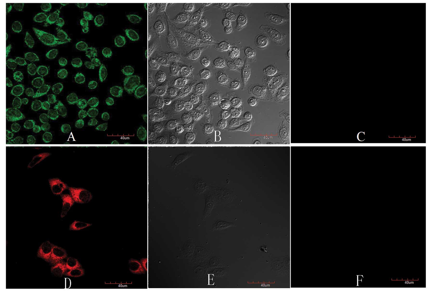
With a laser confocal microscopy,
QD525nm-marked HSP70 was clearly expressed in the
cytoplasm and nucleus of SCC-25 cells, mainly distributed in the
cytoplasm (Fig. 1A). The
QD655nm-marked HSF-1 was expressed in the cytoplasm
(Fig. 1D).
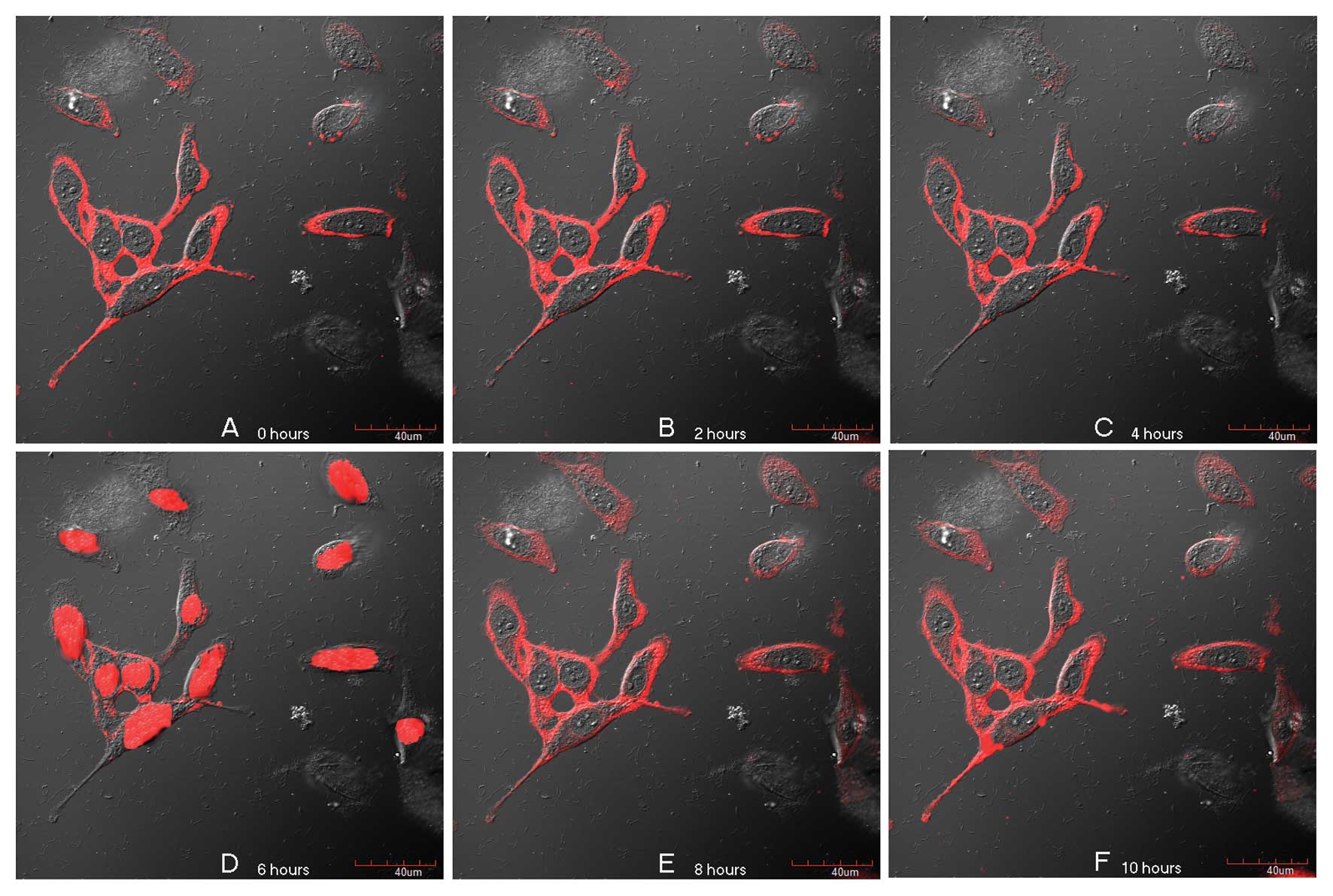
HSP70 kinetics in cells cultured with
normal leucine or limiting leucine
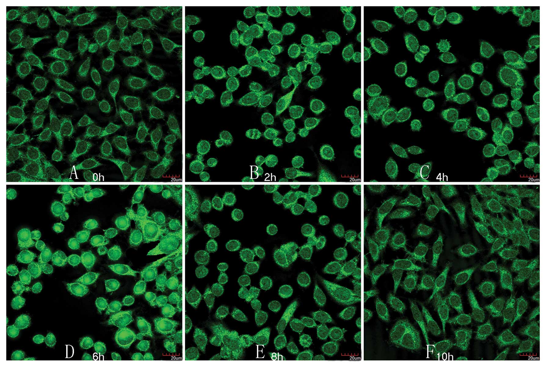
With a laser confocal microscopy, the synthesis of
HSP70 in cells cultured with normal leucine began to increase at 2
h (Fig. 2B), they reached the
maximum value at 6 h, and had a tendency to gather in the nucleus
at 6 h (Fig. 2D).
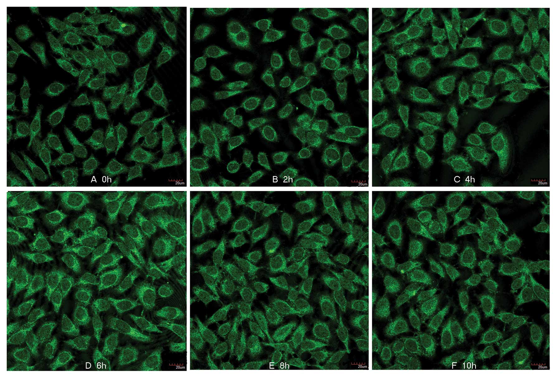
However, the synthesis of HSP70 in cells cultured
with limiting leucine showed a small increase at 6 h (Fig. 3D). The accumulation of HSP70 showed
no tendency to gather in the nucleus (Fig. 3D), whereas cells without leucine
deprivation showed a translocation from the cytoplasm to the
nucleus (Fig. 2D) at 6 h.
HSF-1 kinetics in cells cultured with
normal leucine or limiting leucine
In an attempt to elucidate the difference of the
HSP70 kinetics between the limiting leucine and the normal leucine
group, we investigated the dynamic distribution of HSF-1 under
laser confocal fluorescence microscopy.
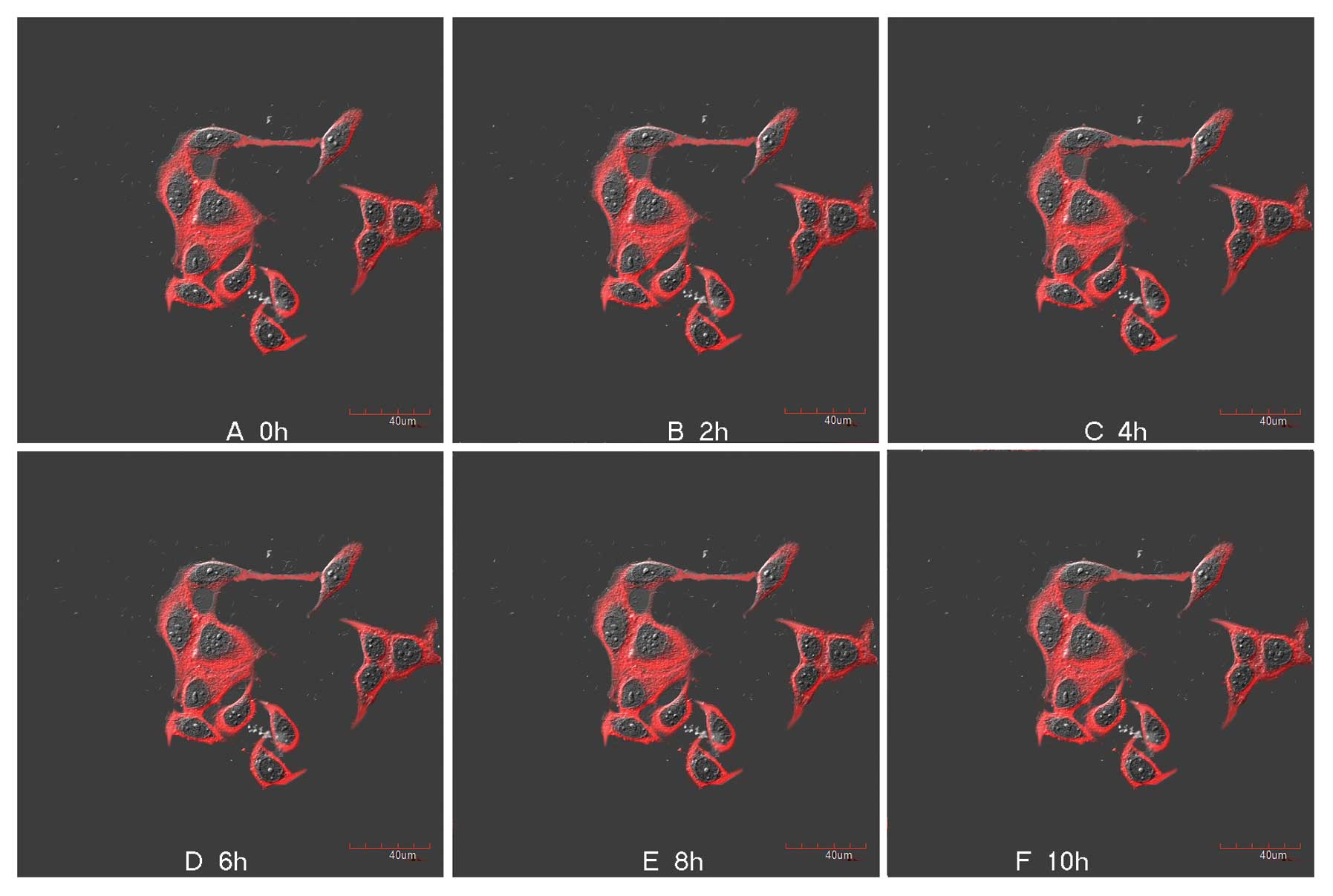
The subcellular localization of HSF-1 in cells
cultured with normal leucine was different during the recovery
periods of heat shock (Fig. 4). The
majority of HSF-1 was found to be localized in the cytoplasm at 0,
2, 4, 8 and 10 h after heating at 42°C for 30 min (32). However, at 6 h, an increasing number
of HSF-1 translocated from the cytoplasm to the nucleus (Fig. 4D), similar to the translocation of
HSP70 (Fig. 2D).
 | Figure 4HSF-1 kinetics in cells cultured with
normal leucine following heat shock at (A) 0 h, (B) 2 h, (C) 4 h,
(D) 6 h, (E) 8 h and (F) 10 h. The majority of HSF-1 was localized
in the plasma membrane cytoplasm at 0, 2, 4, 8 and 10 h. However,
at 6 h after heating at 42°C for 30 min, an increasing number of
HSF-1 translocated from the cytosol to the nucleus, similar to the
translocation of HSP70 (D). |
By contrast, in cells cultured with limiting
leucine, the subcellular localization of HSF-1 presented only a
slight change, HSF-1 was localized in the cytoplasm during the
recovery periods of heat shock, and no translocation was observed
at 6 h after heat shock (Fig.
5D).
Semi-quantitative determination of HSP70
in cells cultured with limiting leucine or normal leucine during
all the recovery periods of heat shock
HSP70 was detected in each group five times and 65
photofluorograms of each group were analyzed by Image-Pro Plus.
Results are expressed as means ± SD.
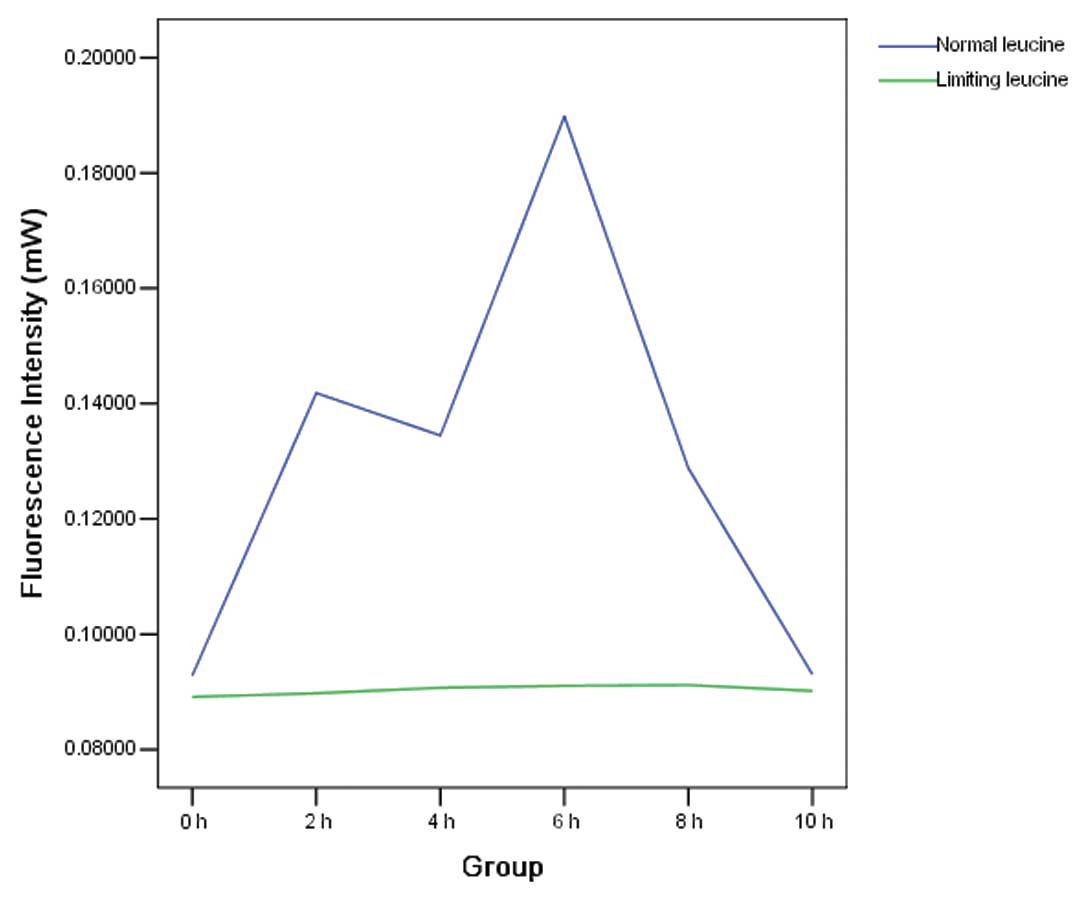
As indicated in Table
I and Fig. 6, the synthesis of
HSP70 in cells cultured with normal leucine had a significant
increase at 2 h after the heat shock (P<0.05). It reached the
maximum value at 6 h (0.1898172±0.00219462 vs. 0.0928257±0.00193721
at 0 h; P<0.05), and it began to decrease at 8 h. At 10 h, it
returned to the level at 0 h.
 | Table IHSP70 expression kinetics in cells
cultured with normal leucine during all the recovery periods of
heat shock (0, 2, 4, 6, 8 and 10 h). |
Table I
HSP70 expression kinetics in cells
cultured with normal leucine during all the recovery periods of
heat shock (0, 2, 4, 6, 8 and 10 h).
| Group | 0 h | 2 h | 4 h | 6 h | 8 h | 10 h |
|---|
| Fluorescent
intensity |
0.0928257±0.00193721 |
0.1418270±0.00437687 |
0.1344760±0.00511160 |
0.1898172±0.00219462 |
0.1287666±0.00353873 |
0.0930118±0.00578231 |
The synthesis of HSP70 in cells cultured with
limiting leucine demonstrated a small increase during all the
recovery periods of heat shock (P>0.05) (Table II and Fig. 6).
| Table IIHSP70 expression kinetics in cells
cultured with limiting leucine during all the recovery periods of
heat shock (0, 2, 4, 6, 8 and 10 h). |
Table II
HSP70 expression kinetics in cells
cultured with limiting leucine during all the recovery periods of
heat shock (0, 2, 4, 6, 8 and 10 h).
| Group | 0 h | 2 h | 4 h | 6 h | 8 h | 10 h |
|---|
| Fluorescent
intensity |
0.0891216±0.00165493 |
0.0897608±0.00237068 |
0.0907190±0.00045941 |
0.0910551±0.00075798 |
0.0911681±0.01056427 |
0.0901642±0.00125764 |
Concurrently, the synthesis of the HSP70 in cells
cultured with normal leucine significantly increased compared to
that of the HSP70 in cells cultured with limiting leucine at 2, 4,
6 and 8 h (P<0.05).
Discussion
Heat shock proteins (HSPs) were first identified as
stress proteins that confer resistance to physical stresses such as
elevated temperatures in all cellular organisms (33–36).
HSP70 expression becomes deregulated in oral cancer. Elevated HSP70
expression leads to resistance to subsequent thermotherapy.
In the present study, we developed the QD-IHC
protocol for HSP70 kinetics in SCC-25 cells cultured with or
without leucine following heat shock. At 6 h after heating at 42°C
for 30 min, the synthesis of the HSP70 in cells without leucine
deprivation increased approximately 2-fold whereas that of cells
with leucine deprivation showed only a small increase. Particularly
the accumulation of HSP70 in cells cultured with normal leucine had
a tendency to gather in the nucleus at 6 h whereas cells with
leucine deprivation showed no translocation. Concurrently, the
synthesis of HSP70 in the normal leucine group was increased
significantly compared to that of the limiting leucine group at 2,
4, 6 and 8 h, respectively (P<0.05).
In an attempt to elucidate the mechanisms of HSP70
kinetics associated with leucine deprivation, we investigated the
dynamic distribution of HSF-1 in SCC-25 cells cultured with or
without leucine following heat shock. HSF-1 is known for its
activation of transcription of the HSP genes during heat shock
(37–42); it possesses a complex modular
structure with several functional domains such as the DNA-binding
domain (DBD; residues 15–120) and trimerization domains (residues
130–203). The trimerization domain is composed of three arrays of
hydrophobic heptad repeats (HR-A/B). When the HSF-1 monomers come
together, they form a leucine zipper, an artifact typically seen in
dimerization. Once the heat shock response is induced, HSF-1 is
activated through trimerization, accumulation in the nucleus,
post-translational modifications and binding to HSP genes through
DBD.
Leucine is one of the three branched chain amino
acids (BCAA) along with isoleucine and valine. Leucine is the most
prominent of the three and is the sole amino acid behind the
branched chain amino acid stimulation of protein synthesis and
anti-catabolic actions. Leucine deprivation results in the
inactivation of HSF-1, and leads to a sharp decrease in the
transcript level of HSF-1 target genes such as HSPA1A (HSP70),
DNAJB1 (HSP40) and HSP90AA1 (43).
In leucine-deprived cells, HSF-1 loses its DBD
activity and the HSF-1 monomer trimerization, accumulation in the
nucleus and post-translational modifications were all inhibited.
Leucine deprivation results in the inactivation of HSF-1, leading
to slight accumulation of HSP70 in the nucleus.
With the valuable research tools for tumor prognosis
and treatment (44–47), protein kinetics with QD provide a
rationale for thermotherapy accompanied by short-term dietary
restriction of leucine in patients with oral tumor.
In conclusion, the HSF-1/HSP system is implicated in
several crucial steps in oncogenesis and tumor progression,
activated HSF-1 and increased HSP levels may aid in the
oncodiagnosis and treatment in the clinic. Leucine deprivation can
disrupt the influence of activation of HSF-1 and the accumulation
of HSP70 following heat shock. Thermotherapy accompanied by
short-term dietary restriction of leucine may be a novel approach
for the treatment of oral cancer.
References
|
1
|
Parkin DM, Pisani P and Ferlay J:
Estimates of the worldwide incidence of eighteen major cancers in
1985. Int J Cancer. 54:594–606. 1993. View Article : Google Scholar : PubMed/NCBI
|
|
2
|
Camisasca DR, Silami MA, Honorato J, Dias
FL, Faria PA and Lourenço SD: Oral squamous cell carcinoma:
clinicopathological features in patients with and without
recurrence. ORL J Otorhinolaryngol Relat Spec. 73:170–176. 2011.
View Article : Google Scholar : PubMed/NCBI
|
|
3
|
Uchida K, Oga A, Nakao M, Mano T, Mihara
M, Kawauchi S, Furuya T, Ueyama Y and Sasaki K: Loss of 3p26.3 is
an independent prognostic factor in patients with oral squamous
cell carcinoma. Oncol Rep. 26:463–469. 2011.PubMed/NCBI
|
|
4
|
Hanash SM, Bobek MP, Rickman DS, Williams
T, Rouillard JM, Kuick R and Puravs E: Integrating cancer genomics
and proteomics in the post-genome era. Proteomics. 2:69–75. 2002.
View Article : Google Scholar : PubMed/NCBI
|
|
5
|
Snyder M and Gerstein M: Genomics.
Defining genes in the genomics era. Science. 300:258–260. 2003.
View Article : Google Scholar : PubMed/NCBI
|
|
6
|
Fields S: Proteomics in genomeland.
Science. 291:1221–1224. 2001. View Article : Google Scholar : PubMed/NCBI
|
|
7
|
Carr KM, Rosenblatt K, Petricoin EF, et
al: Genomic and proteomic approaches for studying human cancer:
prospects for true patient-tailored therapy. Hum Genomics.
1:134–140. 2004. View Article : Google Scholar : PubMed/NCBI
|
|
8
|
Lee SS, Tsai CH, Ho YC and Chang YC: The
upregulation of heat shock protein 70 expression in areca quid
chewing-associated oral squamous cell carcinomas. Oral Oncol.
44:884–890. 2008. View Article : Google Scholar : PubMed/NCBI
|
|
9
|
Markopoulos AK, Deligianni E and
Antoniades DZ: Heat shock protein 70 membrane expression in oral
cancer: a possible new target in antineoplastic therapy?
Chemotherapy. 55:211–214. 2009. View Article : Google Scholar : PubMed/NCBI
|
|
10
|
Zhao JJ, Chen J, Wang ZP, Pan J and Huang
YH: Double labeling and comparison of fluorescence intensity and
photostability between quantum dots and FITC in oral tumors. Mol
Med Report. 4:425–429. 2011.PubMed/NCBI
|
|
11
|
Doty RC, Fernig DG and Levy R: Nanoscale
science: a big step towards the Holy Grail of single molecule
biochemistry and molecular biology. Cell Mol Life Sci.
61:1843–1849. 2004.PubMed/NCBI
|
|
12
|
Hanaki K, Momo A, Oku T, Komoto A,
Maenosono S, Yamaguchi Y and Yamamoto K: Semiconductor quantum
dot/albumin complex is a long-life and highly photostable endosome
marker. Biochem Biophys Res Commun. 302:496–501. 2003. View Article : Google Scholar : PubMed/NCBI
|
|
13
|
McMahon RJ: Chemical reactions involving
quantum tunneling. Science. 299:833–834. 2003. View Article : Google Scholar : PubMed/NCBI
|
|
14
|
Banerjee B, Miedema B and Chandrasekhar
HR: Emission spectra of colonic tissue and endogenous fluorophores.
Am J Med Sci. 316:220–226. 1998. View Article : Google Scholar : PubMed/NCBI
|
|
15
|
Alivisatos P: The use of nanocrystals in
biological detection. Nat Biotechnol. 22:47–52. 2004. View Article : Google Scholar : PubMed/NCBI
|
|
16
|
Garon EB, Marcu L, Luong Q,
Tcherniantchouk O, Crooks GM and Koeffler HP: Quantum dot labeling
and tracking of human leukemic, bone marrow and cord blood cell.
Leuk Res. 31:643–651. 2007. View Article : Google Scholar : PubMed/NCBI
|
|
17
|
Bruchez M Jr, Moronne M, Gin P, Weiss S
and Alivisatos AP: Semiconductor nanocrystals as fluorescent
biological labels. Science. 281:2013–2016. 1998. View Article : Google Scholar
|
|
18
|
Wang HZ, Wang HY, Liang RQ and Ruan KC:
Detection of tumor marker CA125 in ovarian carcinoma using quantum
dots. Acta Biochim Biophys Sin. 36:681–686. 2004. View Article : Google Scholar : PubMed/NCBI
|
|
19
|
Li Z, Wang K, Tan W, Li J, Fu Z, Ma C, Li
H, He X and Liu J: Immunofluorescent labeling of cancer cells with
quantum dots synthesized in aqueous solution. Anal Biochem.
354:169–174. 2006. View Article : Google Scholar : PubMed/NCBI
|
|
20
|
Yezhelyev MV, Gao X, Xing Y, Al-Hajj A,
Nie S and O’Regan RM: Emerging use of nanoparticles in diagnosis
and treatment of breast cancer. Lancet Oncol. 7:657–667. 2006.
View Article : Google Scholar : PubMed/NCBI
|
|
21
|
Tholouli E, Hoyland JA, Di Vizio D,
O’Connell F, Macdermott SA, Twomey D, Levenson R, Yin JA, Golub TR,
Loda M and Byers R: Imaging of multiple mRNA targets using quantum
dot based in situ hybridization and spectral deconvolution in
clinical biopsies. Biochem Biophys Res Commun. 348:628–636. 2006.
View Article : Google Scholar : PubMed/NCBI
|
|
22
|
Azzazy HM, Mansour MM and Kazmierczak SC:
From diagnostics to therapy: prospects of quantum dots. Clin
Biochem. 40:917–927. 2007. View Article : Google Scholar : PubMed/NCBI
|
|
23
|
Wu X, Liu H, Liu J, Haley KN, Treadway JA,
Larson JP, Ge N, Peale F and Bruchez MP: Immunofluorescent labeling
of cancer marker Her2 and other cellular targets with semiconductor
quantum dots. Nat Biotechnol. 21:41–46. 2003. View Article : Google Scholar : PubMed/NCBI
|
|
24
|
Lidke DS, Nagy P, Heintzmann R,
Arndt-Jovin DJ, Post JN, Grecco HE, Jares-Erijman EA and Jovin TM:
Quantum dot ligands provide new insights into erbB/HER
receptor-mediated signal transduction. Nat Biotechnol. 22:198–203.
2004. View
Article : Google Scholar : PubMed/NCBI
|
|
25
|
Jaiswal JK, Mattoussi H, Mauro JM and
Simon SM: Long-term multiple color imaging of live cells using
quantum dot bioconjugates. Nat Biotechnol. 21:47–51. 2003.
View Article : Google Scholar : PubMed/NCBI
|
|
26
|
Chan WC, Maxwell DJ, Gao X, Bailey RE, Han
M and Nie S: Luminescent quantum dots for multiplexed biological
detection and imaging. Curr Opin Biotechnol. 13:40–46. 2002.
View Article : Google Scholar : PubMed/NCBI
|
|
27
|
Hall M, Kazakova I and Yao YM: High
sensitivity immunoassays using particulate fluorescent labels. Anal
Biochem. 272:165–170. 1999. View Article : Google Scholar : PubMed/NCBI
|
|
28
|
Gao X, Cui Y, Levenson RM, Chung LW and
Nie S: In vivo cancer targeting and imaging with semiconductor
quantum dots. Nat Biotechnol. 22:969–976. 2004. View Article : Google Scholar : PubMed/NCBI
|
|
29
|
Medintz IL, Uyeda HT, Goldman ER and
Mattoussi H: Quantum dot bioconjugates for imaging, labelling and
sensing. Nat Mater. 4:435–446. 2005. View
Article : Google Scholar : PubMed/NCBI
|
|
30
|
Sukhanova A, Devy J, Venteo L, Kaplan H,
Artemyev M, Oleinikov V, Klinov D, Pluot M, Cohen JH and Nabiev I:
Biocompatible fluorescent nanocrystals for immunolabeling of
membrane proteins and cells. Anal Biochem. 324:60–67. 2004.
View Article : Google Scholar : PubMed/NCBI
|
|
31
|
Zahavy E, Freeman E, Lustig S, Keysary A
and Yitzhaki S: Double labeling and simultaneous detection of B-
and T cells using fluorescent nano-crystal (q-dots) in
paraffin-embedded tissues. J Fluoresc. 15:661–665. 2005. View Article : Google Scholar : PubMed/NCBI
|
|
32
|
Abe T, Gotoh S and Higashi K: Higher
induction of heat shock protein 72 by heat stress in
cisplatin-resistant than in cisplatin-sensitive cancer cells.
Biochim Biophys Acta. 1445:123–133. 1999. View Article : Google Scholar : PubMed/NCBI
|
|
33
|
Tavassol F, Starke OF, Kokemüller H,
Wegener G, Müller-Tavassol CC, Gellrich NC and Eckardt A:
Prognostic significance of heat shock protein 70 (HSP70) in
patients with oral cancer. Head Neck Oncol. 3:102011. View Article : Google Scholar : PubMed/NCBI
|
|
34
|
Park SR, Lee KD, Kim UK, Gil YG, Oh KS,
Park BS and Kim GC: Pseudomonas aeruginosa exotoxin A
reduces chemoresistance of oral squamous carcinoma cell via
inhibition of heat shock proteins 70 (HSP70). Yonsei Med J.
51:708–716. 2010. View Article : Google Scholar
|
|
35
|
Chen X, Tao Q, Yu H, Zhang L and Cao X:
Tumor cell membrane-bound heat shock protein 70 elicits antitumor
immunity. Immunol Lett. 84:81–87. 2002. View Article : Google Scholar : PubMed/NCBI
|
|
36
|
Thiel UJ, Feltens R, Adryan B, Gieringer
R, Brochhausen C, Schuon R, Fillies T, Grus F, Mann WJ and Brieger
J: Analysis of differentially expressed proteins in oral squamous
cell carcinoma by MALDI-TOF MS. J Oral Pathol Med. 40:369–379.
2011. View Article : Google Scholar : PubMed/NCBI
|
|
37
|
Akerfelt M, Morimoto RI and Sistonen L:
Heat shock factors: integrators of cell stress, development and
lifespan. Nat Rev Mol Cell Biol. 11:545–555. 2010. View Article : Google Scholar : PubMed/NCBI
|
|
38
|
Westerheide SD and Morimoto RI: Heat shock
response modulators as therapeutic tools for diseases of protein
conformation. J Biol Chem. 280:33097–33100. 2005. View Article : Google Scholar : PubMed/NCBI
|
|
39
|
Clos J, Westwood JT, Becker PB, Wilson S,
Lambert K and Wu C: Molecular cloning and expression of a hexameric
Drosophila heat shock factor subject to negative regulation.
Cell. 63:1085–1097. 1990.PubMed/NCBI
|
|
40
|
Sorger PK and Pelham HR: Yeast heat shock
factor is an essential DNA-binding protein that exhibits
temperature-dependent phosphorylation. Cell. 54:855–864. 1988.
View Article : Google Scholar : PubMed/NCBI
|
|
41
|
Wiederrecht G, Seto D and Parker CS:
Isolation of the gene encoding the S. cerevisiae heat shock
transcription factor. Cell. 54:841–853. 1988.PubMed/NCBI
|
|
42
|
Westerheide SD, Raynes R, Powell C, Xue B
and Uversky VN: HSF transcription factor family, heat shock
response, and protein intrinsic disorder. Curr Protein Pept Sci.
13:86–103. 2012. View Article : Google Scholar : PubMed/NCBI
|
|
43
|
Hensen SM, Heldens L, van Enckevort CM,
van Genesen ST, Pruijn GJ and Lubsen NH: Heat shock factor 1 is
inactivated by amino acid deprivation. Cell Stress Chaperones.
17:743–755. 2012. View Article : Google Scholar : PubMed/NCBI
|
|
44
|
Xiao Y and Gao X: Use of IgY antibodies
and semiconductor nanocrystal detection in cancer biomarker
quantitation. Biomark Med. 4:227–239. 2010. View Article : Google Scholar : PubMed/NCBI
|
|
45
|
Sweeney E, Ward TH, Gray N, Womack C,
Jayson G, Hughes A, Dive C and Byers R: Quantitative multiplexed
quantum dot immunohistochemistry. Biochem Biophys Res Commun.
374:181–186. 2008. View Article : Google Scholar : PubMed/NCBI
|
|
46
|
Xing Y, Chaudry Q, Shen C, Kong KY, Zhau
HE, Chung LW, Petros JA, O’Regan RM, Yezhelyev MV, Simons JW, Wang
MD and Nie S: Bioconjugated quantum dots for multiplexed and
quantitative immunohistochemistry. Nat Protoc. 2:1152–1165. 2007.
View Article : Google Scholar : PubMed/NCBI
|
|
47
|
Byers RJ, Di Vizio D, O’connell F,
Tholouli E, Levenson RM, Gossage K, Twomey D, Yang Y, Benedettini
E, Rose J, Ligon KL, Finn SP, Golub TR and Loda M: Semiautomated
multiplexed quantum dot-based in situ hybridization and spectral
deconvolution. J Mol Diagn. 9:20–29. 2007. View Article : Google Scholar : PubMed/NCBI
|