Introduction
Intraductal papillary mucinous neoplasm (IPMN) is a
cystic tumor of the pancreas, which is characterized histologically
by mucin production, varying degrees of cystic dilatation of the
pancreatic ducts and intrapapillary growth (1). The IPMNs account for 7.5% of
clinically diagnosed pancreatic neoplasms and 16.3% of surgically
resected pancreatic tumors (2).
Recent advances in diagnostic imaging modalities are expected to
increase the incidence of pancreatic tumors, including IPMN. In
2006, international guidelines for IPMNs were established, and a
consensus was reached on an algorithm for diagnosis and treatment
strategies (3). However, the
methods of postoperative follow-up, bearing in mind the prognosis
of IPMN patients with a long-term follow-up, choice of surgical
procedures and recurrence, are largely unknown. Since the
epithelial cells lining cystic lesions exhibit variable
histopathological features ranging from benign to malignant, the
presence of an adenomacarcinoma sequence in the carcinogenesis of
pancreatic IPMN has been suggested (4). According to the international
guidelines (3), patients with a
suspected malignant lesion undergo surgical treatment. However,
since their prognosis varies greatly depending on the histological
type and degree of progression (5),
preoperative diagnostic imaging and histopathological examination
are of great significance in the determination of treatment
strategies. Indicators suggestive of malignancy include the degree
of dilation of the pancreatic duct, presence of intracystic nodules
and cyst size (6). Surgical
resection has been recommended if these indicators suggest the
presence of a malignant lesion (3).
In recent years, in addition to the histological grade of
malignancy, subtype classification based on morphological
characteristics has been attempted. Kimura et al(7) classified duodenal papilla cancer into
two subtypes, and reported that there were differences in the
pattern of invasion and prognosis between the two subtypes.
Furukawa et al(8) reported a
new classification of IPMNs into four subtypes (gastric,
intestinal, pancreatobiliary and oncocytic types) based on their
morphological characteristics and mucin expression. In addition,
they reported that these subtypes differed in the location of tumor
origin, histological grade of malignancy, degree of progression,
prognosis and presence or absence of recurrence (9). This subtype classification, like the
histological grade of malignancy, may play a large role in the
determination of treatment strategies for IPMN. If information on
the subtypes of IPMN is available preoperatively, its use in
combination with conventional preoperative imaging modalities may
lead to surgical treatment best suited for the biological
characteristics of the subtypes. Therefore, we classified resected
IPMNs into the four subtypes, clarified their clinicopathological
characteristics, and investigated the possibility of preoperatively
diagnosing these subtypes by immunohistochemical staining of
preoperative biopsy or cytology specimens.
Patients and methods
Sixty specimens that had been surgically resected in
the Department of Surgery, Kurume University School of Medicine
between 1996 and 2012 were examined. The resected specimens were
fixed in 10% buffered formalin, and the whole specimens were cut
into 5-mm slices, from which histologic sections were prepared. The
sections were stained with hematoxylin and eosin (H&E), and the
diameter of the tumor and presence or absence of nodules were
examined histopathologically. In addition, two H&E-stained
sections showing distinct morphological characteristics were
selected for immunohistochemical staining.
Criteria
In terms of the gross appearance, IPMNs were
classified into main-duct, branch-duct and mixed types according to
the already-stated criteria (5).
The histological type of the tumor was determined according to the
criteria of the Japan Pancreas Society (10), and classified as intraductal
papillary mucinous adenoma (IPMA) or intraductal papillary mucinous
carcinoma (IPMC), which was subclassified as non-invasive IPMC,
minimally invasive IPMC or invasive IPMC. However, since the
histopathological definition of ‘minimally invasive’ is ambiguous,
all IPMNs invading beyond the epithelium were classified as
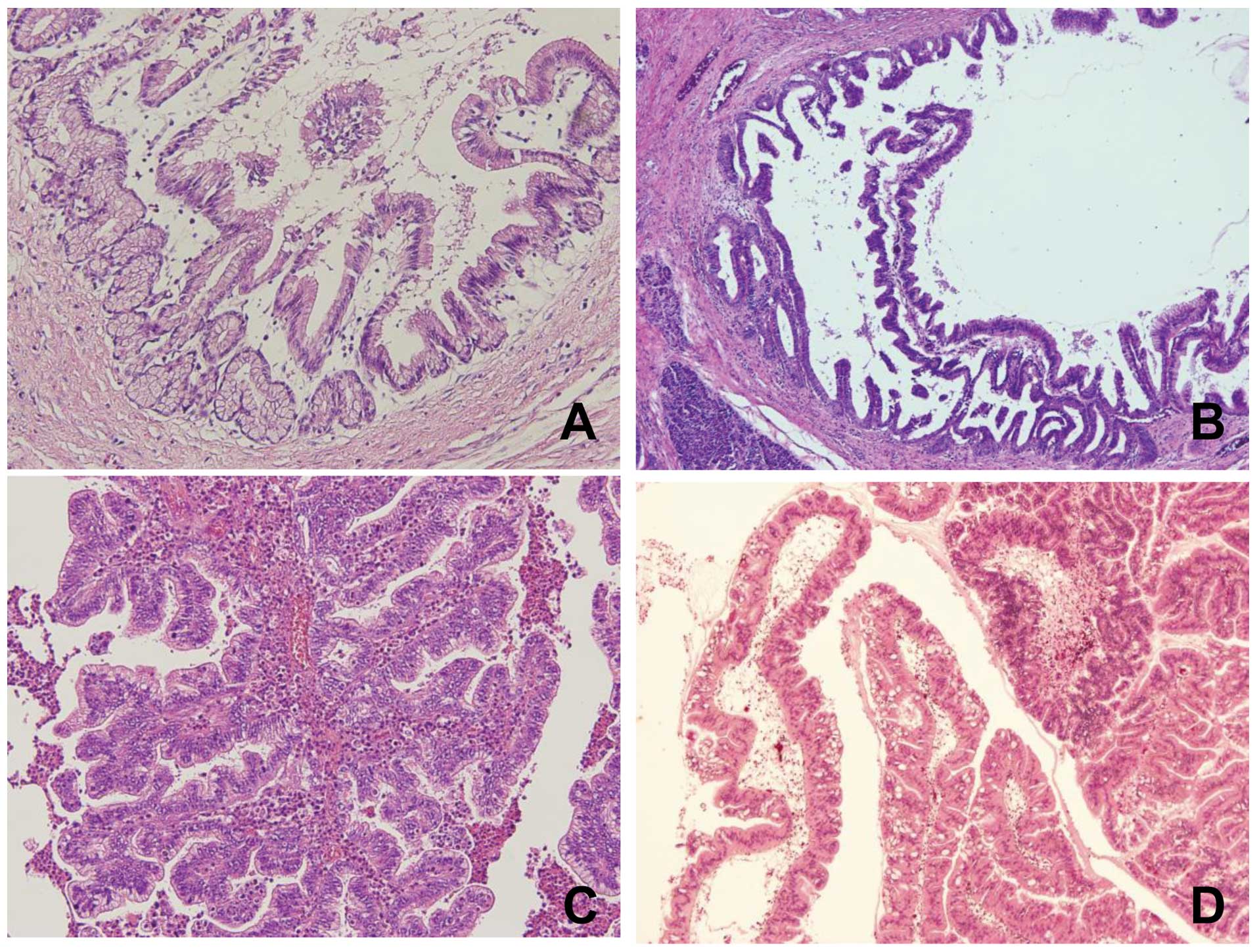
invasive IPMC. Morphologically, IPMNs were classified into gastric,
intestinal, pancreatobiliary and oncocytic types based on their
morphology on H&E- and immunohistochemically-stained sections
according to the report of Furukawa et al(8) (Fig.
1). At first, the subtype of the tumor was determined based on
the tumor morphology on H&E-stained sections. Then, this was
confirmed by immunohistochemical staining as an auxiliary method of
diagnosis. When the immunohistochemical staining pattern did not
correspond to the morphology observed by H&E staining, subtypes
were determined based on the morphological characteristics observed
by H&E staining. As has been shown to date, the gastric type is
positive for MUC-5AC, but negative for MUC-1 and MUC-2; the
intestinal type is positive for MUC-2 and MUC-5AC, but negative for
MUC-1; the pancreatobiliary type is partially positive for MUC-1
and positive for MUC-5AC, but negative for MUC-2; and the oncocytic
type is partially positive for MUC-1 and positive for MUC-5AC, but
negative for MUC-2 (8,15).
Immunohistochemical analysis
Paraffin sections were deparaffinized in xylene,
rehydrated in graded alcohol and transferred to phosphate-buffered
saline (PBS). Endogenous peroxidase was inactivated by incubating
the sections with 0.3% hydrogen peroxide for 30 min at room
temperature. Immunohistochemical staining was performed using a
Vectastain ABC kit (PK-4002; Vector Laboratories, Inc., Burlingame,
CA, USA). Mouse monoclonal antibody against human MUC-1 (clone
Ma695, IgG1; Leica Biosystems Newcastle, Ltd., Newcastle, UK),
mouse monoclonal antibody against human MUC-2 (clone Cep58, IgG1;
Leica Biosystems), and mouse monoclonal antibody against human
MUC-5AC (clone CLH2, IgG1; Leica Biosystems) were used as primary
antibodies at a 1:100 dilution. Peroxidase Substrate kits (SK-4100;
Vector Laboratories) were used for color development. The degree of
immunohistochemical staining was evaluated under a light microscope
at ×10 magnification. When the positive rate of tumor cells was
above or below 5%, the tumor was defined as being mucin-positive
and -negative, respectively.
Attempt at diagnosis of IPMN subtypes in
specimens obtained during preoperative examination
Peroral pancreatoscopy (POPS) can detect minute
intraductal lesions, and is reportedly useful especially in the
diagnosis of the grade of malignancy and degree of extension of
IPMNs in the main pancreatic duct (11). We have been using POPS in our center
since April 2011, and, in the present study, we determined IPMN
subtypes in four biopsy specimens obtained by means of POPS and
subjected them to H&E and immunohistochemical staining. In
addition, cytology specimens obtained during preoperative
endoscopic retrograde cholangiopancreatography (ERCP) were reused
employing the cell transfer technique (12), and subjected to immunocytochemical
staining using the same techniques as those employed for tissue
sections. Since cytology specimens could not be evaluated
morphologically, subtypes were determined as described for
immunohistochemical evaluation.
Statistical analysis
Fisher's exact test or the Chi-square test was used
to evaluate differences between the categorical variables. The
Kruskal-Wallis test was used for quantitative variables in three or
more groups. Survival curves were calculated using the Kaplan-Meier
method. Survival rates were compared using the log-rank test. A
P-value <0.05 was considered to indicate a statistically
significant result.
Results
Surgical treatment was performed in 57 patients with
IPMN, and a total of 60 lesions were examined. Of these lesions,
21, 28, 8 and 3 were of the gastric, intestinal, pancreatobiliary
and oncocytic types, respectively. One patient had 2 benign lesions
at the resection site: one was of the gastric type and the other
was of the intestinal type. Two patients with gastric-type lesions
had concomitant conventional pancreatic cancer. Two patients with
intestinal-type IPMN had recurrence in the remnant pancreas 7 or 9
years after surgery, and underwent resection of the recurrent
lesions, both of which were of the intestinal type. The number of
patients with oncocytic-type IPMN was only 3, making inter-type
comparisons difficult. However, no significant differences were
observed in the gender or age among the patients with different
subtypes. Of the 21 patients with gastric-type IPMN, 20 had adenoma
(IPMA), but 1 had non-invasive IPMC. In contrast, >70% of
patients with intestinal-type IPMN had malignant IPMN: 6 and 14 had
non-invasive IPMC and invasive IPMN, respectively, and 7 had benign
IPMN. The remaining 2 subtypes of IPMN exhibited malignant
features: all 8 patients with pancreatobiliary-type IPMN had
invasive IPMC with frequent extrapancreatic invasion. One and 2 of
the 3 patients with oncocytic-type IPMN had non-invasive and
invasive IPMC, respectively. There were significant differences
among the percentages of histological subtypes. Among the gross
classifications of IPMN, the gastric-type IPMN tended to be of the
branch-duct type, and the other 3 types of IPMN were significantly
more often of the main-duct type. However, some of these 3 types
were classified as the branch-duct type, and, in particular, the
intestinal type was classified as malignant in 3 of the 6 patients.
The diameters of gastric-, intestinal-, pancreatobiliary- and
oncocytic-type tumors were 14.9±6.4, 24.2±12.9, 33.2±18.6 and
23.0±9.9 mm, respectively, showing significant inter-type
differences. Histological nodules were significantly more
frequently observed in gastric-type than in other-type IPMNs, with
no significant difference in nodule diameter. Lymph node metastases
were found only in patients with invasive IPMC: in 2 of 19 patients
with intestinal-type IPMN and 3 of 8 patients with
pancreatobiliary-type IPMN. Two histological types of IPMC were
observed at the invasive front: invasive tubular carcinoma and
invasive colloid carcinoma. All pancreatobiliary and oncocytic-type
IPMNs were invasive tubular carcinoma, whereas 6 and 7 of 13 IPMCs
of the intestinal type were invasive tubular carcinoma and invasive
colloid carcinoma, respectively. A significant difference was noted
in the histological type at the invasive front between the
intestinal and pancreatobiliary types (Table I).
 | Table IMorphological types of IPMN and
clinicopathological features. |
Table I
Morphological types of IPMN and
clinicopathological features.
| Subtype | |
|---|
|
| |
|---|
| Gastric (21) | Intestinal (28) | Pancreatobiliary
(8) | Oncocytic (3) | P-value |
|---|
| Gender |
| Male/female | 17/4 | 19/9 | 5/3 | 2/1 | 0.49 |
| Age (years) | 67.1±7.2 | 67.2±8.4 | 61.8±8.1 | 77.5±3.5 | 0.082 |
| BD/MD/M | 16/4/0 | 7/17/3 | 2/6/0 | 0/2/1 | 0.002 |
| Tomor size (mm) | 14.5±6.1 | 28.3±17 | 31.8±20 | 23.0±9.9 | 0.008 |
| Nodule (−/+) | 13/7 | 5/20 | 1/4 | 0/3 | 0.014 |
| Nodule size (mm) | 9.29±6.7 | 12.6±9.4 | 12.3±10 | 16.0±7.2 | 0.76 |
| Histological
grade |
| A/NI/I | 20/1/0 | 7/6/14 | 0/0/8 | 0/1/2 | 0.0001 |
| Invasion pattern |
| Tubular/colloid | 0/0 | 6/7 | 8/0 | 2/0 | 0.018 |
| Nodal stage |
| pN0/pN1 | 21/0 | 26/2 | 5/3 | 3/0 | 0.029 |
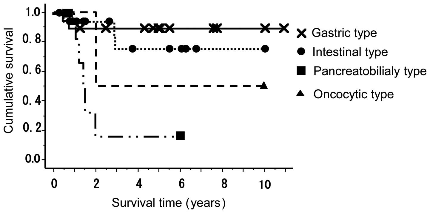
The 5-year cumulative survival rate of patients with
gastric-type IPMN was 91%. However, as described above, the 2
patients with gastric-type IPMN who died had concomitant
conventional pancreatic cancer, and the disease-specific survival
rate was 100%. On the other hand, patients with
pancreatobiliary-type IPMN had a very poor prognosis, with a 5-year
cumulative survival rate of 17%, which was significantly lower than
that of patients with other subtypes of IPMN. The 5-year cumulative
survival rate of patients with intestinal-type IPMN was 75%
(Fig. 2). No patients with adenoma
or carcinoma in situ died of their disease.
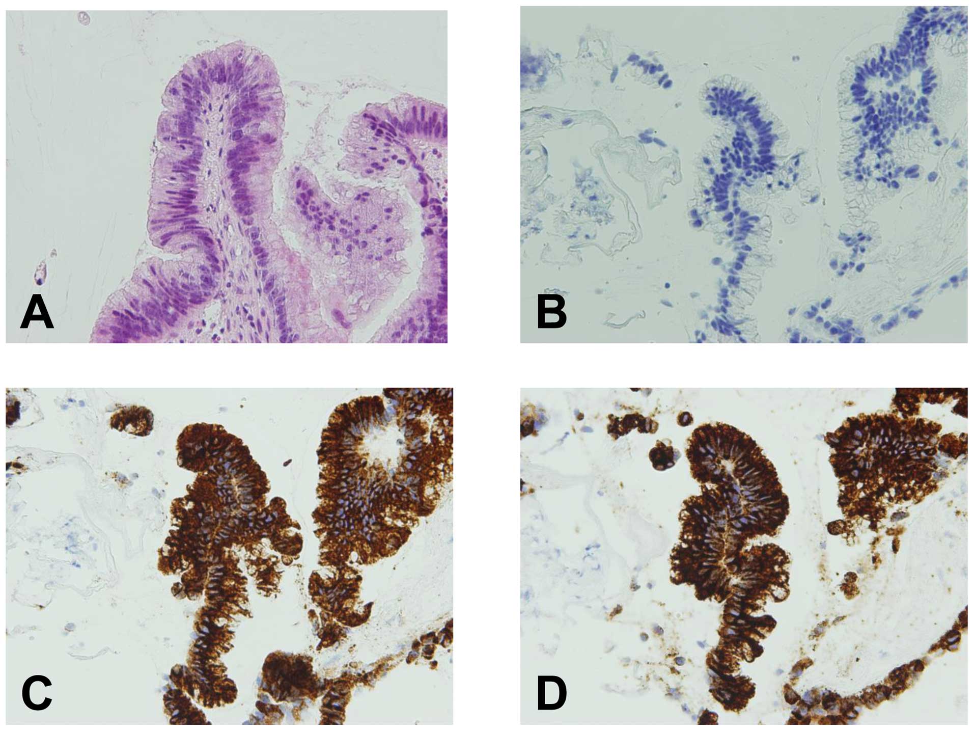
Of the 4 patients who had undergone biopsies during
POPS, three were diagnosed with IPMN, but one could not be
diagnosed because of limited tissue availability. The biopsy
specimens from the three patients were immunohistochemically
positive for MUC-2 and MUC-5AC, and negative for MUC-1, suggesting
intestinal-type IPMN (Fig. 3). The
results of mucin immunohistochemical staining of preoperative
biopsy and surgically resected specimens were in agreement with
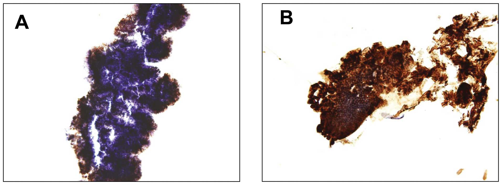
each other. In addition, cytology specimens obtained during
preoperative ERCP from 10 patients were reused employing the cell
transfer technique and subjected to immunocytochemical staining
(Fig. 4).
The 10 patients consisted of 4 with IPMA, 2 with
non-invasive IPMC and 4 with invasive IPMC. All 10 patients, except
1 with pancreatobiliary-type IPMN, had intestinal-type IPMN. Eight
of these 9 patients were positive for MUC-2 and MUC-5AC. These
results agreed closely with those of immunocytochemical staining.
Two of them had branch duct-type IPMN. Unlike the patients positive
for MUC-2 and MUC-5AC, the remaining 1 patient and the patient who
was histopathologically diagnosed with pancreatobiliary-type IPMN
were positive for MUC-5AC alone. On the other hand, the 4 patients
histologically diagnosed with IPMA had no evidence of malignancy
based on Papanicolau's classification and 3 of the 6 patients
histologically diagnosed with malignant IPMN were not so diagnosed
(Table II).
| Table IISummary of the results of cytological
and immunocytochemical analysis performed before surgery. |
Table II
Summary of the results of cytological
and immunocytochemical analysis performed before surgery.
| Papanicolaou's
classification | MUC-1 | MUC-2 | MUC-5AC | Histological
grade | Histologcal
subtype |
|---|
| 1. Class I | (−) | (+) | (+) | IPMA | Intestinal |
| 2. Class I | (−) | (−) | (+) | IPMA | Intestinal |
| 3. Class II | (−) | (+) | (+) | IPMA | Intestinal |
| 4. Class II | (−) | (+) | (+) | IPMA | Intestinal |
| 5. Class V | (−) | (+) | (+) | Non-invasive
IPMC | Intestinal |
| 6. Class V | (−) | (+) | (+) | Non-invasive
IPMC | Intestinal |
| 7. Class I | (−) | (+) | (+) | Invasive IPMC | Intestinal |
| 8. Class I | (−) | (+) | (+) | Invasive IPMC | Intestinal |
| 9. Class I | (−) | (+) | (+) | Invasive IPMC | Intestinal |
| 10. Class V | (−) | (−) | (+) | Invasive IPMC |
Pancreatobiliary |
Discussion
Of the 60 resected lesions, 21 and 28 were gastric-
and intestinal-type IPMNs, respectively, accounting for 80% of all
lesions. These results agreed closely with those previously
reported (9,14), and did not greatly differ in the
mean age or male-to-female ratio. These two subtypes still
predominate, although recent changes in the criteria for surgical
indications for IPMN have resulted in a gradual decrease in the
number of surgical resections for gastric-type IPMN in our center.
On the other hand, pancreatobiliary- and oncocytic-type IPMNs were
often locally invasive, and a large majority of them were difficult
to preoperatively differentiate from conventional pancreatic
cancer. Therefore, it is debatable whether surgery should be
performed for gastric- and intestinal-type IPMNs. Indeed, as
described above, the percentage of malignant gastric-type IPMNs was
significantly different from that of malignant intestinal-type
IPMNs.
The prognosis of all patients with adenoma or
carcinoma in situ was favorable, with a 5-year survival rate
of 100% (data not shown). Therefore, it is important to completely
resect a tumor in the stage of carcinoma in situ.
Gastric-type IPMNs were mostly branch-type lesions, had smaller
diameters, and less often formed nodules, which tended to be
smaller in diameter, than their other-type counterparts. Most
gastric-type IPMNs were benign and 91% of the resected lesions were
adenomas. In addition, one lesion diagnosed as cancer was
non-invasive IPMC, and no patients with gastric-type IPMN died of
the primary disease, with a disease-specific survival rate of 100%.
However, 2 patients with gastric-type IPMN had concomitant
conventional pancreatic cancer, necessitating more careful pre- or
postoperative follow-up of such patients. In the present study, one
of the 2 patients was diagnosed with branch-type IPMN, and
developed a conventional type of pancreatic cancer at a different
site during follow-up. Yamaguchi et al(13) noted that 9.2% of patients with IPMN
had concomitant conventional pancreatic cancer, and stated that
IPMN triggered the diagnosis of early pancreatic cancer. A study
reported that gastric-type IPMNs showed a significantly higher
incidence of KRAS mutations than their intestinal-type
counterparts, indicating an association with the development of
conventional pancreatic cancer (14). Some conventional pancreatic cancers
may develop on the basis of gastric-type IPMN. Compared with
patients with gastric-type IPMN, more patients with intestinal-type
IPMN had main-duct IPMN, with a higher percentage of malignant
tumors. Consensus has been reached as to the need for the surgical
treatment of main-duct IPMN. Indeed, many of the main-duct IPMNs
are thought to be of the intestinal type. The problem here is that
some branch-duct IPMNs are of the intestinal type (9,15). In
the present study, 6 of the 27 intestinal-type IPMNs were of the
branch-duct type, and half of them were classified as malignant.
How to differentiate such IPMNs is a key point for precise
treatment. Invasive cancers derived from intestinal-type IMPC were
invasive tubular carcinoma or invasive colloid carcinoma at the
invasive front, which were observed with the same frequency.
However, histologically, no difference was noted in vascular
invasion, lymph node metastasis or neural invasion (data not
shown). A study by Furukawa et al(9) reported that intestinal-type IPMNs,
many of which were invasive colloid carcinomas, were associated
with a more favorable prognosis than pancreatobiliary- and
oncocytic-type IPMNs, many of which were invasive tubular
carcinomas. Their results are in agreement with ours. The degree of
malignancy of intestinal-type IPMNs may be lower than that of IPMNs
of other types except the gastric type.
All pancreatobiliary-type IPMNs were advanced
carcinoma and were associated with the poorest prognosis. In this
sense, this type is considered the most malignant, which is in
agreement with previous reports (9,15).
Only pancreatobiliary-type IPMNs were often locally invasive, and
some patients with this type of IPMN underwent extended
pancreatectomy or preoperative chemoradiotherapy. Since the
prognosis of pancreatobiliary-type IPMN is as poor as that of
conventional pancreatic cancer, it is imperative to establish an
adjuvant therapy, but, to date, no effective treatment has been
reported. Although its low incidence precludes a large-scale study,
the treatment of this condition is an important problem that should
be addressed in the future.
Although the number of oncocytic-type IPMNs was only
3, the degree of their malignancy was high and often advanced, as
has previously been reported. The 2 oncocytic-type, invasive IPMNs
were invasive tubular carcinoma at the invasive front. A study of a
large cohort of patients with oncocytic-type IPMN reported that
many of these tumors were of the oncocytic type at the invasive
front (16). Although not observed
in the present study, such histological features may be observed at
the invasive front in a study involving a larger number of
patients.
Currently, treatment strategies for IPMN are based
on the presence or absence of symptoms and a comprehensive
evaluation of imaging findings, as defined by the guidelines.
Recently, efforts to obtain information for a decision on treatment
strategies have been reported. Hirono et al(17) measured carcinoembryonic antigen
(CEA) levels in pancreatic juice collected during preoperative
ERCP, and reported that pancreatic CEA levels were high in patients
with malignant tumors. Maker et al(18) measured mucin protein levels in
intraoperatively collected pancreatic juice samples, and reported
that MUC-2 and MUC-4 protein levels were elevated in patients with
highly malignant tumors, indicating a close relationship between
the levels of these proteins and degree of malignancy, and
suggesting the usefulness of these markers for the selection of
patients for surgical treatment. In the present study, the degree
of malignancy depended on the subtype of the tumor, suggesting that
the diagnosis of IPMN subtypes plays a major role in the
determination of treatment strategies for IPMN. Therefore, we
attempted to diagnose IPMN subtypes by immunohistochemical analysis
of preoperative cytology specimens. Although the number of
specimens was small, the diagnoses in preoperative biopsy and
pancreatic juice cytology specimens were in close agreement with
those in resected specimens, suggesting that these methods are very
useful for the diagnosis of IPMN subtypes. These results may play a
large role in the surgical treatment of IPMN. For example, surgery
should be considered even for small, branch duct-type lesions on
imaging, if they are of the highly malignant subtypes other than
the gastric type. As also shown in the present study, the majority
of highly malignant, intestinal-type IPMNs were of the main-duct
type, but some of them were of the branch-duct type and malignant,
making it difficult to distinguish these IPMNs by imaging alone. If
these IPMNs can be distinguished by the immunocytochemical analysis
of cells in the pancreatic juice collected during preoperative
ERCP, it may become possible to perform more precise surgical
treatment. In the present study, despite the reuse of relatively
old specimens, strong MUC-2 expression was observed, which was in
good agreement with the histological features. Immunocytochemically
detected MUC-2-positive cells are very likely to be those of the
intestinal type, as diagnosed immunohistochemically. However, the
immunocytochemical stainability of MUC-1 cannot be evaluated at
this point. The level of MUC-1 expression in pancreatobiliary- and
oncocytic-type IPMNs, as detected by immunohistochemistry, is
thought to be very low (5,15); therefore, it is expected to be
fairly difficult to determine the histological type of IPMN based
on the immunohistochemical expression of MUC-1. Since, in the
present study, many of these two types of IPMN were malignant and
advanced, we did not hesitate in the choice of surgical treatment.
However, studies reported that these IPMNs were resected in the
carcinoma in situ stage (5,15).
Therefore, their diagnosis is an issue to be resolved in the
future. At this point, immunocytochemistry is very useful in
distinguishing gastric-type from intestinal-type IPMN. Needless to
say, immunohistochemistry is difficult to perform in the case of a
small pancreatic duct diameter or branch duct-type IPMNs, and
immunocytochemical diagnosis may be impossible in specimens with a
very small tumor component; thus, these diagnostic techniques
cannot be applied to all cases. However, in recent years,
examination devices in the field of pancreatic diseases have made
progress, and, if thin pancreatic ducts become accessible, more
precise information can be obtained.
In conclusion, we classified resected IPMNs into
subtypes and clarified their clinicopathological characteristics. A
high percentage of IPMNs other than the gastric type were
malignant, and, in particular, many of the pancreatobiliary-type
IPMNs were in an advanced stage, with a very poor prognosis. Many
IPMNs classified as the main duct type on imaging were of the
highly malignant subtypes other than the gastric type, and were
indicated for surgery, but it should be borne in mind that some of
them were of the branch-duct type and malignant. In the future, it
is hoped that preoperative tests for their differential diagnosis
will be established. The immunohistochemical staining of
preoperative biopsy specimens and ERCP-obtained pancreatic juice
may be useful in the differential diagnosis of gastric and
intestinal types of IPMNs. If immunocytochemistry enables the
preoperative diagnosis of IPMN subtypes, its use in combination
with conventional preoperative imaging modalities may lead to
surgical treatment best suited for the biological characteristics
of the four subtypes.
References
|
1
|
Adsay NV, Conlon KC, Zee SY, Brennan MF
and Klimstra DS: Intraductal papillary-mucinous neoplasms of the
pancreas: an analysis of in situ and invasive carcinoma in 28
patients. Cancer. 94:62–77. 2002. View Article : Google Scholar : PubMed/NCBI
|
|
2
|
Furuta K, Watanabe H and Ikeda S:
Differences between solid and duct-ectatic types of pancreatic
ductal carcinomas. Cancer. 69:1327–1333. 1992. View Article : Google Scholar : PubMed/NCBI
|
|
3
|
Tanaka M, Chari S, Adsay NV, Fernandez-del
Castillo C, Falconi M, Shimizu M, Yamaguchi K, Yamao K and Matsuno
S: International consensus guidelines for management of intraductal
papillary mucinous neoplasms and mucinous cystic neoplasms of the
pancreas. Pancreatology. 6:17–32. 2006. View Article : Google Scholar
|
|
4
|
Longnecker DS, Adler G, Hruban RH, et al:
Intraductal papillary mucinous neoplasms of the pancreas. World
Health Organization Classification of Tumors. Pathology and
Genetics of the Digestive System. Hamilton SR and Aaltonen LA: IARC
Press; Lyon: pp. 237–240. 2000
|
|
5
|
Sugiyama M, Suzuki Y, Abe N, Mori T and
Atomi Y: Management of intraductal mucinous neoplasm of the
pancreas. J Gastroenterol. 43:181–185. 2008. View Article : Google Scholar
|
|
6
|
Tanaka M: Intraductal papillary mucinous
neoplasm of the pancreas, diagnosis and treatment. Pancreas.
28:282–288. 2004. View Article : Google Scholar : PubMed/NCBI
|
|
7
|
Kimura W, Futakawa N, Yamagata S, Wada Y,
Kuroda A and Esaki Y: Different clinicopathologic findings in two
histologic types of carcinoma of papilla of Vater. Jpn J Cancer
Res. 85:161–166. 1994. View Article : Google Scholar : PubMed/NCBI
|
|
8
|
Furukawa T, Klöppel G, Volkan Adsay N,
Albores-Saavedra J, Fukushima N, Horii A, Hruban RH, Kato Y,
Klimstra DS, Longnecker DS, Lüttges J, Offerhaus GJ, Shimizu M,
Sunamura M, Suriawinata A, Takaori K and Yonezawa S: Classification
of types of intraductal papillary-mucinous neoplasm of the
pancreas: a consensus study. Virchows Arch. 47:794–799. 2005.
View Article : Google Scholar : PubMed/NCBI
|
|
9
|
Furukawa T, Hatori T, Fujita I, Yamamoto
M, Kobayashi M, Oike N, Morohoshi T, Egawa S, Unno M, Takao S,
Osako M, Yonezawa S, Mino-kenudson M, Lauwers GY, Yamaguchi H, Ban
S and Shimizu M: Prognostic relevance of morphological types of
intraductal papillary mucinous neoplasms of the pancreas. Gut.
60:509–516. 2011. View Article : Google Scholar : PubMed/NCBI
|
|
10
|
Japan Pancreas Society. General Rules for
the Study of Pancreatic Cancer. 6th edition. Kanehara; Tokyo:
2009
|
|
11
|
Yamaguchi T, Hara T, Ysuyaguchi T,
Ishihara T, Tsuchiya S and Saisho H: Peroral pancreatoscopy in the
diagnosis of mucin-producing tumors of the pancreas. Gastrointest
Endosc. 52:67–73. 2000. View Article : Google Scholar : PubMed/NCBI
|
|
12
|
Brown GG and Tao LC: Restoration of broken
cytology slides and creation of multiple slides from a single smear
preparation. Acta Cytol. 36:259–263. 1992.PubMed/NCBI
|
|
13
|
Yamaguchi K, Ouchida J, Ohtsuka T, Nakano
K and Tanaka M: Intraductal papillary-mucinous tumor of the
pancreas concomitant with ductal carcinoma of the pancreas.
Pancreatology. 2:484–490. 2002. View Article : Google Scholar : PubMed/NCBI
|
|
14
|
Mohri D, Asaoka Y, Ijichi H, Miyabayashi
K, kudo Y, Seto M, Ohta M, Tada M, Tanaka Y, Ikenoue T, Tateishi K,
Isayama H, Kanai F, Fukushima N, Tada M, Kawabe T, Omata M and
koike K: Different subtypes of intraductal papillary mucinous
neoplasm in the pancreas have distinct pathways to pancreatic
cancer progression. J Gastroenterol. 47:203–213. 2012. View Article : Google Scholar
|
|
15
|
Takasu N, Kimura W, Moriya T, Hirai I,
Takeshita A, Kamio Y and Nomura T: Intraductal papillary-mucinous
neoplasms of the gastric and intestinal type may have less
malignant potential than the pancreatobiliary type. Pancreas.
39:604–610. 2010. View Article : Google Scholar : PubMed/NCBI
|
|
16
|
Mino-Kenudson M, Fernandez-del Castillo C,
Baba Y, Valsangkar NP, Liss AS, Hsu M, Correa-Gallego C, Ingkakul
T, Johnston RP, Turner BG, Androutsopoulos V, McGrath D, Sahani DV,
Brugge WR, Ogino S, Pitman MB, Warshaw AL and Thayer S: Prognosis
of invasive intraductal papillary mucinous neoplasm depends on
histological and precursor epithelial subtypes. Gut. 60:1712–1720.
2011. View Article : Google Scholar : PubMed/NCBI
|
|
17
|
Hirono S, Tani M, Kawai M, Ina S, Nishioka
R, Miyazawa M, Fujita Y, Uchiyama K and Yamaue H: Treatment
strategy for intraductal papillary mucinous neoplasm of the
pancreas based on malignant predictive factors. Arch Surg.
144:345–349. 2009. View Article : Google Scholar : PubMed/NCBI
|
|
18
|
Maker AV, Katabi N, Gonen M, DeMatteo RP,
D'Angelica MI, Fong Y, Jarnagin WR, Brennan MF and Allen PJ:
Pancreatic cyst fluid and serum mucin levels predict dysplasia in
intraductal papillary mucinous neoplasms of the pancreas. Ann Surg
Oncol. 18:199–206. 2011. View Article : Google Scholar : PubMed/NCBI
|