Introduction
Oral cancer is a life-threatening disease causing
~500,000 deaths annually worldwide (1,2).
Chemotherapy is commonly used to treat a variety of oral cancers,
generally in combination with surgery or radiation (3). One of the chemotherapeutic agents
often used is cis-platinum (II) diammine dichloride (CDDP),
a platinum-based compound. However, like most anticancer agents,
CDDP has potentially severe adverse effects such as nephrotoxicity
(4), neurotoxicity (5), nausea and vomiting (6) and ototoxicity (7). One of its adverse effects in the head
and neck region is xerostomia (dry mouth) (8,9).
Xerostomia results from hypofunction of the salivary
glands and can be caused by several physiological and iatrogenic
factors, autoimmune disorders and infection (10,11).
Although the mechanism and therapeutics of the disease are yet to
be fully elucidated, salivary gland dysfunction can lead to
impaired speech, loss of taste perception and appetite, and
infection, thereby reducing quality of life. Hence most physicians
and basic scientists recognize this disorder as an important issue
for head and neck cancer patients undergoing chemotherapy.
Traditional Chinese medicine has been used for a
variety of diseases for several thousands of years. With this in
mind, an inter-institutional collaborative project involving Showa
University and Tokyo University of Marine Science and Technology
was launched in 2010, in order to elucidate herbal extracts as
potential therapeutic candidates for disorders of the head-and-neck
region (12). The project examined
more than 400 bioactive herbal products. After a preliminary
experiment, in which herbal extracts were examined for prevention
of salivary gland acinar cell death, the root bark of Juncus
effusus (J. effusus, commonly known as soft rush) and
Paeonia suffruticosa (P. suffruticosa, commonly known
as Chinese tree peony) were focused on.
The present in vitro study demonstrated that
the methanolic extracts of these herbs are capable of preventing
apoptotic cell death from CDDP in salivary gland acinar cells, but
not in carcinoma cells. The findings suggest that these herbal
extracts may have potential as novel therapeutic agents for
xerostomia, and may accordingly improve quality of life during
chemotherapy for head and neck cancer patients.
Materials and methods
Preparation of the root bark. J. effusus and P.
suffruticosa were cultivated in China, and the herbal extracts
were prepared in that country before being imported to Japan. A
specimen of each was deposited in the herbarium of the Tokyo
University of Marine Science and Technology. Dry powdered roots
(100 g) were extracted by distilled water and concentrated to 1
mg/ml under reduced pressure.
Cell culture
An immortalized human salivary gland acinar cell
line (NS-SV-Ac), a kind gift from Dr Masayuki Azuma (University of
Tokushima, Japan), was cultured as described elsewhere (13). Acc 2 and Acc M (human adenocystic
carcinoma cell lines) were cultured as described previously
(14). All cells were grown at 37°C
in an atmosphere containing 5% CO2 and 100%
humidity.
Histochemistry
The cells were seeded at a density of
3×10³ cells/well in 48-well cell culture plates. The
next day, 20 μg/ml of CDDP (Nippon Kayaku, Tokyo, Japan) with or
without the herbal extracts (at concentrations of 1 and 10 μg/ml
adjusted by phosphate-buffered saline) were added to the medium.
After 3 days, the cells were fixed and stained with crystal violet
or toluidine blue, as described previously (15).
Cell viability and apoptosis assays
For the
3-[4,5-dimethylthiazol-2-yl]-2,5-diphenyltetrazolium bromide (MTT)
assay, the cells were seeded at a density of 1×10³
cells/well in 96-well cell culture plates. They were treated as
described above, and the MTT assay was performed as described
previously (16). The activities of
caspase 3/7, 8 and 9 were measured using the Caspase-Glo Assay and
GloMax-Multi Plus Detection System (both from Promega Corporation,
Madison, WI, USA), according to the manufacturer’s protocol.
Genomic DNA fragmentation was investigated using a DNA ladder
assay; this was performed with a commercial kit (ApopLadder EX;
Takara, Shiga, Japan), according to the manufacturer’s
protocol.
Western blot analysis
Total cellular protein was prepared as described
previously (17), and the protein
concentration was measured with Quick Start Bradford reagent
(Bio-Rad, Hercules, CA, USA) using bovine serum albumin as a
standard, and aliquots were stored at −80°C until use. Protein
samples (20 μg) were subjected to sodium dodecyl
sulfate-polyacrylamide electrophoresis (SDS-PAGE) in 4–20% gradient
gel (Bio-Rad), and the blots were transferred onto a polyvinylidene
difluoride membrane (Life Technologies, Carlsbad, CA, USA). The
blots were blocked, incubated with primary and horseradish
peroxidase-conjugated secondary antibodies, and washed as
previously described (17).
Subsequently, the signal was visualized using Amersham ECL western
blotting detection reagents (GE Healthcare, UK Ltd.,
Buckinghamshire, UK) and the ChimiDoc XRS Plus ImageLab System
(Bio-Rad). The primary antibodies were purchased from Cell
Signaling Technology, Inc. (Danvers, MA, USA) and Santa Cruz
Biotechnology, Inc. (Santa Cruz, CA, USA), and secondary antibodies
were purchased from GE Healthcare.
Dual luciferase assay (DLA)
Three firefly luciferase reporter vectors (pGL4.24,
pGL4.32 and pGL4.38) were purchased from Promega Corporation.
pGL4.32 and pGL4.38 contain the human κB element and p53 response
element respectively, in a promoter legion of the firefly
luciferase gene, while pGL4.24 has only a minimal promoter (minP)
in the corresponding region. pRL-TK (Promega Corporation) was used
as an internal control for transfection, as described previously
(18). We seeded 5,000 cells in a
96-well culture plate, and the next day, the cells were transfected
with 200 ng of pGL4.24, pGL4.32, or pGL4.38 plus 10 ng of pRL-TK
using Lipofectamine 2000 (Life Technologies), and cultured as
described above. After 3 days, the DLA was performed with the
Dual-Luciferase Reporter Assay System and GloMax-Multi Detection
System (both from Promega Corporation), according to the
manufacturer’s protocol.
Statistical analysis
Unless otherwise specified, all experiments were
repeated at least 3 times and similar results were obtained in the
repeated experiments. Statistical analysis was performed using
repeated measure analysis of paired Student’s t-tests. Data are
expressed as means ± standard deviation of triplicate data.
Results
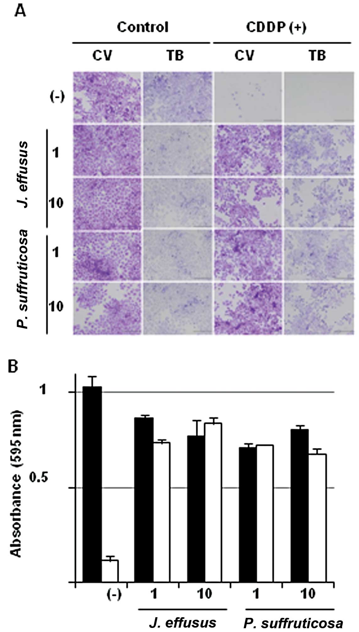
Extracts of J. effusus and P.
suffruticosa protect NS-SV-Ac cells from cytotoxicity induced by
CDDP
As described in our recent study (12), more than 400 bioactive herbal
extracts were subjected to in vitro preliminary screening in
which NS-SV-Ac cells were cultured for 3 days in the presence of
the extracts plus 20 μg/ml of CDDP (data not shown). That study
identified 2 herbal extracts, J. effusus and P.
suffruticosa, as being able to protect cells from CDDP-induced
cell death, at a minimum concentration of 1 μg/ml (Fig. 1A). The present MTT assay reinforced
the histochemical results by showing that 20 μg/ml of CDDP was
sufficiently cytotoxic to induce cell death in NS-SV-Ac cells, but
that cell viability was protected in the presence of 1 or 10 μg/ml
of either extract (Fig. 1B).
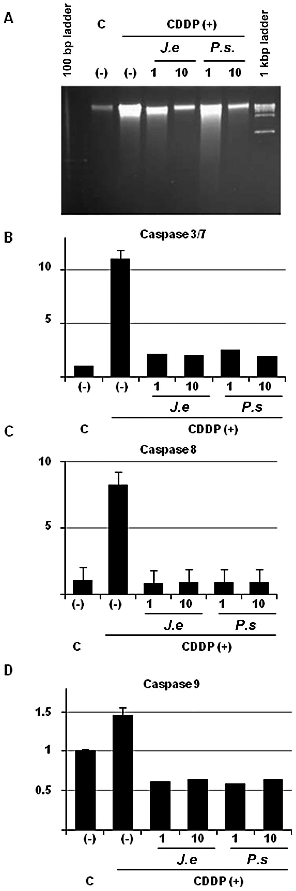
Herbal extracts prevent NS-SV-Ac cells
from apoptosis induced by CDDP
It is well known that CDDP inhibits the synthesis of
DNA and induces apoptotic cell death (4–9). The
DNA ladder assay showed that the genomic DNA of NS-SV-Ac cells in
the CDDP-containing medium was fragmented, and indicated that the
cell death was caused by apoptosis (Fig. 2A). However, addition of the herbal
extracts reduced this fragmentation in a dose-dependent manner
(Fig. 2A). Similarly, although the
activities of caspase 3/7, 8 and 9 were increased by CDDP, addition
of the herbal extracts reduced all these activities to basal
levels, even at the concentration of 1 μg/ml (Fig. 2B–D). The results suggest that the
herbal extracts have the pharmacological capability to protect
normal salivary gland acinar cells from apoptotic cell death
induced by CDDP.
Herbal extracts prevent apoptosis by
upregulating Bcl-2 and Bcl-XL via activation of the Akt
pathway
Next, we investigated the protein(s) involved in the
preventive effects of the herbal extracts on apoptosis.
Accordingly, western blotting for various apoptosis-related
proteins was performed (Fig. 3A).
Addition of CDDP resulted in cleavage of poly(ADP-ribose)
polymerase (PARP) 1; however, this cleavage was almost completely
inhibited by the herbal extracts, regardless of an increase in p53
protein concentration. Regarding mitochondrial apoptosis-related
proteins, expressions of the pro-apoptotic proteins Bax, Bad, Bid
and Bak were very similar among the samples. However, it is
noteworthy that addition of the herbal extracts significantly
increased the expression of Bcl-2 and Bcl-XL
(pro-survival proteins). Expression of other apoptosis-related
proteins was modulated only slightly, if at all, by the extracts
(e.g., FLIP showed minor upregulation, whereas expression of Mcl-1
and XIAP remained very similar). Furthermore, although
phosphorylation of protein kinase B/Akt 1 (Akt 1), which plays a
crucial role in cell survival, was barely observed after addition
of CDDP, this was prevented by addition of the extracts. In
contrast, phosphorylation of nuclear factor-κB (NF-κB)-related
proteins was slightly modulated by the herbal extracts. Next, we
used DLA to investigate the involvement of promoter activity of the
p53 response element and κB element (Fig. 3B). CDDP increased p53 response
element activity, and this activation was abolished by the
extracts. Meanwhile, κB element activity was not modulated by CDDP,
although it was increased slightly by the extracts. The reporter
activity of pGL4.24, a negative control, was not altered,
regardless of the presence or absence of CDDP or the extracts.
Therefore, the results suggest that the reporter activity depends
on the promoter sequence of each vector. Taken together, these
findings suggest that CDDP decreases the phosphorylation of Akt 1
and induces apoptosis through the p53 and mitochondrial apoptotic
pathways, not through NF-κB. The results also suggest that the
herbal extracts are capable of interrupting the p53 and
mitochondrial apoptotic pathways, thereby preventing apoptosis.
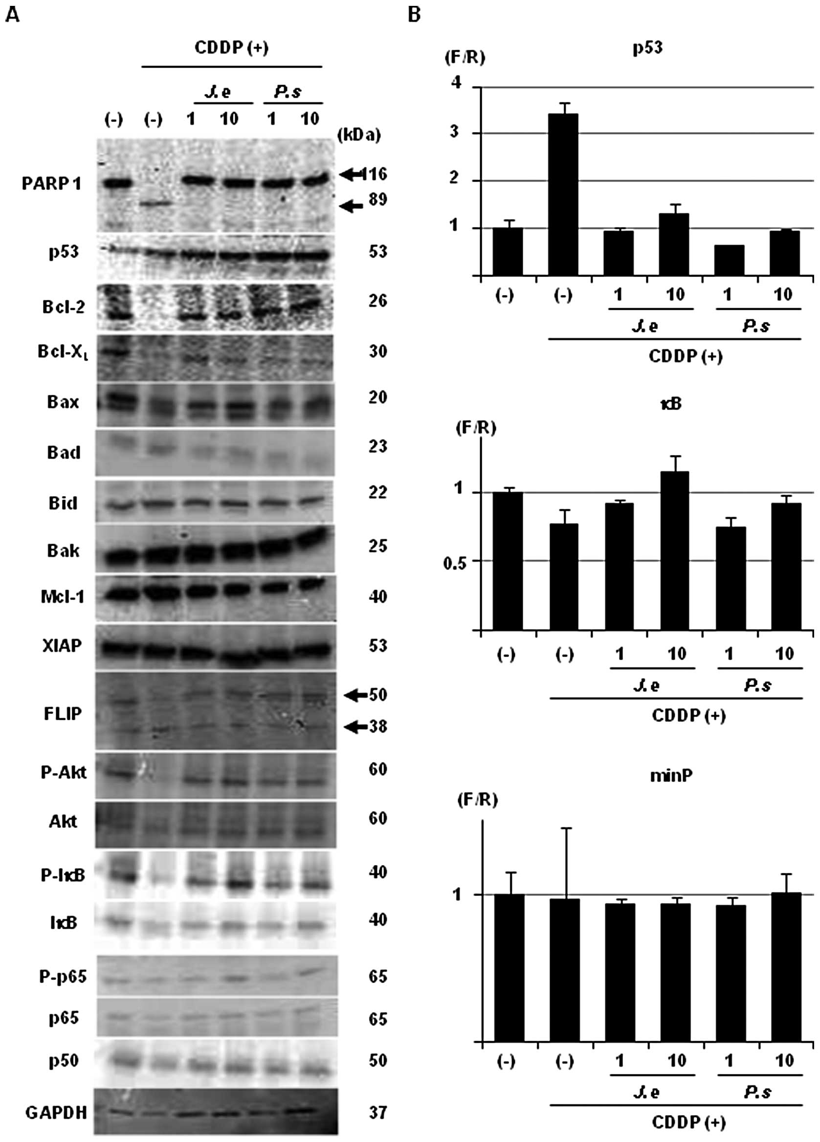 | Figure 3Herbal extracts prevent salivary gland
cells from p53-, Bcl-2/Bax- and Akt 1-mediated apoptosis induced by
CDDP. (A) NS-SV-Ac cells were grown in the absence (−) or presence
of 1 or 10 μg/ml of J. effusus (J. e) or P.
suffruticosa (P. s) for 3 days, and the medium was then
replaced with fresh control or CDDP-containing medium [CDDP (+)].
After 12 h, total cellular protein was purified and subjected to
western blotting for 19 apoptosis-related proteins [PARP 1, p53,
Bcl-2, Bcl-XL, Bax, Bad, Bid, Bak, Mcl-1, XIAP, FLIP,
phosphorylated (P)-Akt, Akt, P-IκB, IκB, P-p65, p65 and p50] as
well as for GAPDH as an internal control. The molecular weights of
interest are shown at the right adjacent side of the panel in kDa.
(B) The cells were treated as described above, and were then
subjected to a dual luciferase assay for the p53 response element
and κB element, and for a minimal promoter (minP) as a negative
control. CDDP, cis-platinum (II) diammine dichloride. |
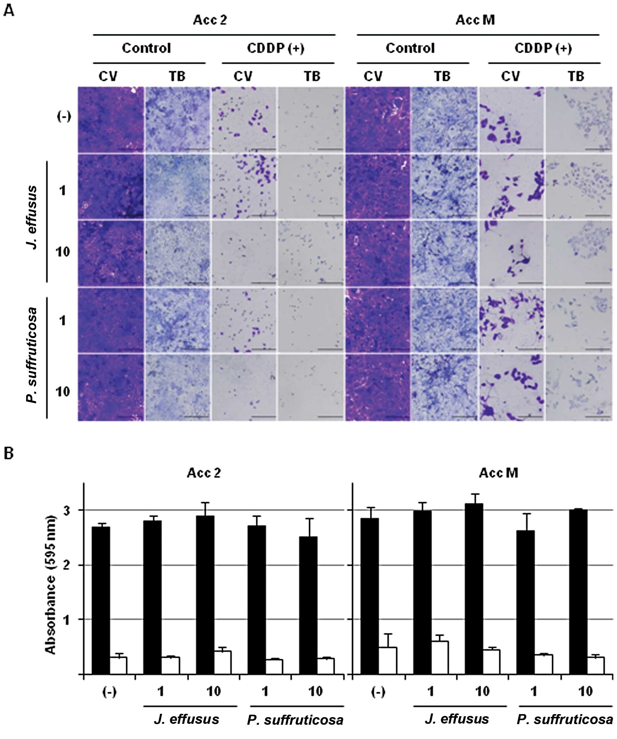
The herbal extracts show no
apoptosis-preventive effects in adenocystic carcinoma cell
lines
In our pilot study (data not shown), several herbal
extracts showed a proliferative effect not only in normal cells,
but also in malignant cell lines. The magnitude of this effect
varied among the herbs tested. We therefore investigated whether
the extracts of J. effusus and P. suffruticosa
exhibit anti-apoptotic or proliferative effects in malignant cells
originating from salivary gland acini. In this experiment, we
utilized 2 adenocystic carcinoma cell lines, Acc 2 and Acc M. As
shown in Fig. 4A, in these cell
lines CDDP induced marked cell death, which was probably apoptotic.
Of note, the herbal extracts showed no protective effect on this
cell death, even at the concentration of 10 μg/ml. The MTT assay
(Fig. 4B) showed the same result as
observed upon histochemistry; i.e., the extracts did not inhibit
the CDDP-induced apoptotic effect. These results suggest a
potential advantage of the herbal extracts as therapeutic agents
for xerostomia during chemotherapy for CDDP.
Discussion
Oral cancer patients usually undergo chemotherapy in
combination with surgery and radiation. Since CDDP
pharmacologically impairs DNA synthesis and transcription, it
exerts cytotoxic and apoptotic effects not only on cancer cells but
also on normal cells. As a result, the salivary glands are damaged,
their function is inhibited, and patients undergoing this type of
chemotherapy often develop xerostomia (10,11).
As chemotherapy-induced xerostomia leads to great restriction of
quality of life, it is important to identify nontoxic agents
capable of counteracting the effect of chemotherapy on salivary
gland function. Several studies have reported that Chinese
traditional herbs (19) and natural
agents (20) are available for the
treatment of xerostomia. Therefore, in the present institutional
collaborative project, we explored herbal extracts that may have
therapeutic effects not only on xerostomia, but also on oral and
skeletal diseases (12).
J. effusus is a species of the Juncus
plant, and traditionally is used as an anti-pyretic and
anti-phlogistic agent (21,22). On the other hand, P.
suffruticosa is used for atherosclerosis, infection,
inflammation, cutaneous disease (23) and diabetes (24), and is also reported to possess
potent anti-oxidant, anti-mutagenic, anti-proliferative,
anti-invasive, anti-arrhythmic, anti-inflammatory, anti-diabetic
and anti-obesity properties (25).
While previous reports on these herbs have addressed
pharmacological availability, we herein revealed that these
extracts are capable of preventing CDDP-induced cell death, which
is mainly caused by apoptosis, in normal salivary gland cells
(Figs. 1 and 2).
p53 (26), Akt 1
(27), several mitochondrial
proteins (e.g., Bcl-2 and Bax) (28) and NF-κB (29) are well known as key regulatory
molecules during apoptotic cell death. In the present study,
expression of p53, Akt 1, and apoptosis-related mitochondrial
proteins was modulated during CDDP-induced apoptosis, and these
changes were prevented by the extracts, while changes in NF-κB were
not induced by CDDP (Fig. 3A and
B). These finding support those of Azuma et al(30), who reported that CDDP-induced
apoptosis was independent of the NF-κB pathway.
Finally, but most importantly, given our goal of
using the extracts to treat xerostomia during chemotherapy, we
investigated whether the herbal extracts had an anti-apoptotic
effect on malignant cells similar to that observed in NS-SV-Ac
cells. Neither histochemistry (Fig.
4A) nor MTT assay (Fig. 4B)
showed that the death of malignant cells was prevented, suggesting
that these extracts could have potential clinical benefit. The
detailed mechanism of the cell-specific preventive effect on
apoptosis remains to be further elucidated. Nonetheless, our
previous study (12), in addition
to previous studies examining a number of herbs, revealed that
various herbal extracts have anticancer effects. Hence, one may
hypothesize a switching mechanism between the triggering of and
prevention of apoptosis, depending on the type of cell. In any
case, further investigation, including isolation and analysis of
bioactive chemicals, detailed molecular and cellular experiments
in vitro, and pre-clinical studies in vivo, is of
course required. Several of these studies are currently underway,
and our findings will be reported in the near future.
Acknowledgements
This study was supported by Grants-in-Aid for
Scientific Research (KAKENHI) (B) (to S.H.) and (C) (to Y.M. and
S.K.) from the Japan Society for the Promotion of Science (JSPS),
and the Nakatomi Foundation (to Y.M.). The authors wish to thank
Drs Yasuto Yoshihama, Tatsuo Shirota, Hiroaki Kamatani, Yasumasa
Yoshizawa, Tomohiko Kutsuna, Sayaka Yoshiba, Daisuke Soga, Daisuke
Sato, Rika Nagasaki, Ryota Kishigami, and Yuji and Sayaka Kurihara
for their helpful suggestions and Ms. Miho Yoshihara for the
secretarial assistance.
References
|
1
|
Choi S and Myers JN: Molecular
pathogenesis of oral squamous cell carcinoma: implications for
therapy. J Dent Res. 87:14–32. 2008. View Article : Google Scholar : PubMed/NCBI
|
|
2
|
Scully C and Bagan JV: Recent advances in
oral oncology 2008; squamous cell carcinoma imaging, treatment,
prognostication and treatment outcomes. Oral Oncol. 45:e25–e30.
2009. View Article : Google Scholar : PubMed/NCBI
|
|
3
|
Price KA and Cohen EE: Current treatment
options for metastatic head and neck cancer. Curr Treat Options
Oncol. 13:35–46. 2012. View Article : Google Scholar : PubMed/NCBI
|
|
4
|
dos Santos NA, Carvalho Rodrigues MA,
Martins NM and dos Santos AC: Cisplatin-induced nephrotoxicity and
targets of nephroprotection: an update. Arch Toxicol. 86:1233–1250.
2012.PubMed/NCBI
|
|
5
|
McWhinney SR, Goldberg RM and McLeod HL:
Platinum neurotoxicity pharmacogenetics. Mol Cancer Ther. 8:10–16.
2009. View Article : Google Scholar
|
|
6
|
Herrstedt J: Antiemetics: an update and
the MASCC guidelines applied in clinical practice. Nat Clin Pract
Oncol. 5:32–43. 2008. View Article : Google Scholar : PubMed/NCBI
|
|
7
|
Rybak LP, Mukherjea D, Jajoo S and
Ramkumar V: Cisplatin ototoxicity and protection: clinical and
experimental studies. Tohoku J Exp Med. 219:177–186. 2009.
View Article : Google Scholar : PubMed/NCBI
|
|
8
|
Rapidis AD, Trichas M, Stavrinidis E, et
al: Induction chemotherapy followed by concurrent chemoradiation in
advanced squamous cell carcinoma of the head and neck: final
results from a phase II study with docetaxel, cisplatin and
5-fluorouracil with a four-year follow-up. Oral Oncol. 42:675–684.
2006.PubMed/NCBI
|
|
9
|
Psyrri A, Kwong M, DiStasio S, et al:
Cisplatin, fluorouracil, and leucovorin induction chemotherapy
followed by concurrent cisplatin chemoradiotherapy for organ
preservation and cure in patients with advanced head and neck
cancer: long-term follow-up. J Clin Oncol. 22:3061–3069. 2004.
View Article : Google Scholar
|
|
10
|
Jensen SB, Pedersen AM, Vissink A, et al:
A systematic review of salivary gland hypofunction and xerostomia
induced by cancer therapies: prevalence, severity and impact on
quality of life. Support Care Cancer. 18:1039–1060. 2010.
View Article : Google Scholar : PubMed/NCBI
|
|
11
|
Napeñas JJ, Brennan MT and Fox PC:
Diagnosis and treatment of xerostomia (dry mouth). Odontology.
97:76–83. 2009.
|
|
12
|
Li C, Yazawa K, Kondo S, et al: The root
bark of Paeonia moutan is a potential anticancer agent in
human oral squamous cell carcinoma cells. Anticancer Res.
32:2625–2630. 2012.
|
|
13
|
Azuma M, Tamatani T, Kasai Y and Sato M:
Immortalization of normal human salivary gland cells with duct-,
myoepithelial-, acinar-, or squamous phenotype by transfection with
SV40 ori- mutant deoxyribonucleic acid. Lab Invest. 69:24–42.
1993.PubMed/NCBI
|
|
14
|
Klosek SK, Nakashiro K, Hara S, Shintani
S, Hasegawa H and Hamakawa H: CD151 forms a functional complex with
c-Met in human salivary gland cancer cells. Biochem Biophys Res
Commun. 336:408–416. 2005. View Article : Google Scholar : PubMed/NCBI
|
|
15
|
Banka S, Mukudai Y, Yoshihama Y, Shirota
T, Kondo S and Shintani S: A combination of chemical and mechanical
stimuli enhances not only osteo- but also chondro-differentiation
in adipose-derived stem cells. J Oral Biosci. 54:188–195. 2012.
View Article : Google Scholar
|
|
16
|
Tsukamoto H, Kondo S, Mukudai Y, et al:
Evaluation of anticancer activities of benzo[c]phenanthridine
alkaloid sanguinarine in oral squamous cell carcinoma cell line.
Anticancer Res. 31:2841–2846. 2011.
|
|
17
|
Yasuda A, Kondo S, Nagumo T, et al:
Anti-tumor activity of dehydroxymethylepoxyquinomicin against human
oral squamous cell carcinoma cell lines in vitro and in vivo. Oral
Oncol. 47:334–339. 2011. View Article : Google Scholar : PubMed/NCBI
|
|
18
|
Mukudai Y, Kubota S, Eguchi T, Kondo S,
Nakao K and Takigawa M: Regulation of chicken ccn2 gene by
interaction between RNA cis-element and putative
trans-factor during differentiation of chondrocytes. J Biol
Chem. 280:3166–3177. 2005.PubMed/NCBI
|
|
19
|
Murakami M, Wei MX, Ding W and Zhang QD:
Effects of Chinese herbs on salivary fluid secretion by isolated
and perfused rat submandibular glands. World J Gastroenterol.
15:3908–3915. 2009. View Article : Google Scholar : PubMed/NCBI
|
|
20
|
Yamamoto T, Staples J, Wataha J, et al:
Protective effects of EGCG on salivary gland cells treated with
γ-radiation or cis-platinum(II)diammine dichloride.
Anticancer Res. 24:3065–3073. 2004.
|
|
21
|
Hanawa F, Okamoto M and Towers GH:
Antimicrobial DNA-binding photosensitizers from the common rush,
Juncus effusus. Photochem Photobiol. 76:51–56. 2002.
View Article : Google Scholar : PubMed/NCBI
|
|
22
|
Behery FA, Naeem ZE, Maatooq GT, et al:
Phenanthrenoids from Juncus acutus L., new natural
lipopolysaccharide-inducible nitric oxide synthase inhibitors. Chem
Pharm Bull. 55:1264–1266. 2007.PubMed/NCBI
|
|
23
|
Hong MH, Kim JH, Na SH, et al: Inhibitory
effects of Paeonia suffruticosa on allergic reactions by
inhibiting the NF-kappaB/I kappaB-alpha signaling pathway and
phosphorylation of ERK in an animal model and human mast cells.
Biosci Biotechnol Biochem. 74:1152–1156. 2010.
|
|
24
|
Poon TY, Ong KL and Cheung BM: Review of
the effects of the traditional Chinese medicine Rehmannia Six
Formula on diabetes mellitus and its complications. J Diabetes.
3:184–200. 2011. View Article : Google Scholar : PubMed/NCBI
|
|
25
|
Choi HS, Seo HS, Kim JH, Um JY, Shin YC
and Ko SG: Ethanol extract of Paeonia suffruticosa Andrews
(PSE) induced AGS human gastric cancer cell apoptosis via
fas-dependent apoptosis and MDM2-p53 pathways. J Biomed Sci.
19:822012.
|
|
26
|
Meulmeester E and Jochemsen AG: p53: a
guide to apoptosis. Curr Cancer Drug Targets. 8:87–97. 2008.
View Article : Google Scholar
|
|
27
|
Manning BD and Cantley LC: AKT/PKB
signaling: navigating downstream. Cell. 129:1261–1274. 2007.
View Article : Google Scholar : PubMed/NCBI
|
|
28
|
Brenner D and Mak TW: Mitochondrial cell
death effectors. Curr Opin Cell Biol. 21:871–877. 2009. View Article : Google Scholar
|
|
29
|
Hayden MS and Ghosh S: Shared principles
in NF-κB signaling. Cell. 132:344–362. 2008.
|
|
30
|
Azuma M, Tamatani T, Ashida Y, Takashima
R, Harada K and Sato M: Cisplatin induces apoptosis in oral
squamous carcinoma cells by the mitochondria-mediated but not the
NF-κB-suppressed pathway. Oral Oncol. 39:282–289. 2003.PubMed/NCBI
|