Introduction
The mediastinum constitutes a compartmentalized
septum or partition that vertically divides the thorax (1). It is anatomically bound on the lateral
side by the parietal pleural reflections along the medial aspects
of both lungs, superiorly by the thoracic inlet, inferiorly by the
diaphragm, anteriorly by the sternum, and posteriorly by the
anterior surfaces of the thoracic vertebral bodies (1–3). When
mediastinal mass lesions are diagnosed using imaging techniques,
image interpretation requires accurate assessment of the lesion
origin, area of existence and extension, and inner structures.
Therefore, it is clinically important to have a standardized method
for classifying the mediastinum into several compartments for the
purpose of tumor description and categorization in differential
diagnosis.
Published methods for classifying mediastinal
compartments include the traditional method, Fraser and Paré
method, Felson method, Heitzman method, Zylak method, and Whitten
method (1–7). However, what is confusing is the fact
that different authors use different terms and methods for the same
thing. Among these methods the classification of the mediastinum
into three compartments by Felson (4) is strictly based on radiology. This
method is practical and user-friendly; however, it utilizes a
lateral chest radiograph, which cannot distinguish between certain
situations. For example, the anterior compartment can include some
masses that are in both the anterior and middle mediastinum, and
the middle compartment can include some masses that are both in the
middle and posterior mediastinum.
X-ray computed tomography (CT) is currently the main
practical clinical examination used for assessing the origin,
existence, and extension of a lesion in complicated mediastinal
structures (8–12). Thus, the mediastinum compartment
needs to be classified based on the transverse plane image.
Our group of diagnostic radiologists from the
Japanese Association for Research on the Thymus (JART) described a
new method for classifying the mediastinal compartment using the
transverse plane image in a recent set of Japanese General Rules
for the Study of Mediastinal Tumors (13).
The purpose of this study was two-fold: firstly, to
present the new method for anatomic mediastinal compartment
classification using transaxial section images acquired by CT; and
secondly, to assess whether the proposed method was user-friendly
and applicable by using it to retrospectively classify a large
number of mediastinal lesions.
Materials and methods
The Ethics Committee of Kurume University approved
this retrospective study (research no. 12002) and waived the
requirement to obtain patient approval or informed consent for the
retrospective review of their records and images. All research was
in compliance with the principles of the Declaration of Helsinki
(version 2008) of the World Medical Association.
We, five radiologists of the Diagnostic Imaging
Committee of the JART, proposed the division of the mediastinum
into the following four compartments that are visible on transverse
CT images: superior portion of mediastinum, anterior mediastinum
(prevascular zone), middle mediastinum (peri-tracheoesophageal
zone), and posterior mediastinum (paravertebral zone) (13). The fundamental basis of this
methodology is that a mass lesion is classified into one of the
four compartments of which the arising organ or tissue of a
mediastinal mass lesion exists and there is a potential space which
a lesion easily extends. The anatomical structure and the virtual
line, which are easy to identify during CT image interpretation and
operation, were set as the index of the boundary. Table I and Figs. 1 and 2 show the details of the classification
method.
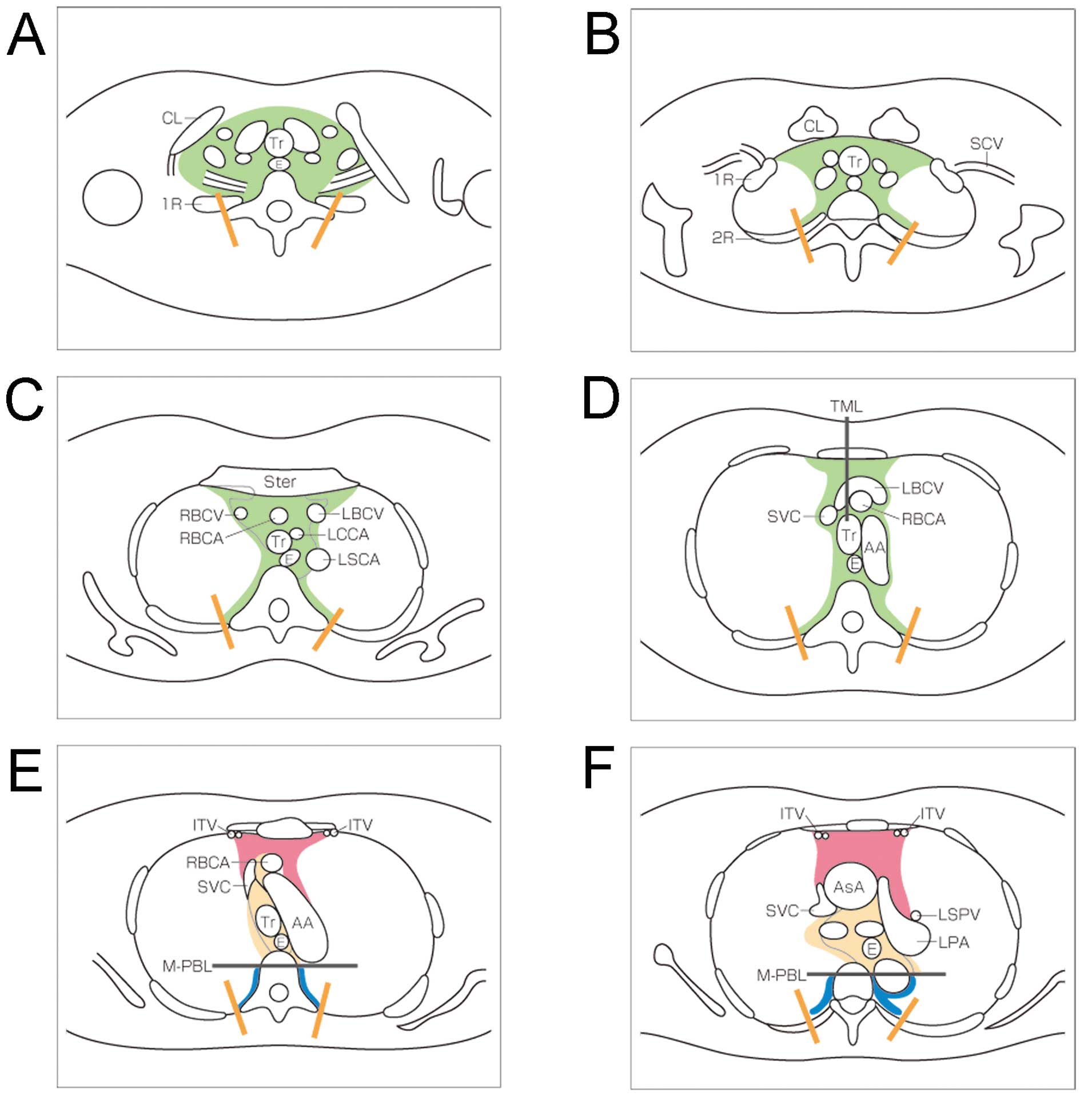 | Figure 1Schematic diagram represents the new
proposal for mediastinal compartment classification according to
the General Rules for Study of Mediastinal Tumors of the Japan
Association for Research on the Thymus (JART). (A) Thoracic inlet,
(B) upper rim of clavicle, (C) sterno-clavicular joint, (D) left
brachiocephalic vein across TML, (E) aortic arch, (F) tracheal
carina, (G) right main pulmonary artery, (H) pulmonary trunk, (I)
left atrium, (J) tricuspid valve. Abbreviations: CL, clavicle; Tr,
trachea; SCA, subclavian artery; SCV, subclavian vein; 1R, first
rib; 2R, second rib; Ster, sternum; E, esophagus; RBCV, right
brachicephalic vein; RBCA, right brachiocephalic artery; LBCV, left
brachicephalic vein; LCCA, left common carotid artery; LSCA, left
subclavian artery; SVC, superior vena cava; AA, aortic arch; AsA,
ascending aorta; LPA, left pulmonary artery; ITV, internal thoracic
vessels; Br, bronchus; LSPV, left superior pulmonary vein; RSPV,
right superior pulmonary vein; PA, pulmonary artery; RA, right
atrium; RIPV, right inferior pulmonary vein; LIPV, left inferior
pulmonary vein; LA, left atrium; TML, tracheal mid-line; M-PBL,
middle-posterior boundary line (see Materials and methods). |
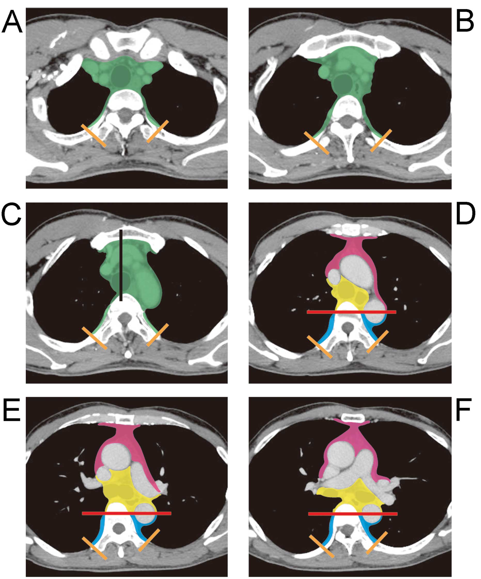 | Figure 2Contrast-enhanced CT images represent
new proposal of mediastinal compartment classification according to
the General Rules for Study of Mediastinal Tumors of the JART. (A)
Thoracic inlet, (B) upper rim of clavicle, (C) sterno-clavicular
joint, (D) left brachiocephalic vein across TML, (E) aortic arch,
(F) tracheal carina, (G) right main pulmonary artery, (H) pulmonary
trunk, (I) left atrium, (J) tricuspid valve, (K) hepatic dome of
diaphragm, (L) middle of 12th thoracic vertebral body.
Abbreviations: TML, tracheal mid-line; M-PBL, middle-posterior
boundaryline (see Materials and methods). |
 | Table IDefinition of the JART mediastinum
compartments |
Table I
Definition of the JART mediastinum
compartments
| Mediastinal
compartment | Definition |
|---|
| Superior portion of
the mediastinum | This compartment
(area highlighted green in Figs. 1
and 2) is defined as the space
between the superior border of the mediastinum (i.e., the thoracic
inlet) and a horizontal plane at the intersection of the caudate
margin of the brachiocephalic vein with the trachea [tracheal
midline line (TML), black line in Figs.
1D and 2C]. This lower border
is the same as the anatomical definition of the right upper
paratracheal (#2R) lymph nodes, based on the lymph node map of the
International Association for Study of Lung Cancer (IASLC)
(14). The area is also
anatomically bound anteriorly by the sternum, laterally by the
parietal (mediastinal) pleural reflections, posteriorly by the
anterior rim of the thoracic vertebral body; and posterolaterally
by a vertical line against the posterior rim of the chest wall at
the lateral rim of the thoracic vertebral transverse process
(orange lines in figures). |
| Anterior mediastinum
(Prevascular zone) | The anatomical
boundaries of this compartment (area highlighted red in Figs. 1 and 2) are as follows: superior, the inferior
boundary of the superior portion of the mediastinum (a horizontal
line at the upper rim of the brachiocephalic vein where it ascends
to the left, crossing in front of the trachea at the midline);
inferior, the diaphragm; anterior, the sternum; lateral, the
parietal (mediastinal) pleural reflections (including the lateral
rims of the bilateral internal thoracic arteries and veins, and
superior and inferior pulmonary veins); and posterior, the
pericardium (including a horizontal line at the posterior rim of
the heart), anterior rims of the left brachiocephalic vein,
superior vena cava, superior and inferior pulmonary veins,
ascending aorta, and the lateral rim of the aortic arch. |
| Middle mediastinum
(Peritracheoesophageal zone) | The anatomical
boundaries of this compartment (area highlighted yellow in Figs. 1 and 2) is anatomically bounded as follows:
superior, the boundary of the superior portion of the mediastinum;
inferior, the diaphragm; anterior, the posterior rim of the left
brachiocephalic vein, superior vena cava, ascending aorta,
bilateral main pulmonary arteries, and the heart; and posterior,
the anterior rim of the descending aorta and a vertical line
connecting a point on each thoracic vertebral body at 1 cm behind
its anterior margin [middle-posterior boundary line (M-PBL), red
lines in Figs. 1 and 2]. This compartment includes mainly the
trachea, bilateral main bronchi and esophagus. |
| Posterior mediastinum
(Paravertebral zone) | This compartment
(area highlighted blue in Figs. 1
and 2) is anatomically bounded as
follows: superior, the boundary of the superior portion of the
mediastinum; inferior, the diaphragm; anterior, the boundary of the
middle mediastinum (M-PBL); and posterio-lateral, a vertical line
against the posterior rim of the chest wall at the lateral rim of
the lateral process of the thoracic spine (orange lines in Figs. 1 and 2). |
From the end of December 2007, each of the five
radiologists checked the medical records retrospectively and
collected respectively 100 consecutive cases with a mediastinal
mass that was proven surgically and/or pathologically in respective
five institutions; thus 500 cases were collected in total. For each
case, each radiologist selected at least one representative CT
image that included the center of a mediastinal lesion, defined as
the geometric center (centroid) in the transverse section showing
the greatest size of the lesion.
Each independent radiologist evaluated each
mediastinal lesion based on the CT image, and assigned each lesion
to one of the proposed four compartments in individual institution.
We further discussed whether each mediastinal lesion could be
satisfactorily categorized into one of the four compartments
without any contradictions, and whether this method was helpful in
differential diagnosis.
Results
Of the 500 selected cases, 55 were excluded for
technical reasons, such as small size, insufficient image
resolution and motion artifact. The remaining 445 cases were
considered to be subjects. They included 246 thymic epithelial
tumors (193 thymomas and 53 thymic carcinomas), 24 thymic malignant
lymphomas, 31 malignant germ cell tumors, 27 mature teratomas, 49
neurogenic tumors, 17 intrathoracic goiters and 51 cystic lesions
(34 bronchogenic cysts, 15 pericardial cysts and 2 esophageal
duplication cysts).
Based on the location of the centroid, it was
possible to satisfactorily classify each mediastinal mass lesion
into one compartment without any inconsistencies (Table II). Most tumors (77.1%, 343/445)
were classified as being located in the anterior mediastinum, while
27 lesions (6.1%) were classified as being located in the superior
portion of the mediastinum, 38 lesions (8.5%) in the middle
mediastinum, and 37 lesions (8.3%) in the posterior
mediastinum.
| Table IIResult of the classification of the
445 mediastinal masses into the JART mediastinal compartment
model. |
Table II
Result of the classification of the
445 mediastinal masses into the JART mediastinal compartment
model.
| Mediastinal
compartments | |
|---|
|
| |
|---|
| Mediastinal
masses | S | A | M | P | Total |
|---|
| Intrathoracic
goiter | 14 | 1 | 2 | - | 17 |
| Thymoma | - | 192 | 1 | - | 193 |
| Thymic carcinoma | - | 52 | 1 | - | 53 |
| Thymic malignant
lymphoma | - | 24 | - | - | 24 |
| Mature teratoma | 2 | 24 | - | 1 | 27 |
| Malignant germ cell
tumors | - | 30 | 1 | - | 31 |
| Pericardial cyst | - | 10 | 5 | - | 15 |
| Bronchogenic
cyst | 1 | 6 | 20 | 7 | 34 |
| Esophageal
duplication cyst | - | - | 2 | - | 2 |
| Neurogenic
tumors | 10 | 4 | 6 | 29 | 49 |
| Total | 27 | 343 | 38 | 37 | 445 |
Almost all thymic epithelial tumors (99%, 244/246),
all 24 thymic malignant lymphomas, and most germ cell neoplasms
(93%, 54/58) were classified as being in the anterior mediastinum
compartment. The majority of intrathoracic goiters (82%, 14/17)
were categorized as being located in the superior portion of the
mediastinum compartment. Of the remaining two goiters, one was
located in the anterior and one was located in the middle
mediastinum. Approximately two-thirds of mass lesions in the middle
mediastinum were cysts, including foregut and pericardial cysts. On
the other hand, two-thirds of pericardial cysts were located in the
anterior mediastinum (particularly in the right cardiophrenic
sinus). Approximately 80% of the 37 mass lesions in the posterior
mediastinum were neurogenic tumors. Accordingly, 29 of the 49
neurogenic tumors (60%) were categorized as being located in the
posterior mediastinum, while 10 (20%) were in the superior portion
of the mediastinum, 4 (8%) were in the anterior mediastinum, and 6
(12%) were in the middle mediastinum.
Discussion
In the present study, we described a new method for
classifying the location of mediastinal lesions according to
compartments seen on transverse plane images. We also investigated
the use of this method in terms of its usefulness and
user-friendliness. We found that this method has several strong
features. First, a lesion’s compartment is defined as the
compartment in which the lesion centroid is found, leaving little
room for misclassification. Second, each compartment is clearly
bound by drawing anatomical boundary reference lines. Finally, each
compartment includes an original organ or structure from which the
mediastinal lesion arose. Using this system, we were able to
satisfactorily classify each of 455 mass lesions into one of the
four compartments, and the compartment characteristics were
considered to be useful in differential diagnoses.
Sone et al (15) assessed potential spaces of
mediastinal compartments using a CT pneumomediastinography and
described discrepancies between the anatomical classification
method and Felson’s method (using a lateral chest radiograph)
(4). Felson did not classify the
mediastinum, but proposed a way to guess the location of a mass
based on its location relative to two drawn lines. In Felson’s
method, the boundary line between the anterior and middle
mediastinum is drawn along the tracheal anterior edge and the
cardiac posterior edge. At the center of the human body (i.e., the
retrosternal space), the sagittal section plane shows the anterior
mediastinal zone to be ahead of the anterior tracheal wall, the
great vessels, and the pericardium. However, at the left side of
the mediastinum, the posterior boundary of the anterior mediastinum
spreads deeply along the aortic arch and left hilum, and this
anatomical complexity produces inconsistency with the Felson’s
method. Namely, the anterior compartment includes some masses
possibly in both the anterior and middle mediastinum on a lateral
chest radiograph. Except for this portion, the anterior mediastinum
(under the superior portion of the mediastinum) in our presently
described classification system corresponds to that in Felson’s
method.
The anterior mediastinum contains the thymus,
mediastinal fatty tissue and anterior mediastinal lymph nodes.
Among these, the thymus is the most important organ in the anterior
mediastinum since a majority of anterior mediastinal masses are
tumors of thymic origin (8–11). After birth, the thymus is a
bi-lobed, triangular gland that occupies the thyropericardiac space
of the anterior mediastinum; it is located anterior to the proximal
ascending aorta, the pulmonary outflow tract, and the superior vena
cava, and it extends caudally sometimes down to the level of the
diaphragm (8). Our proposed
anterior mediastinum compartment corresponds to the normal location
of the thymus. The great vessels (including the left
brachiocephalic vein, superior vena cava, superior and inferior
pulmonary veins, ascending aorta and the lateral rim of the aortic
arch) may be pushed backward and downward by expanding neoplasms of
thymic origin (e.g., thymic epithelial tumors and thymic malignant
lymphomas) and germ cell tumors in the anterior mediastinal zone.
These vascular structures are also barriers to prevent an anterior
mediastinal tumor from extending into the posterior or inferior
area. This is one reason why these structures are the anatomic
posterior boundary of the anterior mediastinum.
Traditional anatomical method divides the
mediastinum into two major compartments (the superior and inferior
mediastinum) by an imaginary line extending from the sternal angle
to the fourth intervertebral (Th4/5) disc. Felson (4) and Sone et al (15) stated that there is no need to
classify the superior mediastinum due to the continuity of the
inferior mediastinal zone. However, classifying the superior
portion of the mediastinum has the advantage of making it easy to
differentiate an intrathoracic goiter or neurogenic tumor of the
thoracic inlet from other mediastinal tumors. We defined the
boundary at the caudate rim of the brachiocephalic vein with the
trachea (tracheal midline). This boundary is familiar to thoracic
radiologists, physicians and surgeons, as it corresponds to an
imaginary boundary of #2R (right upper paratracheal) and #4R (right
lower paratracheal) mediastinal lymph nodes on the IASLC lymph node
map (14). In the present study,
the majority of intrathoracic goiters and one-fifth of neurogenic
tumors were located in the superior portion of the mediastinum, and
these tumors were the main type existing in this zone. Not
classifying this compartment would impact differential diagnosis,
in that many intrathoracic goiters would be categorized as being in
the anterior or middle mediastinum.
Some previously described methods of mediastinal
compartmentalization have stated that the heart and great vessels,
trachea, and main bronchi are located in the middle mediastinum,
while the esophagus is located in the posterior mediastinum
(3,7,12). In
contrast, we did not include the cardiovascular system in the JART
General Rules for Study of Mediastinal Tumors (13). The anterior rims of great vessels,
pericardium and heart are considered to be the posterior boundary
of the anterior mediastinal compartment, while the posterior rims
of the great vessels, pericardium and heart represent the anterior
boundary of the middle mediastinal compartment. We decided that
since the esophagus, trachea, and bronchi share an embryological
origin (foregut, endoderm), these should all be classified into the
same compartment (the middle mediastinum). This anatomical zone
surrounds the tracheobronchoesophageal zone, and is similar to the
‘central zone’ described by Sone et al (15). The majority of the lesions found in
the middle mediastinum included foregut cysts, tracheal and
esophageal tumors, and lymphadenopathy.
It has not been clearly stated whether the
paraaortic area surrounding the thoracic descending aorta is
classified as part of the middle or posterior mediastinum. Our
definition of this boundary is only the M-PBL (the transverse plane
at 1 cm behind the anterior edge of vertebral body). Further
classification techniques may be necessary due to the present lack
of appropriate methods.
Methodologies that have classified the heart and
great vessels as part of the middle mediastinum, have also defined
the front boundary of the posterior mediastinum as the posterior
cardiac edge (1,3,6,7). One
methodology stated that the paravertebral region is bounded at the
front by the anterior surface of the vertebral column and at the
back by the chest wall (16). On
the other hand, Felson’s method stated that the boundary of the
middle mediastinum and posterior mediastinum is drawn along the
line connecting a point on each thoracic vertebral body at 1 cm
behind its anterior margin (4). We
adopted this method as the anterior boundary for the posterior
mediastinum. The posterior mediastinum corresponds to the
paravertebral zone; however, there is no potential space in a
normal situation as the paravertebral area is strongly surrounded
by connective tissue, since air does not pass into this area during
CT pneumomediastinography, Sone et al did not mention this
compartment. However, this space becomes clearly evident when a
mass lesion exists in this zone. Inclusion of this compartment is
useful in diagnosing paravertebral neurogenic tumors using tumor
location. Most neoplasms in the paravertebral zone are neurogenic
tumors that arise from the dorsal root ganglion/neuron, most of
which are located adjacent to the intervertebral foramen. This is
one reason for setting the anterior boundary of the paravertebral
zone at 1 cm behind the anterior margin of the vertebral body.
The literature does not include any previous
descriptions of the posterior and lateral boundaries of the
paravertebral zone. Therefore, based on expediency and experience,
herein we set the bilateral posterolateral boundary lines of the
paravertebral zone as bilateral vertical lines against the
posterior rim of the chest wall at the lateral rim of the thoracic
vertebral transverse process. However, in practice, it might be
somewhat difficult to differentiate a paravertebral neurogenic
tumor from an intercostal neurogenic tumor by using this
boundary.
The present study had several limitations. First, it
was a retrospective study, and there may have been a sample bias as
a majority of the collected cases were anterior mediastinal masses.
However, it is well known that anterior mediastinal masses occur
with a higher incidence than those in other compartments (2,7–10).
Second, we did not analyze inter- and intra-observer agreements for
classifying masses into the four mediastinal compartments. Further
study is needed to analyze reading agreements. Third, our proposal
of this mediastinal compartment classification method has not yet
been validated outside of Japan. The worldwide publication and
review of this proposal will be important for validating this
classification method for use in differential diagnosis of
mediastinal masses in other populations.
In conclusion, we described a method for the
mediastinum compartment classification based on transverse plane
images obtained by CT, with the aim of providing a clinically
practical method that can lead to more consistent and exact
diagnosis of mediastinal masses. We also evaluated this novel
classification method by using it to assess 445 mediastinal mass
lesions. Our results indicate that the new mediastinal compartment
classification method using CT transverse images is sufficiently
user-friendly for practical clinical use and is potentially useful
for the differential diagnoses of mediastinal mass lesions.
Acknowledgements
This study was supported, in part, by a Grant-in-Aid
for Scientific Research (C) (no. 24591799 to K.F.) from the Japan
Society for the Promotion of Science (JSPS) and the National Cancer
Center Research and Development Fund (23-A-25 to M.K., K.F.).
The members of the Editorial Board of the Japan
Association for Research on the Thymus (JARTa) General Rules for Study of Mediastinal
Tumors are as follows: Committee Chair: Dr Yoshitaka Fujii (Chief
editor) from Nagoya City University Graduate School of Medical
Sciences, Nagoya. Committee Co-chairs: Diagnostic Imaging: Dr
Masaki Hara (Section editor) from Nagoya City West Medical Center,
Nagoya; Dr Masahiko Kusumoto from National Cancer Center Hospital,
Tokyo; Dr Fumikazu Sakai from Saitama International Medical Center,
Saitama Medical University, Hidaka; Dr Noriyuki Tomiyama from Osaka
University Graduate School of Medicine, Suita; and Dr Kiminori
Fujimoto from Kurume University School of Medicine and Kurume
University Hospital. TNM Classification and Therapy: Dr Meinoshin
Okumura (Section editor) from Osaka University Graduate School of
Medicine, Suita; Dr Takashi Kondo from Tohoku University, Sendai;
Dr Yuji Matsumura from Ohta Nishinouchi Hospital, Kouriyama; Dr Jun
Nakajima and Dr Tomohiro Murakawa from University of Tokyo Graduate
School of Medicine, Tokyo; Dr Harubumi Kato from Niizashiki Central
General Hospital, Niiza; Dr Masahiro Tsuboi from Yokohama City
University Medical Center, Yokohama; Dr Tomoki Yano, from Nagoya
City University Graduate School of Medical Sciences, Nagoya; Dr
Hiromi Wada from Osaka-Wada Clinic, Suita; Dr Toshiki Hirata and Dr
Hiroshi Date from Kyoto University Hospital, Kyoto; Dr Hiroyuki
Shiono from Nara Hospital Kinki University Faculty of Medicine,
Ikoma; Dr Tomoki Utsumi from NHO Kinki Chuo Medical Center, Sakai;
Dr Shinichi Toyooka from Okayama University Graduate School of
Medicine, Okayama; Dr Kazuya Kondo and Dr Shoji Sakiyama from
University of Tokushima Graduate School, Tokushima; Dr Koichi
Kobayashi and Dr Masafumi Kawamura from Keio University School of
Medicine; Dr Ryosuke Tsuchiya from Cancer Institute Hospital of
JFCR, Tokyo; Dr Ken Kodama from Osaka Medical Center and
Cardiovascular Diseases, Osaka; and Dr Koji Takami from National
Hospital Organization Osaka National Hospital, Osaka.
Histopathology: Dr Kiyoshi Mukai (Section editor), from Tokyo
Saiseikai Central Hospital, Tokyo; Dr Hisashi Tateyama, from
Kasugai Municipal Hospital, Kasugai; and Dr Yoshihiro Matsuno, from
Hokkaido University Hospital, Sapporo.
Notes
a JART, c/o Division of Oncology Immunology and
Surgery, Nagoya City University Graduate School of Medical
Sciences, Mizuho-machi 1, Nagoya 467-8601, Japan.
{ label (or @symbol) needed
for fn[@id='fn2-or-31-02-0565'] } The Editorial Committee of
the Japanese Association for Research on the Thymus (JART) has
granted permission to quote the contents of the guideline book of
the General Rules for Study of Mediastinal Tumors (Kanehara, Tokyo,
2009) (ref. 13) and the members of
the committee are listed in this book.
References
|
1
|
Gatzoulis MA: Mediastinum. Gray’s Anatomy.
The Anatomical Basis of Clinical Practice. Standring S: 40th
edition. Churchill Livingstone (Elsevier); Philadelphia, PA: pp.
939–957. 2008
|
|
2
|
Fraser RS, Müller NL, Colman N and Paré
PD: The mediastinum. Fraser and Paré’s Diagnosis of Diseases of the
Chest. 4th edition. WB Saunders; Philadelphia, PA: pp. 196–234.
1999
|
|
3
|
Fraser RG and Paré JA: The normal chest.
Diagnosis of Diseases of the Chest. 2nd edition. WB Saunders;
Philadelphia, PA: pp. 1–183. 1977
|
|
4
|
Felson B: Chest Roentgenology. WB
Saunders; Philadelphia, PA: 1973
|
|
5
|
Heitzman ER: The Mediastinum. 2nd edition.
Springer-Verlag; New York: 1988, View Article : Google Scholar
|
|
6
|
Zylak CJ, Pallie W and Jackson R:
Correlative anatomy and computed tomography: a module on the
mediastinum. Radiographics. 2:555–592. 1982. View Article : Google Scholar
|
|
7
|
Whitten CR, Khan S, Munneke GJ and Grubnic
S: A diagnostic approach to mediastinal abnormalities.
Radiographics. 27:657–671. 2007. View Article : Google Scholar : PubMed/NCBI
|
|
8
|
Fujimoto K and Müller NL: Anterior
mediastinal masses. Imaging of the Chest. Müller NL and Silva CIS:
2. Saunders; Philadelphia, PA: pp. 1473–1525. 2008, View Article : Google Scholar
|
|
9
|
Tomiyama N, Honda O, Tsubamoto M, et al:
Anterior mediastinal tumors. Diagnostic accuracy of CT and MRI. Eur
J Radiol. 69:280–288. 2009. View Article : Google Scholar : PubMed/NCBI
|
|
10
|
Naidich DP, Zerhouni EA, Siegelman AA and
Kuhn JP: Mediastinum. Computed Tomography and Magnetic Resonance of
The Thorax. 2nd edition. Raven Press; New York: pp. 35–148.
1991
|
|
11
|
Strollo DC, Rosado de Christenson ML and
Jett JR: Primary mediastinal tumors. Part 1: tumors of the anterior
mediastinum. Chest. 112:511–522. 1997. View Article : Google Scholar : PubMed/NCBI
|
|
12
|
Strollo DC, Rosado de Christenson ML and
Jett JR: Primary mediastinal tumors. Part 2: tumors of the middle
and posterior mediastinum. Chest. 112:1344–1357. 1997. View Article : Google Scholar : PubMed/NCBI
|
|
13
|
Japanese Association for Research on the
Thymus (JART). General Rules for Study of Mediastinal Tumors. 1st
edition. Kanehara-Shuppan; Tokyo: 2009, (In Japanese).
|
|
14
|
Rusch VW, Asamura H, Watanabe H, et al:
The IASLC lung cancer staging project. A proposal for a new
international lymph node map in the forthcoming seventh edition of
the TNM classification for lung cancer. J Thorac Oncol. 4:568–577.
2009.PubMed/NCBI
|
|
15
|
Sone S, Higashihara T, Morimoto S, et al:
Potential space of the mediastinum: CT pneumomediastinography. AJR
Am J Roentgenol. 138:1051–1057. 1982. View Article : Google Scholar : PubMed/NCBI
|
|
16
|
Lee KS: Paravertebral masses. Imaging of
the Chest. Müller NL and Silva CIS: 2. Saunders; Philadelphia, PA:
pp. 1551–1570. 2008
|


