Introduction
Gastric cancer is one of the most common causes of
cancer-related mortality, responsible for >700,000 deaths
worldwide per year (1). Although
the main treatment strategy for gastric cancer is surgical or
endoscopic resection, unresectable cases are treated with systemic
chemotherapy. Platinum agents and fluoropyrimidine are the key
therapeutic drugs for advanced gastric cancer (2) and drug resistance is an important
problem accompanying treatment. A number of studies have previously
reported on the mechanisms of gastric cancer chemoresistance
(3) using cultured cells, animal
models and clinical tissue samples. However, the mechanisms of drug
resistance have not been fully elucidated.
It has been reported that both genetic and
epigenetic changes play important roles in carcinogenesis and tumor
progression (4). In various types
of cancer, epigenetic changes are known to be early events in the
multi-steps of carcinogenesis (5).
Promoter hypermethylation is well-known to be important for the
suppression of tumor suppressor gene expression (4). Mechanisms of cancer drug resistance
are considered to be multifactorial; they have epigenetic
alterations (6) and involve
multiple gene functions and signaling pathways. A better
understanding of such mechanisms may provide therapeutic strategies
for gastric cancer.
In the present study, drug-resistant gastric cancer
cell lines were established, and biopsy specimens were obtained
from patients after the acquisition of drug resistance. Genome-wide
analysis of gene expression and DNA methylation with a microarray
for drug-resistant cell lines and endoscopic biopsy specimens of
gastric cancer was performed. Validation with quantitative methods
was also performed.
Materials and methods
Cell culture and 5-aza-2′-deoxycytidine
(5-aza-dC) treatment
AGS was purchased from the American Type Culture
Collection (ATCC) (Manassas, VA, USA), and cultured in RPMI-1640
medium with 10% FBS at 37°C with 5% CO2. For treatment
with 5-aza-dC (decitabine), cells were seeded on day 0, and exposed
to freshly prepared 10 μmol/l 5-aza-dC (Sigma-Aldrich, Tokyo,
Japan) for 24 h on days 1 and 3. After each treatment, the cells
were placed in fresh medium and harvested on day 4 (7).
Drug-resistant gastric cancer cells
Resistant AGS cells were generated by continuous
exposure to increasing concentrations of cisplatin (CDDP) or
5-fluorouracil (5-FU) for 10 months. Viability of cells was
measured by MTS-formazan reduction using CellTiter 96 Aqueous One
Solution Cell Proliferation Assay (Promega, Madison, WI, USA). AGS
cells (2×10³) were cultured using 96-well microplates
for 24 h, and exposed to various concentrations of CDDP or 5-FU for
48 h to calculate the IC50 of CDDP or 5-FU for each cell
line.
Patients and biopsy specimens
Endoscopic biopsy specimens were obtained from two
patients with unresectable advanced gastric cancer who underwent 3
and 4 courses of chemotherapy with oral fluoropyrimidine S-1 plus
CDDP (8) before and after
treatment. Samples after chemotherapy were obtained from lesions
with viable cancer. The present study was approved by the Ethics
Committee of Nagoya University Graduate School of Medicine, and
written informed consent was provided by the patients.
Extraction of DNA and RNA from cell lines
and gastric biopsy specimens
Cells or biopsy specimens were stored at −80°C for
DNA extraction, and we used RNAlater (Ambion, Austin, TX, USA) for
RNA extraction. For extraction of DNA and RNA, DNA Mini kit
(Qiagen, Venlo, The Netherlands) and RNA Mini kit (Qiagen) were
used, respectively.
Gene expression analysis with
microarray
Expression analysis was performed with SurePrint G3
Human GE 8×60K (Agilent, Loveland, CO, USA). Expression of mRNA of
autosomal 17,933 genes was evaluated with 23,856 corresponding
probes. A difference in signal intensity >2-fold was judged to
be significant.
DNA methylation microarray
Bisulfite-converted DNA was used for hybridization
on Infinium HumanMethylation450 BeadChip (Illumina, San Diego, CA,
USA). The β-value [intensity of the methylated allele
(M)/(intensity of the unmethylated allele (U) + intensity of the
methylated allele (M) + 100)] was calculated for each CpG site
(9). Methylation levels of
candidate promoter lesions in CpG islands of 11,692 genes were
evaluated. Genes with a difference in their β-value >0.1 were
extracted. Of these 11,692 genes, expression of 10,365 genes was
also evaluated with expression microarray.
Gene ontology (GO) and pathway
analysis
GO analysis was performed with TargetMine
(http://targetmine.nibio.go.jp/targetmine/begin.do;
National Institute of Biomedical Innovation, Osaka, Japan), and
pathway analysis was performed with the Kyoto Encyclopedia of Genes
and Genomes (KEGG) database. GO terms or pathways with P<0.05
using the Holm-Bonferroni method were judged to be significantly
enriched.
Real-time PCR
Real-time PCR was performed to validate expression
of mRNA with TaqMan Gene Expression Assays and TaqMan Gene
Expression Master Mix (both from Applied Biosystems, Foster City,
CA, USA).
Bisulfite pyrosequencing
Bisulfite treatment was performed with the EpiTect
kit (Qiagen) according to the manufacturer’s protocol. PyroMark PCR
(Qiagen) was used to perform PCR, and bisulfite pyrosequencing was
performed as previously reported (10–12).
In brief, the biotinylated PCR product was captured on
streptavidin-coated beads (Amersham Biosciences, Uppsala, Sweden)
and run on the PSQHS Pyrosequencing System (Biotage, Uppsala,
Sweden) to obtain the degree of methylation.
Results
Resistance to CDDP or 5-FU of established
drug-resistant cell lines
To confirm whether gastric cancer cells cultured in
chemotherapy agents obtained drug resistance, IC50
values were measured. IC50 values of 5-FU were 10 and 56
μM in parent AGS and 5-FU-resistant AGS (5FUr), respectively.
IC50 values of CDDP were 13 and 25 μM in parent AGS and
CDDP-resistant AGS (CDDPr), respectively.
Genes with altered expression in
drug-resistant cells and biopsy specimens
To characterize gene expression profiles of
drug-resistant gastric cancer cells, expression microarray analysis
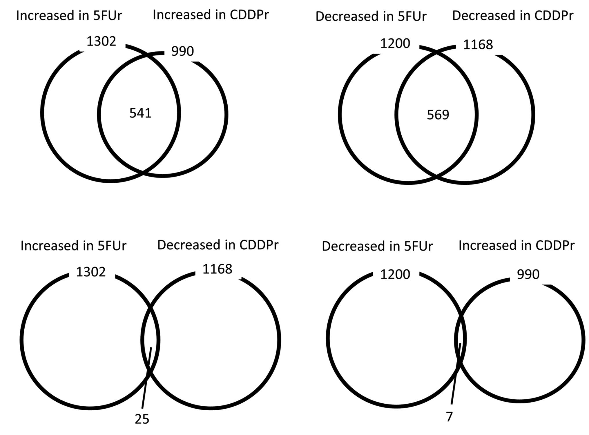
was performed. A comparison of parent AGS, 5FUr and CDDPr change of
expression is shown in Fig. 1. The
expression of 541 genes increased and the expression of 569 genes
decreased in both 5FUr and CDDPr compared with parent AGS. In
contrast, the expression of only 25 genes increased in 5FUr and
decreased in CDDPr, and the expression of only seven genes
decreased in 5FUr and increased in CDDPr. To examine the
characterization of genes with expression altered by drug
treatment, we performed GO analysis and pathway analysis (Table I). Although most enriched GO terms
differed between those changed in 5FUr and those changed in CDDPr,
‘extracellular region’ in GO terms of cellular component was
commonly enriched in both 5FUr and CDDPr. With pathway analysis,
the ‘p53 signaling pathway’ was enriched in both 5FUr and
CDDPr.
 | Table ISignificantly enriched gene ontology
(GO) terms and pathways of genes in which expression was changed in
5-fluorouracil-resistant AGS cells (5FUr) and cisplatin-resistant
AGS cells (CDDPr), compared with parent AGS cells. |
Table I
Significantly enriched gene ontology
(GO) terms and pathways of genes in which expression was changed in
5-fluorouracil-resistant AGS cells (5FUr) and cisplatin-resistant
AGS cells (CDDPr), compared with parent AGS cells.
| A, GO terms
(biological processes) |
|---|
|
|---|
| GO terms (biological
processes) | | P-value | No. of genes |
|---|
| Increased in
5FUr | (No enrichment) | | | |
| Increased in
CDDPr | Response to other
organism | (GO:0051707) | 0.000780 | 45 |
| Cell surface receptor
signaling pathway | (GO:0007166) | 0.00125 | 163 |
| Response to
virus | (GO:0009615) | 0.00180 | 31 |
| Response to biotic
stimulus | (GO:0009607) | 0.00288 | 45 |
| Signal
transduction | (GO:0007165) | 0.00382 | 247 |
| Regulation of signal
transduction | (GO:0009966) | 0.00511 | 125 |
| Positive regulation
of cell communication | (GO:0010647) | 0.00708 | 69 |
| Positive regulation
of signaling | (GO:0023056) | 0.00708 | 69 |
| Immune system
process | (GO:0002376) | 0.00867 | 130 |
| Regulation of cell
motility | (GO:2000145) | 0.0105 | 41 |
| Positive regulation
of signal transduction | (GO:0009967) | 0.0123 | 67 |
| Regulation of cell
migration | (GO:0030334) | 0.0146 | 39 |
| Single-organism
process | (GO:0044699) | 0.0151 | 516 |
| Regulation of
signaling | (GO:0023051) | 0.0159 | 132 |
| Regulation of cell
communication | (GO:0010646) | 0.0182 | 132 |
| Response to
stimulus | (GO:0050896) | 0.0206 | 341 |
| Regulation of
response to stimulus | (GO:0048583) | 0.0212 | 157 |
| Positive regulation
of cell migration | (GO:0030335) | 0.0224 | 27 |
| Regulation of
cellular component movement | (GO:0051270) | 0.0244 | 43 |
| Positive regulation
of cell motility | (GO:2000147) | 0.0308 | 27 |
| Regulation of
localization | (GO:0032879) | 0.0324 | 92 |
| Regulation of
locomotion | (GO:0040012) | 0.0496 | 41 |
| Decreased in
5FUr | Defense response | (GO:0006952) | 2.86E-05 | 110 |
| Immune system
process | (GO:0002376) | 0.000327 | 148 |
| Innate immune
response | (GO:0045087) | 0.000364 | 80 |
| Single-multicellular
organism process | (GO:0044707) | 0.000918 | 290 |
| Immune
response | (GO:0006955) | 0.0131 | 100 |
| Multicellular
organismal process | (GO:0032501) | 0.0161 | 295 |
| Decreased in
CDDPr |
Single-multicellular organism process | (GO:0044707) | 0.0343 | 279 |
| Xenobiotic
catabolic process | (GO:0042178) | 0.0478 | 5 |
|
| B, GO terms
(cellular components) |
|
| GO terms (cellular
components) | | P-value | No. of genes |
|
| Increased in
5FUr | Extracellular
region | (GO:0005576) | 4.64E-07 | 130 |
| Cell periphery | (GO:0071944) | 0.000754 | 280 |
| Plasma
membrane | (GO:0005886) | 0.000907 | 277 |
| Extracellular
region part | (GO:0044421) | 0.00647 | 70 |
| Plasma membrane
part | (GO:0044459) | 0.0134 | 136 |
| Extracellular
space | (GO:0005615) | 0.0271 | 56 |
| Increased in
CDDPr | Extracellular
region | (GO:0005576) | 0.0374 | 92 |
| Decreased in
5FUr | Extracellular
region | (GO:0005576) | 3.24E-05 | 116 |
| Decreased in
CDDPr | Extracellular
region | (GO:0005576) | 7.37E-08 | 121 |
| Cornified
envelope | (GO:0001533) | 0.0124 | 8 |
| Intrinsic to
membrane | (GO:0031224) | 0.0175 | 142 |
| Integral to
membrane | (GO:0016021) | 0.0195 | 138 |
| Cell periphery | (GO:0071944) | 0.0202 | 243 |
| Plasma
membrane | (GO:0005886) | 0.0268 | 240 |
|
| C, GO terms
(molecular functions) |
|
| GO terms (molecular
functions) | | P-value | No. of genes |
|
| Increased in
5FUr | Serine-type
peptidase activity | (GO:0008236) | 3.29E-05 | 24 |
| Serine hydrolase
activity | (GO:0017171) | 5.16E-05 | 24 |
| Serine-type
endopeptidase activity | (GO:0004252) | 0.00325 | 17 |
| Increased in
CDDPr | (No
enrichment) | | |
| Decreased in
5FUr | (No
enrichment) | | | |
| Decreased in
CDDPr | Receptor
binding | (GO:0005102) | 5.08E-04 | 83 |
| Cytokine
activity | (GO:0005125) | 0.0383 | 15 |
|
| D, pathway |
|
| Pathway | | P-value | No. of genes |
|
| Increased in
5FUr | p53 signaling
pathway | | 0.0222 | 14 |
| Increased in
CDDPr | p53 signaling
pathway | | 8.56E-07 | 18 |
| Decreased in
5FUr | (No
enrichment) | | | |
| Decreased in
CDDPr | Cytokine-cytokine
receptor interaction | | 0.00618 | 37 |
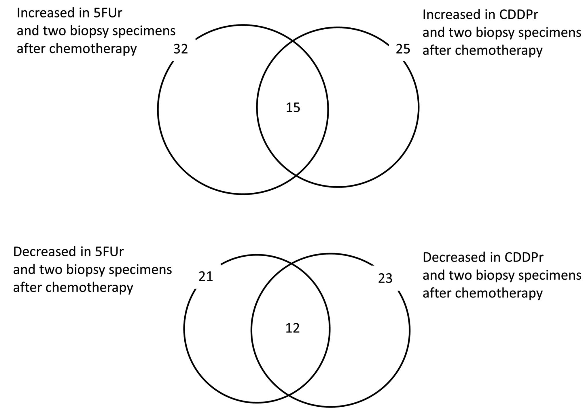
To compare gene expression of gastric cancer before
and after chemotherapy, expression microarray analysis with
endoscopic biopsy specimens was performed. Genes with altered
expression both in drug-resistant cells and in biopsy specimens
after chemotherapy were extracted. The expression of 15 genes
increased 5FUr, CDDPr and two pairs of biopsy specimens, and the
expression of 12 genes decreased (Fig.
2, Table II).
| Table IIList of genes in which expression was
changed commonly in drug-resistant cells and biopsy specimens. |
Table II
List of genes in which expression was
changed commonly in drug-resistant cells and biopsy specimens.
| Increased in 5FUr
and two biopsy specimens after chemotherapy | Decreased in 5FUr
and two biopsy specimens after chemotherapy | Increased in CDDPr
and two biopsy specimens after chemotherapy | Decreased in CDDPr
and two biopsy specimens after chemotherapy | Increased in 5FUr,
CDDPr, and two biopsy specimens after chemotherapy | Decreased in 5FUr,
CDDPr, and two biopsy specimens after chemotherapy |
|---|
| APOC1 | ALPK1 | ACTG2 | ACOXL | APOC1 | ALPK1 |
| BAIAP3 | C17orf110 | ANPEP | ALPK1 | CRYM | CCL21 |
| C4BPA | C4orf47 | APOC1 | BATF | DNAJC28 | CYP2E1 |
| C6orf154 | CCL21 | C9orf123 | BEST4 | HSD17B6 | ETV7 |
| CAPS2 | CYP2E1 | CELA3B | CCL21 | IQCD | FBXO15 |
| CFTR | ETV7 | CRYM | CYP2E1 | KLK13 | GPR110 |
| CRYM | FBXO15 | CTSG | ETV7 | KREMEN2 | NLRC5 |
| DNAJC28 | GPR110 | DNAJC28 | FBXO15 | OLFML3 | PLIN4 |
| FRZB | HEPACAM2 | HOXB3 | GPR110 | OTUD7A | SLC22A20 |
| HSD17B6 IFI44L | HSD17B6 | INSC | PHACTR3 | SLC26A9 | |
| IGF1 | KRT6C | IQCD | KRT31 | PLAT | SLC28A3 |
| IP6K3 | LAMC2 | KLK13 | MUC1 | RARRES2 | SNORA22 |
| IQCD | NLRC5 | KREMEN2 | NCKAP5 | SRI | |
| KCTD7 | OR52K2 | NLRP2 | NLRC5 | TAC3 | |
| KLK13 | PLIN4 | NRG1 | PCSK9 | TNNI3 | |
| KREMEN2 | SLC22A20 | OLFML3 | PLIN4 | | |
| LAMA1 | SLC26A9 | OTUD7A | RAB27B | | |
| LRRC6 | SLC28A3 | PHACTR3 | SLC22A20 | | |
| MSLN | SNORA22 | PLAT | SLC26A9 | | |
| NR2F1 | SPRR3 | RARRES2 | SLC28A3 | | |
| OLFML3 | ZNF750 | RASL10A | SLFNL1 | | |
| OOEP | | SRI | SNORA22 | | |
| OTUD7A | | TAC3 | SYT13 | | |
| PHACTR3 | | TNFSF9 | | | |
| PLAT | | TNNI3 | | | |
| PRODH | | | | | |
| RARRES2 | | | | | |
| SCN2A | | | | | |
| SEPP1 | | | | | |
| SRI | | | | | |
| TAC3 | | | | | |
| TNNI3 | | | | | |
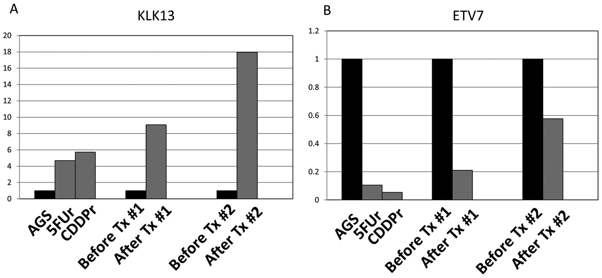
To validate the gene expression change extracted
with microarray, real-time PCR for KLK13 and ETV7 was performed.
Consistent with microarray analysis, KLK13 increased in both
drug-resistant AGS cells and endoscopic biopsy specimens after
chemotherapy (Fig. 3A). In
contrast, ETV7 decreased in both drug-resistant cells and biopsy
specimens after treatment (Fig.
3B).
Integrated analysis of expression and
methylation microarray
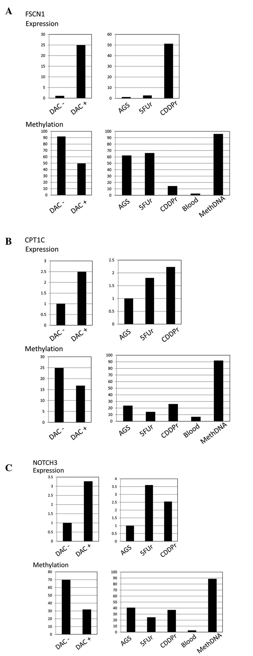
To study whether DNA methylation contributes to gene
expression change caused by chemotherapy agents, we analyzed the
methylation profiles of 5FUr and CDDPr and compared them with
parent AGS. The number of genes that were hypermethylated and
decreased in expression and that were hypomethylated and increased
in expression was 74.
Next, to evaluate whether alterations in the DNA
methylation of these genes was related to expression change, gene
expression change was measured by treatment with a demethylating
agent. Twenty-one of those 74 genes increased in expression after
treatment with decitabine (Table
III). Furthermore, gene expression and methylation were
validated with quantitative methods, TaqMan PCR and bisulfite
pyrosequencing, respectively, for FSCN1, CPT1C and NOTCH3.
Expression of these three genes increased after treatment with
decitabine (Fig. 4). FSCN1 revealed
increased expression and hypomethylation in CDDPr compared with
parent AGS cells. Regarding CPT1C, 5FUr showed hypomethylation and
increased expression. CDDPr also showed increased expression,
although the methylation level did not change. NOTCH3 showed
increased expression and hypomethylation, especially in 5FUr.
| Table IIIList of genes in which expression
increased and methylation levels decreased, or expression decreased
and methylation levels increased. |
Table III
List of genes in which expression
increased and methylation levels decreased, or expression decreased
and methylation levels increased.
| Increased
expression and hypo- methylation in 5FUr | Decreased
expression and hyper- methylation in 5FUr | Increased
expression and hypo- methylation in CDDPr | Decreased
expression and hyper- methylation in CDDPr |
|---|
| ARC | ATP2C2 | ABCG4 | ATP2C2 |
| C12orf34 | C15orf60 | C12orf34 | C15orf60 |
| CPT1C | PRAME | CARD9 | FRMD6 |
| CST6 | ZNF773 | CST6 | SECTM1 |
| CYB5R2 | | FES | TNFSF12 |
| KCNH8 | | FSCN1 | ZNF773 |
| MESP1 | | KCNH8 | |
| NOTCH3 | | MESP1 | |
| RASGRP2 | | VGF | |
| TNFSF12 | | | |
Discussion
The gene expression and the DNA methylation of
drug-resistant cell lines and biopsy specimens were evaluated
before and after chemotherapy, and some genes revealed altered
expression and altered methylation. In drug-resistant cells,
treatment with 5-FU and CDDP caused consistent expression change in
>1,000 genes. In contrast, the expression of only a small number
of genes changed reciprocally (Fig.
1), and those genes were considered to be potentially related
with drug-specific sensitivity. In GO analysis, only a small number
of GO terms are commonly enriched in both 5-FU-resistant cells and
CDDP-resistant cells, and enriched pathways related to the two
drugs are also different. These findings may be related to
differences in mechanisms of resistance to each drug.
Expression change of genes before and after
chemotherapy was also evaluated using endoscopic biopsy specimens,
and revealed that profiles of changes were different in the two
patients. Expression of some genes increased or decreased, both in
drug-resistant cells and biopsy specimens after chemotherapy
(Fig. 2, Table II). Such genes are considered to be
candidates as key molecules for drug resistance, and may be useful
as biomarkers of drug-sensitivity.
KLK13 is one member of the tissue kallikrein (KLK)
family which includes 15 genes (KLK1-KLK15) and plays a role in
tumor cell invasion and migration (13). KLK13 has already been reported to be
upregulated in gastric cancer cells after exposure to
antineoplastic agents, including epirubicin and methotrexate
(14). It has also been reported
that overexpression of KLK13 results in an increase of malignant
cell behavior, and that knockdown of its endogenous gene expression
causes a significant decrease in cell migratory and invasive
properties (13). We found that
KLK13 increased in both drug-resistant cells and biopsy specimens,
a finding suggesting that KLK13 may play a role in 5-FU and CDDP
resistance in gastric cancer.
In contrast to KLK13, expression of ETV7 decreased
in drug-resistant cells. ETV7 is a member of the Ets transcription
factor family and is reported to act as an inhibitor of
differentiation (15). Since ETV7
was downregulated in drug-resistant gastric cancer in the present
study, it may be related with mechanisms of gastric cancer
drug-sensitivity.
It has been reported that epigenetic profiles are
useful for identifying molecular mediators for cancer drug
sensitivity (6). In terms of a
correlation between gene expression and DNA methylation, we also
performed expression and methylation microarray analyses, and found
some genes with altered methylation levels and expression levels.
FSCN1 revealed increased expression and hypomethylation in
CDDP-resistant cells. FSCN1 has been reported to play an important
role in cancer development and is associated with invasion and
metastasis (16). It has also been
reported that higher intensity FSCN1 staining correlated with
more-advanced cancer stages, and inversely correlated with survival
rates in gastric adenocarcinoma (17). This suggests that FSCN1 may
influence patient survival through acquisition of resistance to
chemotherapy drugs.
In our experiment, CPT1C was increased in expression
in 5-FU and CDDP-resistant cells, and demethylated in
5-FU-resistant cells. CPT1C has been reported to promote tumor
growth and rapamycin resistance (18). CPT1C expression correlates inversely
with mammalian target of rapamycin (mTOR) pathway activation,
contributes to rapamycin resistance in murine primary tumors, and
is frequently upregulated in human lung tumors (18). To our knowledge, there has been no
report of a relationship between CPT1C and 5-FU or CDDP.
Notch is a transmembrane heterodimeric receptor with
4 distinct members (NOTCH1 to NOTCH4) present in humans. NOTCH3 is
one of the Notch family members. It has been reported that NOTCH1,
another molecule in Notch family members, expression is associated
with cell aggressiveness and 5-FU drug resistance in human
esophageal squamous cell carcinoma cell lines in vitro, and
also with poor survival in human esophageal squamous cell
carcinomas (19). It has also been
reported that expression levels of Notch3 were increased in rat
tracheal epithelial cells after treatment with 5-FU (20). NOTCH3 knockdown enhanced the
sensitivity of nasopharyngeal carcinoma cells to CDDP treatment
(21), and NOTCH3 overexpression
correlated with shorter progression-free/overall survival in
patients with advanced stage ovarian carcinoma treated with
platinum and taxane (22). In our
data, NOTCH3 was upregulated in drug-resistant cells. In gastric
cancer, NOTCH3 may be related to drug resistance.
However, we could not find a significant difference
in methylation levels among biopsy samples. One of the limitations
is that biopsy specimens may not represent the characteristics of
the whole tumor, since gastric cancer is known to be biologically
heterogeneous.
In the present study, genes with altered expression
and DNA methylation were extracted after treatment with
chemotherapeutic agents in gastric cancer. These alterations may be
related to mechanisms of gastric cancer drug resistance, and may be
useful as biomarkers that predict drug sensitivity. Further studies
with a large number of clinical samples are necessary.
Acknowledgements
The authors thank Ms. Chie Moriyama for her
technical support. This study was funded by the Ministry of
Education, Culture, Sports, Science and Technology of Japan (no.
20378053).
References
|
1
|
Ferlay J, Shin HR, Bray F, Forman D,
Mathers C and Parkin DM: Estimates of worldwide burden of cancer in
2008: GLOBOCAN 2008. Int J Cancer. 127:2893–2917. 2010. View Article : Google Scholar : PubMed/NCBI
|
|
2
|
Okines A, Verheij M, Allum W, Cunningham D
and Cervantes A; ESMO Guidelines Working Group. Gastric cancer:
ESMO Clinical Practice Guidelines for diagnosis, treatment and
follow-up. Ann Oncol. 21(Suppl 5): v50–v54. 2010. View Article : Google Scholar : PubMed/NCBI
|
|
3
|
Zhang D and Fan D: New insights into the
mechanisms of gastric cancer multidrug resistance and future
perspectives. Future Oncol. 6:527–537. 2010. View Article : Google Scholar : PubMed/NCBI
|
|
4
|
Taby R and Issa JP: Cancer epigenetics. CA
Cancer J Clin. 60:376–392. 2010. View Article : Google Scholar : PubMed/NCBI
|
|
5
|
Ushijima T: Epigenetic field for
cancerization. J Biochem Mol Biol. 40:142–150. 2007. View Article : Google Scholar
|
|
6
|
Shen L, Kondo Y, Ahmed S, et al: Drug
sensitivity prediction by CpG island methylation profile in the
NCI-60 cancer cell line panel. Cancer Res. 67:11335–11343. 2007.
View Article : Google Scholar : PubMed/NCBI
|
|
7
|
Yamashita S, Takahashi S, McDonell N, et
al: Methylation silencing of transforming growth factor-β receptor
type II in rat prostate cancers. Cancer Res. 68:2112–2121.
2008.
|
|
8
|
Koizumi W, Narahara H, Hara T, et al: S-1
plus cisplatin versus S-1 alone for first-line treatment of
advanced gastric cancer (SPIRITS trial): a phase III trial. Lancet
Oncol. 9:215–221. 2008. View Article : Google Scholar : PubMed/NCBI
|
|
9
|
Bibikova M, Barnes B, Tsan C, et al: High
density DNA methylation array with single CpG site resolution.
Genomics. 98:288–295. 2011. View Article : Google Scholar : PubMed/NCBI
|
|
10
|
Colella S, Shen L, Baggerly KA, Issa JP
and Krahe R: Sensitive and quantitative universal Pyrosequencing
methylation analysis of CpG sites. Biotechniques. 35:146–150.
2003.PubMed/NCBI
|
|
11
|
An B, Kondo Y, Okamoto Y, et al:
Characteristic methylation profile in CpG island methylator
phenotype-negative distal colorectal cancers. Int J Cancer.
127:2095–2105. 2010. View Article : Google Scholar : PubMed/NCBI
|
|
12
|
Shen L, Guo Y, Chen X, Ahmed S and Issa
JP: Optimizing annealing temperature overcomes bias in bisulfite
PCR methylation analysis. Biotechniques. 42:48–58. 2007. View Article : Google Scholar : PubMed/NCBI
|
|
13
|
Chou RH, Lin SC, Wen HC, Wu CW and Chang
WS: Epigenetic activation of human kallikrein 13 enhances
malignancy of lung adenocarcinoma by promoting N-cadherin
expression and laminin degradation. Biochem Biophys Res Commun.
409:442–447. 2011. View Article : Google Scholar : PubMed/NCBI
|
|
14
|
Florou D, Mavridis K and Scorilas A: The
kallikrein-related peptidase 13 (KLK13) gene is
substantially up-regulated after exposure of gastric cancer cells
to antineoplastic agents. Tumour Biol. 33:2069–2078. 2012.
|
|
15
|
Gu X, Shin BH, Akbarali Y, et al: Tel-2 is
a novel transcriptional repressor related to the Ets factor
Tel/ETV-6. J Biol Chem. 276:9421–9436. 2001. View Article : Google Scholar : PubMed/NCBI
|
|
16
|
Hanker LC, Karn T, Holtrich U, et al:
Prognostic impact of fascin-1 (FSCN1) in epithelial ovarian cancer.
Anticancer Res. 33:371–377. 2013.PubMed/NCBI
|
|
17
|
Tsai WC, Jin JS, Chang WK, et al:
Association of cortactin and fascin-1 expression in gastric
adenocarcinoma: correlation with clinicopathological parameters. J
Histochem Cytochem. 55:955–962. 2007. View Article : Google Scholar : PubMed/NCBI
|
|
18
|
Reilly PT and Mak TW: Molecular pathways:
tumor cells co-opt the brain-specific metabolism gene CPT1C
to promote survival. Clin Cancer Res. 18:5850–5855. 2012.
View Article : Google Scholar : PubMed/NCBI
|
|
19
|
Liu J, Fan H, Ma Y, et al: Notch1 is a
5-fluorouracil resistant and poor survival marker in human
esophagus squamous cell carcinomas. PLoS One. 8:e561412013.
View Article : Google Scholar : PubMed/NCBI
|
|
20
|
Ma XB, Jia XS, Liu YL, et al: Expression
and role of Notch signalling in the regeneration of rat tracheal
epithelium. Cell Prolif. 42:15–28. 2009. View Article : Google Scholar : PubMed/NCBI
|
|
21
|
Man CH, Wei-Man Lun S, Wai-Ying Hui J, et
al: Inhibition of NOTCH3 signalling significantly enhances
sensitivity to cisplatin in EBV-associated nasopharyngeal
carcinoma. J Pathol. 226:471–481. 2012. View Article : Google Scholar : PubMed/NCBI
|
|
22
|
Rahman MT, Nakayama K, Rahman M, et al:
Notch3 overexpression as potential therapeutic target in advanced
stage chemoresistant ovarian cancer. Am J Clin Pathol. 138:535–544.
2012. View Article : Google Scholar : PubMed/NCBI
|