Introduction
Verrucous carcinoma was first described by Ackerman
as a variant of squamous cell carcinoma of the oral cavity
(1). Clinically, it is usually a
warty, papillary exophytic, firm, non-ulcerating lesion with a
broad base and a red, white, or variegated red and white surface
(2). Histologically, it is
characterized by verrucous proliferation of the squamous epithelium
with wide and elongated rete ridges exhibiting a pushing border
invasion into the underlying connective tissue with epithelial
dysplasia. A mixed chronic inflammatory cell infiltrate composed of
lymphocytes, plasma cells and histiocytes may also be prominent in
the stroma (3,4). Oral verrucous hyperplasia (OVH) is a
benign lesion without malignant features and cellular atypia. A
properly oriented histological section including normal margin
tissue is used for the morphological differentiation of OVH and
oral verrucous carcinoma (OVC). In addition, immunohistochemistry
such as p53, which is used to determine expression of mutant p53
protein, and Ki67, a proliferating marker, are used to diagnose
OVC. Positive signals for p53 and Ki67 are usually increased in
carcinoma specimens including OVC, whereas OVH alone does not show
significant positive signals for these proteins. Pathologists are
sometimes presented with specimens in which a clear distinction
between the benign or malignant state is difficult to determine,
and these are tentatively termed as oral verrucous borderline
lesions (OVLs). The presence of some degree of epithelial dysplasia
along with fewer positive signals of p53 and Ki67 protein makes it
difficult to distinguish these lesions. Moreover, they share
certain morphological similarities both clinically and
histopathologically. However, it is vitally important to
distinguish between OVC and its morphologically similar benign
lesions considering the significance of therapeutic and prognostic
implications of such lesions.
HuR is a ubiquitously expressed mRNA-binding
protein. Intracellularly, HuR is localized predominantly in the
nucleus but it shuttles between the nucleus and the cytoplasm. The
export of HuR is mediated by its association with transportin 1
(Trn1) and transportin 2 (Trn 2) via the shuttling sequence
termed’HNS’ in the hinge region and by its association with pp32,
APRIL and SET α/β protein, which includes the nuclear export signal
recognized by the export receptor chromosome maintenance region 1
(CRM1) (5–7). AU-rich elements (ARE) are located in
the untranslated regions of many proto-oncogenes, growth factors
and cytokine mRNAs as the core sequence of AUUUA. HuR binds to AREs
to protect ARE-mRNAs against rapid degradation. Owing to
nucleocytoplasmic translocation of HuR being necessary for its
activity and the cytoplasmic presence of HuR found in several
carcinomas, it has been hypothesized that cytoplasmic HuR
expression may be a prognostic marker in cancer patients (8,9). Our
previous study showed that export of HuR may be used as a
diagnostic marker for oral cancers (10). In the present study, we analyzed the
expression of p53, Ki67 along with HuR proteins in 48 cases
diagnosed as OVH, OVC or OVL and compared them with
clinicopathological parameters. The OVL cases were further
investigated to determine the association of protein expression and
malignant transformation. Thus, we aimed to evaluate whether the
cytoplasmic expression of HuR protein facilitates the
differentiation of true malignant lesions among OVLs.
Materials and methods
Patients and clinicopathological
investigation
The present study examined 6 cases of OVH, 17 cases
of OVC and 25 OVLs. All samples were collected from Hokkaido
University Hospital between 1985 and 2010. Diagnosis was based on
histological examination. Cases with epithelial verrucous
hyperplasia without cellular dysplasia were diagnosed as OVH
whereas OVCs were characterized by verrucous proliferation of
squamous epithelium with wide and elongated rete ridges exhibiting
a pushing border invasion into the underlying connective tissue
with epithelial dysplasia. However, for OVLs we used the following
diagnostic criteria: epithelial hyperplasia with hyperkeratosis and
a verrucous surface, non-invasion of the hyperplastic epithelium
into the lamina propria compared with adjacent normal mucosal
epithelium, and lesions with varying degrees of epithelial
dysplasia. The cases with OVLs were further followed up for 3 years
to evaluate their potential for malignant transformation. Detailed
demographic and clinical data for the cases of OVH, OVC and OVLs
are listed in Table I.
 | Table IDemographic and clinical data of the
patients with oral verrucous hyperplasia (OVH), oral verrucous
carcinoma (OVC) and oral verrucous lesions (OVLs). |
Table I
Demographic and clinical data of the
patients with oral verrucous hyperplasia (OVH), oral verrucous
carcinoma (OVC) and oral verrucous lesions (OVLs).
| Characteristics | Total cases | Lesion types | P-value |
|---|
|
|---|
| OVH | OVL | OVC |
|---|
| All cases, n (%) | 48 | 6 (12.5) | 25 (52.1) | 17 (35.4) | |
| Age (years) | | | | | 0.586 |
| Mean | 65±17 | 53±15 | 68±17 | 65±16 | |
| Median | 69 | 51 | 71 | 64 | |
| Range | 23–96 | 32–73 | 23–95 | 47–96 | |
| Gender, n (%) | | | | | 0.774 |
| Female | 28 (58.3) | 3 (10.7) | 14 (50.0) | 11 (39.3) | |
| Male | 20 (41.7) | 3 (15.0) | 11 (55.0) | 6 (30.0) | |
| Location, n (%) | | | | | 0.196 |
| Gingiva | 19 (39.6) | 2 (10.5) | 7 (36.8) | 10 (52.6) | |
| Tongue | 14 (29.2) | 2 (14.3) | 10 (71.4) | 2 (14.3) | |
| Buccal mucosa | 5 (10.4) | 1 (20.0) | 4 (80.0) | 0 (0.0) | |
| Lip | 6 (12.5) | 0 (0.0) | 3 (50.0) | 3 (50.0) | |
| Palate | 3 (6.3) | 1 (33.3) | 0 (0.0) | 2 (66.7) | |
| FOM | 1 (2.1) | 0 (0.0) | 1 (100.0) | 0 (0.0) | |
Immunohistochemical analysis
The paraffin blocks of the specimens were cut in
5-μm sections and examined immunohistochemically. Sections were
deparaffinized in xylene, rehydrated in graded alcohol and
subjected to antigen retrieval by heat treatment in Tris-EDTA (TE)
buffer. To inhibit endogenous peroxidase activity, the slides were
then immersed in 3% H2O2 for 5 min followed
by blocking solution [1% BSA in phosphated-buffered saline (PBS)]
for 30 min. The immunohistochemical detection of HuR was carried
out using anti-HuR monoclonal antibodies (1:6,000 dilution; Santa
Cruz, Santa Cruz, CA, USA) in PBS in blocking solution in a
humidified chamber at 4°C overnight. The sections were then
subjected to Simple Stain Max PO (M) (Nichirei Bioscience, Tokyo,
Japan) at 37°C for 30 min. Careful rinses were performed with
several changes of PBS between the stages of the procedure.
Visualization was carried out using the ChemMate EnVision kit/HRP
(Dako, Tokyo, Japan), and the sections were counterstained with
hematoxylin. The same tissues were immunostained with monoclonal
antibodies against p53 and Ki-67 (1:100). The stained slides were
examined by light microscopy, and the positive cell distribution in
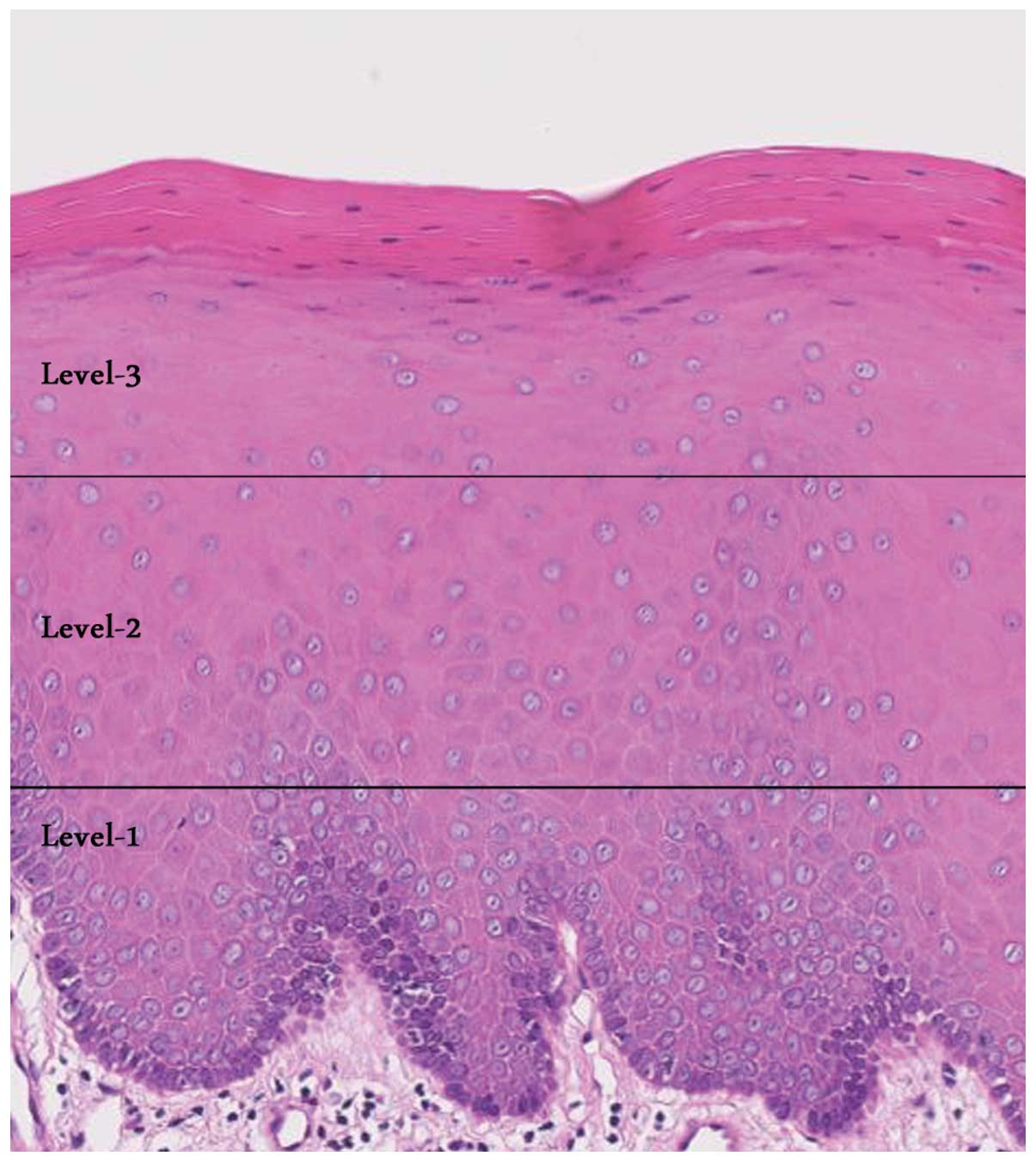
the different levels of the epithelium was observed. As shown in
Fig. 1, the epithelium was divided
into 3 levels: level 1 (lower one-third of the epithelium), level 2
(lower two-thirds of the epithelium) and level 3 (extending to the
upper one-third of the epithelium).
Nuclear staining was considered as positive for p53
and Ki67 proteins, whereas cytoplasmic staining was considered as
positive for HuR. The sections were initially scanned at low power,
at least 3 high-power fields were then chosen randomly, and at
least 1,000 cells were counted in level 1 of the epithelium for
each case. The labeling indices (LIs) of cytoplasmic or nuclear
staining of each antibody were defined as a ratio of
immunostaining-positive cells to the total number of cells counted.
Normal oral epithelium (NE) and oral squamous cell carcinoma (SCC)
were used as negative and positive controls, respectively.
Statistical analysis
The Chi-square test was applied to compare the
association of the patient basal characteristics (age, gender,
lesion location) and the distribution pattern of the target
proteins (HuR, Ki67 and p53) in all lesions (OVH, OVC and OVL). The
mean LIs of the three examined proteins were compared by the
Student’s paired t-test. The association between the expression of
proteins and malignant transformation of 25 OVLs was also assessed
by Chi-square analysis. SPSS for Windows release 17.0 (SPSS) was
used for statistical analysis. A value of P<0.05 was considered
to indicate a statistically significant result.
Results
The basal characteristics (age, gender and lesion
location) of 48 patients and their association with 3 different
types of oral lesions (OVH, OVL and OVC) are presented in Table I. The age range of the patients with
OVH, OVC and OVLs was 32–73 years (mean age, 51), 47–96 years (mean
age, 64) and 23–95 years (mean age, 71), respectively. Gingiva was
the common site for all lesions (39.6%) followed by the tongue
(29.2%). The male to female ratios were 1:1.8 and 1:1.3 for OVC and
OVLs, respectively. Both lesions had a female predilection;
however, there was no statistically significant association between
basal characteristics and types of oral lesions.
Immunohistochemical findings are summarized in Tables II and III and illustrated in Figs. 2–5.
Distribution of all the three examined proteins (HuR, Ki67 and p53)
in the different levels of the epithelium had a significant
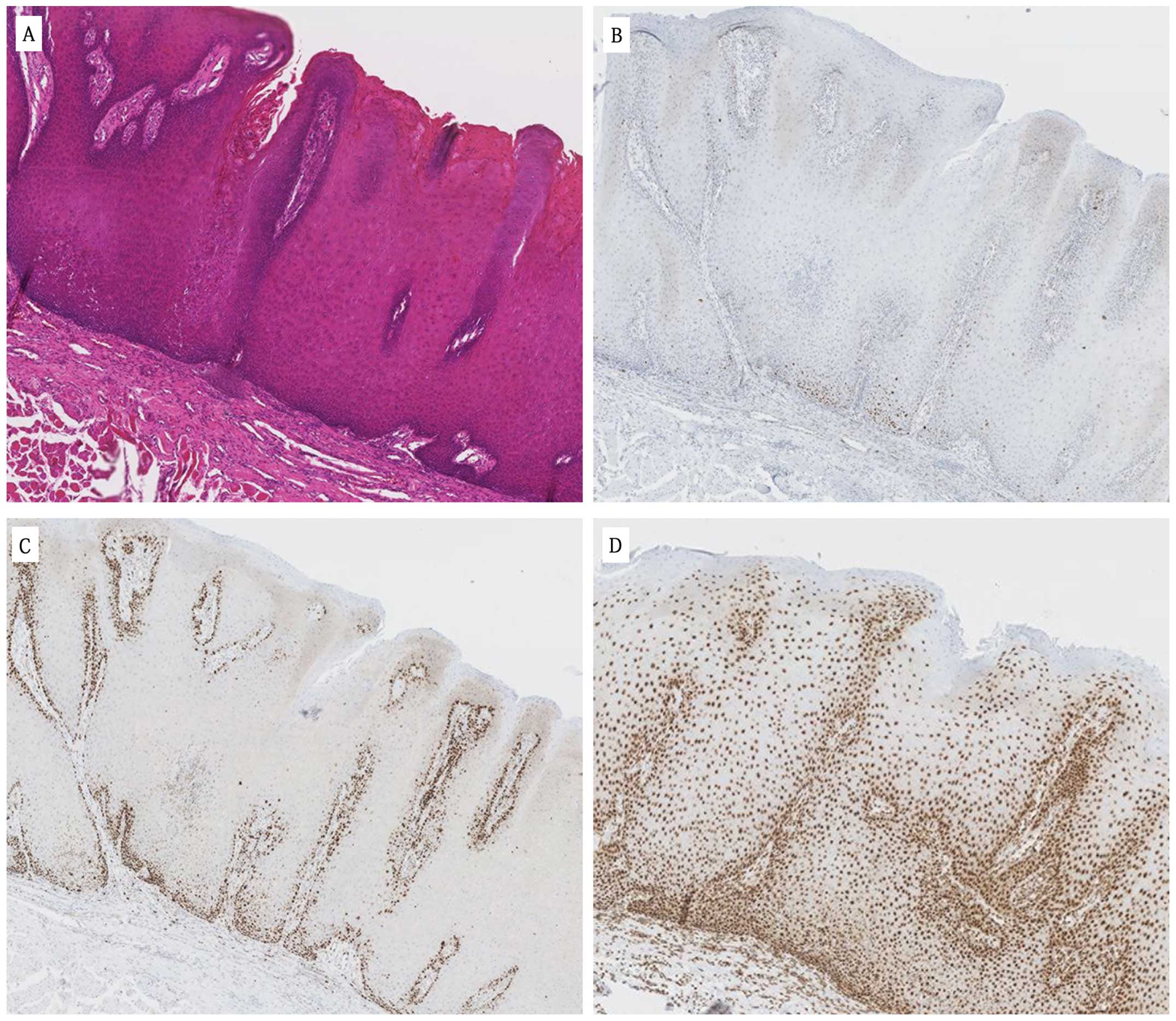
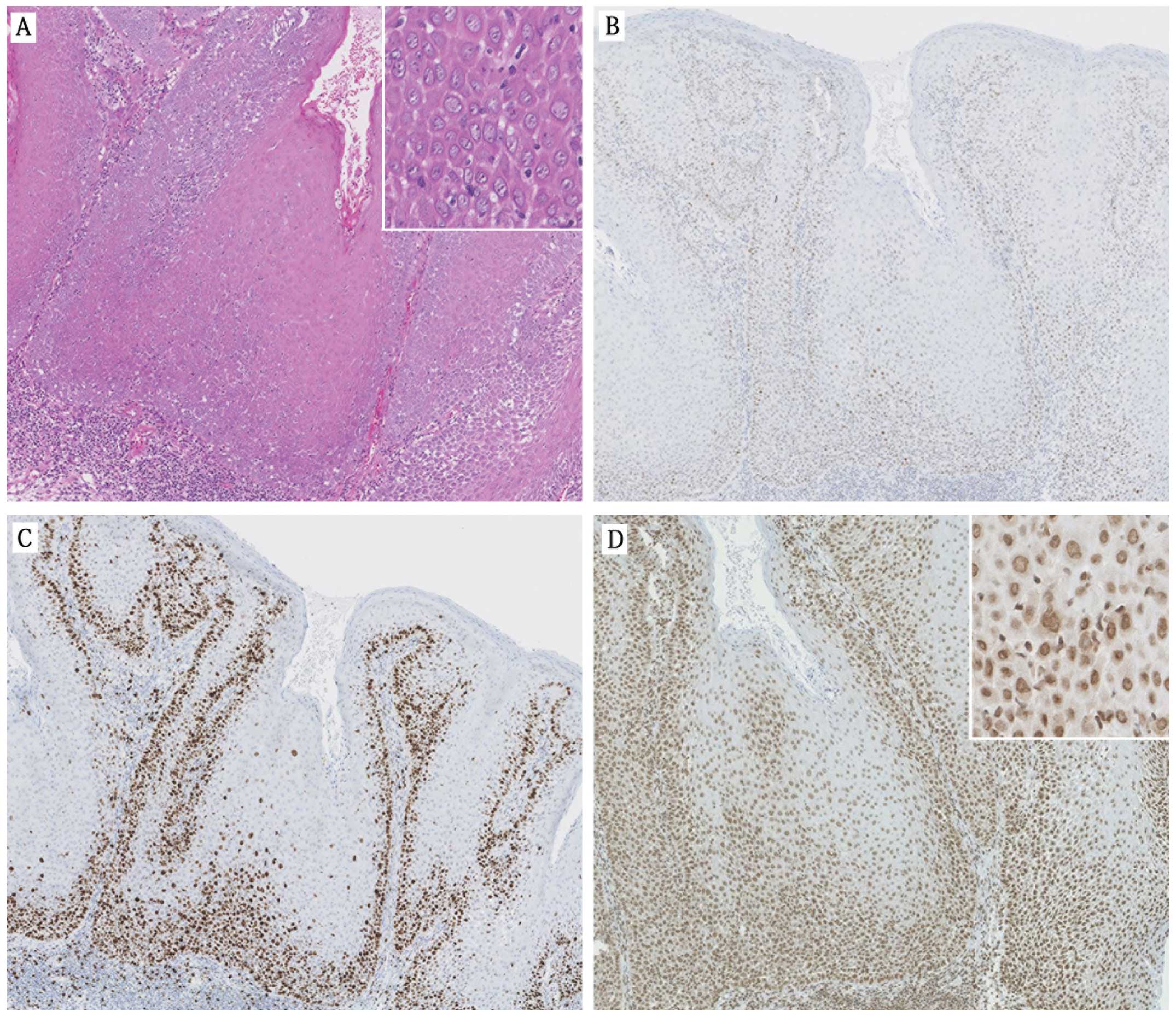
association with the oral lesions (Table II). The major finding was that in
all OVH cases, positive signals for all proteins were restricted to
level 1 of the epithelium (Fig. 3
and Table II) whereas there was a
general trend for a more diffuse staining pattern in OVCs compared
to OVHs and OVLs. More specifically, expression of HuR (64.7%) and
p53 (52.9%) proteins extended up to levels 3 and 2 of the
epithelium, respectively (Fig. 4
and Table II). In addition, 64.7%
of OVCs showed Ki67-positive signals in level 2 of the epithelium.
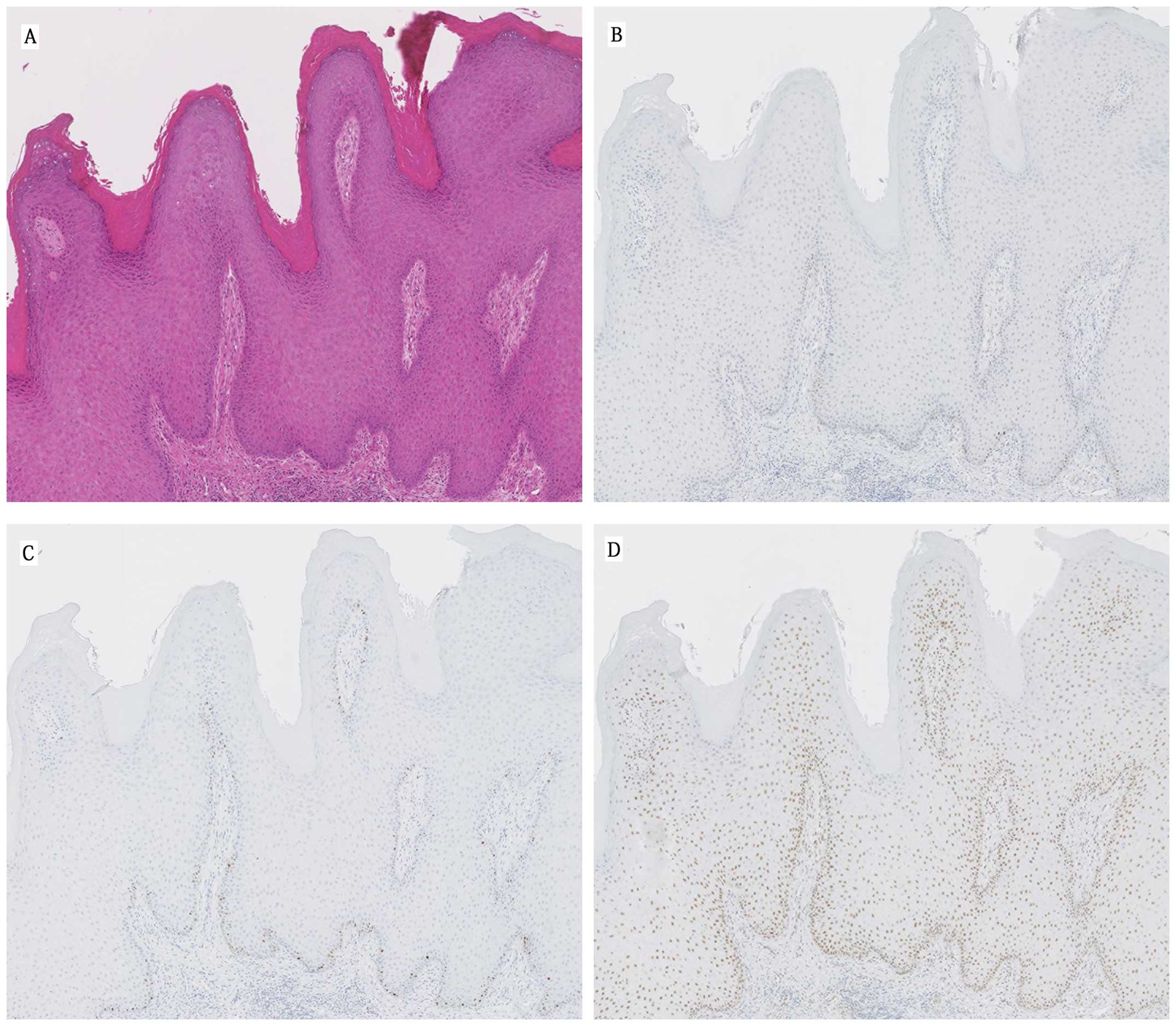
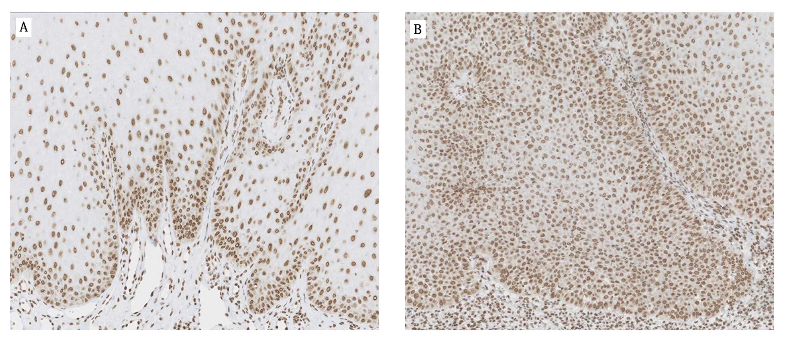
All OVL cases predominantly showed positive staining with HuR
(64%), Ki67 (80%) and p53 (100%) in level 1 of the epithelium
(Fig. 5 and Table II). However, in some cases HuR
(35.3%) and Ki67 (20%) expression was observed in level 2 of the
epithelium.
| Table IIDistribution of p53, Ki67 and HuR
proteins in the different levels of the epithelium in the OVH, OVL
and OVC cases. |
Table II
Distribution of p53, Ki67 and HuR
proteins in the different levels of the epithelium in the OVH, OVL
and OVC cases.
| | Protein expression in
the different levels of the epithelium | |
|---|
| |
| |
|---|
| Examined
proteins | No. of cases | Level 1 | Level 2 | Level 3 | P-value |
|---|
|
|
|
|---|
| n (%) | n (%) | n (%) |
|---|
| HuR | | | | | 0.000 |
| OVH | 6 | 6 (100) | - | - | |
| OVL | 25 | 16 (64) | 9 (36.0) | - | |
| OVC | 17 | - | 6 (35.3) | 11 (64.7) | |
| Ki67 | | | | | 0.002 |
| OVH | 6 | 6 (100) | - | - | |
| OVL | 25 | 20 (80.0) | 5 (20.0) | - | |
| OVC | 17 | 6 (35.3) | 11 (64.7) | - | |
| p53 | | | | | 0.000 |
| OVH | 6 | 6 (100) | - | - | |
| OVL | 25 | 25 (100) | - | - | |
| OVC | 17 | 8 (47.1) | 9 (52.9) | - | |
| Table IIICategorization of the 25 oral
verrucous lesions (OVLs) based on the labeling indices (LIs) of the
examined proteins. |
Table III
Categorization of the 25 oral
verrucous lesions (OVLs) based on the labeling indices (LIs) of the
examined proteins.
| Examined
proteins | Category | No. (%) of cases in
the different categories (n=25) | Mean LI (%) in the
different categories | No. (%) of cases
with positive staining in the different categories |
|---|
|
|---|
| Level 1 | Level 2 |
|---|
| HuR | High LI (LI
>27) | 10 (40) | 38.3±7.1 | 1 (10) | 9 (90) |
| Low LI (LI
≤27) | 15 (60) | 19.2±3.7 | 15 (100) | 0 (0) |
| Ki-67 | High LI (LI
>26) | 7 (28) | 34.6±3.3 | 2 (29) | 5 (71) |
| Low LI (LI
≤26) | 18 (72) | 21.3±4.6 | 18 (100) | 0 (0) |
| p53 High | LI (LI >11) | 10 (40) | 12.7±0.5 | 10 (100) | 0 (0) |
| Low LI (LI
≤11) | 15 (60) | 9.6±1.3 | 15 (100) | 0 (0) |
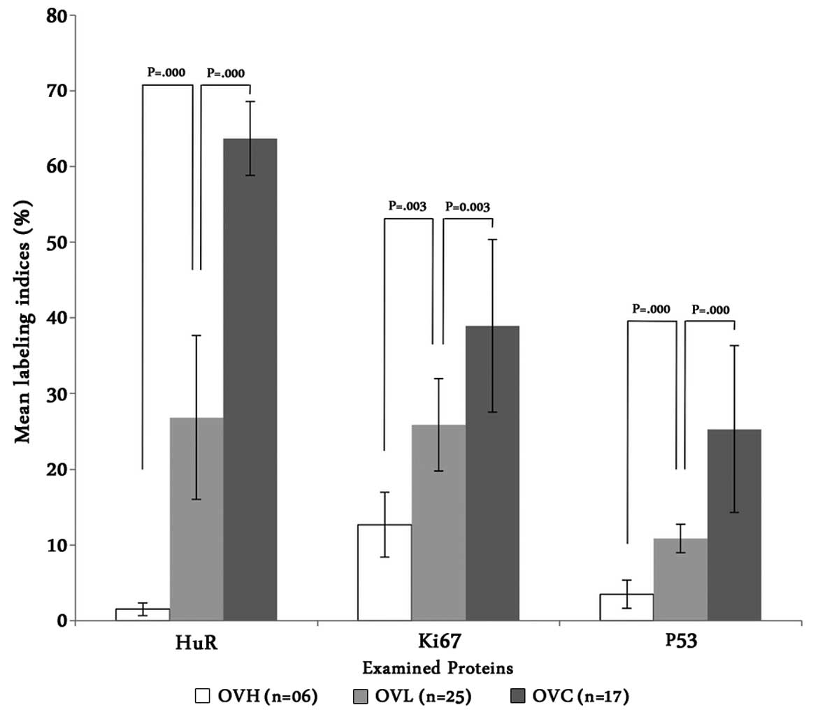
The mean LI (percentage of positive staining) of the
examined proteins was analyzed in all lesions. The mean LI of the
OVLs was significantly greater than that of the OVHs and lower than
that of the OVCs (Fig. 2). The LI
of HuR, Ki67 and p53 in the OVCs was 42.7-, 3.0- and 7.1-fold
higher than the LI of the OVHs, and 2.4-, 1.5- and 2.3-fold higher
than the OVL cases, respectively. When compared to the OVCs, only
HuR expression markedly differed between the OVH and OVL cases. The
OVL cases were also divided into two groups, high and low based on
their mean LI for the respective protein (Table III). The mean LI for the HuR
protein in the high group was 2-fold higher than that for the
corresponding low group, whereas for Ki67 and p53 these differences
were 1.6- and 1.3-fold, respectively. In addition, 90% of the cases
having high HuR LIs showed a positive signal in level 2 of the
epithelium (Table III).
Expression of HuR in the high and low group cases is also displayed
in Fig. 6. All 25 OVLs were further
followed-up for 3 years to determine their risk to develop
malignancy. Association between oral cancer development and
expression of the examined proteins was analyzed and is presented
in Table IV.
| Table IVAssociation of the expression of the
examined proteins and the development of malignancy in 25 oral
verrucous lesions (OVLs). |
Table IV
Association of the expression of the
examined proteins and the development of malignancy in 25 oral
verrucous lesions (OVLs).
| Prognostic
factors | No. of cases | Cases with
malignant transformation | P-value |
|---|
|
|---|
| n (%) |
|---|
| Association with
LIs of individual proteins | | | |
| HuR | | | 0.001 |
| High LI | 10 | 6 (60.0) | |
| Low LI | 15 | 0 (0.0) | |
| Ki67 | | | 0.169 |
| High LI | 7 | 3 (42.9) | |
| Low LI | 18 | 3 (16.7) | |
| p53 | | | 0.702 |
| High LI | 10 | 2 (20.0) | |
| Low LI | 15 | 4 (26.7) | |
| Association with
combination of the examined proteins | | | |
| HH-KH-PH | 5 | 2 (40.0) | 0.005 |
| HH-KL-PL | 3 | 3 (100.0) | |
| HH-KH-PL | 1 | 1 (100.0) | |
| HH-KL-PH | 1 | 0 (0.0) | |
| HL-KL-PL | 10 | 0 (0.0) | |
| HL-KH-PL | 1 | 0 (0.0) | |
| HL-KH-PH | 0 | 0 (0.0) | |
| HL-KL-PH | 4 | 0 (0.0) | |
| Association with
the distribution pattern of the examined proteins in the
epitheliuma | | | |
| HuR | | | 0.000 |
| Level 1 | 16 | 0 (0.0) | |
| Level 2 | 9 | 6 (66.7) | |
| Ki67 | | | 0.035 |
| Level 1 | 20 | 3 (15.0) | |
| Level 2 | 5 | 3 (60.0) | |
Six out of 25 (24%) OVLs underwent malignant
transformation, and all of them had a high HuR LI (60%) and
positive staining at level 2 (66.7%) of the epithelium (Table IV). This association was highly
significant in terms of malignant transformation. However, no
significant association was found when the LIs of Ki67 and p53 were
analyzed. The lethal combination of the three examined proteins in
relation to malignancy was also analyzed. As shown in Table IV, all of the cases that underwent
malignant transformation had a high HuR LI in combination with
either high or low Ki67 and p53. It is noteworthy that none of the
cases having a low HuR LI transformed into malignancy. Consistent
with these results, it is evident that OVLs with a high LI and wide
distribution of HuR protein in the epithelium had a significantly
higher oral cancer incidence than those with lower expression.
Discussion
OVC and OVH are commonly diagnosed by morphological
analysis, and expression levels of p53 and Ki67 are usually
considered as biomarkers to facilitate the diagnosis of OVC. The
p53 gene is reported to be the most frequent target for genetic
alterations leading to cancer, and its mutation has been
demonstrated in epithelial neoplasms. Although the p53 protein is
expressed constitutively in all cells, it normally cannot be
detected by immunohistochemical methods due to its short half-life
and quick disintegration. Mutated p53 on the other hand, being more
stable, accumulates in the cell and is readily detected by
immunohistochemistry (11).
Overexpression of p53 has been shown to occur in squamous
premalignant lesions of the head and neck region, upper
aerodigestive tract, the breast, and the urogenital tract, possibly
in response to carcinogen-induced DNA damage (12–14).
Our results are in agreement with previous studies, which
demonstrated increased positivity for p53 protein (mean LI, 25%)
extending up to the lower two-thirds of the epithelium (level 2),
whereas OVH showed very few positive signals (mean LI, 3.5%)
restricted to the basal cell layer of the epithelium (level 1).
Ki67 is a proliferation-associated protein that is
believed to play a critical role in regulation of the cell cycle.
The protein is expressed strictly during the active parts of the
cell cycle, including the G1, S and G2/M phases, but not in resting
cells in the G0 phase (15–17). Immunostaining with Ki67 highlights
cells that are actively involved in proliferation. Positive
expression of Ki67 has been used as a tool to estimate the
proliferative potential in head and neck cancers and patient
prognosis (18). Diffuse expression
of Ki67 has been demonstrated throughout the entire thickness of
the epithelium in invasive squamous carcinomas (19). Non-dysplastic acanthotic epithelium,
in contrast, shows expression limited to the basal cell layer
(20). In the present study, OVC
cases showed increased positive staining (mean LI, 39%) extending
to the lower two-thirds of the epithelium (level 2), whereas there
were few positive cells in the OVH cases (mean LI, 13%) and they
were localized in the lower one-third of the epithelium (level
1).
Oral verrucous borderline lesions (OVLs) are a much
greater diagnostic and therapeutic challenge. Lesions showing mild
to moderate dysplasia with few positive signals for p53 and Ki67
make diagnosis difficult, and pathologists often hesitate to
categorize them as benign or already transformed malignant lesions
considering the possible extensive surgical exposure of the cancer
patient, whereas benign lesions are treated conservatively. In the
present study, OVLs showed mild to moderate epithelial dysplasia
(Fig. 5), and expression levels of
Ki67 and p53 proteins (mean LI, 26 and 11%, respectively) were
higher than OVHs but lower than OVCs. Analogous to OVHs,
distribution of the positive cells was restricted predominantly to
the lower one-third of the epithelium (level 1). In such borderline
cases, morphological analysis and expression of p53 and Ki67 may
not help to distinguish true malignant lesions or those that have
the potential to transform into a malignancy.
It would be a great achievement in cancer diagnosis
if we could evaluate the histochemical parameters for the risk of
carcinoma in OVLs. Cytoplasmic HuR expression has been implicated
in the malignancy of colon, ovarian, breast, salivary gland,
uterine, larynx and prostate cancers and has been postulated to
contribute to the cancerous malignant phenotype (21–25).
Under physiological conditions, cellular stress induces HuR to bind
AU-rich element (ARE)-containing mRNAs, and the complex is
transported from the nucleus to the cytoplasm in a CRM1-dependent
manner. In contrast, our previous study showed that HuR and
ARE-mRNA in oral cancer cells are exported to the cytoplasm without
CRM1 suggesting the possible role of cytoplasmic HuR expression in
cell malignancy (10). In the
present study, we investigated whether the cytoplasmic HuR
expression pattern in OVLs may assist to discern true malignant
lesions.
As expected, OVCs showed diffuse cytoplasmic
expression of HuR (mean LI, 64%) throughout the epithelium, whereas
expression was rarely noted in the OVHs (mean LI, 1.5%) and
distributed in the lower one-third of the epithelium (level 1).
Although HuR expression was restricted to level 1 in more than half
(64%) of the OVLs, the expression was extended to the lower
two-thirds (level 2) of the epithelium in the remaining 36% cases
which is comparable to the expression of HuR in OVCs (35.3%).
Notably, HuR expression in OVLs (mean LI, 27%) was 2.4-fold lower
than that in the OVCs, whereas in OVHs the expression was 42.7-fold
lower. This considerable difference between OVHs and OVLs compared
to OVCs provide additional information regarding the role of HuR in
cancer development of OVL cases.
We further followed up the OVLs to understand the
risk of malignant transformation. All 25 OVL cases were divided
into high and low group based on their LIs for the three examined
protein, and their association with cancer development was
assessed. As shown in Table IV,
the OVL cases (6 out of 25) that were transformed into a malignancy
had a high HuR LI and had a more diffuse staining pattern in the
epithelium. None of the cases having a low HuR LI transformed into
a malignancy. These observations are in line with the hypothesis
that during tumorigenesis, HuR expression translocates from the
nucleus to the cytoplasm. This specific finding indicates the need
for proper treatment and careful follow-up for OVLs with high HuR
expression. Our results suggest the strong possibility that
expression of HuR in mild and moderate dysplasia may help to
identify lesions with a high potential for malignant
transformation, even when the positive signals for p53 and Ki67 are
not significant. These findings indicate that the degree of HuR
expression in OVLs may be an effective diagnostic factor that
determines the potential of a lesion for malignant transformation.
In conclusion, it is important to emphasize that appropriate
diagnosis of OVLs can prevent wide surgical resection of the
lesions as recommended for certain cases of oral
well-differentiated squamous cell carcinoma.
References
|
1
|
Ackerman LV: Verrucous carcinoma of the
oral cavity. Surgery. 23:670–678. 1948.PubMed/NCBI
|
|
2
|
Fertilo A and Recher G: Ackerman’s tumor
(verrucous carcinoma) of the larynx: a clinicopathologic study of
77 cases. Cancer. 46:1617–1630. 1980.
|
|
3
|
Batsakis JG, Hybels R, Crissman JD and
Rice DH: The pathology of head and neck tumors: verrucous
carcinoma, Part 15. Head Neck Surg. 5:29–38. 1982. View Article : Google Scholar : PubMed/NCBI
|
|
4
|
Medina JE, Dichtel W and Luna MA:
Verrucous-squamous carcinomas of the oral cavity. A
clinicopathological study of 104 cases. Arch Otolaryngol.
110:437–440. 1984. View Article : Google Scholar : PubMed/NCBI
|
|
5
|
Rebane A, Aab A and Steitz JA: Transportin
1 and 2 are redundant nuclear import factors for hnRNP A1 and HuR.
RNA. 10:590–599. 2004. View Article : Google Scholar : PubMed/NCBI
|
|
6
|
Fan XC and Steitz JA: HNS, a
nuclear-cytoplasmic shuttling sequence in HuR. Proc Natl Acad Sci
USA. 95:15293–15298. 1998. View Article : Google Scholar : PubMed/NCBI
|
|
7
|
Brennan CM, Gallouzi IE and Steitz JA:
Protein ligands to HuR modulate its interaction with target mRNAs
in vivo. J Cell Biol. 151:1–14. 2000. View Article : Google Scholar : PubMed/NCBI
|
|
8
|
Erkinheimo TL, Lassus H, Sivula A, et al:
Cytoplasmic HuR expression correlates with poor outcome and with
cyclooxygenase 2 expression in serous ovarian carcinoma. Cancer
Res. 63:7591–7594. 2003.PubMed/NCBI
|
|
9
|
Denkert C, Weichert W, Pest S, et al:
Overexpression of the embryonic-lethal abnormal vision-like protein
HuR in ovarian carcinoma is a prognostic factor and is associated
with increased cyclooxygenase 2 expression. Cancer Res. 64:189–195.
2004. View Article : Google Scholar : PubMed/NCBI
|
|
10
|
Hasegawa H, Kakuguchi W, Kuroshima T, et
al: HuR is exported to the cytoplasm in oral cancer cells in a
different manner from that of normal cells. Br J Cancer.
100:1943–1948. 2009. View Article : Google Scholar : PubMed/NCBI
|
|
11
|
Szymańska K and Hainaut P: TP53 and
mutations in human cancer. Acta Biochim Pol. 50:231–238. 2003.
|
|
12
|
Pavelic ZP, Li YQ, Stambrook PJ, et al:
Overexpression of p53 protein is common in premalignant head and
neck lesions. Anticancer Res. 14:2259–2266. 1994.PubMed/NCBI
|
|
13
|
Shin DM, Kim J, Ro JY, Hittelman J, Roth
JA, Hong WK and Hittelman WN: Activation of p53 gene
expression in premalignant lesions during head and neck
tumorigenesis. Cancer Res. 54:321–326. 1994.
|
|
14
|
Wang LD, Shi ST, Zhou Q, et al: Changes in
p53 and cyclin D1 protein levels and cell proliferation in
different stages of human esophageal and gastric-cardia
carcinogenesis. Int J Cancer. 59:514–519. 1994. View Article : Google Scholar : PubMed/NCBI
|
|
15
|
MacCallum DE and Hall PA: The location of
pKi67 in the outer dense fibrillary compartment of the nucleolus
points to a role in ribosome biogenesis during the cell division
cycle. J Pathol. 190:537–544. 2000. View Article : Google Scholar : PubMed/NCBI
|
|
16
|
Gerdes J, Lemke H, Baisch H, Wacker HH,
Schwab U and Stein H: Cell cycle analysis of a cell
proliferation-associated human nuclear antigen defined by the
monoclonal antibody Ki-67. J Immunol. 133:1710–1715.
1984.PubMed/NCBI
|
|
17
|
Braun N, Papadopoulos T and
Müller-Hermelink HK: Cell cycle dependent distribution of the
proliferation-associated Ki-67 antigen in human embryonic lung
cells. Virchows Arch B Cell Pathol Incl Mol Pathol. 56:25–33. 1988.
View Article : Google Scholar : PubMed/NCBI
|
|
18
|
Woźniak A, Golusiński W, Kaczmarek E and
Kaczmarek J and Kaczmarek J: Prognostic significance of Ki 67 and
PCNA expression in laryngeal squamous cell carcinoma (morphometric
evaluation of labelling index-L1). Otolaryngol Pol. 56:437–443.
2002.(In Polish).
|
|
19
|
Gimenez-Conti IB, Collet AM, Lanfranchi H,
et al: p53, Rb, and cyclin D1 expression in human oral verrucous
carcinomas. Cancer. 8:17–23. 1996. View Article : Google Scholar : PubMed/NCBI
|
|
20
|
Saito T, Nakajima T and Mogi K:
Immunohistochemical analysis of cell cycle-associated proteins p16,
pRb, p53, p27 and Ki-67 in oral cancer and precancer with special
reference to verrucous carcinomas. J Oral Pathol Med. 28:226–232.
1999. View Article : Google Scholar : PubMed/NCBI
|
|
21
|
Cho NP, Han HS, Soh Y, Lee KY and Son HJ:
Cytoplasmic HuR over-expression is associated with increased
cyclooxygenase-2 expression in laryngeal squamous cell carcinomas.
Pathology. 39:545–550. 2007. View Article : Google Scholar : PubMed/NCBI
|
|
22
|
Cho NP, Han HS, Soh Y and Son HJ:
Overexpression of cyclooxygenase-2 correlates with cytoplasmic HuR
expression in salivary mucoepidermoid carcinoma but not in
pleomorphic adenoma. J Oral Pathol Med. 36:297–303. 2007.
View Article : Google Scholar : PubMed/NCBI
|
|
23
|
Heinonen M, Bono P, Narko K, et al:
Cytoplasmic HuR expression is a prognostic factor in invasive
ductal breast carcinoma. Cancer Res. 65:2157–2161. 2005. View Article : Google Scholar : PubMed/NCBI
|
|
24
|
Niesporek S, Kristiansen G, Thoma A, et
al: Expression of the ELAV-like protein HuR in human prostate
carcinoma is an indicator of disease relapse and linked to COX-2
expression. Int J Oncol. 32:341–347. 2008.PubMed/NCBI
|
|
25
|
Gallouzi IE and Steitz JA: Delineation of
mRNA export pathways by the use of cell-permeable peptides.
Science. 294:1895–1901. 2001. View Article : Google Scholar : PubMed/NCBI
|