Introduction
Breast cancer (BC) is the most frequently occurring
malignant disease among women in the Western hemisphere, inflicting
one in eight women (1). Despite
marked progress in disease management, morbidity and mortality
remain a public health concern, prompting efforts to advance our
understanding of BC biology, with the aim of developing innovative
approaches. In this respect, particular attention should be given
to promoters of cell growth and microenvironment.
Macrophage migration inhibitory factor (MIF) is a
pleiotropic inflammatory cytokine of 12.5-kDa monomeric molecular
weight originally described as a T cell lymphokine modulating
macrophage motility. Subsequently, MIF was shown to be produced by
a variety of immune and non-immune cells such as B- and
T-lymphocytes as well as endocrine, endothelial and epithelial
cells of diverse histogenetic origin. Pathophysiologically, MIF
plays a pivotal role in various autoimmune and inflammatory
disorders, such as rheumatoid arthritis, systemic lupus
erythematosus, septic shock and atherosclerosis (2,3). In
addition, there is growing evidence that MIF is involved in
cancerogenesis and progression. Currently, there is a general
consensus that MIF promotes tumor growth by several mechanisms; it
stimulates cancer cell proliferation by triggering the
MAPK/PI3K/Akt pathways, inhibits induction of p53-dependent
apoptosis, increases production of vascular endothelial growth
factor (VEGF) and inhibits the antitumor immune response (4–6).
Moreover, it modulates metastatic behavior of tumor cells and
affects tumor stromal cells (7,8). On
the cellular level, MIF is stored in the cytoplasmic compartment
and is released in response to several stimuli. In breast cancer
cells, signaling is triggered by its receptor CD74, then channeled
via the Akt pathway, with the involvement of Src and PI3K (9). Additionally, CD44 can be recruited to
the complex with CD74 and G-protein coupled receptors CXCR2 and
CXCR4 can act as receptors, inducing rapid activation of integrins
(10). Within the cell, c-Jun
activation domain-binding protein-1 (JAB1) serves as binding
partner, thereby reducing secretion and autocrine growth
stimulation (11). MIF has also
been reported to inhibit apoptosis by binding to p53 (12).
MIF serum levels are elevated in breast cancer
patients (13) and MIF has been
shown to be overexpressed in breast cancer tissue compared to
normal breast. Correlations with classical histoprognostic factors
remain controversial (14–16).
Looking at a receptor, CD74 is expressed in breast
cancer tissue and its presence appears to be correlated with lymph
node invasion and triple-negative tumors (17–19)
making a correlation study attractive.
Due to the pro-tumoral activities, the MIF pathway
might be considered as a potential therapeutic target. Of note,
tumor-activated HSP90 chaperone complex protects MIF from
degradation, suggesting that HSP90 inhibitors could serve as
anti-MIF therapeutic agents. Indeed, the HSP90 inhibitor
17-N-allylamino-17-demethoxygeldanamycin (17-AAG) inhibits growth
of MIF-expressing breast tumors in mice (20). Regarding the MIF receptor,
milatuzumab, a humanized anti-CD74 antibody, has clinical activity
on lymphomas and has been tested in vitro with some success
as an antibody-drug conjugate on solid cancer cell lines positive
for CD74 (21).
These considerations led us to an
immunohistochemical assessment of expression of MIF and CD74 in
serial sections of human breast cancer tumor specimens, mapping
their profiles in cancer and stromal cells. In parallel, the serum
level of MIF was determined in breast cancer patients.
Materials and methods
Breast cancer patients and healthy
women
Formalin-fixed, paraffin-embedded, residual tissue
material of diagnostic biopsies of 96 breast cancer tumors
(Table I), which were available for
retrospective analysis by immunohistochemistry, were examined for
MIF expression and 59 of them for CD74. In each case, the
pathological stage and histological grade were defined according to
the criteria of the World Health Organization 2012. Estrogen
receptor (ER), progesterone receptor (PR) status, Ki-67 labeling
index and HER2 expression were evaluated at the time of the
original diagnosis by immunohistochemistry, as previously described
(22–24). Positivity for ER and PR as well as
HER2 score has been defined previously (25). The characteristics of the tumors are
outlined in Table I. Residual
tumor-free breast tissue blocks from 16 breast plasties for
esthetic purposes were used as reference specimens of healthy
tissue.
 | Table ITumor characteristics. |
Table I
Tumor characteristics.
| MIF data | CD74 data |
|---|
|
|
|
|---|
| Variable | n (%) n=96 | Level of sign. | n (%) n=59 | Level of sign. |
|---|
| Tumor size | | S | | NS |
| T1 | 43 (45) | | 23 (39) | |
| T2 | 40 (42) | | 27 (45) | |
| T3 | 10 (11) | | 7 (12) | |
| T4 | 3 (3) | | 2 (3) | |
| Histological
type | | NS | | NS |
| Invasive
ductal | 84 (87) | | 50 (85) | |
| Invasive
lobular | 11 (11) | | 9 (15) | |
| Other | 1 (1) | | 0 | |
| Lymph node
status | | NS | | NS |
| N0 | 62 (65) | | 35 (59) | |
| N0–3 | 34 (35) | | 20 (34) | |
| NE | | | 4 | |
| Histological
grade | | NS | | NS |
| G1–2 | 74 (77) | | 50 (85) | |
| G3 | 22 (23) | | 8 (15) | |
| NE | | | 1 | |
| Estrogen receptor
status | | NS | | S |
| Negative | 17 (18) | | 8 (14) | |
| Positive
(>1%) | 79 (82) | | 50 (85) | |
| NE | 1 | | | |
| Progesterone
receptor status | | NS | | NS |
| Negative | 25 (26) | | 12 (20) | |
| Positive
(>1%) | 71 (74) | | 46 (78) | |
| NE | | | 1 | |
| Triple receptor
negative status | | NS | | S |
| Negative | 83 (86) | | 52 (88) | |
| Positive
(>1%) | 13 (14) | | 6 (10) | |
| NE | | | 1 | |
| Ki-67 | | NS | | NS |
| Low (≤15%) | 47 (49) | | 29 (49) | |
| High
(>15%) | 49 (51) | | 29 (49) | |
| NE | | | 1 | |
| HER-2 | | NS | | NS |
| Amplified | 7 (7) | | 5 (8) | |
| Non amplified | 89 (93) | | 53 (90) | |
| NE | | | | 1 |
Blood samples from 36 newly diagnosed early breast
cancer female patients (BCP) were obtained prospectively for
determining serum level of MIF, prior to any breast cancer
treatment (Table II). Twenty-two
healthy women (HW) were also enrolled in this prospective study as
a control group. In both cohorts, 10 ml of blood were obtained,
centrifuged at 4°C and sera stored at −20°C until assaying.
| Table IICharacteristics of patients/tumors
for MIF serum measurements. |
Table II
Characteristics of patients/tumors
for MIF serum measurements.
|
Characteristics | No. |
|---|
| Total no. of
patients | 36 |
| Total no. of
tumors | 37 |
| Mean age
(years) | 56 (range
30–80) |
| Tumor histological
type | no./37 |
| Invasive carcinoma
NST | 34 |
| Lobular invasive
carcinoma | 2 |
| Others | 1 |
| T1 | 22 |
| T2 | 13 |
| T3–T4 | 2 |
| Positive node | 19 |
| Grade III | 16 |
| Ki-67 index
>15% | 12 |
| Positive hormone
receptor | 29 |
| Triple receptor
negative | 7 |
| HER2 amplified | 4 |
| Neoadjuvant
chemotherapy | 17 |
This study was approved by the Ethics Committee of
Erasme Hospital, Brussels, Belgium, according to the international
and Belgian laws (P2008/314 and A2013/016).
Determination of MIF serum levels
Serum concentration was assayed by a sandwich
enzyme-linked immunosorbent assay (ELISA) using a commercial kit
(DuoSet ELISA Development kit, R&D Systems, Minneapolis, MN,
USA). The assays were carried out according to the instructions
provided by the supplier. MIF concentrations in serum samples were
determined by interpolation from a reference curve established with
increasing concentrations of recombinant human MIF.
Immunohistochemistry on tissue specimens
and assessment
For immunostaining of MIF, after antigen retrieval
by microwave treatment, sections were pretreated with hydrogen
peroxide to block endogenous peroxidase activity. Thereafter, they
were exposed to casein to avoid false-positive staining. These
steps were followed by sequential incubations with (i) primary
antibody (rabbit polyclonal anti-human MIF (26), (ii) post-blocking (Immunologic, The
Netherlands), (iii) poly-dextran secondary antibody against rabbit
immunoglobulins. Immunocomplexes were finally visualized by
exposure to the chromogen diaminobenzidine in the presence of
H2O2. Sections were counterstained with luxol
fast blue prior to light microscopy examination.
For immunostaining of CD74 (rabbit polyclonal
anti-human CD74 FL-296, Santa Cruz Biotechnology, Santa Cruz, CA,
USA), immunohistochemistry was performed using a procedure similar
to that described previously for MIF (27).
MIF and CD74 expressions in the glandular and in the
stromal compartments were assessed by light microscopy and
quantified according to a modified Allred score (22).
Statistical analysis
Non parametric analysis was carried out using the
Mann-Whitney test and Spearman’s rank order correlations was
used.
Results
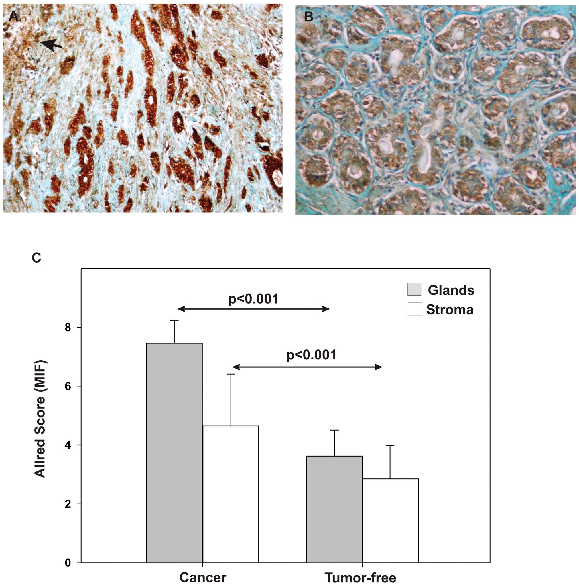
MIF expression is increased in carcinoma
cells and in the stroma of breast cancer tissue
The intensity of immunohistochemical staining for
MIF was assessed in tumor-free breast tissues (tumor-free, n=16;
Fig. 1B) and in invasive ductal and
lobular carcinomas (cancer, n= 96; Fig.
1A). The staining intensity, quantified using modified Allred
scores, was markedly increased in carcinomas compared to tumor-free
specimens (Mann-Whitney test, p<0.001; Fig. 1C). With regard to the distribution
of the immunohistochemical signal, it was strong and homogeneous in
carcinoma cells and appeared essentially extra-nuclear (i.e.
cytoplasmic and membrane staining). By contrast, the signal
intensity was weaker and its presentation more heterogeneous in the
stromal compartment (Fig. 1A),
although it showed the same extra-nuclear distribution. In the
latter compartment, peritumoral fibroblasts were the most intensely
stained, whereas lymphocytes and macrophages as well as endothelial
cells showed weaker immunostaining.
There was no significant correlation of staining
with histological type, nodal status, histological grade, HER2
amplification, hormonal receptor status and the Ki-67 index
(Mann-Whitney test NS, data not shown). However, we found a
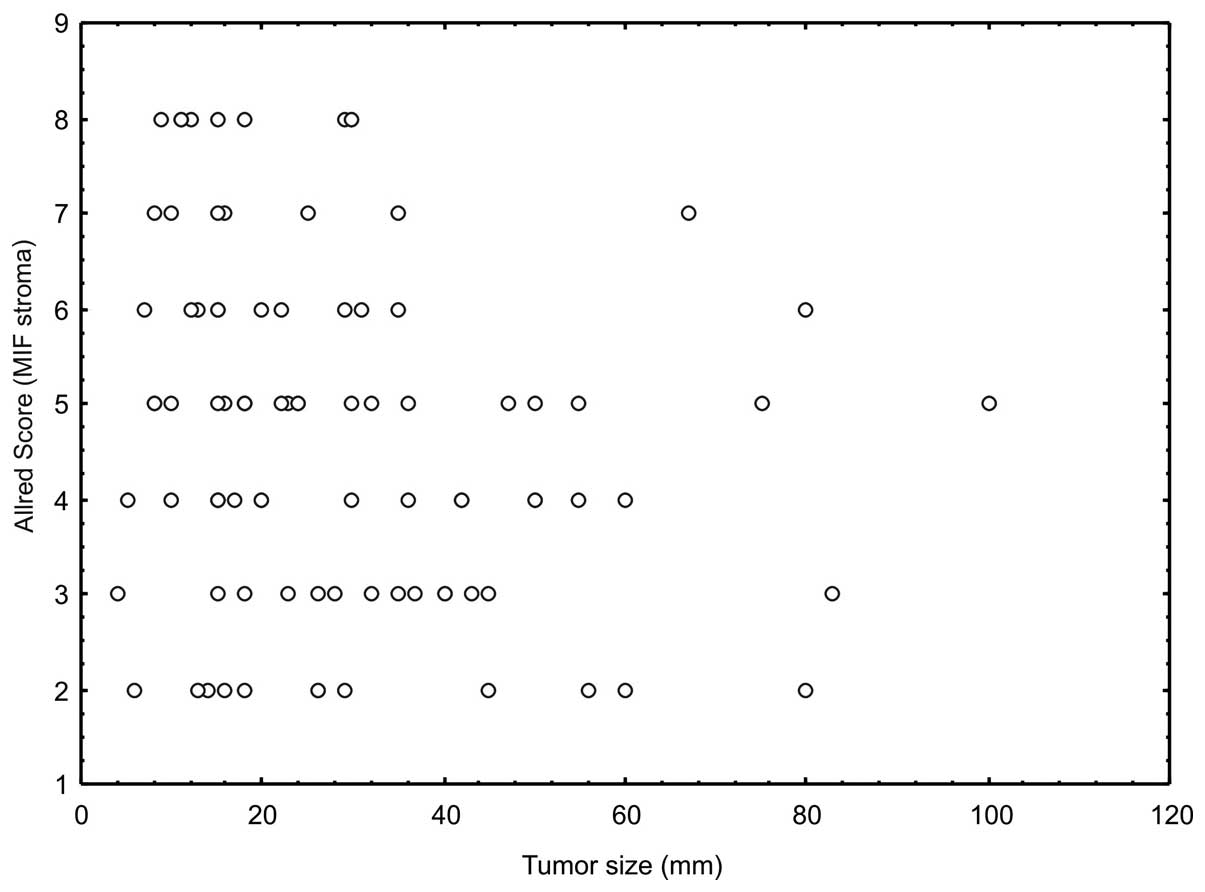
significant inverse correlation between tumor size and stromal
positivity (Spearman’s rank test, R=−0,238, n=91, p=0.02; Fig. 2).
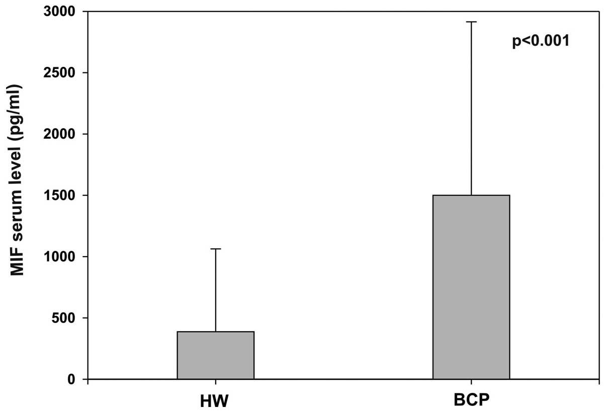
Serum MIF levels are increased in breast
cancer patients
In addition to the immunohistochemical analysis of
MIF expression in breast tumors, we used an ELISA to compare MIF
levels in serum of 36 patients with BC (Table II) to those of 22 healthy
individuals. Fig. 3 shows the
average serum level for each group. In healthy individuals, we
found a mean level of 387 pg/ml. In contrast, the mean
concentration in patients reached 1,500 pg/ml, a concentration
approximately four-fold higher than that recorded in healthy
individuals (Mann-Whitney test, p<0.001; Fig. 3). We did not find any significant
variation of the serum levels according to the tumor
characteristics.
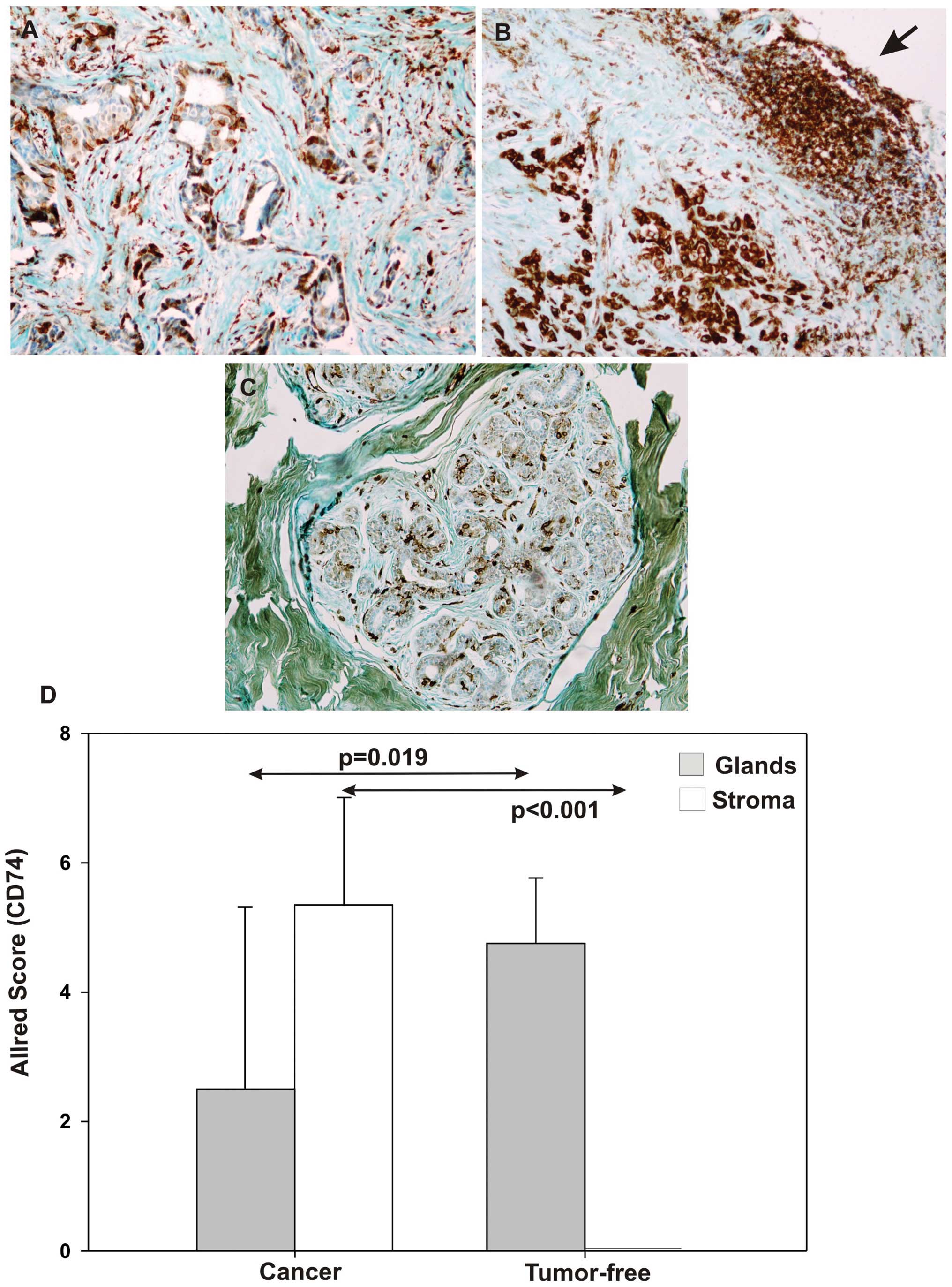
CD74 expression in breast cancer
tissues
CD74 expression was evaluated by
immunohistochemistry in tumor specimens (cancer, n=59; Fig. 4A) and compared to that observed in
tumor-free breast tissues (tumor-free, n=15; Fig. 4C). Compared with tumor-free tissues,
immunostaining intensity analysis of cancer breast biopsies,
quantified according to a modified Allred scoring, revealed a
significant decrease in CD74 expression in the neoplastic glandular
compartment (Mann-Whitney test, p=0.019; Fig. 4D), contrasting with an increased
expression in the peritumoral stroma (Mann-Whitney test,
p<0.001; Fig. 4D). In cancer
cells, immunoreactivity for CD74 signal was weak and heterogeneous,
mostly located on the membrane and in the cytoplasm. CD74
positivity was observed in the stroma surrounding carcinomas,
namely in lymphocytes (arrow, Fig.
4B), macrophages and vessel endothelium, with a similar
intracellular distribution.
There was no significant variation according to
histological type, tumor size, nodal status, histological grade,
HER2 amplification and Ki-67 index in either compartment. However,
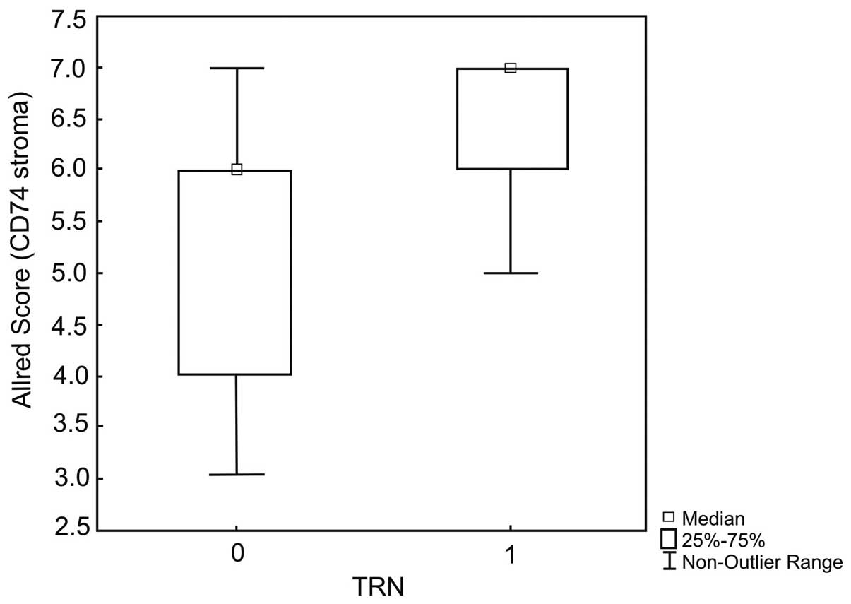
stromal CD74 expression appeared significantly correlated with
triple-negative receptor (TRN) status and the absence of estrogen
receptors (Mann-Whitney test, p=0.02; Fig. 5).
Discussion
Numerous clinical and experimental data suggest that
MIF can play a role in the pathogenesis of various solid tumors. We
here combined serum level assessment with immunohistochemical
detection. In this respect, our study is the first to report the
semi-quantitative immunohistochemical evaluation of both expression
of MIF and CD74 in serial sections of biopsy specimens, as well as
their distribution between cancer cells and the peritumoral stroma.
Our results allow us to consider suggestions on the involvement of
this cytokine in breast tumor biology.
We confirmed the significant increase of MIF serum
levels in BCP, observed previously by others (15,16).
In a set of 98 serum proteins, MIF was able to discriminate normal
tissue from breast cancer (28),
although the authors considered the elevation of serum MIF more
indicative of the inflammatory response to breast tumor than to the
tumor itself. Correlations between MIF and classical
histoprognostic factors remain controversial. A negative
correlation with the number of involved nodes and a positive
correlation with poor response to neoadjuvant chemotherapy have
been reported (13,15). Recent data in mice suggest an
immunosuppressive role of MIF favoring BC metastasis (29). Furthermore, this cytokine could also
contribute to the systemic metabolic disturbances associated with
BC (30). Thus, the increased serum
level of MIF in BCP could be a non-specific signature of a systemic
response to breast cancer with immune and metabolic
implications.
Our data extended the data basis for an increased
expression of MIF in breast cancer tumors (14–16).
Of note, upregulation concerns tumor cells and peritumoral stroma.
Verjans et al described a significant increase of MIF in
breast carcinoma, this increase showing a positive correlation with
the ER/PR status and a negative one with tumor size, in association
with better overall survival. This data set intimated a beneficial
role of intracellular MIF, whereas extracellular MIF is
pro-oncogenic by promoting cancer cell-stroma interactions
(14). Increased MIF positivity in
cancer and stromal cells, including tumor-associated macrophages,
correlated inversely with nodal involvement and also led to
suggestions for a role of MIF in tumor-stroma interactions
(15). We observed an inverse
correlation between stromal MIF expression and tumor size, as well
as an elevated MIF presence in fibroblasts surrounding the tumor
tissue. This is consistent with the hypothesis that MIF could
modulate the tumor size by inhibiting recruitment of
cancer-associated fibroblasts (CAFs)/myofibroblasts, eventually
resulting in retardation of tumor growth (7). These CAFs are known to be the source
for effectors shaping a pro-tumoral microenvironment such as the
chemokine CXCL-1 and interleukins-6 and -8 and may be involved in
tumorigenesis (31,32).
Following profiling of MIF expression, we proceeded
to map CD74 in breast cancer and in tumor-free tissue. We showed
that the CD74 positivity is significantly increased in lymphocytes,
macrophages and endothelial cells but heterogeneous in neoplastic
cells. A correlation was detected between tumor stromal CD74
expression and the tumor triple receptor-negative status (TRN),
including the absence of ER itself. A correlation between CD74
expression in BC, TRN status and lymph node invasion has previously
been reported (17,18).
To date, co-expression of MIF and CD74 has not been
studied in breast cancer but it has been described in prostate and
non-small cell lung cancer. In prostate cancer, MIF was intense but
CD74 staining was weak and patchy (33). In lung cancer, CD74 was mainly
detected in the stromal compartment or in stromal and epithelial
cells. Co-expression of MIF and CD74 was associated with greater
vascularity and higher levels of pro-angiogenic CXC chemokines
(34). We suggest that, in breast
cancer, high-level expression of CD74 could be a marker of
increased vascularity in stroma. Since bevacizumab, an anti-VEGF
antibody, appears to be more efficient in TRN breast cancer
(35), stromal CD74 expression
could be examined as a predictive marker of response to
bevacizumab-based therapy.
Our findings showed an increased extent of MIF
expression in cancer cells and in stromal fibroblasts of BC tumor,
in contrast to a less uniform increase of CD74 expression mainly in
stromal lymphocytes, macrophages and endothelium. This could
suggest a pro-oncogenic role of MIF in BC tumors taking place
predominantly in the stromal compartment. MIF, secreted by tumor
cells, could then modulate the immune microenvironment and its
neovascularization, favoring escape from immune surveillance and
tumor cell dissemination. Experimental data published show that MIF
suppression in breast cancer cell lines does not affect their
proliferation in vitro but causes delayed tumor growth in
mice, increasing the prevalence of an immune suppressive
myeloid-derived population within the tumor (28,36).
Xu et al showed that MIF might promote angiogenesis in BC
tumors (16). Finally, the
discordance between the MIF and CD74 expression on cancer cells
suggests that MIF could act on cells through other types of
receptors, such as CXCR4 and CXCR7 (7,37).
In vitro studies on MIF/CD74 (CD44) knockdown by siRNA
approach, as reported for clear cell renal carcinoma (38), shed light on this aspect.
In conclusion, our data support the concept of a
functional role of MIF in human breast cancer. In addition to auto-
and paracrine effects on cancer cells, MIF could contribute to
shape the microenvironment leading to immunomodulation and
angiogenesis, these aspects deserving further investigations.
Interfering with MIF effects in breast tumors in a therapeutic
perspective remains an attractive but complex challenge, notably
depending on the development of suitable MIF inhibitors. Level of
co-expression of MIF and CD74 could be a surrogate marker for
efficacy of anti-angiogenic drugs, particularly in TRN breast
cancer tumors.
Acknowledgements
Mrs. Nadege Kindt is the recipient of a grant
(‘Televie’) from the National Fund for Scientific Research.
Professors C. Decaestecker and G. Laurent are Senior Research
Associates of the National Fund for Scientific Research (Belgium).
Tumor blocks were kindly provided by Dr D. Faverly from CMP
Pathology (Brussels).
References
|
1
|
Altekuse SF, Kosary CL, Krapcho M, et al:
SEER Cancer Statistics Review, 1975–2007. NCI; Bethesda: 2010
|
|
2
|
Grieb G, Merk M, Bernhagen J and Bucala R:
Macrophage migration inhibitory factor (MIF): a promising
biomarker. Drug News Perspect. 23:257–264. 2010. View Article : Google Scholar : PubMed/NCBI
|
|
3
|
Santos LL and Morand EF: Macrophage
migration inhibitory factor: a key cytokine in RA, SLE and
atherosclerosis. Clin Chim Acta. 399:1–7. 2009. View Article : Google Scholar : PubMed/NCBI
|
|
4
|
Conroy H, Mawhinney L and Donnelly SC:
Inflammation and cancer: macrophage migration inhibitory factor
(MIF) - the potential missing link. QJM. 103:831–836. 2010.
View Article : Google Scholar : PubMed/NCBI
|
|
5
|
Mitchell RA: Mechanisms and effectors of
MIF-dependent promotion of tumourigenesis. Cell Signal. 16:13–19.
2004. View Article : Google Scholar : PubMed/NCBI
|
|
6
|
Rendon BE, Willer SS, Zundel W and
Mitchell RA: Mechanisms of macrophage migration inhibitory factor
(MIF)-dependent tumor microenvironmental adaptation. Exp Mol
Pathol. 3:180–185. 2009. View Article : Google Scholar : PubMed/NCBI
|
|
7
|
Tarnowski M, Grymula K, Liu R, et al:
Macrophage migration inhibitory factor is secreted by
rhabdomyosarcoma cells, modulates tumor metastasis by binding to
CXCR4 and CXCR7 receptors and inhibits recruitment of
cancer-associated fibroblasts. Mol Cancer Res. 8:1328–1343. 2010.
View Article : Google Scholar
|
|
8
|
Martinez LM, Vallone VB F, Labovsky V, et
al: Changes in the peripheral blood and bone marrow from untreated
advanced breast cancer patients that are associated with the
establishment of bone metastases. Clin Exp Metastasis. 31:213–232.
2014. View Article : Google Scholar : PubMed/NCBI
|
|
9
|
Lue H, Thiele M, Franz J, et al:
Macrophage migration inhibitory factor (MIF) promotes cell survival
by activation of the AKT pathway and role for CSN5/JAB1 in the
control of autocrine MIF activity. Oncogene. 26:5046–5059. 2007.
View Article : Google Scholar : PubMed/NCBI
|
|
10
|
Bernhagen J, Krohn R, Lue H, et al: MIF is
a noncognate ligand of CXC chemokine receptors in inflammatory and
atherogenic cell recruitment. Nat Med. 13:587–596. 2007. View Article : Google Scholar : PubMed/NCBI
|
|
11
|
Zernecke A, Bernhagen J and Weber C:
Macrophage migration inhibitory factor in cardiovascular disease.
Circulation. 117:1594–1602. 2008. View Article : Google Scholar : PubMed/NCBI
|
|
12
|
Mitchell RA, Liao H, Chesney J,
Fingerle-Rowson G, Baugh J, David J and Bucala R: Macrophage
migration inhibitory factor (MIF) sustains macrophage
proinflammatory function by inhibiting p53: regulatory role in the
innate immune response. Proc Natl Acad Sci USA. 99:345–350. 2002.
View Article : Google Scholar : PubMed/NCBI
|
|
13
|
Fersching DMI, Nagel D, Siegele B, Salat
C, Heinemann V, Holdenrieder S and Stoetzer OJ: Apoptosis-related
biomarkers sFAS, MIF, ICAM-1 and PAI-1 in serum of breast cancer
patients undergoing neoadjuvant chemotherapy. Anticancer Res.
32:2047–2058. 2012.PubMed/NCBI
|
|
14
|
Verjans E, Noetzel E, Bektas N, et al:
Dual role of macrophage migration inhibitory factor (MIF) in human
breast cancer. BMC Cancer. 9:2302009. View Article : Google Scholar : PubMed/NCBI
|
|
15
|
Bando H, Matsumoto G, Bando M, et al:
Expression of macrophage migration inhibitory factor in human
breast cancer: association with nodal spread. Jnp J Cancer Res.
93:389–396. 2002.PubMed/NCBI
|
|
16
|
Xu X, Wang B, Ye C, et al: Overexpression
of macrophage migration inhibitory factor induces angiogenesis in
human breast cancer. Cancer Lett. 261:147–157. 2008. View Article : Google Scholar : PubMed/NCBI
|
|
17
|
Tian B, Zhang Y, Li N, Liu X and Dong J:
CD74: a potential novel target for triple-negative breast cancer.
Tumour Biol. 33:2273–2277. 2012. View Article : Google Scholar : PubMed/NCBI
|
|
18
|
Greenwood C, Metodieva G, Al-Janabi K, et
al: Stat1 and CD74 overexpression is co-dependent and linked to
increased invasion and lymph node metastasis in triple-negative
breast cancer. J Proteomics. 75:3031–3040. 2012. View Article : Google Scholar : PubMed/NCBI
|
|
19
|
Metodieva G, Correa Nogueira-de-Souza N,
Greenwood C, Al-Janabi K, Leng L, Bucala R and Metodiev MV:
CD74-dependent deregulation of the tumor suppressor scribble in
human epithelial and breast cancer cells. Neoplasia. 15:660–668.
2013.PubMed/NCBI
|
|
20
|
Schulz R, Marchenko ND, Holemboswki, et
al: Inhibiting the HSP90 chaperone destabilizes macrophage
migration inhibitory factor and thereby inhibits breast tumor
progression. J Exp Med. 209:275–289. 2012. View Article : Google Scholar
|
|
21
|
Govindan SV, Cardillo TM, Sharkey RM, Tat
F, Gold DV and Goldenberg DM: Milatuzumab-SN-38 conjugates for the
treatment of CD74+cancers. Mol Cancer Ther. 12:968–978.
2013. View Article : Google Scholar : PubMed/NCBI
|
|
22
|
Hammond EH, Hayes DF, Dowsett M, et al:
American Society of Clinical Oncology/College of American
Pathologists guideline recommendations for immunohistochemical
testing of estrogen and progesterone receptors in breast cancer
(unabridged version). Arch Pathol Lab Med. 134:48–72. 2010.
|
|
23
|
Dowset M, Nielsen TO, A’Hern R, et al:
Assesment of Ki67 in breast cancer: recommendations from the
international Ki67 in breast cancer working group. J Natl Cancer
Inst. 103:1656–1664. 2011. View Article : Google Scholar : PubMed/NCBI
|
|
24
|
Schrohl AS, Pedersen HC, Jensen SS,
Nielsen S and Brünner N: Human epidermal growth factor receptor
(HER2) immunoreactivity: specificity of three pharmacodiagnostic
antibodies. Histopathology. 59:975–983. 2011. View Article : Google Scholar
|
|
25
|
Preat F, Simon P and Noel JC: Differences
in breast carcinoma immunohistochemical subtypes between immigrant
Arab and European women. Diagn Pathol. 9:262014. View Article : Google Scholar : PubMed/NCBI
|
|
26
|
Cludts S, Decaestecker Ch, Johnson B, et
al: Increased expression of macrophage migration inhibitory factor
during progression to hypopharyngeal squamous cell carcinoma.
Anticancer Res. 30:3313–3319. 2010.PubMed/NCBI
|
|
27
|
Kindt N, Lechien J, Decaestecker C, et al:
Expression of macrophage migration-inhibitory factor is correlated
with progression in oral cavity carcinomas. Anticancer Res.
32:4499–4505. 2012.PubMed/NCBI
|
|
28
|
Jesneck JL, Mukherjee S, Yurkovetsky Z,
Clyde M, Marks JR, Lorkshin AE and Lo JY: Do serum biomarkers
really measure breast cancer? BMC Cancer. 9:1642009. View Article : Google Scholar : PubMed/NCBI
|
|
29
|
Simpson KD, Templeton DJ and Cross JV:
Macrophage migration inhibitory factor promotes tumor growth and
metastasis by inducing myeloid-derived suppressor cells in the
tumor microenvironment. J Immunol. 189:5533–5540. 2012. View Article : Google Scholar
|
|
30
|
Kim H, Lee S, Kim JH, et al: Elevated
levels of macrophage migration inhibitory factor in women with
metabolic syndrome. Horm Metab Res. 43:642–645. 2011. View Article : Google Scholar : PubMed/NCBI
|
|
31
|
Kolar M, Szabo P, Dvorankova B, et al:
Upregulation of IL-6, IL-8 and CXCL-1 production in dermal
fibroblasts by normal/malignant epithelial cells in vitro:
Immunohistochemical and transcriptomic analyses. Bio Cell.
104:738–751. 2012. View Article : Google Scholar
|
|
32
|
Catteau X, Simon P, Vanhaeverbeek M and
Noël JC: Variable stromal periductal expression of CD34 and smooth
muscle actin (SMA) in intraductal carcinoma of the breast. PLoS
One. 8:e57732013. View Article : Google Scholar : PubMed/NCBI
|
|
33
|
Meyer-Siegler KL, Iczkowski KA and Vera
PL: Further evidence for increased macrophage migration inhibitory
factor expression in prostate cancer. BMC Cancer. 5:732005.
View Article : Google Scholar : PubMed/NCBI
|
|
34
|
McClelland M, Zhao L, Carskadon S and
Arenberg D: Expression of CD74, the receptor for macrophage
migration inhibitory factor, in non-small cell lung cancer. Am J
Pathol. 174:638–646. 2009. View Article : Google Scholar : PubMed/NCBI
|
|
35
|
von Minckwitz G, Eidtmann H, Rezai M, et
al: Neoadjuvant chemotherapy and bevacizumab for HER-2 negative
breast cancer. N Engl J Med. 366:299–309. 2012.PubMed/NCBI
|
|
36
|
Simpsons KD and Cross JV: MIF
metastasis/MDSC-inducing factor? Oncoimmunology. 2:32013.PubMed/NCBI
|
|
37
|
Zhang L, Ye SB, Ma G, et al: The
expressions of MIF and CXCR4 protein in tumor microenvironment are
adverse prognostic factors in patients with esophageal squamous
cell carcinoma. J Transl Med. 11:602013. View Article : Google Scholar : PubMed/NCBI
|
|
38
|
Du W, Wright BM, Li X, et al:
Tumor-derived macrophage migration inhibitory factor promotes an
autocrine loop that enhances renal cell carcinoma. Oncogene.
32:1469–1474. 2013. View Article : Google Scholar : PubMed/NCBI
|