Introduction
Esophageal cancer is the eighth most common cancer
worldwide with distinct geographical and ethnic characteristics
(1). In China, it ranks as the
fourth leading cause of cancer related mortality (2). Esophageal squamous cell carcinoma
(ESCC) is the dominant type of esophageal malignancy in China. ESCC
carcinogenesis is a multistage process characterized by
morphological changes from normal esophagus to basal cell
hyperplasia, dysplasia, carcinoma in situ and squamous cell
carcinoma (SCC). It involves complex interactions between the
environment and genetic factors (3). Although early intervention, as well as
surgical, medical and radiotherapy techniques have undergone great
achievements to date, the 5-year survival rate of ESCC patients
remains low (4,5). Since most patients are at an advanced
stage at initial diagnosis, the chance of surgery is lost (6). Chemotherapy-based comprehensive
treatment is the main treatment for advanced patients. Yet,
chemotherapeutic outcomes are not optimistic due to the poor
selection and serious adverse reactions of standard chemotherapy
drugs. Therefore, targeted therapy drugs that can avoid the
chemotherapy-induced adverse reactions and elevate the treatment
effect are critical to reduce the morbidity and mortality of
ESCC.
Insulin-like growth factor-1 receptor (IGF-1R) is a
tyrosine kinase receptor implicated in the pathogenesis of multiple
cancers (7). Overexpression of
IGF-IR has been reported in a range of human solid tumors such as
breast, non-small cell lung and prostate cancer, sarcomas,
hepatocellular carcinoma and pancreatic, ovarian and
gastrointestinal cancers (8–11).
After ligand binding, IGF-1R can initiate the activation of the
PI3K/AKT/mTOR signaling and Ras/Raf/MEK/MAPK pathways resulting in
the activation of multiple transcription factors such as ELK-1,
CREB and AP-1 to modulate cell proliferation, survival,
differentiation, motility, invasion and angiogenesis (12,13).
Furthermore, more and more evidence indicates that IGF-1R is a
prerequisite for tumor progression and is involved in the critical
steps of the metastatic cascade (14,15).
Therefore IGF-1R is one of the most attractive targets for cancer
therapeutic interventions (16).
In addition to IGF-1R, the IGF signaling system
consists of another two ligands, IGF-1 and IGF-II; another two cell
membrane receptors, IGF-IIR and insulin receptor; and six IGF
binding proteins (IGFBP 1–6) (17).
High circulating levels of IGF-I and a decreased level of IGFBP3
have been linked to increased cancer risk and have been identified
as prognostic markers for various cancers such as breast, prostate,
colon and lung cancer (18). Almost
90% of the circulating IGFs bind to IGFBP-3. IGFBP-3 inhibits IGF
action at the cellular level by competitively binding IGFs and
thereby preventing their binding to the IGF-1R. The molar ratio of
IGF-I to IGFBP-3 has been regarded as an important index of IGF-I
bioavailability. IGF-1R is associated with the sensitivity to
chemotherapy in several types of cancers including lung, colon and
head and neck cancer (19–21). The serum concentrations of IGF1 and
IGFBP-3 may also play a role in the response to the
chemotherapies.
IGF-1R has been reported as a marker for prognosis
and as a therapeutic target in human ESCC (22). IGF-I autocrine system could
stimulate tumor growth and chemoresistance in human esophageal
carcinoma cells (23). However, the
detailed biological function of IGF-1R in ESCC still requires
investigation, and the implications of IGF-1R in treatment are
still unclear. In this study, we determined the expression of
IGF-1R in ESCC patients and investigated the biological function of
IGF-1R in ESCC cell lines in a xenograft animal model. The overall
objective of this study was to identify the potential application
of IGF-1R in ESCC clinical treatment.
Materials and methods
Ethics statement
This study was approved by the Institutional Review
Boards of the First Affiliated Hospital of Zhengzhou University.
Written informed consent was obtained from all participants for the
use of their blood or tissue samples. Full consent was obtained
from the Institutional Review Boards of the First Affiliated
hospital of Zhengzhou University. Animal care and experiments were
approved by the Institute Animal Care and Use Committee of the
Cancer Institute and Hospital, Chinese Academy of Medical Sciences
and Peking Union Medical College.
Human ESCC cell lines and ESCC patient
samples
ESCC cell lines EC9706, EC109 and NEC were gifts
from the Department of Etiology and Carcinogenesis, Cancer
Institute and Hospital, Chinese Academy of Medical Sciences and
Peking Union Medical College. All the cells were cultured in
RMPI-1640 medium with 10% fetal bovine serum (FBS), in 5%
CO2 at 37°C.
Eighty human ESCC tissues used for
immunohistochemical staining were collected from the First
Affiliated Hospital of Zhengzhou University with the exact
post-surgical pathological examination. Eighteen normal
tumor-adjacent tissues were acquired from these patients at least 3
cm away from the primary tumor. These patients consisted of 52
females and 28 males. The mean age was 60 years. According to
histological grade, 22 patients were in grade I, 37 patients were
in grade II, and 21 patients were in grade III. According to
clinical stage 6 patients were in stage I, 35 patients were in
stage II, 32 patients were in stage III and 7 patients were in
stage IV. There were 26 patients without lymph node metastasis and
53 patients with lymph node metastasis.
Serum samples used for the ELISA study were
collected from 120 patients. These patients were diagnosed with
ESCC between 2007 to 2009 at The First Affiliated Hospital of
Zhengzhou University, Henan Cancer Hospital and Zhengzhou Central
Hospital. None of these patients received any therapy before the
two courses of chemotherapy treatment. The pre-therapy serum was
collected 1–3 days before treatment, and the post-therapy serum was
collected at the end of the treatments. These patients consisted of
51 females and 121 males. The mean age was 60 years. According to
clinical stage, 43 patients were in stage IIB, 70 patients were in
stage III and 59 patients were in stage IV. The chemotherapy effect
was divided into 4 grades according to the Response Evaluation
Criteria in Solid Tumors (RECIST): complete response (CR), all
lesions disappeared lasting for at least 4 weeks; partial response
(PR), the lesion volume shrank >50% lasting for at least 4
weeks; stable disease (SD), no significant change, shrinking
<50% and enlargement <25%; progressive disease (PD), the
lesion enlarged >25%.
Bioinformatic analysis
The microarray data GSE 33426 and GSE 23400 were
downloaded from the GEO database: (http://www.ncbi.nlm.nih.gov/gds/). The differentially
expressed genes were identified using GEO2R tools on GEO. The
heatmap was generated using Genesis 1.0 (Graz, Austria). The
biofunction and pathway analysis was performed with IPA®
Software (http://www.ingenuity.com).
Immunohistochemistry
Fresh tumor tissues were fixed with formalin,
embedded and finally cut into 4-μm sections. The sections were
deparaffinized by dimethylbenzene and graded ethanol. Then the
sections were incubated with 3% H2O2, goat
serum, anti-IGF-1R antibody, biotin-labeled secondary antibody,
peroxidaze-conjugated streptavidin in order before DAB staining.
All the reagents were purchased from ZSGB-Bio (Beijing, China). The
staining intensity of IGF-1R was quantified into 3 groups according
to the percentage of stained cells: negative, 0–5% cells were
stained; weak positive, >5–50% cells were stained; strong
positive, >50–100% cells were stained.
Western blot analysis
Protein was extracted from the cells using lysis
buffer (50 mM Tris-HCl pH 7.4, 150 mM NaCl, 1 mM EDTA, 1% Triton
X-100). The protein concentration was measured using the the
Bradford method. The protein was separated by SDS-PAGE and
transferred to a polyvinylidene fluoride (PVDF) membrane
(Millipore, Billerica, MA, USA). After incubation with 10% milk,
the primary IGF-1R antibody, and the secondary antibody, the blot
signals were detected by chemiluminescence using the ECL kit
(Applygen, Beijing, China). All the antibodies were purchased from
Santa Cruz (Dallas, TX, USA).
Stable IGF-1R-knockdown cell line
pGCsilencer™ U6/Neo/GFP/RNAi-IGF-1R plasmid used for
the knockdown of IGF-1R and the pGCsilencer™U6/Neo/GFP negative
control were obtained from Genechem Co. (Shanghai, China). Both
plasmids were transfected into EC9706 cells using Lipofectamine™
2000 (Invitrogen, Carlsbad, CA, USA). The cells were digested and
seeded into a new 24-well plate, diluted 5 times, 24 h after the
transfection. Then, the regular medium was changed to medium
containing 600 μg/ml G418 for stable transfection colony screening.
After all of the cells without transfection died 4 days later, the
remained cells were sustained by medium containing 200 μg/ml G418
and accepted as the stable transfection cell lines:
IGF-1R-knockdown cell line and blank transfection control cell
line. Western blotting and RT-PCR verified the knockdown of IGF-1R
in the cell lines.
Cell proliferation study
Cells were seeded in a 96-well plate at a density of
4,000 cells/well. The next day, 10 μl of 5mg/ml MTT was added to
the medium for a 3-h incubation, and the time point was set as zero
then detected every 24 h for 6 days. Then the medium was removed,
and 150 μl DMSO was added. Finally the OD value was read at 490 nm
on a Model 680 microplate reader (Bio-Rad, Hercules, CA, USA) after
a 10-min incubation.
Flow cytometry
Cells were digested by trypsin, washed twice in cold
PBS, fixed with ice cold 70% methanol, and incubated at 4°C
overnight. Cells were then washed in PBS and incubated with 25
μg/ml propidium iodide containing 30 μg/ml ribonuclease for 30 min
at room temperature. Cells were analyzed on the FACSCalibur Cell
Sorting System (Becton Dickinson, Franklin Lakes, NJ, USA) using BD
FACSuite™ Software in triplicate.
Cell colony formation study
Cells were seeded onto 6-well plates at a density of
300 cells/well. Following 12 days, visible colonies were fixed with
methanol and stained with 0.4% crystal violet solution. Colonies
were counted, and the number of visible colonies in each well was
determined. Each assay was performed in triplicate.
Mouse ESCC xenograft study
Male BALB/c nude mice 5–6 weeks old were purchased
from the Institute of Laboratory Animal Sciences, Chinese Academy
of Medical Sciences and Peking Union Medical College. The mouse
were divided into three groups and were inoculated with stable
IGF-1R-knockdown cells, empty vector control cells or wild-type
EC9706 cells, respectively (six mice in each group). A total of
2×10⁶ cells were inoculated subcutaneously into the
right dorsal flank. The size of the tumor was measured twice per
week using a digital vernier caliper. The tumor volume was
determined from the orthogonal dimensions (d1, d2, d3) using the
formula (d1 × d2 × d3) × π/6. Five weeks after inoculation, the
mice were euthanized by cervical dislocation, and xenograft tumors
were collected.
ELISA
Collected venous blood was centrifuged for 10 min at
2,000 rpm. The upper tube of serum was then taken for
cryopreservation and stored at −40°C for ELISA assay. IGF-1 and
IGFBF-3 ELISA kits were obtained from Senxiong Biotech (Shanghai,
China). The standard curve was established according to the
manufacturer’s instructions. Serum samples (100 μl) were incubated
in each well for 120 min, followed by 50 μl primary antibody buffer
for 60 min, 100 μl secondary primary antibody buffer for 60 min,
100 μl substrate buffer for 10 min and 1 drop stop solution. Then
the plate was read at 490 nm on the Model 680 microplate reader.
The serum concentrations of IGF-1 and IGFBP3 were calculated by the
standard curves.
Statistical analysis
All statistical analyses were performed using the
SPSS 13.0 package. Pearson’s χ2 test was used to detect
the expression difference in the cancer and normal tissues. All
tests of statistical significance were two-sided, and P<0.05 was
considered to indicate a statistically significant difference. All
quantitative data are presented as means ± SD. Independent samples
t-test and one-way ANOVA were adopted to compare the means.
Results
Bioinformatic analysis of IGF-1R in
ESCC
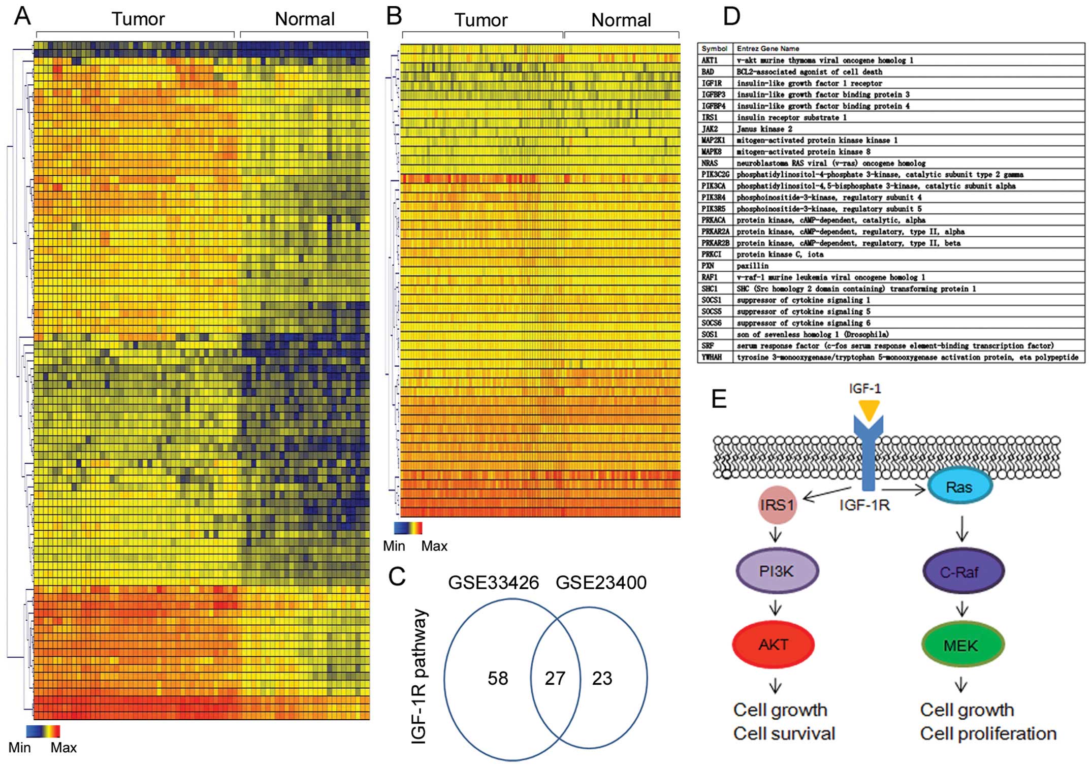
In order to study the expression and potential
function of IGF-1R in ESCC, we took advantage of publicly available
gene expression data (GSE33426 and GSE23400). In the referenced
studies, tumor cluster and normal cells separated by laser-capture
microscope or tumor tissues and adjacent normal tissues were
compared by cDNA microarray. IGF-1R was identified as a
differentially expressed gene in both studies. Moreover, we found
85 and 50 genes, respectively, related with the IGF-1R signaling
pathway (Fig. 1A and B).
Twenty-three genes in the IGF-1R signaling pathway were identified
in both studies (Fig. 1C and D).
After IPA analysis, we found that these genes were mainly mapped to
AKT and MEK cascade and were related to cell growth, proliferation
and survival (Fig. 1E). The
bioinformatic analysis supported our hypothesis that IGF-1R plays
an important role in ESCC.
IGF-1R is highly expressed in human ESCC
cancer tissues
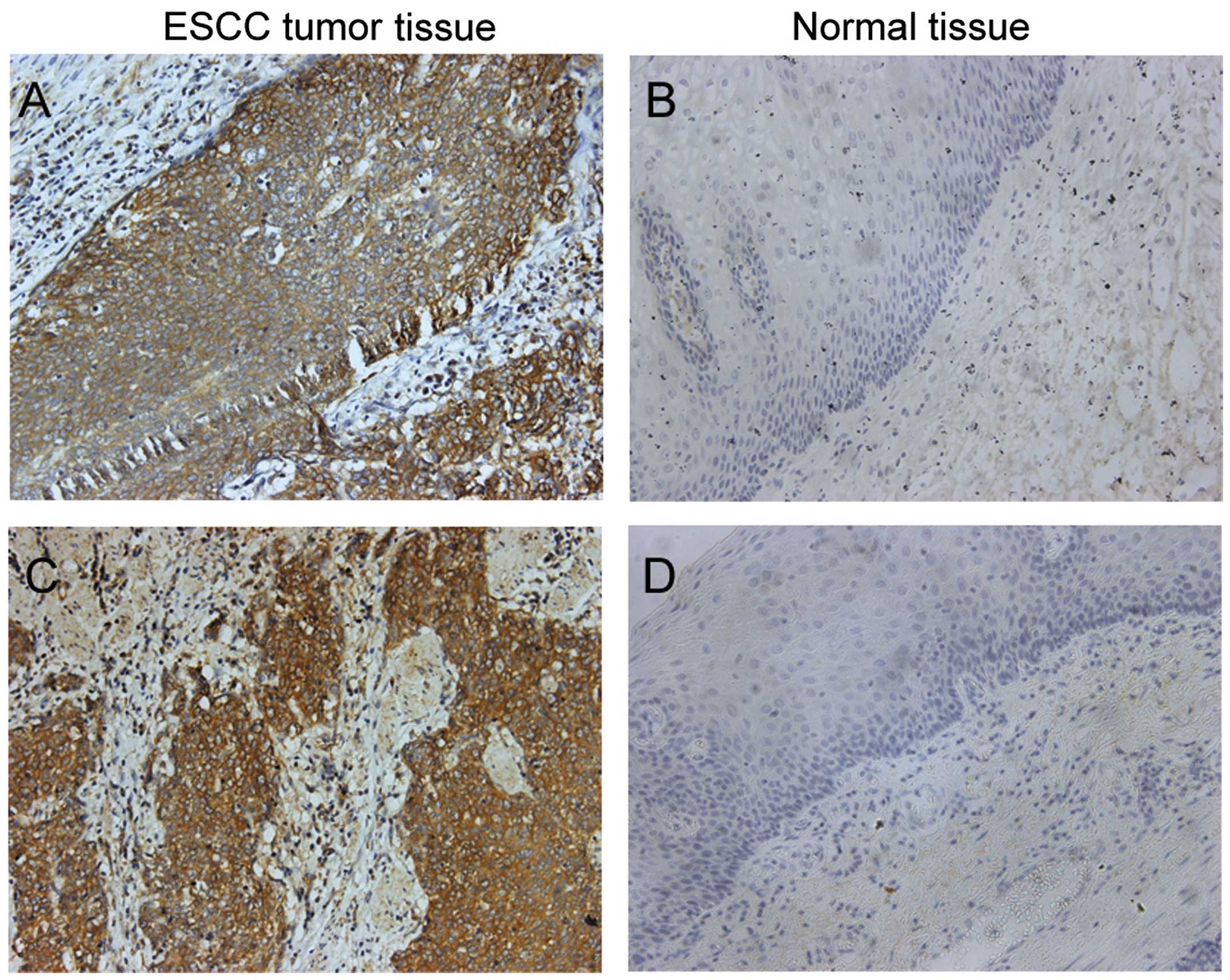
Firstly, we used immunohistochemical (IHC) staining
to detect the expression of IGF-1R in ESCC tumor tissues (Fig. 2A and C) and adjacent normal tissues
(Fig. 2B and D). After IHC
staining, IGF-1R showed strong staining in the cancer cell
membrane. Of the 80 cancer specimens, 11 cases were negative, 28
cases were weakly positive and 41 cases were strongly positive. The
total positive rate was 86.25% and the strong positive rate was
51.25%. Of the 18 normal tumor-adjacent tissues, 7 cases were
negative, 9 cases were weakly positive and 2 cases were strongly
positive. The total positive rate was 61.11% and the strong
positive rate was 11.11%. Both the total positive rate and the
strong positive rate were significantly higher in the ESCC tissues
than in the normal tumor-adjacent esophageal tissues (total
positive rate, χ2=4.630, P<0.05; strong positive
rate, χ2=9.614, P<0.01). In addition, we found a
relationship between the expression of IGF-1R and cancer patient
characteristics (Table I). We found
that high expression of IGF-1R was associated with more aggressive
lymph node metastasis, lower histological grade and advanced
clinical stage. Both positive and strong positive rates of IGF-1R
expression were also higher in patients with lymph node metastasis
than those without lymph node metastasis (P<0.01), and were also
higher in patients with advanced clinical stage (III–IV stage) than
those with early clinical stage (I–II stage). The expression of
IGF-1R was also increased from a low differentiation grade to a
moderate differentiation stage and high differentiation grade
(P<0.05 between each of the two groups). No significant
difference was noted in relation to age or gender (P>0.05).
These results showed that IGF-1R was highly expressed in ESCC
tissues, and the high expression of IGF-1R in the ESCC tissues was
related to tumor progression.
 | Table IAssociation of IGF-1R with clinical
characteristics. |
Table I
Association of IGF-1R with clinical
characteristics.
| Clinical
characteristics | Negative n (%) | Weak positive n
(%) | Strong positive n
(%) | P-value (total
positive) | P-value (strong
positive) |
|---|
| Gender |
| Female | 8 (10.7) | 19 (32.2) | 25 (57.2) | >0.05 | >0.05 |
| Male | 3 (15.4) | 9 (36.5) | 16 (48.1) | | |
| Age, years |
| <51 | 1 (7.7) | 5 (38.5) | 7 (53.8) | >0.05 | >0.05 |
| 51–65 | 5 (11.6) | 18 (38.3) | 24 (51.1) | | |
| >65 | 5 (25.0) | 5 (25.0) | 10 (50.0) | | |
| Lymph node
metastasis |
| Yes | 3 (5.6) | 11 (20.3) | 40 (74.1) | <0.05 | <0.05 |
| No | 8 (30.8) | 17 (65.3) | 1 (3.9) | | |
| Histological
grade |
| Low | 1 (4.8) | 3 (14.2) | 17 (81.0) | <0.05 | <0.05 |
| Moderate | 3 (8.1) | 16 (43.2) | 18 (48.7) | | |
| High | 7 (30.2) | 9 (40.9) | 6 (27.3) | | |
| Clinical stage |
| I–II | 10 (24.7) | 18 (43.9) | 13 (31.7) | <0.01 | <0.01 |
| III–IV | 1 (2.6) | 10 (25.6) | 28 (71.8) | | |
IGF-1R acts as an oncogene in ESCC in
vitro and in vivo
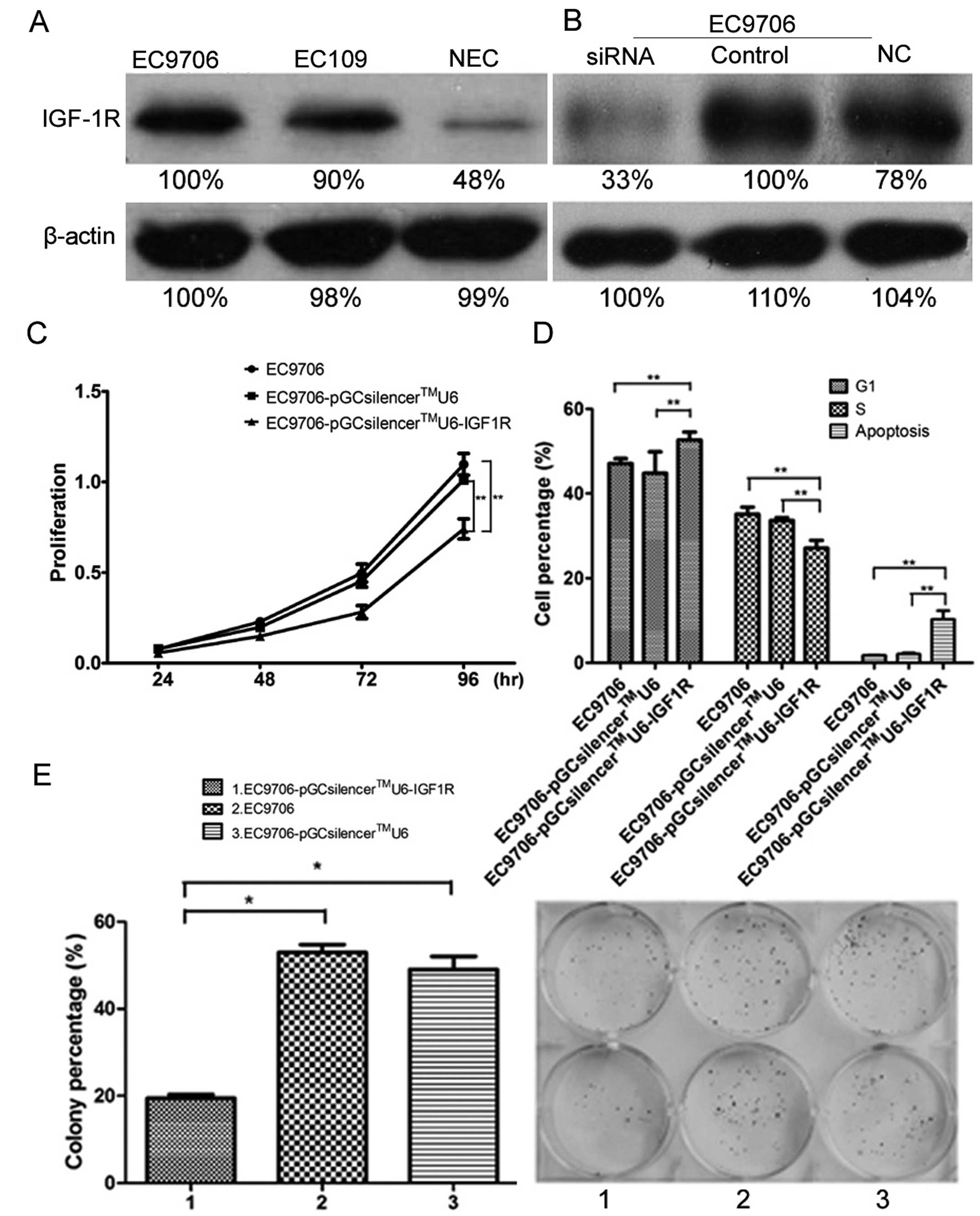
We determined the expression of IGF-1R in EC9706,
EC109 and NEC cells and found that IGF-1R was relatively highly
expressed in these three ESCC cell lines. We established stable
IGF-1R-knockdown cells by transfection of
pGCsilencer™U6/Neo/GFP/RNAi IGF-1R in EC9706 cells. Western
blotting and RT-PCR verified the successful knockdown of IGF-1R in
these cells (Fig. 3A and B).
Compared with the empty vector control, the cell proliferation was
inhibited and the doubling time was extended significantly in the
IGF-1R-knockdown cells (Fig. 3C).
Significant cell cycle arrest occured in the IGF-1R-knockdown cells
with an increased percentage of G1 phase cells (P<0.05) and a
decreased percentage of S phase cells (P<0.01) (Fig. 3D). In addition, a higher apoptosis
rate (P<0.05) and weaker clonogenesis ability (P<0.05) were
found after IGF-IR-knockdown (Fig.
3E). All of these results showed that IGF-1R acts as an
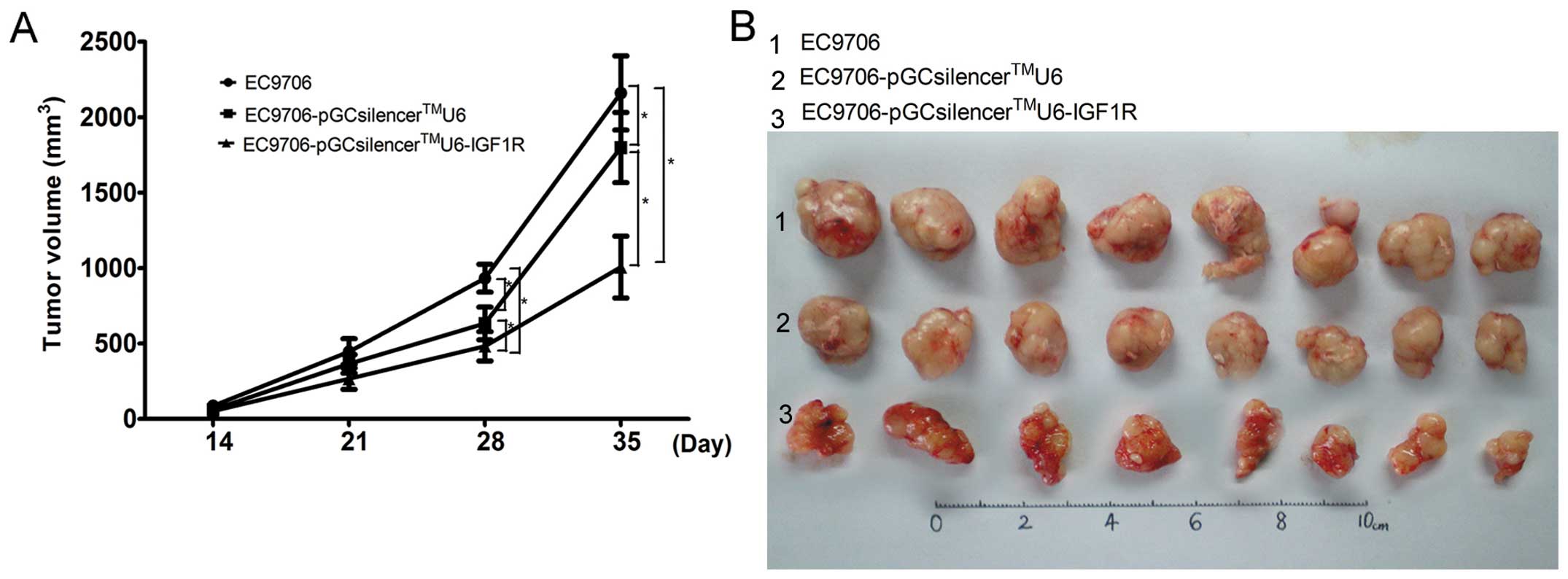
oncogene in ESCC in vitro. We also used a mouse ESCC
xenograft model to detect the tumor growth ability of IGF-1R in
vivo. Ten days after cell inoculation, subcutaneous nodules
were noted and finally developed into tumors. Although all three
cell lines induced visible tumors in the nude mouse, the size of
the tumors induced by the stable IGF-1R-knockdown cells was
significantly smaller than those formed by the empty control
(P<0.05) and wild-type cells (P<0.05) (Fig. 4A and B). The animal assay showed
that IGF-1R can induce tumor growth in vivo.
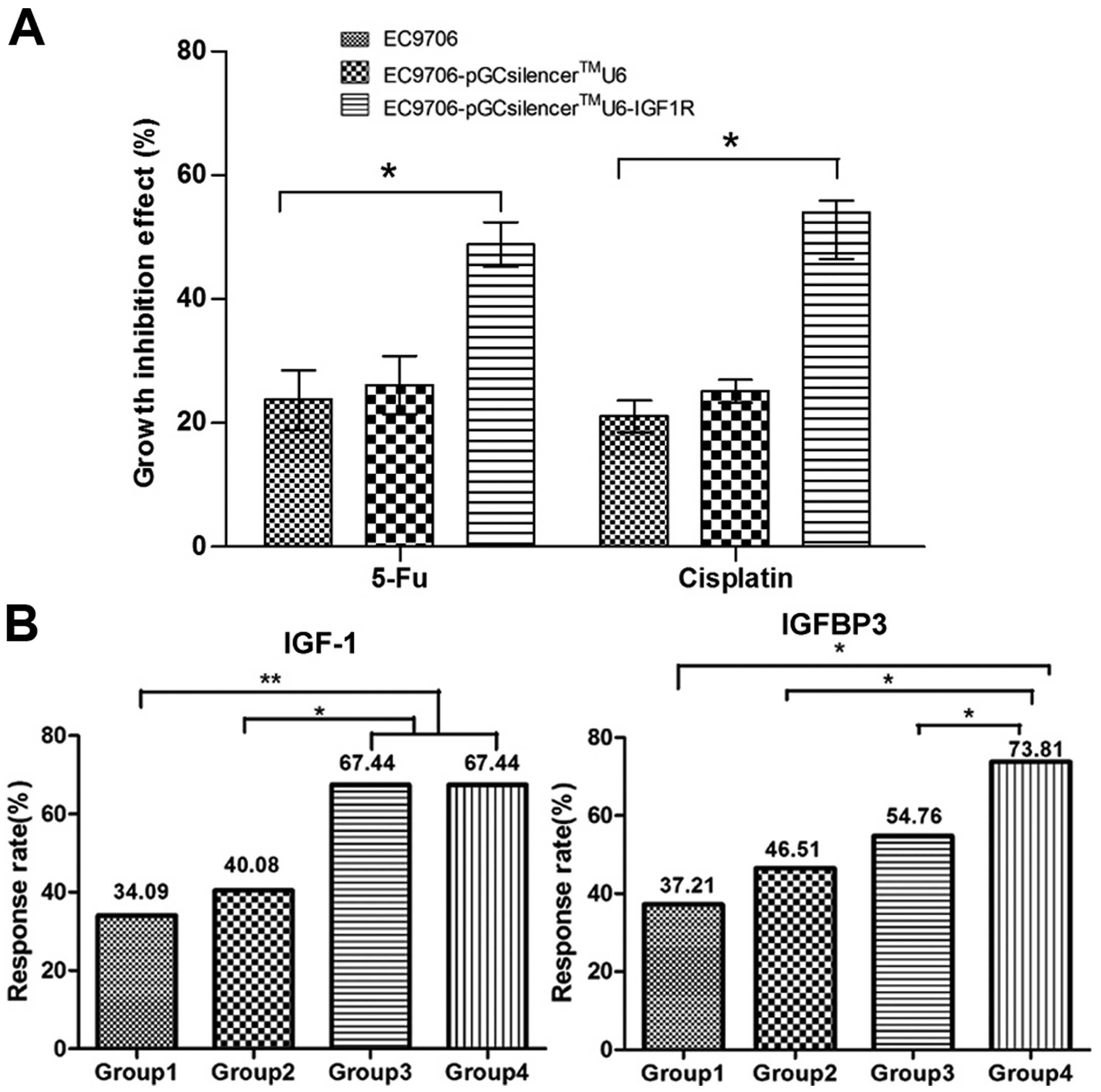
High expression of IGF-1R is related to
low sensitivity to chemotherapy in the ESCC cell lines
We compared the sensitivity to 5-FU and cisplatin
between the IGF-1R-knockdown cells and the empty vector control. We
found that the growth inhibition rate of 5-FU and cisplatin in the
pGCsilencer™U6-IGF-1R-transfected cells was higher than that in the
empty vector-transfected and the non-transfected cells (P<0.05)
(Fig. 5A). The high expression of
IGF-1R facilitates cancer cell resistance to chemotherapy.
Serum concentrations of IGF-1/IGFBP-3 are
associated with the chemotherapy response
We determined the serum concentrations of IGF-1 and
IGFBP-3 in 172 patients both before and after chemotherapy
treatment (Fig. 5B). The mean serum
concentrations of IGF-1 pre-therapy and post-therapy in all
patients were 268.87±61.66 and 266.42±49.98 ng/ml, respectively,
without significant difference. The mean serum concentrations of
IGFBP-3 pre-therapy and post-therapy in all patients were
2523.2±469.83 and 2598.8±563.56 ng/ml, respectively, without
significant difference. In contrast, the serum concentration of
IGFBP-3 increased (P<0.01) and the IGF-1/IGFBP-3 ratio decreased
(P=0.01) significantly after chemotherapy in the chemotherapy
responsive group (CR and PR). The serum concentration of IGF-1
decreased after chemotherapy in the responsive groups but without
significance (P>0.05). Accordingly, in the chemotherapy
unresponsive group (SD and PD) the IGF-1/IGFBP-3 ratio increased
significantly (P<0.05) after chemotherapy; the concentration of
IGFBP-3 decreased but without significance (P>0.05). There was
no variation in IGF-1 (Table II).
These results showed that the IGF-1/IGFBP-3 ratio can be used to
predict the chemotherapeutic effect; a decreased IGF-1/IGFBP-3
molar ratio presents a better chemotherapeutic effect. We also
evaluated the chemotherapy response rate in 4 groups divided
according to the quartiles of plasma IGF-1 and IGFBP-3.
Chemotherapy response rate increased from low concentrations of
IGF-1 and IGFBP-3 to high concentrations of IGF-1 and IGFBP-3.
These results showed that increased serum levels of IGF-1 and
IGFBP-3 are associated with significantly higher rates of tumor
response (P<0.05).
| Table IISerum concentration of IGF-1, IGFBP-3
and IGF-1/IGFBP-3 ratio. |
Table II
Serum concentration of IGF-1, IGFBP-3
and IGF-1/IGFBP-3 ratio.
| Groups | n | IGF-1 (ng/ml) | IGFBP-3
(ng/ml) | IGF-1/IGFBP-3 |
|---|
| Chemotherapy
responsive |
| Pre-therapy | 90 | 284.5±56.5 | 2599.4±444.6 | 0.11391±0.034 |
| Post-therapy | 90 | 270.0±50.1 | 2854.3±559.0 | 0.10030±0.036 |
| P-value | | 0.07 | 0.001 | 0.01 |
| Chemotherapy
unresponsive |
| Pre-therapy | 82 | 251.7±62.8 | 2439.5±485.1 | 0.10723±0.034 |
| Post-therapy | 82 | 262.5±49.9 | 2318.5±418.4 | 0.11820±0.035 |
| P-value | | 0.226 | 0.089 | 0.045 |
Discussion
In the present study, we determined the expression
of IGF-1R in an ESCC cohort and verified the high expression of
IGF-1R in ESCC tumor tissues when compared with that in adjacent
normal tissues. High expression of IGF-1R was found to be
associated with aggressive lymph node metastasis, lower
histological grade and advanced clinical stage. After knockdown of
IGF-1R in ESCC cell lines and the mouse xenograft assay, we
verified the pro-proliferation, apoptosis inhibition and tumor
growth function of IGF-1R in vitro and in vivo. We
also found that IGF-1R was associated with the response to standard
chemotherapy drugs 5-FU and cisplatin in the ESCC cell lines. More
importantly, we found that the serum concentration of IGF-1/IGFBP3
can be used for the predicting chemotherapeutic effect. Increased
serum levels of IGF-1 and IGFBP-3 were found to be associated with
significantly higher rates of tumor response.
High expression of IGF-1R has been reported in
several cancers including ESCC. Imsumran et al used IHC to
detect the expression of IGF-1R in ESCC tumor tissues and found
that ~50% of tissues showed high expression of IGF-1R which was
associated with invasive depth, metastasis, advanced tumor stage
and recurrence (22). In this
study, we also detected high expression of IGF-1R in ESCC tissues
compared with that in adjacent normal tissues. We found that high
expression of IGF-1R was associated with more aggressive lymph node
metastasis, lower histological grade and advanced clinical stage,
similar to the results of previous research. We suggest that the
expression of IGF-1R can be used as a biomarker for the diagnosis
and tumor progression of ESCC.
IGF-1R plays an important role in carcinogenesis
mainly based on the activation of the PI3K/AKT/mTOR signaling and
the Ras/Raf/MEK/MAPK pathways. The activation of AKT and MAPK
pathways help cancer cells acquire the ability for proliferation,
evasion of apoptosis, insensitivity to antigrowth signals,
unlimited replicative potential, metastasis and angiogenesis
(24,25). Both of these pathways are reported
to be activated in ESCC. After using siRNA to knock down the
expression of IGF-1R, we found significant inhibition of cell
growth, cell cycle arrest, reduced apoptosis and fewer colonies.
These results are the same as previous findings. Various strategies
such as anti-IGF-1R antibodies, IGF-1 mimetic peptides, antisense
strategies, IGF-1R-specific peptide aptamers, targeted degradation
of IGF-1R and expression of dominant-negative IGF-1R mutants have
been explored to inhibit IGF-1R signaling (26). AVE1642, a humanized version of the
murine monoclonal antibody can bind specifically and with high
affinity to human IGF-1R preventing IGF-1R binding to its ligand
resulting in receptor inactivation (27). It has been reported to delay the
growth of tumor xenografts and to prolong the survival of
tumor-bearing nude mice. An IGF-1R inhibitor NVP-AEW541 showed
significant inhibition of pancreatic cell lines, and is being used
for an in vivo study (28).
Another small molecular IGF-1R inhibitor BMS-554417 showed
antitumor activity in breast cancer in vitro and in
vivo (29). The small molecular
inhibitor for IGF-1R and IGF-1R antibodies both can be used for
ESCC treatment (30). In further
research, we will utilize the IGF-1R antibody or a small molecular
inhibitor for IGF-1R to treat cells or animals to verify new
strategies for the targeted therapy for IGF-1R.
Chemotherapy resistance is a serious obstacle to
cancer therapy. Any cellular and molecular events associated with
chemotherapeutic effects are possible targets for better outcomes
from treatment. We found that high expression of IGF-1R facilitated
ESCC cell resistance to chemotherapy. Liu et al reported
that IGF-I prevented the apoptosis in ESCC CE81T/VGH cells induced
by chemotherapeutic drugs, such as cisplatin, 5-FU and camptothecin
(23). Thus, interruption of IGF-IR
function may provide a strategy by which to retard tumor growth and
increase the sensitivity of esophageal carcinoma to chemotherapy.
Due to the important bioactivities of the ligand for IGF-1R, IGF1,
we hypothesized that chemotherapy may influence the serum levels of
IGF-1 and IGFBP-3. In order to prove this hypothesis, we compared
the serum concentrations of IGF-1 and IGFBP3 in ESCC patients
before and after chemotherapy. Our results showed that the
IGF-1/IGFBP-3 ratio was significantly decrease after chemotherapy
in the chemotherapy responsive group (CR and PR) and was increased
in the chemotherapy unresponsive group (SD and PD). The
IGF-1/IGFBP3 ratio can be used to predict the chemotherapeutic
effect. The serum concentration of IGFBP3 was increased in the
chemotherapy responsive group and had a tendency to decrease in the
chemotherapy unresponsive group. We divided patients into 4 groups
according to quartiles of serum IGF-1 and IGFBP-3 and then compared
the chemotherapy response rate in these 4 groups. We found that the
chemotherapy response rate increased from low concentrations of
IGF-1 and IGFBP-3 to high concentrations of IGF-1 and IGFBP-3.
These results showed that increasing serum IGF-1 and IGFBP-3 are
associated with significantly higher rates of tumor response
(P<0.05). Since almost 90% of circulating IGFs bind with
IGFBP-3, we believe that the concentration of IGF-1/IGFBP-3 is more
effective for predicting the chemotherapeutic effect. We believe
that the potential function of IGF-1/IGFBP-3 to predict
chemotherapeutic effect is also related to the function of
IGF-1R.
In conclusion, in the present study we verified the
oncogenic function of IGF-1R in vitro and in vivo. We
proved that a high serum concentration of IGF-1R is associated with
chemotherapy resistance. This study offers strong evidence for the
application of IGF-1R as a new target for ESCC therapy.
Acknowledgements
We thank all the patients who participated in this
study. We thank Dr Jane Yu for the suggestions concerning the
bioinformatic analysis.
References
|
1
|
Jemal A, Bray F, Center MM, et al: Global
cancer statistics. CA Cancer J Clin. 61:69–90. 2011. View Article : Google Scholar
|
|
2
|
He J, Gu D, Wu X, et al: Major causes of
death among men and women in China. N Engl J Med. 353:1124–1134.
2005. View Article : Google Scholar : PubMed/NCBI
|
|
3
|
Stoner GD and Gupta A: Etiology and
chemoprevention of esophageal squamous cell carcinoma.
Carcinogenesis. 22:1737–1746. 2001. View Article : Google Scholar : PubMed/NCBI
|
|
4
|
Qi YJ, Chao WX and Chiu JF: An overview of
esophageal squamous cell carcinoma proteomics. J Proteomics.
75:3129–3137. 2012. View Article : Google Scholar : PubMed/NCBI
|
|
5
|
Holmes RS and Vaughan TL: Epidemiology and
pathogenesis of esophageal cancer. Semin Radiat Oncol. 17:2–9.
2007. View Article : Google Scholar : PubMed/NCBI
|
|
6
|
Lin Q, Gao XS, Qiao XY, et al: Phase I
trial of escalating-dose cisplatin with 5-fluorouracil and
concurrent radiotherapy in Chinese patients with esophageal cancer.
Acta Med Okayama. 62:37–44. 2008.PubMed/NCBI
|
|
7
|
Liao Y, Abel U, Grobholz R, et al:
Up-regulation of insulin-like growth factor axis components in
human primary prostate cancer correlates with tumor grade. Hum
Pathol. 36:1186–1196. 2005. View Article : Google Scholar : PubMed/NCBI
|
|
8
|
Tsuta K, Mimae T, Nitta H, et al:
Insulin-like growth factor-1 receptor protein expression and gene
copy number alterations in non-small cell lung carcinomas. Hum
Pathol. 44:975–982. 2013. View Article : Google Scholar : PubMed/NCBI
|
|
9
|
Rikhof B, de Jong S, Suurmeijer AJ, Meijer
C and van der Graaf WT: The insulin-like growth factor system and
sarcomas. J Pathol. 217:469–482. 2009. View Article : Google Scholar : PubMed/NCBI
|
|
10
|
Ucar DA, Magis AT, He DH, et al:
Inhibiting the interaction of cMET and IGF-1R with FAK effectively
reduces growth of pancreatic cancer cells in vitro and in vivo.
Anticancer Agents Med Chem. 13:595–602. 2013. View Article : Google Scholar : PubMed/NCBI
|
|
11
|
Jia Y, Zhang Y, Qiao C, et al: IGF-1R and
ErbB3/HER3 contribute to enhanced proliferation and carcinogenesis
in trastuzumab-resistant ovarian cancer model. Biochem Biophys Res
Commun. 436:740–745. 2013. View Article : Google Scholar : PubMed/NCBI
|
|
12
|
Sharmila G, Bhat FA, Arunkumar R, et al:
Chemopreventive effect of quercetin, a natural dietary flavonoid on
prostate cancer in in vivo model. Clin Nutr. Sep 3–2013.(Epub ahead
of print). View Article : Google Scholar
|
|
13
|
Wilson S and Chia SK: IGF-1R inhibition:
right direction, wrong pathway? Lancet Oncol. 14:182–183. 2013.
View Article : Google Scholar : PubMed/NCBI
|
|
14
|
Zhao X, Dou W, He L, et al: MicroRNA-7
functions as an anti-metastatic microRNA in gastric cancer by
targeting insulin-like growth factor-1 receptor. Oncogene.
32:1363–1372. 2013. View Article : Google Scholar : PubMed/NCBI
|
|
15
|
Werner H and Bruchim I: The insulin-like
growth factor-I receptor as an oncogene. Arch Physiol Biochem.
115:58–71. 2009. View Article : Google Scholar : PubMed/NCBI
|
|
16
|
Wei Z, Hurtt R, Gu T, et al: GRK2
negatively regulates IGF-1R signaling pathway and cyclins’
expression in HepG2 cells. J Cell Physiol. 228:1897–1901.
2013.PubMed/NCBI
|
|
17
|
Fürstenberger G, Senn E, Morant R,
Bolliger B and Senn HJ: Serum levels of IGF-1 and IGFBP-3 during
adjuvant chemotherapy for primary breast cancer. Breast. 15:64–68.
2006.PubMed/NCBI
|
|
18
|
Ozkan EE: Plasma and tissue insulin-like
growth factor-I receptor (IGF-IR) as a prognostic marker for
prostate cancer and anti-IGF-IR agents as novel therapeutic
strategy for refractory cases: a review. Mol Cell Endocrinol.
344:1–24. 2011. View Article : Google Scholar
|
|
19
|
Thariat J, Bensadoun RJ, Etienne-Grimaldi
MC, et al: Contrasted outcomes to gefitinib on tumoral IGF1R
expression in head and neck cancer patients receiving postoperative
chemoradiation (GORTEC trial 2004-02). Clin Cancer Res.
18:5123–5133. 2012. View Article : Google Scholar
|
|
20
|
Peled N, Wynes MW, Ikeda N, et al:
Insulin-like growth factor-1 receptor (IGF-1R) as a biomarker for
resistance to the tyrosine kinase inhibitor gefitinib in non-small
cell lung cancer. Cell Oncol. 36:277–288. 2013. View Article : Google Scholar : PubMed/NCBI
|
|
21
|
Jones HE, Gee JM, Barrow D, et al:
Inhibition of insulin receptor isoform-A signalling restores
sensitivity to gefitinib in previously de novo resistant
colon cancer cells. Br J Cancer. 95:172–180. 2006. View Article : Google Scholar : PubMed/NCBI
|
|
22
|
Imsumran A, Adachi Y, Yamamoto H, et al:
Insulin-like growth factor-I receptor as a marker for prognosis and
a therapeutic target in human esophageal squamous cell carcinoma.
Carcinogenesis. 28:947–956. 2007. View Article : Google Scholar : PubMed/NCBI
|
|
23
|
Liu YC, Leu CM, Wong FH, et al: Autocrine
stimulation by insulin-like growth factor I is involved in the
growth, tumorigenicity and chemoresistance of human esophageal
carcinoma cells. J Biomed Sci. 9:665–674. 2002. View Article : Google Scholar : PubMed/NCBI
|
|
24
|
Dhillon AS, Hagan S, Rath O and Kolch W:
MAP kinase signalling pathways in cancer. Oncogene. 26:3279–3290.
2007. View Article : Google Scholar : PubMed/NCBI
|
|
25
|
Hanahan D and Weinberg RA: Hallmarks of
cancer: the next generation. Cell. 144:646–674. 2011. View Article : Google Scholar : PubMed/NCBI
|
|
26
|
Negi A, Ramarao P and Kumar R: Recent
advancements in small molecule inhibitors of insulin-like growth
factor-1 receptor (IGF-1R) tyrosine kinase as anticancer agents.
Mini Rev Med Chem. 13:653–681. 2013. View Article : Google Scholar : PubMed/NCBI
|
|
27
|
Soria JC, Massard C, Lazar V, et al: A
dose finding, safety and pharmacokinetic study of AVE1642, an
anti-insulin-like growth factor-1 receptor (IGF-1R/CD221)
monoclonal antibody, administered as a single agent and in
combination with docetaxel in patients with advanced solid tumours.
Eur J Cancer. 49:1799–1807. 2013. View Article : Google Scholar
|
|
28
|
Ioannou N, Seddon AM, Dalgleish A,
Mackintosh D and Modjtahedi H: Treatment with a combination of the
ErbB (HER) family blocker afatinib and the IGF-IR inhibitor,
NVP-AEW541 induces synergistic growth inhibition of human
pancreatic cancer cells. BMC Cancer. 13:412013. View Article : Google Scholar : PubMed/NCBI
|
|
29
|
Haluska P, Carboni JM, Loegering DA, et
al: In vitro and in vivo antitumor effects of the dual insulin-like
growth factor-I/insulin receptor inhibitor, BMS-554417. Cancer Res.
66:362–371. 2006. View Article : Google Scholar : PubMed/NCBI
|
|
30
|
Ucar DA, Cox A, He DH, et al: A novel
small molecule inhibitor of FAK and IGF-1R protein interactions
decreases growth of human esophageal carcinoma. Anticancer Agents
Med Chem. 11:629–637. 2011. View Article : Google Scholar : PubMed/NCBI
|