Introduction
According to the Surveillance, Epidemiology, and End
Results program in the United States, the 10-year survival rate of
patients with ampullary cancer after resection is 41.8% (1). Combining surgery and chemoradiotherapy
can improve survival rates; however, the 3-year survival rate among
patients with metastatic cancer is only 20% (2). Several histopathological markers have
been identified that predict prognosis, including tumor stage,
nodal metastasis, lymphovascular invasion, pancreaticobiliary
subtype, pancreatic invasion and resection margin (3–5).
Despite these predictors, the metastasis of ampullary cancer is
unpreventable.
The molecular mechanisms of carcinogenesis in
ampullary cancer include the inhibition of tumor suppressors,
mutation of oncogenes, depletion of adhesion molecules,
infiltration of tumor-associated macrophages and activation of
cancer stem cells (6–10). Cancer stem cells (CSCs) remain
dormant to escape from chemoradiotherapy (11). The cytoskeleton is modified in
metastatic CSCs; this process has been termed
epithelial-mesenchymal transition (EMT) (12,13).
Scholars have observed the concomitant expression of EMT- and
CSC-related genes (14–16). Certain stemness factors are modified
during EMT of cancer (13,14). Nestin is an intermediate filament
protein that exhibits stemness characteristics in brain tumors.
Nestin+/CD133+ tumor cells are regarded as
CSCs (17). Nestin has been found
to be overexpressed in several types of cancers (18–20).
Therefore, nestin provides a link between CSCs and EMT, and
overexpression of nestin may play a role in cancer metastasis. This
study assessed the functions of nestin in ampullary adenocarcinoma
and its correlation with clinical outcomes of patients. We
hypothesize that nestin modulates cancer progression and is
associated with patient survival.
Materials and methods
Patients
A total of 102 patients who were diagnosed with
ampullary adenocarcinoma and who underwent radical resection from
January 1990 to April 2013 at the National Cheng Kung University
Hospital (NCKUH) were enrolled. Patients who received conservative
treatment or exhibited other cell types of ampullary cancer were
excluded. Demographics, histopathological information and clinical
outcomes were collected by conducting a retrospective review of the
patient charts.
Immunohistochemical (IHC) staining
Tissue of ampullary adenocarcinoma and the normal
duodenum were fixed in 4% formalin and embedded in paraffin. Serial
sections were cut from each sample. All samples were acquired from
the Human Biobank at the Research Center of Clinical Medicine of
NCKUH after obtaining appropriate informed consent. The study was
approved by the Institutional Review Board (NCKUH IRB no:
A-ER-100-395).
IHC staining was performed using a monoclonal mouse
anti-human nestin antibody (Santa Cruz Biotechnology, Inc., Dallas,
TX, USA). The sections were incubated using an avidin-biotin
complex reagent (Dako, Carpinteria, CA, USA) and final color was
developed with 3-amino-9-ethyl carbazole (Zymed Laboratories, Inc.,
San Francisco, CA, USA). The sections were counterstained with
hematoxylin. The internal positive controls consisted of
endothelial cells, and primary or secondary antibodies were omitted
to serve as negative controls. The immunoreactivity of the nestin
protein was assessed using the semi-quantitative method and scaled
according to the immunoreactive score (IRS) established by Remmele
and Schicketanz (21). The IRS
points ranged from 0 to 12 and were divided into 4 classifications:
negative, weak, mild and strong. One researcher assessed all
lesions (H.-P.H).
Semi-quantitative reverse transcription
polymerase chain reaction (RT-PCR)
The fresh cancer tissues and normal duodenum were
obtained for RT-PCR. The total RNA was extracted from the fresh
tissues, and single-stranded complementary DNA (cDNA) was
synthesized using oligo(dT) as the random primer. The cDNA was
amplified using the primers for β-actin and nestin genes: β-actin
sense, 5′-AGC GGG AAA TCG TGC GTG-3′ and antisense, 5′-CAG GGT ACA
TGG TGG TGG TGC C-3′; nestin sense, 5′-TTG ACC AGG AGA TAG CTA GAC
CTC-3′ and antisense, 5′-GAC TTT CCT TGT CTA CCT CCT CTG-3′. The
RT-PCR products were analyzed using agarose gel electrophoresis,
and the nestin bands were semi-quantified using densitometric
analysis and subsequently normalized relative to the β-actin
bands.
cDNA microarray
Fresh tissues of paired ampullary adenocarcinoma and
normal duodenum were analyzed using a cDNA microarray study. The
RNA from the normal duodenum was labeled with Cy3 (PerkinElmer,
Waltham, MA, USA) and the RNA from the ampullary adenocarcinoma was
labeled with Cy5 during the in vitro transcription process.
The Cy-labeled complementary RNA was hybridized to an Agilent
SurePrint G3 Human GE 8×60K microarray (Agilent Technologies, Santa
Clara, CA, USA). The microarrays were scanned at 535 nm for Cy3 and
625 nm for Cy5. The scanned images were assayed and substantially
normalized using the rank-consistency filtering Lowess method. The
data were analyzed using GeneSpring software. An Ingenuity Pathway
Analysis (IPA 6.0; Ingenuity Systems, Mountain View, CA, USA) was
used for the networks of the interacting genes.
Western blotting
Total protein lysates from the tumor specimens or
cells were obtained, and the protein concentration of the
supernatants was measured using the amido-black method. Equal
amounts of protein (30 μg) were separated on 10–15% polyacrylamide
gels by SDS-gel electrophoresis and transferred to polyvinylidene
difluoride membranes. Immunodetection was performed using an
antibody against interacting proteins of nestin [cyclin-dependent
kinase 5 (CDK5); Ras-related C3 botulinum toxin substrate 1 (Rac1)]
and α-tubulin (Cell Signaling Technology, Inc., Danvers, MA, USA).
Protein expression was visualized by ECL chemiluminescence
(Promega, Madison, WI, USA) and quantitated by comparison with
α-tubulin.
Statistical analysis
All statistical analyses were conducted using SPSS
version 12.0 (SPSS Inc., Chicago, IL, USA). A univariate analysis
of the categorical variables was performed using the Chi-square
test. The continuous variables were compared using the
nonparametric Kruskal-Wallis H test. Any association between
specific markers and the recurrence-free survival of patients was
assessed using the Kaplan-Meier method, and the level of
significance was tested using the log-rank test. The Cox
proportional hazard regression model was used to evaluate multiple
predictors of recurrence-free survival. Each model included age and
gender as covariates. A P-value <0.05 was considered
statistically significant.
Results
Expression of nestin mRNA in ampullary
adenocarcinoma
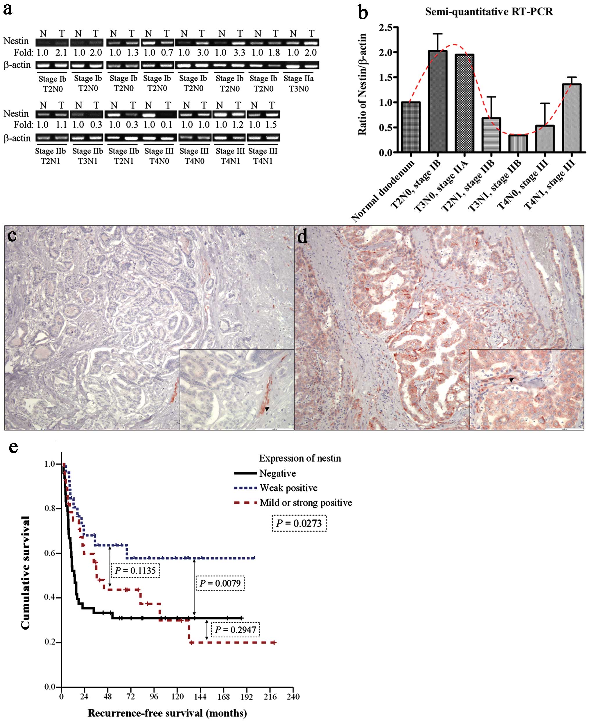
The level of nestin mRNA expression was detected in
the 15 pairs of cancer and normal tissue samples (Fig. 1a). Fig.
1b shows the quantitative ratio of nestin to β-actin
expression. An increased nestin:β-actin ratio was found in the
early stages of cancer without lymph node metastasis (T2N0 or T3N0)
and in advanced cancer with lymph node metastasis (T4N1). However,
the nestin:β-actin ratio was low in T2N1, T3N1 and T4N0 cancers
(Fig. 1a). The fluctuating curve
shown in Fig. 1b demonstrated the
various function of nestin in early and advanced ampullary
adenocarcinoma.
Expression of nestin protein in ampullary
adenocarcinoma
IHC staining of nestin was performed to confirm the
results of the RT-PCR and indicated that intratumor lymphatic ducts
or vessels also exhibited nestin expression (Fig. 1c). The immuno-reactivity of nestin
was primarily detected in the cytoplasm of cancer cells; Fig. 1d displays the strongest
immunoreactivity of nestin (IRS 12 points). Fig. 2 shows examples of IHC staining,
which were matched with the RT-PCR results. The level of nestin
expression in cancer cells was defined as negative (IRS 0–1 points;
48 patients), weak (IRS 2–3 points; 26 patients), mild (IRS 4–8
points; 25 patients), or strong (IRS 9–12 points; 3 patients).
Patients who exhibited strong nestin expression were grouped with
those with mild expression as these groups were small. Weak nestin
expression was correlated with negative lymphovascular invasion,
decreased perineural invasion, absence of pancreatic invasion, free
resection margin and an early stage (Table I). Patients with mild or strong
nestin expression had a small tumor, pancreatic invasion, a
microscopically positive margin and advanced cancer stage (Table I).
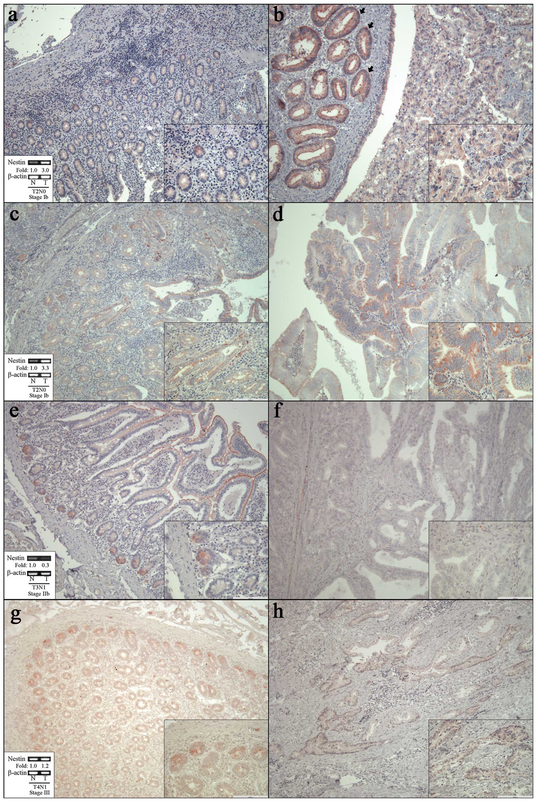 | Figure 2Expression of nestin in ampullary
adenocarcinoma by immunohistochemistry (IHC) staining. The main
images were captured at ×100 magnification and the small images
located at the right lower quadrant of each figure were captured at
×400. (a, c, e and g) The boxes at the left lower quadrant contain
the semi-quantitative results of RT-PCR. (a, c, e and g) Expression
of nestin protein in normal duodenum and (b, d, f and h) ampullary
adenocarcinoma. (a and c) Weak expression of nestin in normal
duodenal mucosa and (b and d) strong or mild expression of nestin
in T2N0, stage IB ampullary adenocarcinoma of the same patient. (b)
Strong expression of nestin in adjacent adenoma is shown to the
left side of the arrows. (e) Weak expression in normal duodenal
mucosa and (f) negative expression in T3N1, stage IIB ampullary
adenocarcinoma. The nestin mRNA is downregulated in stage IIB
cancer (T3N1) in RT-PCR (Fig. 1a)
and (f) immunoreactivity of nestin is nearly undetectable in cancer
with (e) preservation in normal duodenum. (g) Weak expression in
normal duodenal mucosa and (h) weak expression in T4N1, stage III
ampullary adenocarcinoma. The IHC results in T4N1 disease revealed
equal density of nestin in cancer and normal duodenum, which was
the same as the results in RT-PCR. Expression of nestin protein in
IHC staining was correlated with detection of nestin mRNA in
RT-PCR. |
 | Table ICorrelation of nestin expression with
the patient clinicopathological findings in ampullary
adenocarcinoma. |
Table I
Correlation of nestin expression with
the patient clinicopathological findings in ampullary
adenocarcinoma.
| | Nestin expression
by IHC staining | |
|---|
| |
| |
|---|
| All cases
(n=102) | Negative
(n=48) | Weak expression
(n=26) | Mild or strong
expression (n=28) | P-value |
|---|
| Age at surgery
(years)a | 65 (32–90) | 62 (32–90) | 64 (36–78) | 67 (35–83) | 0.131 |
| Gender, n (%) | | | | | 0.350 |
| Female | 41 (40) | 19 (40) | 8 (31) | 14 (50) | |
| Male | 61 (60) | 29 (60) | 18 (69) | 14 (50) | |
| Tumor type,
grossly, n (%) | | | | | 0.419 |
| Polypoid | 59 (58) | 25 (52) | 14 (54) | 20 (71) | |
| Ulcerative | 21 (21) | 10 (21) | 6 (23) | 5 (18) | |
| Mixed | 22 (22) | 13 (27) | 6 (23) | 3 (11) | |
| Histological
differentiation, n (%) | | | | | 0.568 |
| Well | 45 (46) | 18 (38) | 12 (50) | 15 (56) | |
| Moderate | 47 (48) | 26 (54) | 11 (46) | 10 (37) | |
| Poor | 7 (7) | 4 (8) | 1 (4) | 2 (7) | |
| pT status, n
(%) | | | | | 0.051 |
| pT1 | 12 (12) | 3 (6) | 4 (15) | 5 (18) | |
| pT2 | 42 (41) | 16 (33) | 16 (62) | 10 (36) | |
| pT3 | 32 (31) | 18 (38) | 5 (19) | 9 (32) | |
| pT4 | 16 (16) | 11 (23) | 1 (4) | 4 (14) | |
| Tumor size
(cm)a | 2.4 (0.7–8.0) | 2.5 (0.8–8.0) | 2.5 (1.0–6.5) | 1.7 (0.7–4.0) | 0.010 |
| Pancreatic
invasion, n (%) | | | | | 0.018 |
| Negative | 54 (54) | 21 (44) | 20 (77) | 13 (50) | |
| Positive | 46 (46) | 27 (56) | 6 (23) | 13 (50) | |
| Lymphovascular
invasion, n (%) | | | | | 0.003 |
| Negative | 40 (50) | 11 (28) | 14 (70) | 15 (60) | |
| Positive | 44 (50) | 28 (72) | 6 (30) | 10 (40) | |
| pN status, n
(%) | | | | | 0.508 |
| pN0 | 55 (60) | 26 (55) | 16 (70) | 13 (62) | |
| pN1 | 36 (40) | 21 (45) | 7 (30) | 8 (38) | |
| Perineural
invasion, n (%) | | | | | 0.030 |
| Negative | 46 (68) | 15 (52) | 14 (88) | 17 (74) | |
| Positive | 22 (32) | 14 (48) | 2 (12) | 6 (26) | |
| Resection margin, n
(%) | | | | | 0.031 |
| Free | 91 (89) | 41 (85) | 26 (100) | 24 (86) | |
| Microscopically
positive | 11 (11) | 7 (15) | 0 | 4 (14) | |
| AJCC TNM
stageb, n (%) | | | | | 0.038 |
| Stage I | 45 (44) | 15 (31) | 17 (65) | 13 (46) | |
| Stage II | 41 (40) | 22 (46) | 8 (31) | 11 (39) | |
| Stage III | 16 (16) | 11 (23) | 1 (4) | 4 (14) | |
Correlation of nestin expression with the
outcomes of the patients
Patients with weak nestin expression had a decreased
incidence of recurrence, and particularly local recurrence or
recurrence within the 12 months after operation (Table II). Patients with negative or mild
to strong nestin expression experienced an increased risk of
recurrence (Table II). Patients
with weak nestin expression had a more favorable prognosis (5-year
recurrence-free survival rate of 63.5%) than those with negative or
mild to strong nestin expression (31.0 and 43.6%) (Fig. 1e). The regimens of adjuvant therapy
were similar among the patients with negative, weak or mild to
strong nestin expression (data not shown). A multivariate analysis
was conducted to examine the survival predictors (Table III). Histological differentiation,
tumor stage, nodal metastasis, pancreatic invasion, resection
margin, and American Joint Committee on Cancer (AJCC) tumor, node
and metastases (TNM) staging system have been used as predictors in
previous studies (3–5). In our results, resection margin and
nestin expression were prognostic predictors. A microscopically
positive margin predicted recurrence; however, weak nestin
expression predicted a favorable prognosis (P=0.030) (Table III).
| Table IIRecurrence in patients with ampullary
adenocarcinoma after radical resection, as compared with nestin
expression. |
Table II
Recurrence in patients with ampullary
adenocarcinoma after radical resection, as compared with nestin
expression.
| Nestin expression
by IHC staining | |
|---|
|
| |
|---|
| Negative
(n=48) | Weak expression
(n=26) | Mild or strong
expression (n=28) | P-value |
|---|
| Liver metastasis, n
(%) | 17 (35) | 6 (23) | 5 (18) | 0.199 |
| Local recurrence, n
(%) | 21 (44) | 4 (15) | 12 (43) | 0.024 |
| Peritoneal
carcinomatosis, n (%) | 9 (19) | 2 (8) | 2 (7) | 0.219 |
| Bone metastasis, n
(%) | 5 (10) | 2 (8) | 1 (4) | 0.523 |
| Other
metastasisa, n (%) | 11 (23) | 2 (8) | 5 (18) | 0.217 |
| Late recurrence, n
(%) (> postoperative 12 months) | 10 (21) | 5 (19) | 11 (39) | 0.155 |
| Early recurrence, n
(%) (≤ postoperative 12 months) | 23 (48) | 5 (19) | 7 (25) | 0.020 |
| Subtotalb, n (%) | 33 (69) | 10 (38) | 18 (64) | 0.035 |
| Table IIIMultivariate analysis of the
prognostic factors for recurrence-free survival in patients with
ampullary adenocarcinoma who underwent radical resection. |
Table III
Multivariate analysis of the
prognostic factors for recurrence-free survival in patients with
ampullary adenocarcinoma who underwent radical resection.
| HR | 95% CI | P-value |
|---|
| Age (years) | 1.008 | 0.985–1.032 | 0.499 |
| Gender | | | 0.874 |
| Female | 1 | | |
| Male | 0.958 | 0.561–1.634 | |
| Resection
margin | | | 0.002 |
| Free | 1 | | |
| Microscopically
positive | 3.080 | 1.523–6.229 | |
| Nestin expression
in IHC | | | 0.061 |
| Negative | 1 | | |
| Weak positive | 0.446 | 0.215–0.926 | 0.030 |
| Mild or strong
positive | 0.636 | 0.350–1.159 | 0.139 |
Gene interaction network of nestin in
ampullary adenocarcinoma
A microarray analysis of paired samples was
conducted to identify the interacting genes associated with nestin.
A total of 5 patients with ampullary adenocarcinoma were enrolled.
One of the 2 patients with T2N0 (stage IB cancer) and 2 of the 3
patients with T3N0 (stage IIA cancer) developed recurrence. The
Notch pathway is located upstream of nestin (22) and the NOTCH2 and NOTCH3 genes were
overexpressed in ampullary cancer (Fig.
3a). The interaction between nestin and CDK5 modulates
downstream molecules, including RAC1 and NOS3 [epithelial nitric
oxide synthase (eNOS)] (23–25).
Four of the 5 patients exhibited increased nestin expression and 3
of the 5 patients exhibited decreased CDK5 or RAC1 expression. The
RAC1 pathway was suppressed among certain patients with early
ampullary cancer.
In advanced cancer, multiple oncogenic pathways were
activated and tumor suppressors were inhibited (Fig. 3b). The transforming growth factor-β1
(TGF-β1)/Smad pathway enhances cell migration through nestin/CDK5
signaling. Activation of the platelet-derived growth factor
receptor (PDGFR) pathway and supression of phosphatase and tensin
homolog (PTEN) signaling promote cell migration. Regarding the
patients with negative nestin expression, the activation of NOTCH,
TGF-β1 or PDGFR pathways could induce cancer metastasis (Fig. 3b).
Fresh tissue specimens of paired cancer and normal
mucosa from 7 patients were examined by western blotting, including
4 patients without recurrence and 3 with recurrence (Fig. 3c). CDK5 and Rac1 were key proteins
in the downstream signaling of nestin, and quantitative results of
the CDK5/GAPDH or Rac1/GAPDH ratio are shown in Fig. 3d. An increased ratio of CDK5/GAPDH
with a decreased ratio of Rac1/GAPDH was detected in patients
without recurrence; however, the phenomenon disappeared in patients
with recurrence. The result of the western blotting confirmed that
the RAC1 pathway was suppressed in certain patients with early
ampullary cancer.
Discussion
Ampullary adenocarcinoma is the most common
malignancy in the small intestine. This is the first study to
discuss nestin expression in ampullary adenocarcinoma; the findings
indicated that nestin expression was low in normal duodenum,
upregulated in the early stages of cancer, downregulated in the
intermediate stages of cancer, and upregulated in locally advanced
ampullary adenocarcinoma. Patients with nestin expression in cancer
cells had varied prognosis: those with weak nestin expression had a
favorable recurrence-free survival, whereas those with mild to
strong nestin expression had an unfavorable survival.
In normal physiological development, nestin is
expressed in neurogenic or myogenic cells, and is replaced by
tissue-specific intermediate filaments after differentiation
(26). In pathological situations,
nestin expression is reinduced (27). Nestin is overexpressed in cancer and
is correlated with nodal metastasis and cancer invasiveness
(18–20). Nestin was found to modulate
metastasis in cancer and cell migration was inhibited by
nestin-shRNA (28). Su et al
(29) confirmed that nestin
overexpression enhances cell migration and EMT through the
TGF-β1/Smad4-mediated pathway. In this study, nestin mRNA was
overexpressed in the early stages of cancer without lymph node
metastasis and in the late stages of cancer with lymph node
metastasis, but not in the intermediate stages of cancer (Fig. 1). The results were reconfirmed using
IHC staining (Fig. 2). Weak nestin
expression was correlated with favorable tumor features (Table I). The patients with weak nestin
expression demonstrated a better recurrence-free survival rate
(Fig. 1e and Table III). However, mild or strong
nestin expression was correlated with unfavorable tumor
characteristics and an increased risk of recurrence (Table II). This result contradicted
previous studies that have explored nestin expression among other
cancers (18–20). We speculate that nestin exhibits
various functions in the early and advanced stages of ampullary
adenocarcinoma. Certain oncogenic proteins involved in ampullary
cancer exhibit functions distinct from those exhibited in other
cancers. EpCAM is expressed in normal epithelium and is upregulated
in epithelial cancer. EpCAM regulates c-myc and cyclins to enhance
cell proliferation (30). However,
the loss of EpCAM is related to an aggressive tumor phenotype of
ampullary cancer, which is different from other types of cancer
(10). The findings in the present
study suggest that nestin plays various roles in early and in
advanced ampullary adenocarcinoma.
Nestin is regulated by CDK5, and CDK5 regulates
apoptosis, senescence, angiogenesis, differentiation and migration
(31,32). Accumulation of CDK5 under oxidative
stress triggers apoptosis and cell death (31). However, activation of CDK5 in cancer
induces nestin reorganization, and inhibition of CDK5 reduces tumor
growth and metastasis (23,33). The varying behaviors of CDK5 may
explain why nestin plays various roles in early and in advanced
ampullary adenocarcinoma.
A microarray analysis that compared cancerous and
normal cells was conducted to explore the regulators of nestin
(Fig. 3). A small GTPase protein,
RAC1, is connected with nestin through CDK5 (Fig. 3a). RAC1 facilitates the
proliferation of intestinal stem cells, formation of intestinal
adenoma, initiation of colorectal cancer and migration of cancer
cells (34,35). Oxidative stress suppresses the
kinase activity of RAC1, induces activation of CDK5 and results in
senescence of cells (24). Fig. 3a shows inconsistent patterns of
nestin, CDK5 and RAC1 expression in the cDNA microarray and western
blotting is shown in Fig. 3c. In
the patients without recurrence, an increased CDK5/GAPDH ratio and
a decreased Rac1/GAPDH ratio were detected (Fig. 3c and d). Lacking adequate
neovascularization, hypoxia may develop during growth of a primary
tumor in early ampullary cancer; thus, nestin/CDK5 temporarily
suppresses RAC1. The cell migration and cancer metastasis are
transiently suppressed and the tumor grows locally. This may be the
reason that patients with weak nestin expression in ampullary
adenocarcinoma had a favorable prognosis after undergoing radical
resection.
The function of nestin/CDK5 changes in advanced
cancer (33). Nestin behaves as a
stemness protein to induce proliferation, migration and metastasis
(28,29). In pancreatic cancer with nestin
overexpression, a K-ras mutation was found to induce persistent
activation of CDK5 (36).
Suppression of the nestin/CDK5 complex reduces proliferation,
migration and metastasis in cancer (33,36).
In this study, nestin was a predictor of distant metastasis and
poor prognosis in the ampullary cancer patients with mild to strong
nestin expression (Table II and
Fig. 1e). Inhibition of nestin in
the select patients with ampullary adenocarcinoma could provide a
therapeutic solution.
Furthermore, the activation of multiple oncogenic
pathways and the knockdown of tumor suppressors form a complex
network in cancer cells. The existence of crosstalk between the
WNT, BMP, Hedgehog and Notch pathways in cancer has been verified.
Accumulation of mutations in these pathways contributes to tumor
heterogeneity (37). Furthermore,
the inhibition of Notch attenuates nestin expression (22,38).
In this study, the microarray data indicated overexpression of the
NOTCH2 and NOTCH3 genes (Fig. 3a).
Deregulation of the PDGFR mutation, activation of the TGF-β1/Smad
pathway and inhibition of PTEN also enhance cell migration
(Fig. 3b). Among patients with
negative nestin expression, the activation of Notch, TGF-β1 or
PDGFR pathways and suppression of PTEN may induce cancer
metastasis.
Nestin expression was upregulated in the early and
locally advanced stages of ampullary adenocarcinoma, but was not
upregulated in the intermediate stage. Weak nestin expression was
correlated with favorable tumor characteristics and a good
prognosis. The nestin/CDK5 complex may suppress RAC1 and contribute
to a favorable prognosis in patients with weak nestin expression.
Activation of multiple oncogenic pathways in patients who exhibited
or did not exhibit nestin overexpression increased the likelihood
of a poor prognosis. Blocking nestin may prevent metastasis in
patients with strong nestin expression.
Acknowledgements
The study was supported by grants from the National
Science Council (grant NSC-101-2314-B-006-028) and the National
Cheng Kung University Hospital (to H.-P.H.). We are grateful for
the support from the Human Biobank, Research Center of Clinical
Medicine, National Cheng Kung University Hospital. We were blessed
with support from the late superintendent, Professor Pin-Wen Lin.
Furthermore, we thank Professor Li-Tzong Chen, Dr Meng-Chi Yen and
Miss Tzu-Wen Wang for their support.
Abbreviations:
|
AJCC TNM stage
|
American Joint Committee on Cancer
tumor, node and metastasis staging system
|
|
CDK5
|
cyclin-dependent kinase 5
|
|
CSC
|
cancer stem cell
|
|
EMT
|
epithelial-mesenchymal transition
|
|
eNOS
|
endothelial nitric oxide synthase
|
|
IHC
|
immunohistochemistry
|
|
PDGFR
|
platelet-derived growth factor
receptor
|
|
PTEN
|
phosphatase and tensin homolog
|
|
Rac1
|
Ras-related C3 botulinum toxin
substrate 1
|
|
RT-PCR
|
reverse transcription-polymerase chain
reaction
|
|
TGF-β
|
transforming growth factor β
|
References
|
1
|
O’connell JB, Maggard MA, Manunga J, et
al: Survival after resection of ampullary carcinoma: a national
population-based study. Ann Surg Oncol. 15:1820–1827. 2008.
View Article : Google Scholar
|
|
2
|
Jiang ZQ, Varadhachary G, Wang X, et al: A
retrospective study of ampullary adenocarcinomas: overall survival
and responsiveness to fluoropyrimidine-based chemotherapy. Ann
Oncol. 24:2349–2353. 2013. View Article : Google Scholar : PubMed/NCBI
|
|
3
|
Hsu HP, Shan YS, Hsieh YH, Yang TM and Lin
PW: Predictors of recurrence after pancreaticoduodenectomy in
ampullary cancer: comparison between non-, early and later
recurrence. J Formos Med Assoc. 106:432–443. 2007. View Article : Google Scholar : PubMed/NCBI
|
|
4
|
Shroff RT: Pathologic Markers of Prognosis
in Ampullary Carcinoma. 2012, UT GSBS Dissertations and Theses
(Open Access). Paper 246. http://digitalcommons.library.tmc.edu/utgsbs_dissertations/246/.
|
|
5
|
Hsu HP, Yang TM, Hsieh YH, Shan YS and Lin
PW: Predictors for patterns of failure after
pancreaticoduodenectomy in ampullary cancer. Ann Surg Oncol.
14:50–60. 2007. View Article : Google Scholar
|
|
6
|
Imai Y, Oda H, Tsurutani N, Nakatsuru Y,
Inoue T and Ishikawa T: Frequent somatic mutations of the APC and
p53 genes in sporadic ampullary carcinomas. Jpn J Cancer Res.
88:846–854. 1997. View Article : Google Scholar : PubMed/NCBI
|
|
7
|
Yamazaki K, Hanami K, Nagao T, Asoh A,
Sugano I and Ishida Y: Increased cyclin D1 expression in cancer of
the ampulla of Vater: relevance to nuclear beta catenin
accumulation and k-ras gene mutation. Mol Pathol. 56:336–341. 2003.
View Article : Google Scholar : PubMed/NCBI
|
|
8
|
Hsu HP, Shan YS, Jin YT, Lai MD and Lin
PW: Loss of E-cadherin and beta-catenin is correlated with poor
prognosis of ampullary neoplasms. J Surg Oncol. 101:356–362.
2010.PubMed/NCBI
|
|
9
|
Hsu HP, Shan YS, Lai MD and Lin PW:
Osteopontin-positive infiltrating tumor-associated macrophages in
bulky ampullary cancer predict survival. Cancer Biol Ther.
10:144–154. 2010. View Article : Google Scholar : PubMed/NCBI
|
|
10
|
Piscuoglio S, Lehmann FS, Zlobec I, et al:
Effect of EpCAM, CD44, CD133 and CD166 expression on patient
survival in tumours of the ampulla of Vater. J Clin Pathol.
65:140–145. 2012. View Article : Google Scholar
|
|
11
|
Visvader JE and Lindeman GJ: Cancer stem
cells in solid tumours: accumulating evidence and unresolved
questions. Nat Rev Cancer. 8:755–768. 2008. View Article : Google Scholar : PubMed/NCBI
|
|
12
|
Thiery JP: Epithelial-mesenchymal
transitions in tumour progression. Nat Rev Cancer. 2:442–454. 2002.
View Article : Google Scholar : PubMed/NCBI
|
|
13
|
Mani SA, Guo W, Liao MJ, et al: The
epithelial-mesenchymal transition generates cells with properties
of stem cells. Cell. 133:704–715. 2008. View Article : Google Scholar : PubMed/NCBI
|
|
14
|
Chen C, Wei Y, Hummel M, et al: Evidence
for epithelial-mesenchymal transition in cancer stem cells of head
and neck squamous cell carcinoma. PLoS One. 6:e164662011.
View Article : Google Scholar : PubMed/NCBI
|
|
15
|
Raimondi C, Gradilone A, Naso G, et al:
Epithelial-mesenchymal transition and stemness features in
circulating tumor cells from breast cancer patients. Breast Cancer
Res Treat. 130:449–455. 2011. View Article : Google Scholar : PubMed/NCBI
|
|
16
|
Chiou SH, Wang ML, Chou YT, et al:
Coexpression of Oct4 and Nanog enhances malignancy in lung
adenocarcinoma by inducing cancer stem cell-like properties and
epithelial-mesenchymal transdifferentiation. Cancer Res.
70:10433–10444. 2010. View Article : Google Scholar : PubMed/NCBI
|
|
17
|
Calabrese C, Poppleton H, Kocak M, et al:
A perivascular niche for brain tumor stem cells. Cancer Cell.
11:69–82. 2007. View Article : Google Scholar : PubMed/NCBI
|
|
18
|
Liu C, Chen B, Zhu J, et al: Clinical
implications for nestin protein expression in breast cancer. Cancer
Sci. 101:815–819. 2010. View Article : Google Scholar
|
|
19
|
Ryuge S, Sato Y, Wang GQ, et al:
Prognostic significance of nestin expression in resected non-small
cell lung cancer. Chest. 139:862–869. 2011. View Article : Google Scholar
|
|
20
|
Kawamoto M, Ishiwata T, Cho K, et al:
Nestin expression correlates with nerve and retroperitoneal tissue
invasion in pancreatic cancer. Hum Pathol. 40:189–198. 2009.
View Article : Google Scholar :
|
|
21
|
Remmele W and Schicketanz KH:
Immunohistochemical determination of estrogen and progesterone
receptor content in human breast cancer. Computer-assisted image
analysis (QIC score) vs. subjective grading (IRS). Pathol Res
Pract. 189:862–866. 1993. View Article : Google Scholar : PubMed/NCBI
|
|
22
|
Shih AH and Holland EC: Notch signaling
enhances nestin expression in gliomas. Neoplasia. 8:1072–1082.
2006. View Article : Google Scholar
|
|
23
|
Pallari HM, Lindqvista J, Torvaldsona E,
et al: Nestin as a regulator of Cdk5 in differentiating myoblasts.
Mol Biol Cell. 22:1539–1549. 2011. View Article : Google Scholar : PubMed/NCBI
|
|
24
|
Alexander K, Yang HS and Hinds PW:
Cellular senescence requires CDK5 repression of Rac1 activity. Mol
Cell Biol. 24:2808–2819. 2004. View Article : Google Scholar : PubMed/NCBI
|
|
25
|
Lee CH, Wei YW, Huang YT, et al: CDK5
phosphorylates eNOS at Ser-113 and regulates NO production. J Cell
Biochem. 110:112–117. 2010.PubMed/NCBI
|
|
26
|
Iwatsuki H and Suda M: Seven kinds of
intermediate filament networks in the cytoplasm of polarized cells:
structure and function. Acta Histochem Cytochem. 43:19–31. 2010.
View Article : Google Scholar : PubMed/NCBI
|
|
27
|
Chou YH, Khuon S, Herrmann H and Goldman
RD: Nestin promotes the phosphorylation-dependent disassembly of
vimentin intermediate filaments during mitosis. Mol Biol Cell.
14:1468–1478. 2003. View Article : Google Scholar : PubMed/NCBI
|
|
28
|
Kleeberger W, Bova GS, Nielsen ME, et al:
Roles for the stem cell associated intermediate filament Nestin in
prostate cancer migration and metastasis. Cancer Res. 67:9199–9206.
2007. View Article : Google Scholar : PubMed/NCBI
|
|
29
|
Su HT, Weng CC, Hsiao PJ, et al: Stem cell
marker nestin is critical for TGF-β1-mediated tumor progression in
pancreatic cancer. Mol Cancer Res. 11:768–779. 2013. View Article : Google Scholar : PubMed/NCBI
|
|
30
|
Patriarca C, Macchi RM, Marschner AK and
Mellstedt H: Epithelial cell adhesion molecule expression (CD326)
in cancer: A short review. Cancer Treat Rev. 38:68–75. 2012.
View Article : Google Scholar
|
|
31
|
Dhavan R and Tsai LH: A decade of CDK5.
Nat Rev Mol Cell Biol. 2:749–759. 2001. View Article : Google Scholar : PubMed/NCBI
|
|
32
|
Contreras-Vallejos E, Utreras E and
Gonzalez-Billault C: Going out of the brain: non-nervous system
physiological and pathological functions of Cdk5. Cell Signal.
24:44–52. 2012. View Article : Google Scholar
|
|
33
|
Feldmann G, Mishra A, Hong SM, et al:
Inhibiting the cyclin-dependent kinase CDK5 blocks pancreatic
cancer formation and progression through the suppression of Ras-Ral
signaling. Cancer Res. 70:4460–4469. 2010. View Article : Google Scholar : PubMed/NCBI
|
|
34
|
Myant KB, Cammareri P, McGhee EJ, et al:
ROS production and NF-κB activation triggered by RAC1 facilitate
WNT-driven intestinal stem cell proliferation and colorectal cancer
initiation. Cell Stem Cell. 12:761–773. 2013. View Article : Google Scholar : PubMed/NCBI
|
|
35
|
Yang WH, Lan HY, Huang CH, et al: RAC1
activation mediates Twist1-induced cancer cell migration. Nat Cell
Biol. 14:366–374. 2012. View Article : Google Scholar : PubMed/NCBI
|
|
36
|
Eggers JP, Grandgenett PM, Collisson EC,
et al: Cyclin-dependent kinase 5 is amplified and overexpressed in
pancreatic cancer and activated by mutant K-Ras. Clin Cancer Res.
17:6140–6150. 2011. View Article : Google Scholar : PubMed/NCBI
|
|
37
|
Bertrand FE, Angus CW, Partis WJ and
Sigounas G: Developmental pathways in colon cancer: crosstalk
between WNT, BMP, Hedgehog and Notch. Cell Cycle. 11:4344–4351.
2012. View Article : Google Scholar : PubMed/NCBI
|
|
38
|
Hu YY, Zheng MH, Cheng G, et al: Notch
signaling contributes to the maintenance of both normal neural stem
cells and patient-derived glioma stem cells. BMC Cancer. 11:822011.
View Article : Google Scholar : PubMed/NCBI
|