Introduction
Head and neck squamous cell carcinoma (HNSCC) is the
sixth most common cancer worldwide. In China, statistical data
reveal that the incidence of HNSCC in males and females is 8.17 and
4.6/100,000 individuals, respectively, and HNSCC accounts for 5.1
and 2.7% of all malignant tumors each year. Statistically, oral
squamous cell carcinoma (OSCC) is the most common HNSCC and
accounts for 2–3% for all malignant tumors. Tongue squamous cell
carcinoma (TSCC) is the most common carcinoma among OSCC cases,
accounting for 25–40% of mouth neoplasms (1). The incidence of TSCC has increased in
recent years, making it a major public health concern. In spite of
the wide use of combination therapy, such as
surgery-radio-chemotherapy, the mortality rate of TSCC remains
high. The 5-year survival rate of TSCC patients is less than 50%
(2). To our knowledge, the primary
cause of therapy failure is recurrence in situ and distant
metastases. Therefore, it is important to identify new biomarkers
for therapeutic targets.
The metastasis-associated in colon cancer 1 (MACC1)
gene was identified in a genome-wide screen of human colon cancer,
and its expression is closely related to the metastasis of colon
cancer (3). MACC1 is a key
regulator of the hepatocyte growth factor (HGF)-MET signaling
pathway, which is related to colon cancer metastasis (4,5). MACC1
downregulation inhibits tumorigenicity through the Akt/β-catenin
signaling pathway in nasopharyngeal carcinoma cells (6). Recently, many studies have reported
that MACC1 is involved in cancer development and progression by
promoting tumor cell proliferation, migration and invasion. Further
studies have demonstrated that MACC1 upregulates cell growth and
metastatic progression in several solid tumors, including lung
cancer (7,8), hepatocellular carcinoma (4,9–11),
ovarian carcinoma (12), pancreatic
cancer (13), breast cancer
(14) and colon cancer (4,15,16).
To our knowledge, no research regarding the function of MACC1 in
TSCC has been reported. In the present study, we determined that
the overexpression of MACC1 was significantly correlated with the
poor overall survival of the studied patients. The knockdown of
MACC1 inhibited cell proliferation, migration and invasion, the
knockdown of MACC1 also attenuated cisplatin resistance in TSCCA
cells and contributed to apoptosis. Furthermore, we demonstrated
that MACC1 influences the invasion and migration of TSCCA cells by
secreting and activating urokinase-type plasminogen activator
system (uPA) but not metalloproteinase 2 (MMP2).
Materials and methods
Cell culture
TSCCA cells were purchased from the College of
Stomatology, Shanghai Jiao Tong University (Shanghai, China) and
cultured in RPMI-1640 medium (Gibco, Carlsbad, CA, USA) containing
10% fetal bovine serum (Biological Industries, Israel) at 37°C with
5% CO2.
Small interfering RNA (siRNA)
transfection
siRNA duplexes were synthesized and purified by
GenePharma (Shanghai). The siRNA sequences for MACC1 were as
follows: sense, 5′-AAGAUUGGACUUGUACACUGCTT-3′ and antisense,
5′-TTUUCUAACCUGAACAUGUGACG-3′. The transfection of MACC1-siRNA was
performed using the Lipofectamine 2000 reagent (Invitrogen,
Carlsbad, CA, USA) following the manufacturer’s instructions. The
transfection of MACC1-NC was conducted in the same manner to serve
as the control. In this study, the experimental group was
transfected with MACC1-siRNA, whereas the control group was
transfected with MACC1-NC.
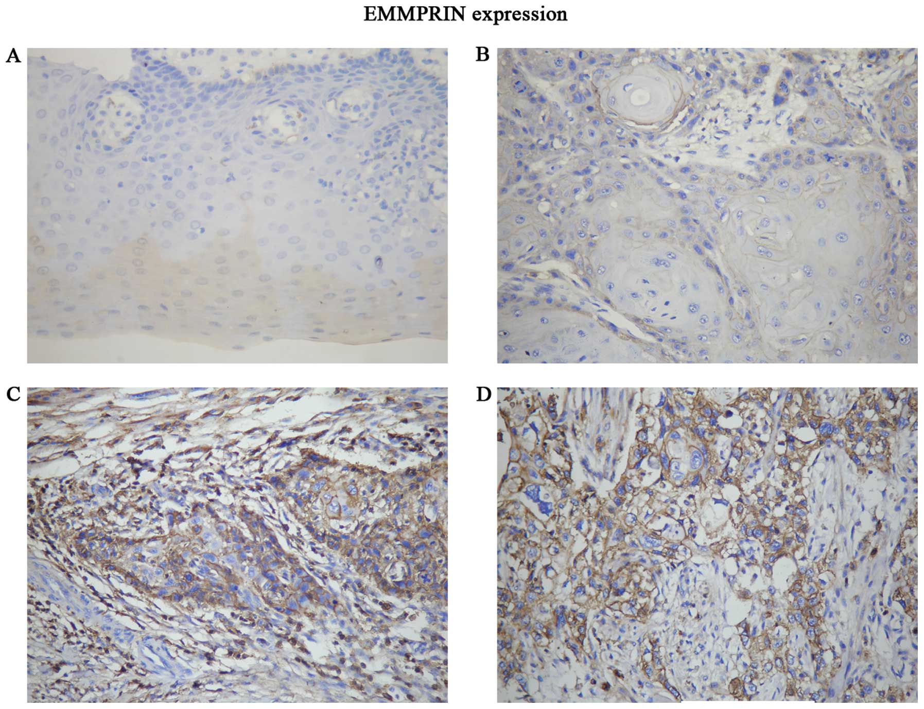
Immunohistochemistry (IHC)
Sixty-five paraffin-embedded specimens of TSCC and
the adjacent non-cancerous tissues were collected from 1996 to 2006
at the Department of Oral and Maxillofacial Surgery, Sun Yat-Sen
Memorial Hospital. The study was approved by the Institutional
Research Ethics Committee of Sun Yat-Sen Memorial Hospital of Sun
Yat-Sen University. Consent was obtained from the patients, and the
clinical research materials were collected from the patients prior
to clinical treatment. Immunohistochemical staining was performed
with a polyclonal anti-MACC1 antibody (Abcam Ltd., Hong Kong) and a
polyclonal anti-extracellular matrix metalloproteinase inducer
(EMMPRIN) antibody (Abgent, Suzhou, China) according to standard
protocols. Briefly, the tissue sections were blocked sequentially
with 3% H2O2 and normal serum and then
incubated with MACC1 (1:100) and EMMPRIN (1:100) at 4°C overnight.
The tissue sections were incubated with a biotinylated secondary
antibody and conjugated with a streptavidin-HRP complex (ready to
use SP kit; Zhongshan Co., Beijing, China). Finally, the sections
were visualized with 3-3′-diaminobenzidine and counterstained with
hematoxylin. Sections of human colon tissues stained positive with
these antibodies were used as the positive control for MACC1, TSCC
tissue known to have positive staining for EMMPRIN were used as a
positive control, and PBS was used instead of the primary
antibodies for the negative controls.
Evaluation of IHC staining
The IHC evaluation of the tissues was conducted by 2
pathologists (H-F.L. and H-G.L.) who assessed the number of
positive cells and the intensity of staining. The positive results
were judged by semi-quantitative points. The staining intensity
scores were 0 (negative), 1 (weak), 2 (medium) and 3 (strong). The
integral of the rate of positive cells was 0 (0%), 1 (1–25%), 2
(26–50%) and 3 (>50%). The staining intensity score and the
proportional score were added to obtain the total score (17). A total score ≥3 was considered to
represent high MACC1 expression. A total score <3 was considered
to represent low MACC1 expression. For the EMMPRIN staining
evaluation, samples in which >10% of tumor cells were stained
positive with anti-EMMPRIN antibodies were evaluated as strongly
positive (++), ≤10% cells were positive (+) and no detectable
staining was negative (−) (2). The
clinical features of the patients are summarized in Table I, and the overall survival time of
the patients is analyzed in Fig.
2.
 | Table IAssociation of MACC1 expression with
the clinicopathological features of the tongue squamous cell
carcinoma patients. |
Table I
Association of MACC1 expression with
the clinicopathological features of the tongue squamous cell
carcinoma patients.
| MACC1
expression | |
|---|
|
| |
|---|
|
Characteristics | Low (%) | High (%) | P-value |
|---|
| Age (years) |
| ≤40 | 7 (53.8) | 6 (46.2) | 0.615 |
| >40 | 32 (61.5) | 20 (38.5) | |
| Gender |
| Male | 22 (53.7) | 19 (46.3) | 0.176 |
| Female | 17 (70.8) | 7 (29.2) | |
|
Differentiation |
| Well | 8 (66.7) | 4 (33.3) | 0.553 |
| Moderate | 29 (58.0) | 21 (42.0) | |
| Poor | 2 (66.7) | 1 (33.3) | |
| Lymphatic
metastasis |
| No | 34 (70.8) | 14 (21.2) | 0.003 |
| Yes | 5 (29.4) | 12 (70.6) | |
| Clinical stage |
| I | 13 (86.7) | 2 (13.3) | 0.088 |
| II | 14 (51.9) | 13 (48.1) | |
| III | 4 (44.4) | 5 (55.6) | |
| IV | 8 (57.1) | 6 (42.9) | |
| EMMPRIN |
| (−) | 17 (85.0) | 3 (15.0) | 0.003 |
| (+) | 10 (58.8) | 7 (41.2) | |
| (++) | 12 (42.9) | 16 (57.1) | |
Western blot analysis
After transfection, the cells were collected, and
the total protein was extracted from the cells. Thirty microliters
of protein was loaded and separated using 10% sodium dodecyl
sulfate polyacrylamide gel electrophoresis (SDS-PAGE) (Beyotime,
Shanghai, China) and transferred to Immobilon-P transfer membranes
(PVDF) (Beyotime). The membranes were blocked with 5% non-fat milk
in Tris-buffered saline (TBS) containing 0.1% Tween-20 for 1 h at
room temperature. The blots were probed with the relevant primary
antibodies overnight at 4°C, washed in TBST and probed with a
species-specific horseradish peroxidase-conjugated secondary
antibody (anti-rabbit; Shanghai ExCell Biology, Inc., China). The
anti-MACC1 antibody (Abcam), anti-uPA antibody (GeneTex, Wuhan,
China) and the anti-MMP2 antibody (GeneTex) were used to probe the
alterations of the protein. GAPDH (KangCheng-Biotech, Shanghai,
China) was used as a loading control. The western blot analysis
process was performed according to standard protocols.
CCK-8 assays
After 24 h of transfection, the cells were
collected, and 5×10³ cells/well were plated into 96-well
plates. All of the cells were incubated overnight. After 48, 72 and
96 h of transfection, the cell proliferation was determined by the
Cell Counting Kit-8 (Dojindo, Kyushu, Japan). The medium of each
well was removed and a mixture of 10 μl CCK-8 and 90 μl of 10% FBS
RPMI-1640 was added. The plates were incubated for a further 3 h,
and the absorbance at 450 nm was measured by a microplate reader
(Multiskan MK3; Thermo Electric, Shanghai, China). All of the
groups at each time-point had 5 wells. The relative cell survival
(%) was determined by the following formula:
(ODsiRNA/ODscramble) × 100%.
After 24 h of transfection, the TSCCA cells were
seeded at a density of 1×10⁴ cells/well into 96-well
plates and incubated overnight. Six concentrations of cisplatin (0,
2.5, 5, 10, 20 and 40 μmol) were added into each well with equal
volume. After 48 h, the cell proliferation was determined using the
CCK-8. The medium from each well was removed, and a mixture of 10
μl CCK-8 and 90 μl of 10% FBS RPMI-1640 was added. The plates were
incubated for a further 3 h, and the absorbance at 450 nm was
measured by a microplate reader (Multiskan MK3; Thermo Electric).
All of the groups treated with each concentration had 5 wells. The
relative cell survival (%) was determined by the following formula
(ODsiRNA/ODscramble) × 100%.
Apoptosis assays
Cells were seeded at a density of
5×10⁵/well in 6-well plates. The experimental group and
the control group each had 3 wells. After incubating overnight, the
experimental and the control groups of TSCCA cells were transfected
with MACC1-NC and MACC1-siRNA, respectively. After 24 h of
transfection, 5 μmol cisplatin was added to each well to induce
apoptosis; the plates were then incubated for 48 h. The TSCCA cells
were collected, including the supernatant of the culture medium,
and the total cells in each well were washed with PBS twice, and
then 300 μl 1X buffer diluents was added to each sample. A total of
300 μl of the cell suspension was divided into 200 μl for the
apoptosis analysis group and 100 μl for the loading control. The
former group was incubated with 2.5 μl of 7-AAD and 2.5 μl of PE
(both from BD Biosciences, Franklin Lakes, NJ, USA) antibodies for
15 min at room temperature in the dark. All of the samples were
analyzed for apoptosis using the Becton-Dickinson FACSVerse™ (BD
Biosciences).
In vitro migration and invasion
assays
For the Transwell migration assays, 3×10⁵
cells were seeded into the upper chamber of a polycarbonate
Transwell plate (Corning, Suzhou, China). The lower chamber of the
Transwell plate contained 10% FBS RPMI-1640. The cells were
incubated at 37°C with 5% CO2 for 18 h. For the
Transwell invasion assays, we mixed 10 μl of Matrigel basement
membrane matrix (BD Biosciences) and 90 μl of cold serum-free
RPMI-1640 per well and then placed the mixture into the upper
chamber of the Transwell. After 4–6 h of incubation at 37°C with 5%
CO2, 1×10⁵ cells were seeded into the upper
chamber of the polycarbonate Transwell plate; the lower chamber of
the Transwell plate contained 20% FBS RPMI-1640. The cells were
incubated at 37°C with 5% CO2 for 48 h. Subsequently,
the cells inside of the upper chamber were removed by cotton swabs.
Migratory and invasive cells on the lower chamber surface were
fixed in 4% formaldehyde, stained with 0.5% crystal violet and
counted (3 random × 40 fields/well). The same experiments were
performed a minimum of 3 times.
ELISA assays
Sample preparation. After 24 h of transfection and
incubation in 6-well plates with MACC1-NC and MACC1-siRNA,
respectively, the supernatant of the culture medium of the TSCCA
cells was collected and centrifuged at 1,000 × g for 20 min at room
temperature. The uPA and MMP2 concentrations in the supernatant of
the culture medium were quantified using a uPA ELISA kit
(Cloud-Clone Corp., Houston, TX, USA) and an MMP2 ELISA kit (ExCell
Biology Inc.) according to the manufacturers’ recommended
protocols. The sample analyses were executed in triplicate and were
repeated a minimum of 3 times.
Statistical analysis
All of the statistical analyses were conducted using
SPSS 13.0 statistical software. The Wilcoxon X, Mann-Whitney U-test
and Kruskal-Wallis analyses were used to analyze the correlation
between MACC1 expression and the clinicopathological
characteristics. The relationship between MACC1 and EMMPRIN
expression was assessed by the Spearman method. The survival curves
were plotted using the Kaplan-Meier method and were compared with
the log-rank test. Student’s t-test was used to compare the levels
of cell proliferation, cisplatin resistance, migration and invasion
ability between the different groups. P<0.05 was considered to
indicate a statistically significant result.
Results
MACC1 overexpression is associated with
lymphatic metastasis and EMMPRIN expression and contributes to poor
overall survival in TSCC
Sixty-five paraffin-embedded specimens of TSCC and
their adjacent non-cancerous tissues were examined by IHC. The
results showed that MACC1 expression in TSCC was significantly
higher than that in the non-cancerous epithelial tissue adjacent to
the carcinoma. MACC1-positive products were mainly localized in the
cytoplasm of TSCC. The overexpression of MACC1 was closely
associated with lymphatic metastasis and EMMPRIN expression
(P=0.003, P=0.003). Our previous study demonstrated that EMMPRIN
expression was localized in the cytomembrane and its expression was
significantly associated with tumor diameter and poor survival
(2). No significant association was
found between MACC1 overexpression and other clinicopathological
characteristics, including age, gender and differentiation
(Fig. 1 and Table I). We did not find a significant
difference between MACC1 expression and clinical stage (Table I, P=0.088), however, the data
demonstrated a positive trend. High MACC1 expression levels tended
to occur often in patients with advanced stages (III+IV) of TSCC.
We suggest that the unclear relationship between the high
expression of MACC1 and the clinical stage may be elucidated if we
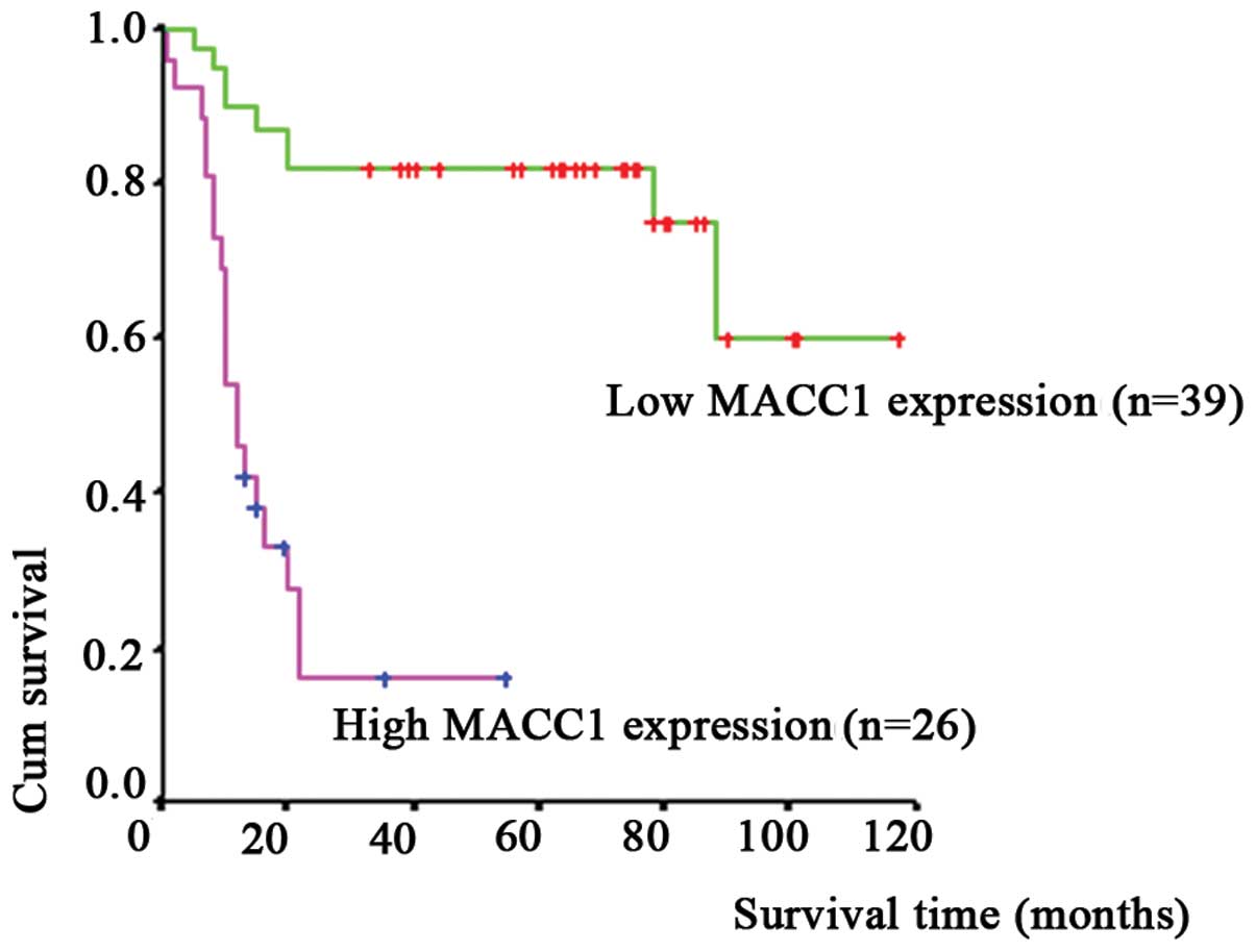
include more cases in our further studies. Furthermore, we followed
up the patients who were diagnosed with TSCC in this study. We
found that the patients with MACC1 overexpression were often
suffering from distant metastasis and were approaching death. The
survival curves were generated using the Kaplan-Meier method and
were compared with the log-rank test. Multivariate analysis
demonstrated that MACC1 and EMMPRIN (CD147) overexpression were
closely related with the overall survival time of the patients
(Table II, P=0.0002, P=0.0042)
respectively. Thirty-nine patients with low MACC1 expression levels
were followed up from 1 to 117 months, with a median overall
survival of 66 months. Twenty-six patients with high MACC1
expression levels were followed up from 1 to 55 months, with a
median overall survival of 12 months. Conclusively, the patients
with high MACC1 expression levels demonstrated a significantly
shorter survival time (Fig. 2 and
Table II, P<0.05).
| Table IIMultivariate analysis of prognostic
factors associated with survival time in the 68 patients with
TSCC. |
Table II
Multivariate analysis of prognostic
factors associated with survival time in the 68 patients with
TSCC.
| Variable | B | S.E | Wald | Df | Sig | R | Exp (B) |
|---|
| Gender | 0.746 | 0.461 | 2.618 | 1 | 0.106 | 0.053 | 2.109 |
| Age (years) | −0.694 | 0.434 | 2.561 | 1 | 0.110 | −0.050 | 0.500 |
|
Differentiation | −0.063 | 0.354 | 0.037 | 1 | 0.847 | 0.000 | 0.934 |
| Lymphatic
metastasis | 0.102 | 0.512 | 0.040 | 1 | 0.842 | 0.000 | 1.1073 |
| Clinical stage | 0.205 | 0.248 | 0.681 | 1 | 0.409 | 0.0171 | 1.227 |
| EMMPRIN
expression | 0.987 | 0.345 | 8.188 | 1 | 0.0042 | 0.167 | 2.684 |
| MACC1
expression | 1.888 | 0.503 | 14.072 | 1 | 0.0002 | 0.233 | 6.606 |
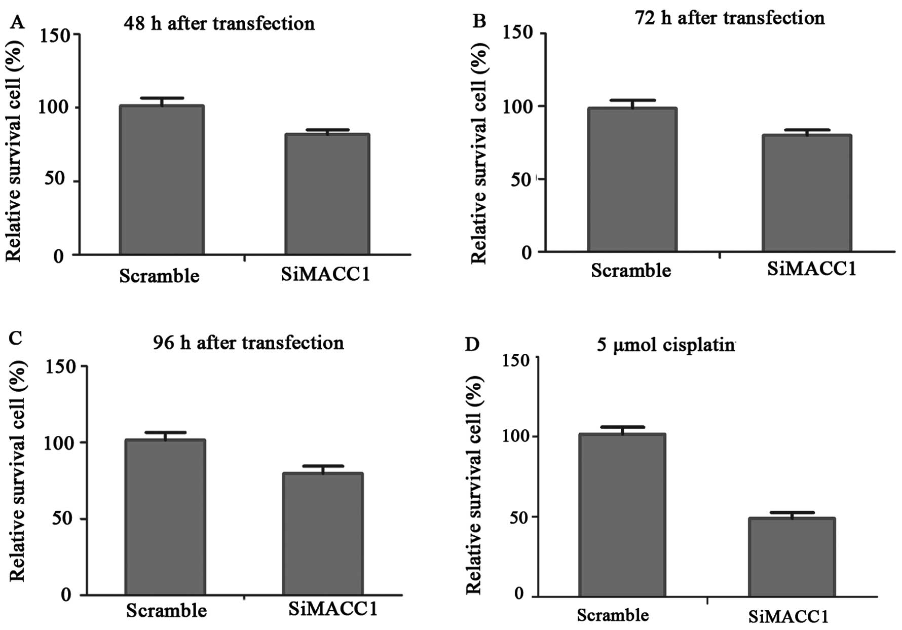
Downregulation of MACC1 significantly
inhibits cell proliferation, attenuates cisplatin resistance and
induces apoptosis of TSCC in vitro
The western blot analysis showed that the knockdown
of MACC1 resulted in the inhibition of MACC1 protein expression in
TSCCA cells (Fig. 3G). We tested
three time-points (48, 72 and 96 h) after the knockdown of MACC1.
The results of the cell proliferation tested by CCK-8 showed that
MACC1-siRNA-transfected TSCCA cells exhibited decreased cell growth
ability (P<0.05) compared with the control group (Fig. 3A–C, P<0.05). The inhibition rate
of MACC1 knockdown on proliferation was ~25%. Drug resistance to
cisplatin assays revealed that MACC1-siRNA-transfected TSCCA cells
had attenuated drug resistance to cisplatin, especially at the
concentrations of 10 and 5 μmol cisplatin (Fig. 3D and E, P<0.05). The apoptosis
assay showed that after transfection with MACC1-siRNA, the early
apoptosis rate increased from 8.45±1.30 to 12.66±1.29%. The
difference between the 2 groups was statistically significant
(Fig. 3F, P<0.05). Our studies
demonstrated that the downregulation of MACC1 significantly
inhibited proliferation, attenuated cisplatin resistance and
induced apoptosis in the TSCCA cells in vitro.
Downregulation of MACC1 contributes to
the inhibition of migration and invasion of TSCC in vitro
The Transwell penetration assay of migration
demonstrated that the number of migratory cells transfected with
MACC1-NC and MACC1-siRNA at 18 h was 837±31 and 532±36/field of
view, respectively. The Transwell penetration assay of invasion had
the same trend as the migration assay; the number of invasive cells
transfected with MACC1-NC and MACC1-siRNA at 48 h was 1223±79 and
749±34/field of view, respectively. The experiments were carried
out a minimum of 3 times. The differences in the 2 groups
(MACC1-siRNA and MACC1-NC) were statistically significant (Fig. 4A and B, P<0.05). We conclude that
knockdown of MACC1 contributed to the inhibition of migration and
invasion of TSCCA cells in vitro.
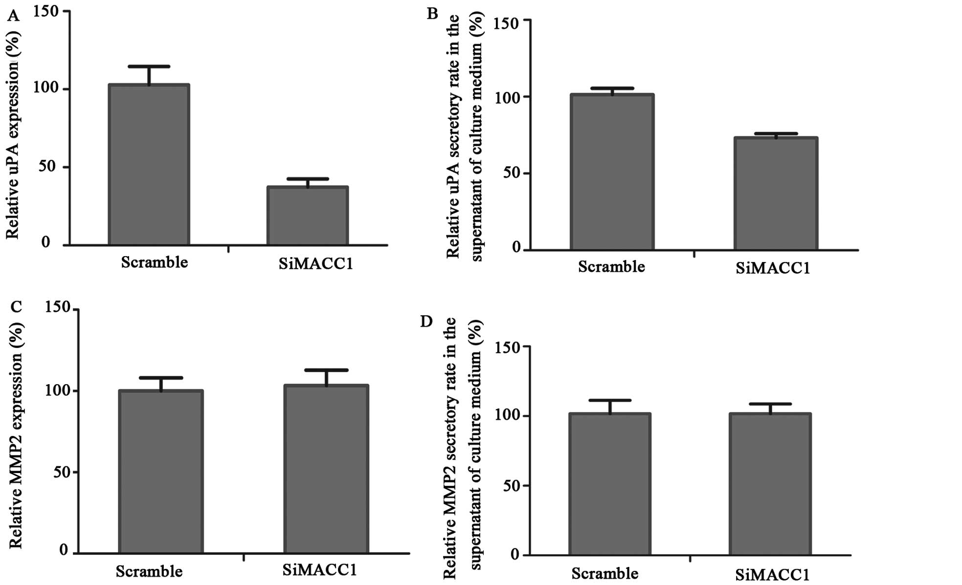
Knockdown of MACC1 markedly reduces the
invasive ability through suppression of uPA but not of MMP2
expression and secretion in TSCC in vitro
The ELISA assay demonstrated that the MACC1-siRNA
transfection decreased the uPA secretion by nearly 25%. The uPA
protein expression was extremely attenuated after transfection with
MACC1-siRNA compared with the MACC1-NC group. The difference in uPA
secretion and expression between the experimental and the control
group was statistically significant (Fig. 5A, B and F, P<0.05). The MMP2
expression analyzed by western blot analysis and the MMP2 secretion
detected by ELISA had no significant changes (Fig. 5C, D and G; P>0.05). Based on the
results, we primarily concluded that the downregulation of MACC1
inhibited invasion by attenuating uPA rather than by attenuating
MMP2 expression and secretion.
Discussion
Although progress has been made in the treatment of
TSCC, the prognosis remains poor, with a 5-year survival rate
<50% (2). To our knowledge,
local recurrences and distant metastases are responsible for the
failure of treatment in TSCC. It is important to identify
biomarkers to predict the prognosis of TSCC to reduce mortality and
medical costs.
MACC1 is a newly identified oncogene related to
malignant proliferation and metastases in several tumor types. The
downregulation of MACC1 has been reported to inhibit proliferation
and tumorigenicity of nasopharyngeal carcinoma cells through the
Akt/β-catenin signaling pathway (6). MACC1 is a key regulator of the HGF-MET
signaling pathway, which is related to colon cancer metastases
(4,5). Increased MACC1 expression may be a
possible biomarker for evaluating postoperative recurrence in
patients with lung cancer (7,8). MACC1
is involved in the progression of breast cancer and represents a
potentially useful biomarker for prognosis (14). In other cancers, such as colon
carcinoma (4,5), pancreatic cancer (13), ovarian cancer (12) and hepatocellular carcinoma (9–11),
MACC1 has been shown to be a new oncogene associated with the
malignant biological behaviors of tumors. Whether or not MACC1
expression is related to TSCC has not been previously elucidated.
This study determined that MACC1 expression in TSCC was
significantly higher than that in the non-cancerous epithelium
adjacent to the carcinomas of tongue. Increased MACC1 expression
was highly associated with lymphatic metastases and EMMPRIN
expression and predicted a poor overall survival of the TSCC
patients. Silencing of the MACC1 gene downregulated cell
proliferation, migration and invasion and it attenuated the
cisplatin resistance of TSCCA cells and contributed to apoptosis.
MACC1 influenced the invasion and migration of TSCCA cells by
secreting and activating uPA instead of MMP2.
Previous researchers found MACC1 overexpression in
various tumor tissues (6,10,12)
and pancreatic cancer patient serum samples (13). In nasopharyngeal carcinoma, high
MACC1 expression was significantly related to UICC stage (P=0.005)
and N classification (P<0.05) (6). In pancreatic cancer patients, a high
serum level of MACC1 expression was positively correlated with
lymph node metastases (P=0.035), distant metastases (P=0.005) and
more advanced TNM stages (P=0.024) (13). In our study, we examined 68 TSCC
tissues and the non-cancerous epithelial tissues adjacent to the
TSCC using IHC. We found that MACC1 expression in TSCC was
significantly higher than that in the non-cancerous epithelium
adjacent to the TSCC. We analyzed the relationship between clinical
characteristics of the patients and MACC1 expression. We discovered
that high MACC1 expression was correlated with lymphatic
metastases. The multivariate analysis revealed that MACC1 was
significantly associated with the overall survival time of TSCC
patients. High MACC1 expression contributed to a shorter survival
time in TSCC patients. We did not find a significant difference
between MACC1 expression and the clinical tumor stage (Table I, P=0.088), but the data did
demonstrate a positive trend. High MACC1 expression tended to occur
in later stages (III+IV). We suggest that the unclear relationship
between high expression of MACC1 and clinical stage could be
resolved if we include more cases in our further studies.
Researchers have reported that RNA interference against MACC1
resulted in antitumor proliferation and progression in ovarian
carcinoma. MACC1 knockdown may be involved in the inhibition of the
HGF/MET and MEK/ERK pathways (12).
Studies have proposed that the downregulation of MACC1 inhibits the
proliferation and tumorigenicity of NCC through the Akt/β-catenin
signaling pathway (6). The
regulation of the Ras/ERK pathway by MACC1 in pancreatic cancer
affects metastasis and progression (13). The expression of MACC1 is associated
with colon cancer tumorigenesis and metastasis by regulating the
HGF/Met signaling pathway (4,16). The
mechanism of MACC1 regulation of TSCC remains unclear. To explore
the role of MACC1 in TSCC, we employed siRNA to downregulate MACC1
expression and to determine its effect on proliferation, migration,
invasion, chemoresistance and apoptosis. A CCK-8 test on TSCCA
cells showed that the proliferation was markedly inhibited and that
apoptosis was induced after downregulation of MACC1 expression.
TSCCA cell sensitivity to cisplatin increased after silencing of
the MACC1 gene. The Transwell experiment demonstrated that
migration and invasion abilities of the TSCCA cell line were
significantly downregulated after transfection with MACC1-siRNA.
The results of this study are consistent with the results of
previous studies (6,12). We suggest that regulation,
proliferation, apoptosis and chemoresistance by MACC1 may involve
the HGF/MET, MEK/ERK, Akt/β-catenin or Ras/ERK pathway in TSCC.
However, the actual mechanism requires further investigation.
Previous studies have demonstrated that EMMPRIN
which is a plasma membrane protein of the immunoglobulin (Ig)
superfamily, enhances cell proliferation, invasion and metastasis
of tumors and contributes to poor overall survival (18,19).
In our previous study, EMMPRIN expression, especially EMMPRIN
isoform 2 expression, was associated with the metastases of head
and neck cancer (20).
Overexpression of EMMPRIN promoted proliferation, invasion and
metastases of cancer, resulting in poor overall survival (19,21).
EMMPRIN expression was significantly associated with tumor
diameter, clinical stage and poor survival time in TSCC (2). MACC1 was correlated with carcinoma
progression and metastases. It was previously not known whether
there is a relationship between MACC1 expression and EMMPRIN
expression in TSCC. This study demonstrated that increased MACC1
expression was positively associated with EMMPRIN expression
(P=0.003). We suggest that MACC1 and EMMPRIN may cooperate in
carcinogenesis in TSCC. The underlying regulatory mechanism between
these genes remains to be further elucidated. Clinically, the
failure of treatment in TSCC is due to recurrence and distant
metastasis, which seriously affects the prognosis of patients.
MACC1 has been identified as an oncogene that is correlated with
the metastasis of malignant tumors. The underlying mechanisms
involved in the invasion and metastasis of tongue tumors remain
unknown. The activation of MMPs, particularly MMP2, has been
reported to upregulate invasion and metastases in several malignant
carcinomas (22–24). The activation of MMP2 is an
important factor mediating invasion in tongue squamous carcinomas
(25,26). EMMPRIN stimulates the activation and
expression of MMP2, which results in the promotion of invasion
(27–29). We previously demonstrated that
EMMPRIN activates MMP2 and uPA to enhance invasion in TSCC
(20). The uPA has been shown to be
closely associated with invasion and metastases of carcinomas
(29–32). IHC revealed that MACC1 and EMMPRIN
are correlated in predicting malignancy and poor progression. Since
EMMPRIN was involved in tumor malignancy and progression, we
hypothesized that MACC1 promotes metastasis through the expression
and secretion of MMP2 and/or uPA. We used western blot analysis to
examine the MMP2 and uPA expression levels. We used ELISA to detect
the secretion of MMP2 and uPA in the supernatant of the culture
medium. The results showed that uPA expression and secretion were
significantly reduced after the knockdown of MACC1 in TSCC cells.
No significant variation was found in the MMP2 expression tested by
western blot analysis and in the MMP2 secretion detected by ELISA.
Therefore, we primarily suggest that the downregulation of MACC1
inhibited invasion by attenuating uPA instead of MMP2 expression
and secretion. The actual mechanism needs further
investigation.
In conclusion, MACC1 overexpression contributes to
metastasis and poor overall survival in TSCC patients. The
downregulation of MACC1 inhibits invasion, migration and
proliferation, attenuates cisplatin resistance and induces
apoptosis in TSCC. MACC1 downregulates the invasion of TSCC by
attenuating uPA instead of MMP2 expression and secretion. MACC1 may
serve as a novel biomarker and therapeutic target for TSCC.
Acknowledgements
This study was supported by the Fundamental Research
Funds for the Central Universities (no. 13ykpy26), the Guangdong
Province Natural Science Foundation (no. S2013010014794) and the
National Natural Science Fund of China (81101592).
Abbreviations:
|
TSCC
|
tongue squamous cell carcinoma
|
|
MACC1
|
metastasis-associated in colon
cancer
|
|
EMMPRIN
|
extracellular matrix metalloproteinase
inducer
|
|
MACC1-siRNA
|
MACC1 small interfering RNA
|
|
IHC
|
immunohistochemistry
|
|
HNSCC
|
head and neck squamous cell
carcinoma
|
|
OSCC
|
oral squamous cell carcinoma
|
References
|
1
|
Amar A, Rapoport A, Curioni OA, et al:
Prognostic value of regional metastasis in squamous cell carcinoma
of the tongue and floor of mouth. Braz J Otorhinolaryngol.
79:734–737. 2013.(In English, and Portuguese). View Article : Google Scholar
|
|
2
|
Huang Z, Huang H, Li H, et al: EMMPRIN
expression in tongue squamous cell carcinoma. J Oral Pathol Med.
38:518–523. 2009. View Article : Google Scholar : PubMed/NCBI
|
|
3
|
Qu JH, Chang XJ, Lu YY, et al:
Overexpression of metastasis-associated in colon cancer 1 predicts
a poor outcome of hepatitis B virus-related hepatocellular
carcinoma. World J Gastroenterol. 18:2995–3003. 2012. View Article : Google Scholar : PubMed/NCBI
|
|
4
|
Stein U, Burock S, Herrmann P, et al:
Circulating MACC1 transcripts in colorectal cancer patient plasma
predict metastasis and prognosis. PLoS One. 7:e492492012.
View Article : Google Scholar : PubMed/NCBI
|
|
5
|
Stein U, Walther W, Arlt F, et al: MACC1,
a newly identified key regulator of HGF-MET signaling, predicts
colon cancer metastasis. Nat Med. 15:59–67. 2009. View Article : Google Scholar
|
|
6
|
Meng FJ, Li H, Shi HJ, et al: MACC1
down-regulation inhibits proliferation and tumourigenicity of
nasopharyngeal carcinoma cells through Akt/β-catenin signaling
pathway. PLoS One. 8:e608212013. View Article : Google Scholar
|
|
7
|
Shimokawa H, Uramoto H, Onitsuka T, et al:
Overexpression of MACC1 mRNA in lung adenocarcinoma is associated
with postoperative recurrence. J Thorac Cardiovasc Surg.
141:895–898. 2011. View Article : Google Scholar
|
|
8
|
Chundong G, Uramoto H, Onitsuka T, et al:
Molecular diagnosis of MACC1 status in lung adenocarcinoma by
immunohistochemical analysis. Anticancer Res. 31:1141–1145.
2011.PubMed/NCBI
|
|
9
|
Xie C, Wu J, Yun J, et al: MACC1 as a
prognostic biomarker for early-stage and AFP-normal hepatocellular
carcinoma. PLoS One. 8:e642352013. View Article : Google Scholar : PubMed/NCBI
|
|
10
|
Qiu J, Huang P, Liu Q, et al:
Identification of MACC1 as a novel prognostic marker in
hepatocellular carcinoma. J Transl Med. 9:1662011. View Article : Google Scholar : PubMed/NCBI
|
|
11
|
Yang YP, Qu JH, Chang XJ, et al: High
intratumoral metastasis-associated in colon cancer-1 expression
predicts poor outcomes of cryoablation therapy for advanced
hepatocellular carcinoma. J Transl Med. 11:412013. View Article : Google Scholar : PubMed/NCBI
|
|
12
|
Zhang R, Shi H, Chen Z, et al: Effects of
metastasis-associated in colon cancer 1 inhibition by small hairpin
RNA on ovarian carcinoma OVCAR-3 cells. J Exp Clin Cancer Res.
30:832011. View Article : Google Scholar : PubMed/NCBI
|
|
13
|
Wang G, Kang MX, Lu WJ, et al: MACC1: A
potential molecule associated with pancreatic cancer metastasis and
chemoresistance. Oncol Lett. 4:783–791. 2012.PubMed/NCBI
|
|
14
|
Huang Y, Zhang H, Cai J, et al:
Overexpression of MACC1 and its significance in human breast cancer
progression. Cell Biosci. 3:162013. View Article : Google Scholar : PubMed/NCBI
|
|
15
|
Zhang Y, Wang Z, Chen M, et al:
MicroRNA-143 targets MACC1 to inhibit cell invasion and migration
in colorectal cancer. Mol Cancer. 11:232012. View Article : Google Scholar : PubMed/NCBI
|
|
16
|
Migliore C, Martin V, Leoni VP, et al:
MiR-1 downregulation cooperates with MACC1 in promoting MET
overexpression in human colon cancer. Clin Cancer Res. 18:737–747.
2012. View Article : Google Scholar
|
|
17
|
Guo T, Yang J, Yao J, Zhang Y, Da M and
Duan Y: Expression of MACC1 and c-Met in human gastric cancer and
its clinical significance. Cancer Cell Int. 13:1212013. View Article : Google Scholar : PubMed/NCBI
|
|
18
|
Zheng HC, Takahashi H, Murai Y, et al:
Upregulated EMMPRIN/CD147 might contribute to growth and
angiogenesis of gastric carcinoma: a good marker for local invasion
and prognosis. Br J Cancer. 95:1371–1378. 2006. View Article : Google Scholar : PubMed/NCBI
|
|
19
|
Vigneswaran N, Beckers S, Waigel S, et al:
Increased EMMPRIN (CD 147) expression during oral carcinogenesis.
Exp Mol Pathol. 80:147–159. 2006. View Article : Google Scholar
|
|
20
|
Huang Z, Tan N, Guo W, et al:
Overexpression of EMMPRIN isoform 2 is associated with head and
neck cancer metastasis. PLoS One. 9:e915962014. View Article : Google Scholar : PubMed/NCBI
|
|
21
|
Zhong WD, Han ZD, He HC, et al: CD147,
MMP-1, MMP-2 and MMP-9 protein expression as significant prognostic
factors in human prostate cancer. Oncology. 75:230–236. 2008.
View Article : Google Scholar : PubMed/NCBI
|
|
22
|
Zheng PC, Chen X, Zhu HW, et al: Capn4 is
a marker of poor clinical outcomes and promotes nasopharyngeal
carcinoma metastasis via nuclear factor-κB-induced matrix
metalloproteinase 2 expression. Cancer Sci. 105:630–638. 2014.
View Article : Google Scholar : PubMed/NCBI
|
|
23
|
Huang Q, Lan F, Wang X, et al:
IL-1β-induced activation of p38 promotes metastasis in gastric
adenocarcinoma via upregulation of AP-1/c-fos, MMP2 and MMP9. Mol
Cancer. 13:182014. View Article : Google Scholar
|
|
24
|
Moroz A, Delella FK, Almeida R, et al:
Finasteride inhibits human prostate cancer cell invasion through
MMP2 and MMP9 downregulation. PLoS One. 8:e847572013. View Article : Google Scholar
|
|
25
|
Xiao W, Jiang M, Li H, Li C, Su R and
Huang K: Knockdown of FAK inhibits the invasion and metastasis of
Tca8113 cells in vitro. Mol Med Rep. 8:703–707. 2013.PubMed/NCBI
|
|
26
|
Wang X, Sun W, Zhang C, et al: TGF-β1
inhibits the growth and metastasis of tongue squamous carcinoma
cells through Smad4. Gene. 485:160–166. 2011. View Article : Google Scholar : PubMed/NCBI
|
|
27
|
Zucker S, Hymowitz M, Rollo EE, et al:
Tumorigenic potential of extracellular matrix metalloproteinase
inducer. Am J Pathol. 158:1921–1928. 2001. View Article : Google Scholar : PubMed/NCBI
|
|
28
|
Sienel W, Polzer B, Elshawi K, et al:
Cellular localization of EMMPRIN predicts prognosis of patients
with operable lung adenocarcinoma independent from MMP-2 and MMP-9.
Mod Pathol. 21:1130–1138. 2008. View Article : Google Scholar : PubMed/NCBI
|
|
29
|
Bao YN, Cao X, Luo DH, et al:
Urokinase-type plasminogen activator receptor signaling is critical
in nasopharyngeal carcinoma cell growth and metastasis. Cell Cycle.
13:1958–1969. 2014. View
Article : Google Scholar : PubMed/NCBI
|
|
30
|
Shi H, Liu L, Liu L, Geng J, et al:
beta-Elemene inhibits the metastasis of B16F10 melanoma cells by
downregulation of the expression of uPA, uPAR, MMP-2 and MMP-9.
Melanoma Res. 24:99–107. 2014. View Article : Google Scholar : PubMed/NCBI
|
|
31
|
Quemener C, Gabison EE, Naimi B, et al:
Extracellular matrix metalloproteinase inducer up-regulates the
urokinase-type plasminogen activator system promoting tumor cell
invasion. Cancer Res. 67:9–15. 2007. View Article : Google Scholar : PubMed/NCBI
|
|
32
|
Lescaille G, Menashi S, Cavelier-Balloy B,
et al: EMMPRIN/CD147 up-regulates urokinase-type plasminogen
activator: implications in oral tumor progression. BMC Cancer.
12:1152012. View Article : Google Scholar : PubMed/NCBI
|