Introduction
BID (BH3-interacting domain death agonist) protein
participates in extrinsic apoptotic signaling (1) and is also able to mediate DNA damage
response (2,3). The level of BID is critical for the
viability of numerous types of cancer cells since its silencing
makes them resistant to apoptosis induced by death receptor ligands
(4,5), whereas overexpression sensitizes
certain types of cancer cells to TRAIL (6,7) or
etoposide (8). Due to the above
features, BID has been considered for therapeutic exploitation
(4). One of the obstacles
associated with its application is the lack of a stringent control
of the cellular level of BID, when it is expressed using an
adenovirus (7) or pcDNA vectors
(3,4), which are commonly used to deliver the
protein to cells. As a result, BID could be overexpressed and
eventually toxic to cells treated with the vectors. In a previous
study, we showed that recombinant BID fused with TAT cell
penetrating peptide (TAT-BID) could be delivered to cells in a
controlled manner (9). We
demonstrated that by using an appropriate dose of TAT-BID it was
possible to maintain its concentration at the level which was not
toxic alone but sensitized cancer cells to apoptosis, this way
increasing the possibility of the potential therapeutic use of the
protein. We found that the extent of the effect caused by TAT-BID
depended on the cell line used and the anticancer drug employed to
induce apoptosis. These results suggested that further studies on
the combinations of TAT-BID, an anticancer drug and a cell line
might reveal the optimal efficacy of TAT-BID as a potential
therapeutic agent. Following that line of thought, in the present
study, we examined the sensitization of different cancer cell lines
to doxorubicin.
Doxorubicin (DOX; alternative name: Adriamycin) is
an anthracycline compound, commonly used as an anticancer drug
alone or in combined therapies. Its anticancer molecular action is
complex and includes, among others: inhibition of topoisomerases I
and II, formation of DNA adducts and free radicals, interaction
with membrane proteins, histone eviction and ceramide
overproduction (reviewed in refs. 10,11).
It has also been shown that administration of DOX alters a
transcription profile of the cell (12,13)
and that it sensitizes cells to other anticancer drugs (14,15).
Due to the broad spectrum of processes affected by DOX, its effects
in combined therapies are difficult to predict but rather they need
experimental examination. We present here the experimental evidence
that TAT-BID strongly sensitized HeLa and PC3 cells to DOX, whereas
sensitization of A549 and LNCaP cell lines was moderate or
negligible, respectively.
Materials and methods
Reagents
Doxorubicin (DOX), RPMI-1640 medium, Dulbecco’s
modified Eagle’s medium (DMEM), trypsin inhibitor from soybean,
fetal bovine serum (FBS), penicillin and streptomycin, D-glucose,
sodium pyruvate, MTT reagent and anti-GAPDH antibodies were
obtained from Sigma-Aldrich (St. Louis, MO, USA); F12K medium from
the American Type Culture Collection (ATCC; Manassas, VA, USA);
anti-HA antibodies from Santa Cruz Biotechnology (Dallas, TX, USA);
anti-caspase-3 antibodies from Cell Signaling Technology (Danvers,
MA, USA); anti-PARP1 and PE-anti-caspase-3 antibodies from
Beckton-Dickinson (San Jose, CA, USA); DRAQ5 from BioStatus Ltd.
(Shepshed, UK); Dako fluorescent mounting medium from Dako North
America (Carpinteria, CA, USA); and Giemsa’s azur and May-Grünwald
dyes were from Merck Millipore (Billerica, MA, USA).
Cell culture
Four cancer cell lines were used in the present
study: two prostate cancer cell lines (PC3 and LNCaP) and two
non-prostate cancer cell lines: non-small human lung cancer (A549)
and cervical carcinoma (HeLa). All cell lines were purchased from
the European Collection of Cell Cultures (ECACC). Prostate cancer
cells were cultured in RPMI-1640 medium. LNCaP medium contained
additionally high D-glucose (4.5 mg/ml), 10 mM HEPES and 1 mM
sodium pyruvate. The human non-small lung cancer A549 cell line and
cervical carcinoma HeLa cell line were cultured in F12K or DMEM,
respectively. All media were supplemented with 10% FBS, penicillin
(100 U/ml) and streptomycin (100 μg/ml). Cultures were
maintained in a 95% humidified atmosphere of 5% CO2 at
37°C. For the experiments, the cells were seeded in 96-well plates,
dishes or cover glasses.
Plasmid construction, mutagenesis,
expression, isolation and purification of the recombinant
proteins
Plasmids pET28a/TAT-BID encoding BID proteins
(wild-type and T59AS76A-mutated variant) fused with the TAT domain
were constructed as previously described (9). T59A/S76A variant of TAT-BID was
mutated in a manner which made it unphosphorylable by CK2 kinase.
Both mentioned proteins were tagged with the His-tag used for
purification and with the HA-tag used for simple identification of
the protein in the cell. His-tag and the TAT peptide used for the
cell penetration were localized at the N-terminal end of the
protein; HA tags were placed at the C-terminal end. TAT-BID
proteins were expressed, isolated and purified as previously
described (9). The protein
concentration in the samples used in the experiments was quantified
by densitometry after SDS-polyacrylamide gel electrophoresis using
ChemiDocXRS (Bio-Rad Laboratories).
Uptake of the recombinant proteins by the
cells
To examine the time-dependent kinetics of uptake and
degradation of the recombinant protein by HeLa cells, TAT-BID was
added directly to the culture medium at a concentration of 40
μg/ml in the presence of the trypsin inhibitor from soybean
(final concentration 0.005%). Whole cell extracts were prepared as
previously described (9), and
western blot analysis was carried out with the aid of anti-HA
antibodies (1:2,000). The membranes were subsequently stripped of
the primary antibody and re-probed with anti-GAPDH antibodies
(1:100,000). The results were quantified by densitometry using
ChemiDocXRS.
Analysis of cell viability
To examine the effect of doxorubicin (DOX) on cancer
cell viability, the PC3, LNCaP, HeLa and A549 cells were seeded in
96-well plates and incubated with different doses of DOX (ranging
from 0 to 20 μM) for 24 or 48 h. Similarly, to examine the
effect of DOX combined with TAT-BID on cancer cell viability, all
tested cell lines were seeded in 96-well plates and treated with 30
μg/ml TAT-BID (for PC3 and LNCaP cells) or 40 μg/ml
TAT-BID (for HeLa and A549 cells) alone or in combination with
either 0.5 μM or 1 μM DOX for either 24 or 48 h. The
same set of experiments were conducted for the wild-type and
mutated (T59A/S76A) variant of TAT-BID. Cell viability was analyzed
with MTT assay performed as previously described (9).
Apoptotic assays: procaspase-3 activation
and PARP1 cleavage
Procaspase-3 activation and PARP1 cleavage were
identified using western blot analysis. HeLa cells were seeded in
60-mm dishes, cultured for 24 h and then treated with 40
μg/ml TAT-BID, 1 μM DOX or both of these agents
together for 15 or 18 h, respectively. Next, both floating and
adherent cells were collected, washed with PBS, lysed in Laemmli
buffer and boiled for 20 min at 95°C. The proteins were then
separated on 12% polyacrylamide gel, transferred onto a PVDF
membrane, and procaspase-3 (31 kDa) and its active form caspase-3
(large fragment of caspase-3 resulting from cleavage, 17 kDa) were
identified using specific anti-caspase-3 antibodies (1:1,000).
PARP1 (116 kDa) and its apoptotic fragment (85 kDa) were identified
using specific anti-PARP1 antibodies (1:2,000). GAPDH protein was
detected as a loading control using anti-GAPDH antibodies
(1:100,000). The results were quantified by densitometry using
ChemiDocXRS.
Immunofluorescent staining for active
caspase-3
Procaspase-3 activation was also identified using
immunofluorescent staining for confocal microscopy observations.
HeLa cells were seeded on glass coverslips and cultured for 24 h
and then treated with 40 μg/ml TAT-BID, 1 μM DOX or
both of these agents together for 15 h. The cells were then washed
twice with PBS, fixed with 4% PFA for 10 min at room temperature
and washed twice with PBS. Next cells were permeabilized with 0.1%
Triton X-100 in PBS for 5 min at room temperature and blocked in 3%
BSA in PBS for 3 h at room temperature. After blocking, the cells
were incubated with PE-anti-caspase-3 antibodies (1:100 in blocking
buffer) for 3 h at room temperature in the dark in a humidified
chamber. The cells were then washed twice with 0.05% Tween-20 in
PBS, stained with DRAQ (1:1,000 in PBS) and washed again with 0.05%
Tween-20 in PBS. Finally the coverslips were mounted on glass
microscope slides. Microscopy observations were carried out on a
Nikon A1R confocal laser scanning microscope equipped with a Plan
Apochromat VC 60x/1.40 oil DIC objective (Nikon Instruments,
Melville, NY, USA). For detecting green fluorescence of PE, 488 nm
excitation line and 525 nm emission filter were used. For detecting
red fluorescence of DRAQ, 641 nm excitation line and 700 nm
emission filter were used. Data were analyzed with NIS-Elements
imaging software version 4.0 (Nikon Instruments).
Microscopic observations
To observe morphological changes, the HeLa cells
were seeded in 35-mm dishes, cultured for 24 h and then treated
with 40 μg/ml TAT-BID, 0.5 μM DOX or both of these
agents together for 24 or 48 h, respectively. The cells were then
washed twice with PBS and fixed with ice-cold methanol for 10 min
at 4°C. Next, the cells were washed twice with PBS and stained with
0.25% May-Grünwald for 3 min at room temperature. After that 0.1 M
phosphate buffer pH 7.0 was added (1:1) and left for 5 min. Next,
the cells were stained with 0.76% Giemsa’s azur for 15 min at room
temperature, washed at least three times and dried. Images
(magnification, x100) were captured using a Nikon eclipse TE200
microscope equipped with a Nikon Digital Camera DXM 1200 (Nikon
Instruments).
Data analysis
All experiments carried out as cell viability
measurements were repeated at least five times, and for each
individual point at least five independent measurements were made.
The results are shown as an average ± SD. Western blot analysis and
microscopic observations were carried out in triplicate and
representative results are presented. Differences between groups
were calculated using the Studen’ts t-test. A P-value <0.05 was
considered to indicate a statistically significant result. The
following ranges were defined: P<0.05, P<0.01 and P<0.001.
Statistica version 10 software (StatSoft, Inc., Krakow, Poland) was
used for analysis.
Results
Previously, we found that TAT-BID moderately
sensitized PC3 and A459 cells, and slightly sensitized HeLa cells
to TRAIL, while it was ineffective in the sensitization of LNCaP
cells (9). We also observed slight
sensitization of PC3 cells but not the remaining cell lines to
camptothecin. Due to the diversified effects of TAT-BID on the
above mentioned cell lines as previously observed, we examined in
the present study the same cell lines in terms of their
sensitization by TAT-BID to DOX.
The sensitization of cancer cells to TRAIL and
camptothecin was previously determined to be optimally visible for
concentrations of both drugs that, when administered alone,
resulted in a relatively small decrease in cell viability (9). To exploit the same idea in the present
study, we tested the effects of DOX acting alone on the viability
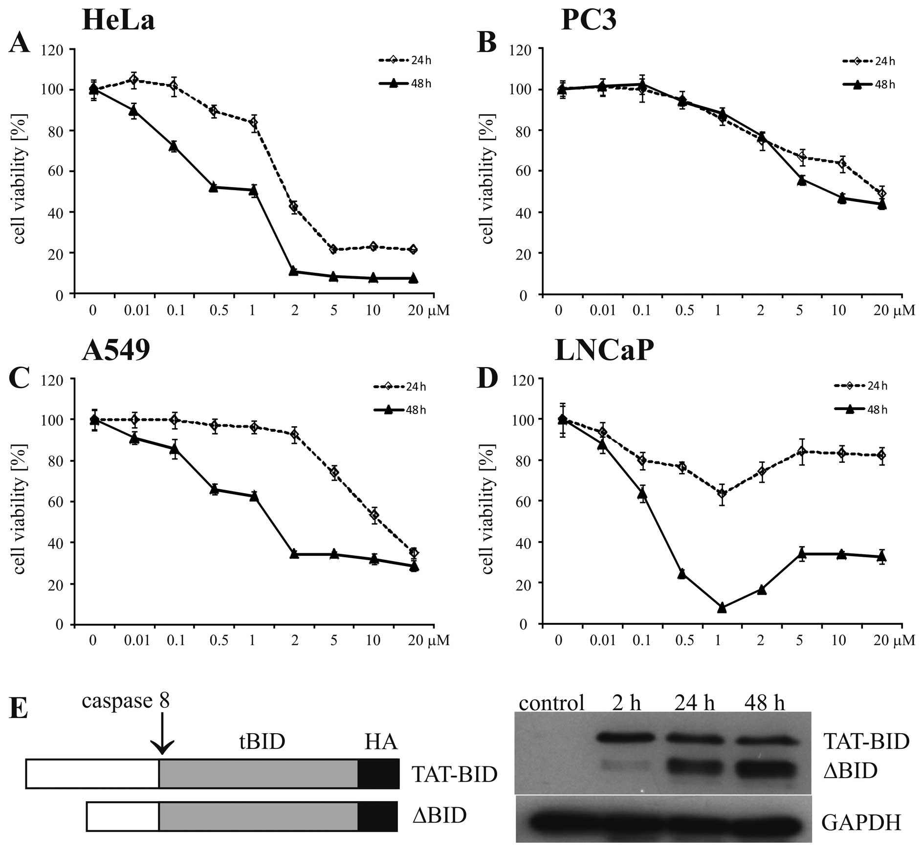
of particular cell lines. There was a broad range of sensitivity of
the examined cell lines to DOX (Fig.
1A–D and Table I). Moreover,
different types of sensitivity patterns were observed for different
cell lines in terms of their dose- and time-dependence of cell
viability (Fig. 1A–D). The decrease
in cell viability of the HeLa and A549 cells treated with DOX was
both dose- and time-dependent. Sensitivity patterns of prostate
cancer cells to DOX were different from those of the HeLa and A549
cell lines. In the case of PC3, there was no time-dependent effect
on the viability of PC3 cells treated with DOX; the decrease in
viability after a 24-h treatment was similar or the same as after a
48-h treatment (Fig. 1B). In the
case of LNCaP cells, the effects of DOX depended on the time of the
treatment; however, a simple dose-dependence was not observed. The
maximal reduction in cell viability was at 1 μM DOX,
followed by weakening of the effect observed for higher
concentrations of DOX (Fig. 1D).
Such a pattern suggested an additional factor that appeared at
higher concentrations of DOX and counteracted the lethal action of
DOX. It has been previously shown that treatment of prostate cancer
cells with DOX results in the elevated expression of
multidrug-resistance proteins (16). It is, thus, possible that such a
process occurred in the LNCaP cells and interfered with the lethal
action of DOX at concentrations >1 μM.
 | Table IIC50 values calculated for
different cell lines treated for 48 h with DOX. |
Table I
IC50 values calculated for
different cell lines treated for 48 h with DOX.
| Cell line | IC50
(μM) |
|---|
| PC3 | 8.00 |
| A549 | 1.50 |
| HeLa | 1.00 |
| LNCaP | 0.25 |
Based on the above observations, we selected the
concentration of 0.5 μM DOX to be used in the further
experiments. When this concentration was used for a 24-h treatment,
a small or moderate decrease in viability was noted in all cell
lines. To be closer to the effective range of DOX, we additionally
used 1 μM DOX. Moreover, we also tested a 48-h
administration of DOX at both concentrations to observe the effects
resulting from a long-term DOX treatment (12–15) on
the sensitization by TAT-BID. The latter conditions raised a
question of whether TAT-BID remained stable for >24 h to
influence the apoptosis induced by DOX after that time. In fact, we
observed degradation of TAT-BID inside the cells. The amount of
degraded TAT-BID accounted only for 23% of the TAT-BID present in
the cells after 2 h but this value increased up to 62% after 24 h
and to 75% after 48 h (Fig. 1E).
However, the total amount of TAT-BID in the cells increased during
the treatment and was 1.5-fold and 2-fold higher after 24 and 48 h,
respectively, than after 2 h of the treatment. As a result, an
absolute amount of intact TAT-BID in the cells after 48 h decreased
by only ~30% as compared to the amount present in the cells after a
2-h treatment. Moreover, the majority of degraded TAT-BID remained
in the form of a 26–27 kDa polypeptide (ΔBID) comprising 222–230
C-terminal residues as it was detected by anti-HA antibodies
(Fig. 1E). This means that ΔBID
included the active tBID fragment and a sequence accessible to
caspase-8. Taken together, we conclude that the recombinant active
form of BID (tBID) was available and influenced apoptotic signaling
during the entire period of the experiment.
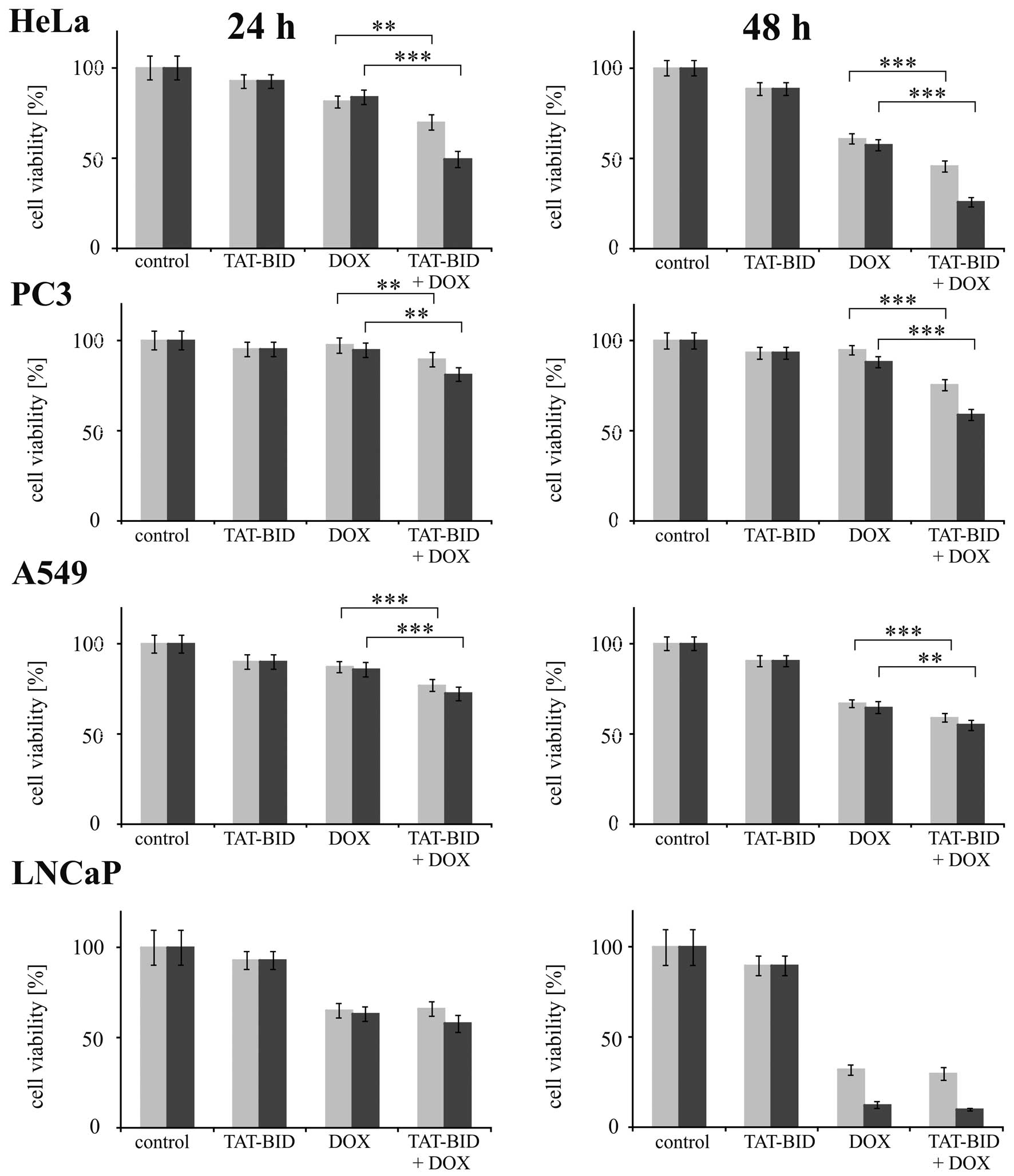
TAT-BID sensitized particular cell lines to DOX to
different extents (Fig. 2 and
Table II). Statistically
significant changes were observed for HeLa, PC3 and A549 but not
for LNCaP cells (Fig. 2). Among the
first group, the highest sensitization was found for HeLa and PC3
cells. This was reflected by a reduction in the cell viability
observed when TAT-BID appeared in addition to DOX, which was ~30%
for both cell lines treated with 1 μM DOX for 48 h. In the
A549 cell line such a reduction was only slightly >10% (Fig. 2). This was also reflected by the
synergy between DOX and TAT-BID, calculated here as the coefficient
of drug interaction (CDI) (17). A
slight synergistic effect (CDI, 0.8–0.9) was observed in the PC3
and HeLa cells treated with 0.5 μM DOX for 48 h. A moderate
synergistic effect (CDI, 0.7–0.8) was found for PC3 cells treated
with 1 μM DOX for 48 h and a significant synergistic effect
(CDI<0.7) for HeLa cells treated with 1 μM DOX for either
24 or 48 h (Table II). In the
latter case, the synergistic effect for TAT-BID and DOX was more
pronounced than any effect calculated for previously described
(9) combinations of TAT-BID with
other anticancer agents: TRAIL and camptothecin (Table II).
| Table IICoefficients of drug interaction (CDI)
for TAT-BID and DOX administered to different cell lines under
different conditions. |
Table II
Coefficients of drug interaction (CDI)
for TAT-BID and DOX administered to different cell lines under
different conditions.
| Cells | DOX 0.5 μM, 24
h | DOX 0.5 μM, 48
h | DOX 1 μM, 24
h | DOX 1 μM, 48
h | TRAILa | CPTa |
|---|
| PC3 | 0.9961 | 0.8518 | 0.9001 | 0.7165 | 0.7663 | 0.8831 |
| A549 | 0.9813 | 0.9719 | 0.9392 | 0.9360 | 0.7929 | 0.9347 |
| HeLa | 0.9296 | 0.8485 | 0.6400 | 0.5129 | 0.8653 | 0.9418 |
| LNCaP | 0.9719 | 1.0341 | 0.9360 | 0.9102 | 0.9697 | 0.9572 |
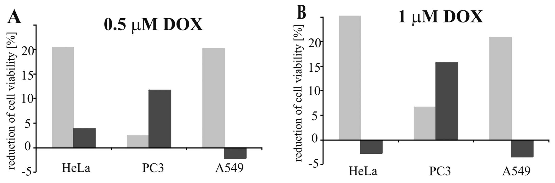
A distinct feature of the sensitization of HeLa and
A549 cell lines to DOX was that in both cases the extent of
sensitization by TAT-BID was poorly dependent on whether the cells
were treated for 24 or 48 h. This was illustrated by an additional
increased reduction in cell viability observed when DOX was
supplemented by TAT-BID which was relatively stable upon
prolongation of treatment time. This was in contrast to the
reduction in cell viability observed upon addition of DOX alone
which was clearly time-dependent (Fig.
3). The reverse pattern was found for PC3 cells. A decrease in
cell viability observed upon addition of DOX alone was independent
on time, whereas an additional increased reduction in cell
viability observed when TAT-BID was combined with DOX was
time-dependent (Fig. 3).
We also tested the sensitization of all cell lines
by the mutant TAT-BIDT59A/S76A that cannot be
inactivated by cellular CK2 kinase. It has been shown that
phosphorylation of BID at T59 and S76 might protect BID from
activation by caspase-8 cleavage and eventually from further
processing of the apoptotic signal (18). However, we previously demonstrated
that the mutant TAT-BIDT59A/S76A is as effective in the
sensitization of cells to TRAIL and camptothecin as the wild-type
TAT-BID (9). In the present study,
we found the same effectiveness of the mutant in the sensitization
of all the cell lines to DOX (data not shown).
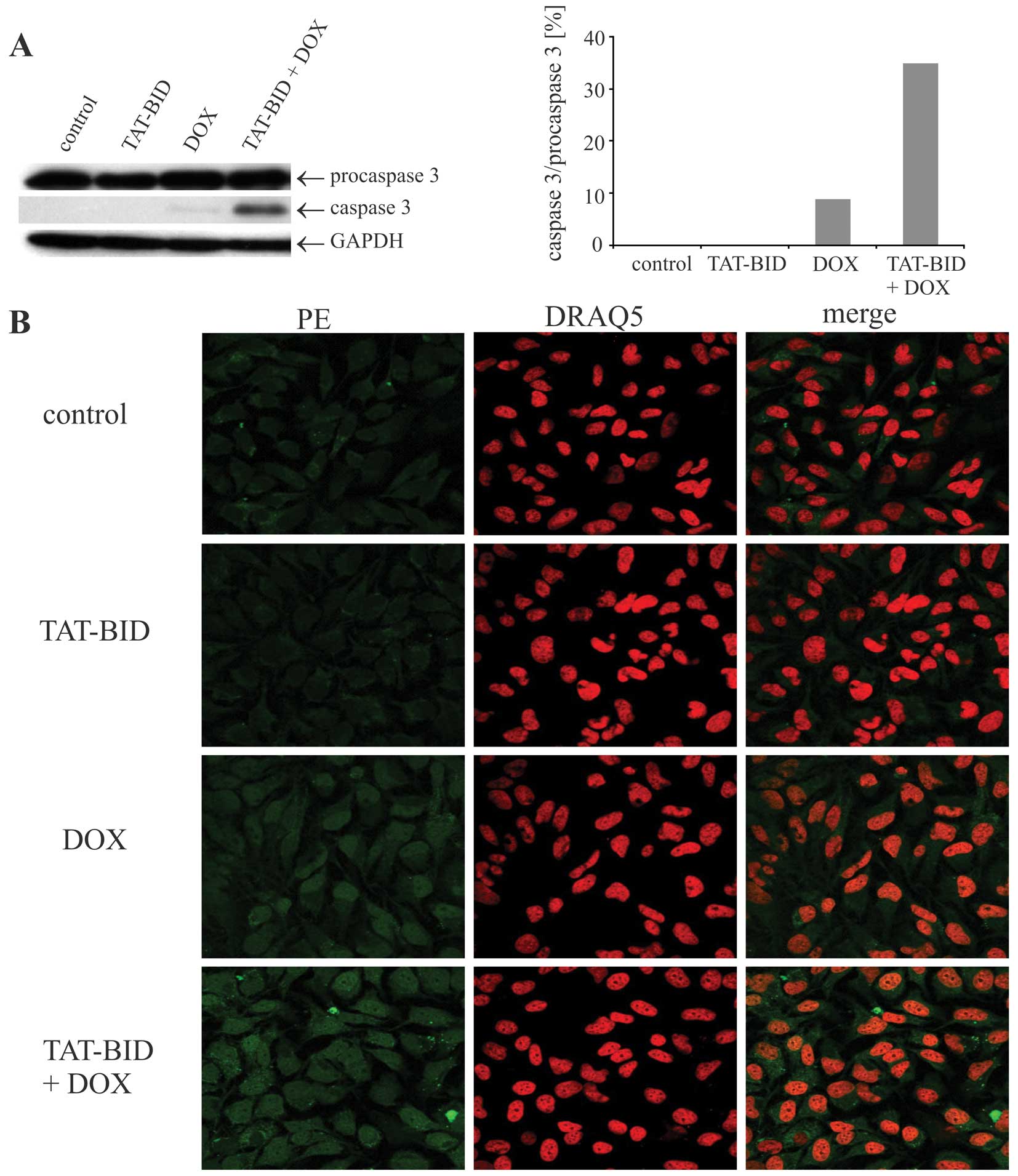
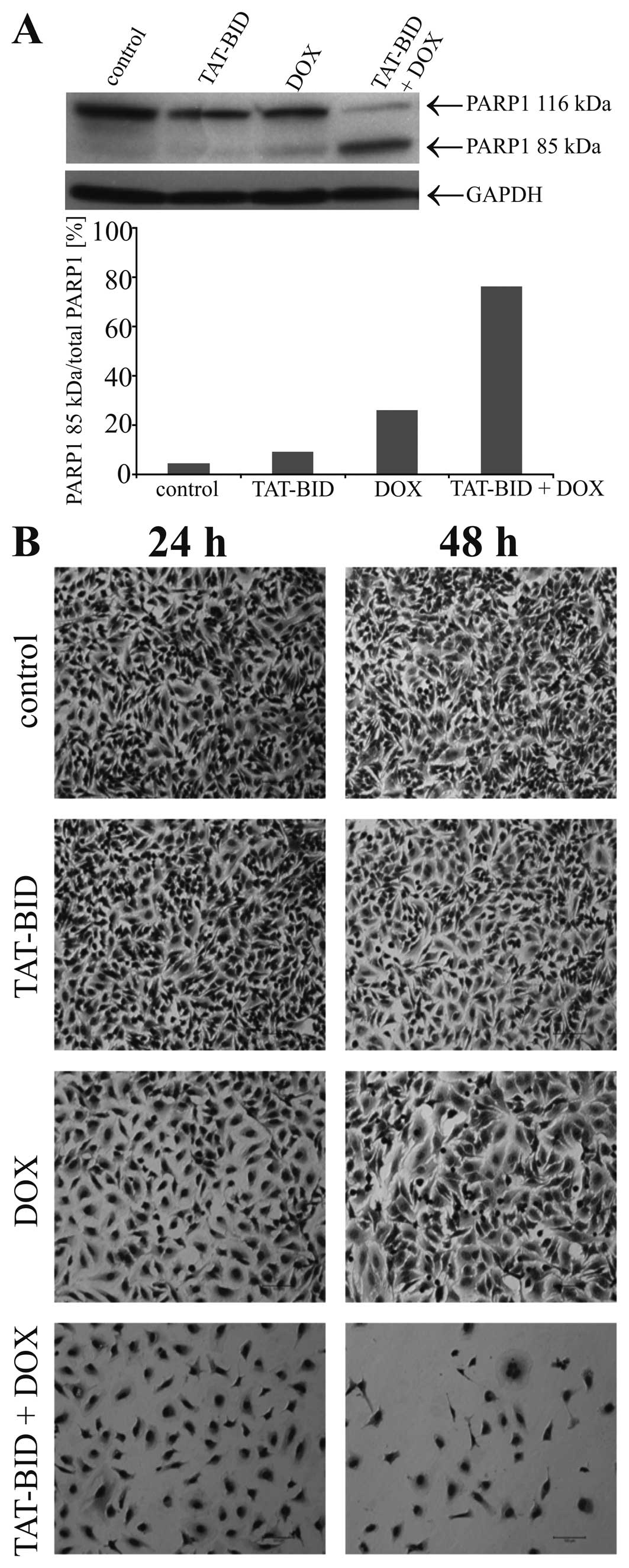
To gain a deeper insight into the mode of action of
TAT-BID combined with DOX, we performed additional experiments on
the HeLa cell line which was chosen since it was effectively
sensitized by TAT-BID to DOX (Fig.
2), and since the synergistic effect between TAT-BID and DOX
was high for this cell line (Table
II). We found that sensitization of the cells as revealed by
MTT assay resulted from increased apoptosis achieved upon combined
treatment. In the presence of both TAT-BID and DOX, after a 15-h
treatment we observed activation of procaspase-3, both by western
blot analysis (Fig. 4A) and
microscopic observations (Fig. 4B).
This was followed by enhanced PARP1 cleavage (18-h treatment;
Fig. 5A), and eventually by
pronounced changes in cell appearance and a decrease in their
number (24- and 48-h treatment; Fig.
5B).
Discussion
The main finding presented in the present study is
that externally delivered recombinant BID was unexpectedly
efficient in killing HeLa and PC3 cells when it was combined with
DOX. This observation is the first of all relevant findings
supporting the potential use of TAT-BID + DOX as a therapeutic
combination. In a previous study (9), we found that TAT-BID combined with
TRAIL was effective against PC3 and A549 cells, suggested its
anticancer potential in the treatment of advanced prostate cancer
and non-small human lung cancer. This study showed that cervical
carcinoma and prostate cancer cells appear even a better target for
TAT-BID when it is combined with DOX.
DOX is widely clinically used in the treatment of
different types of cancers. However, toxicity in the brain, liver,
kidneys and particularly in the heart are serious issues associated
with DOX chemotherapy (10).
Therefore, reducing the DOX dosage by combined administration with
a synergistically acting factor is an effective therapeutic option
(19). The combination of TAT-BID
and DOX exhibited the most pronounced synergy at 1 μM DOX
(Table II). This was the
concentration at which DOX acting alone decreased only weakly or at
most moderately the viability of HeLa and PC3 cells, and higher
doses of DOX were needed to achieve an effect similar to that of
the TAT-BID + DOX combination (compare Figs. 1 and 2). Therefore, TAT-BID may be considered as
a factor that potentially reduces the side-effects of DOX owing to
a lower drug dosage.
In addition to suggesting the potential therapeutic
use of TAT-BID in combination with DOX, this study presents some
observations and raises various questions concerning the role of
BID in the apoptosis induced by DOX in different types of cancer
cells. The main observation is that the extent of sensitization of
particular cell lines by TAT-BID is specific for DOX and does not
overlap with those observed previously for TRAIL or camptothecin
(9). Several different models have
been proposed for DOX-mediated cell death (10–15).
It has been suggested that the specific pathway to cell death
induced by DOX depends on the concentration of the drug, the time
of treatment and cancer type (10).
This is in agreement with the distinct sensitivities of different
cell lines to DOX (Table I), and
with the different patterns of dependence of DOX effects on the
time of treatment and on the DOX dose (Fig. 1), observed here when DOX was
administered alone. This is also in agreement with the specific DOX
sensitization of particular cell lines by TAT-BID. The only
exception to the latter feature is the lack of sensitization to all
examined agents [(9) and the
present study] observed for LNCaP cells which most possibly results
from impairment in signaling localized upstream from BID (20).
Questions raised by the above observation include:
why the effect of TAT-BID was more pronounced in PC3 and HeLa than
in A549 cells, and why extra BID delivered to HeLa cells was so
efficient in supporting the apoptosis induced by DOX and not by
TRAIL or camptothecin.
Concerning the PC3 cells, meaningful synergy was
observed only after a 48-h and not after a 24-h treatment (Table II). There may have been a specific
time-dependence in the effects caused in PC3 cells by DOX acting
alone or in the presence of TAT-BID, distinct from those observed
for HeLa and A549 cells. The effects of DOX on PC3 cells were not
dependent on time when DOX acted alone but they were when TAT-BID
was present (Fig. 3). This pattern
suggests that there were finite resources of a critical factor
necessary for the progression of DOX-induced apoptosis in the PC3
cells which, however, were exhausted within the first 24 h of the
treatment but were still present when TAT-BID was added. It also
means that the critical factor was BID protein. Such a sharp
dependence of the effect of DOX on the BID level may explain the
reason for the pronounced sensitivity of PC3 cells to TAT-BID
combined with DOX observed after 48 h. A specific feature of PC3
cells making them distinct from the other cells used here is the
lack of active p53 (21). p53 has
been shown to be critical for the effectiveness of DOX due to
regulation of the accumulation of DOX in the cells (22). We previously linked a lack of active
p53 in PC3 cells with higher sensitivity of these cells to TAT-BID
combined with camptothecin due to the absence of the p53-dependent
apoptotic pathway induced by DNA damage (9). Such a mechanism may also be the reason
for the specific time-dependence of DOX effects in PC3 cells
observed in the absence and in the presence of TAT-BID, and
eventually in pronounced sensitivity to TAT-BID combined with
DOX.
In regards to the HeLa cells, no conclusive data can
explain its marked sensitization to DOX by TAT-BID. Detailed
studies on the mechanism of DOX-induced apoptosis in HeLa cells
identified no alteration in expression of basic apoptotic proteins,
BID, BAX, Bcl-2, Bcl-xL and p53, during the first 48-h treatment
(23). Another report demonstrated
a small but significant decrease in Bcl-2 and an increase in the
BAD level observed in HeLa cells after a 18-h treatment with DOX
(19). Since BAD increases the
sensitivity of cells to BID (24),
alterations in Bcl-2 and BAD levels after long-term treatment might
together contribute to the sensitization of HeLa cells to DOX.
Acknowledgments
We thank Dr Alicja Czubaty from the Department of
Molecular Biology (University of Warsaw) for providing anti-PARP1
antibodies, Dr Katarzyna Piwocka from the Laboratory of Cytometry
(Nencki Institute of Experimental Biology) for PE-anti-caspase-3
antibodies and Professor Barbara Tudek from the Institute of
Genetics and Biotechnology (University of Warsaw) for
anti-caspase-3 antibodies. We are also grateful to Dr Bohdan
Paterczyk from the Laboratory of Electron and Confocal Microscopy
(University of Warsaw) for help with the confocal microscopy.
References
|
1
|
Kantari C and Walczak H: Caspase-8 and
Bid: Caught in the act between death receptors and mitochondria.
Biochim Biophys Acta. 1813:558–563. 2011. View Article : Google Scholar : PubMed/NCBI
|
|
2
|
Liu Y, Bertram CC, Shi Q and Zinkel SS:
Proapoptotic Bid mediates the Atr-directed DNA damage response to
replicative stress. Cell Death Differ. 18:841–852. 2011. View Article : Google Scholar :
|
|
3
|
Song G, Chen GG, Chau DKF, Miao J and Lai
PBS: Bid exhibits S phase checkpoint activation and plays a
pro-apoptotic role in response to etoposide-induced DNA damage in
hepatocellular carcinoma cells. Apoptosis. 13:693–701. 2008.
View Article : Google Scholar : PubMed/NCBI
|
|
4
|
Tsuno T, Mejido J, Zhao T, Phillips T,
Myers TG, Bekisz J and Zoon KC: BID is a critical factor
controlling cell viability regulated by IFN-α. J Immunother.
35:23–31. 2012. View Article : Google Scholar
|
|
5
|
Goncharenko-Khaider N, Lane D, Matte I,
Rancourt C and Piche A: The inhibition of Bid expression by Akt
leads to resistance to TRAIL-induced apoptosis in ovarian cancer
cells. Oncogene. 29:5523–5536. 2010. View Article : Google Scholar : PubMed/NCBI
|
|
6
|
Miao J, Chen GG, Chun SY, Yun JP, Chak EC,
Ho RL and Lai PB: Adenovirus-mediated tBid overexpression results
in therapeutic effects on p53-resistant hepatocellular carcinoma.
Int J Cancer. 119:1985–1993. 2006. View Article : Google Scholar : PubMed/NCBI
|
|
7
|
Fukazawa T, Walter B and Owen-Schaub LB:
Adenoviral Bid overexpression induces caspase-dependent cleavage of
truncated Bid and p53-independent apoptosis in human non-small cell
lung cancers. J Biol Chem. 278:25428–25434. 2003. View Article : Google Scholar : PubMed/NCBI
|
|
8
|
Li Y, Dai C, Li J, Wang W and Song G:
Bid-overexpression regulates proliferation and phosphorylation of
Akt and MAPKs in response to etoposide-induced DNA damage in
hepatocellular carcinoma cells. Onco Targets Ther. 5:279–286.
2012.PubMed/NCBI
|
|
9
|
Orzechowska EJ, Kozlowska E, Czubaty A,
Kozlowski P, Staron K and Trzcinska-Danielewicz J: Controlled
delivery of BID protein fused with TAT peptide sensitizes cancer
cells to apoptosis. BMC Cancer. 14:7712014. View Article : Google Scholar : PubMed/NCBI
|
|
10
|
Tacara O, Sriamornsak P and Dass CR:
Doxorubicin: an update on anticancer molecular action, toxicity and
novel drug delivery systems. J Pharm Pharmacol. 65:157–170. 2012.
View Article : Google Scholar
|
|
11
|
Yang F, Teves SS, Kemp CJ and Henikoff S:
Doxorubicin, DNA torsion, and chromatin dynamics. Biochim Biophys
Acta. 1845.84–89. 2014.
|
|
12
|
Leung KL and Wang TT: Differential effects
of chemotherapeutic agents on the Bcl-2/Bax apoptosis pathway in
human breast cancer cell line MCF-7. Basic Cancer Res Treat.
55:73–83. 1999. View Article : Google Scholar
|
|
13
|
Watanabe M, Naraba H, Sakyo T and Kitagawa
T: DNA damage-induced modulation of GLUT3 expression is mediated
through p53-independent extracellular signal-regulated kinase
signaling in HeLa cells. Mol Cancer Res. 8:1547–1557. 2010.
View Article : Google Scholar : PubMed/NCBI
|
|
14
|
Kelly MM, Hoel BD and Voelkel-Johnson C:
Doxorubicin pretreatment sensitizes prostate cancer cell lines to
TRAIL induced apoptosis which correlates with the loss of c-FLIP
expression. Cancer Biol Ther. 1:520–527. 2002. View Article : Google Scholar : PubMed/NCBI
|
|
15
|
Kang J, Bu J, Hao Y and Chen F: Subtoxic
concentration of doxorubicin enhances TRAIL-induced apoptosis in
human prostate cancer cell line LNCaP. Prostate Cancer Prostatic
Dis. 8:274–279. 2005. View Article : Google Scholar : PubMed/NCBI
|
|
16
|
Sánchez C, Mendoza P, Contreras HR,
Vergara J, McCubrey JA, Huidobro C and Castellón EA: Expression of
multidrug resistance proteins in prostate cancer is related with
cell sensitivity to chemotherapeutic drugs. Prostate. 69:1448–1459.
2009. View Article : Google Scholar : PubMed/NCBI
|
|
17
|
Wong FC, Woo CC, Hsu A, Kwong B and Tan
BK: The anticancer activities of Vernonia amygdalina extract in
human breast cancer cell lines are mediated through
caspase-dependent and p53-independent pathways. PLoS One.
8:e780212013. View Article : Google Scholar
|
|
18
|
Desagher S, Osen-Sand A, Montessuit S,
Magnenat E, Vilbois F, Hochmann A, Journot L, Antonsson B and
Martinou JC: Phosphorylation of Bid by casein kinases I and II
regulates its cleavage by caspase 8. Mol Cell. 8:601–611. 2001.
View Article : Google Scholar : PubMed/NCBI
|
|
19
|
Lee SJ, Hwang SO, Noh EJ, Kim DU, Nam M,
Kim JH, Nam JH and Hoe KL: Transactivation of bad by
vorinostat-induced acetylated p53 enhances doxorubicin-induced
cytotoxicity in cervical cancer cells. Exp Mol Med. 46:e762014.
View Article : Google Scholar : PubMed/NCBI
|
|
20
|
Zhang X, Jin TG, Yang H, DeWolf WC,
Khosravi-Far R and Olumi AF: Persistent c-FLIP(L) expression is
necessary and sufficient to maintain resistance to tumor necrosis
factor-related apoptosis-inducing ligand-mediated apoptosis in
prostate cancer. Cancer Res. 64:7086–7091. 2004. View Article : Google Scholar : PubMed/NCBI
|
|
21
|
He Z, Zhang Y, Mehta SK, Pierson DL, Wu H
and Rohde LH: Expression profile of apoptosis related genes and
radio-sensitivity of prostate cancer cells. J Radiat Res.
52:743–751. 2011. View Article : Google Scholar : PubMed/NCBI
|
|
22
|
Hait WN and Yang JM: The individualization
of cancer therapy: the unexpected role of p53. Trans Am Clin
Climatol Assoc. 117:85–101. 2006.
|
|
23
|
Bien S, Rimmbach C, Neumann H, Niessen J,
Reimer E, Ritter CA, Rosskopf D, Cinatl J, Michaelis M, Schroeder
HW and Kroemer HK: Doxorubicin-induced cell death requires
cathepsin B in HeLa cells. Biochem Pharmacol. 80:1466–1477. 2010.
View Article : Google Scholar : PubMed/NCBI
|
|
24
|
Howells CC, Baumann WT, Samuels DC and
Finkielstein CV: The Bcl-2-associated death promoter (BAD) lowers
the threshold at which the Bcl-2-interacting domain death agonist
(BID) triggers mitochondria disintegration. J Theor Biol.
271:114–123. 2011. View Article : Google Scholar
|