Introduction
The regenerating gene (Reg) was originally isolated
from a complementary DNA library of rat regenerating pancreatic
islets (1). Thereafter, its human
homologue REG Iα was suggested to be involved in the
pathophysiology of not only the gastrointestinal inflammation but
also its associated cancer (2–4).
Moreover, we previously clarified that REG Iα protein acts as a
trophic and/or anti-apoptotic factor in the development of gastric
cancer (5). With regard to the
clinical significance of REG Iα expression, it has been reported
that REG Iα is a useful marker for predicting the response to
chemotherapy or prognosis in patients with gastric cancer (6–8).
Gastric cancer has a poor prognosis because of its
marked propensity for invasion and metastasis. Gastric cancer
tissues are composed of not only cancer cells but also stromal
cells, and their interaction is thought to be crucial for tumor
progression. Regarding the role of REG Iα protein, accumulating
evidence suggests that the REG Iα receptor (EXTL3; exostoses
like-3) is ubiquitously expressed in gastric cancer cells (9) and that REG Iα protein secreted from
the cells promotes tumor cell growth or survival through an
autocrine or paracrine mechanism (5). However, the effect of REG Iα protein
on stromal cells remains unclear. Endothelial cells, which are an
important stromal component in the tumor microenvironment, play a
role in angiogenesis by interacting with the tumor cells, resulting
in tumor progression (10).
Therefore, in the present study, we investigated whether REG Iα
protein promotes the growth and survival of the endothelial cells
and examined the intracellular signaling by which REG Iα protein
affects endothelial cell growth and survival. Moreover, to clarify
the significance of REG Iα protein in angiogenesis, we investigated
the expression of REG Iα and microvessel density (MVD) in gastric
cancer tissues.
Materials and methods
Reagents and cell culture
Anti-Akt, anti-phospho-specific Akt (p-Akt; Ser473),
anti-ERK, and anti-phospho-specific ERK (p-ERK) antibodies were
purchased from Cell Signaling Technology (Beverly, MA, USA).
Anti-β-actin antibody was purchased from Sigma.
The human umbilical vein endothelial cells (HUVECs)
were obtained from Lonza (Walkersville, MD, USA) and maintained in
EGM-2 medium containing the bullet kit according to the supplier’s
instructions. All the cells were incubated at 37°C in a humidified
atmosphere of 5% CO2.
Recombinant REG Iα protein was generated in insect
cells using the Bac-to-Bac expression system (Invitrogen, Carlsbad,
CA, USA) by Kitayama Labes (Ina, Japan). Full length human REG Iα
cDNA was cloned and inserted into the pFastBac vector (Invitrogen).
The constructed vector was then transformed into E. coli DH
10Bac, and recombinant Bacmid-REG Iα was produced by transposition.
Then, Spodoptera frugiperda (Sf9) insect cells were
infected with Bacmid-REG Iα to generate the recombinant
baculoviruses carrying human REG Iα cDNA. The recombinant
baculovirus particles were harvested in the culture supernatant,
and used to infect Sf9 insect cells in a large volume (1 L)
of culture medium. The supernatant (crude extract) including the
secreted REG Iα protein was then incubated with Ni-NTA agarose
(Qiagen) and purified by elution through SP-Sepharose (GE
Healthcare Life Science).
RNA extraction and reverse
transcription-polymerase chain reaction (RT-PCR)
Total RNA was extracted from each cell line using
Trizol reagent (Invitrogen). Five micrograms of total RNA were
reverse-transcribed using oligo dT primer (Applied Biosystems,
Branchburg, NJ, USA) and 200 U of Superscript™ II reverse
transcriptase (Invitrogen) in a total volume of 20 μl. For
the following PCR, pairs of oligonucleotide primers for human EXTL3
(11) and human
glyceraldehyde-3-phosphate dehydrogenase (GAPDH) (9) were prepared. Human EXTL3:
5′-CAACCGATTCTTACCCTGG-3′ (sense) and 5′-GGAAGTTCATGGCAATGTCC-3′
(antisense); human GAPDH: 5′-GGCTGCTTTTAACTCTGGTA-3′ (sense and
5′-ATGCCAGTGAGCTTCCCGT-3′ (antisense). One microliter of RT product
(cDNA) was amplified by PCR as previously described (11).
Western blot analysis
Following treatment with or without a reagent, the
cells were lysed in protein extraction buffer as previously
reported (12). Protein extract (25
μg) was fractionated by sodium dodecyl sulfate
polyacrylamide gel electrophoresis and transferred to a
polyvinylidene difluoride membrane. The membrane was incubated with
a primary antibody and then with a peroxidase-conjugated secondary
antibody. Proteins were detected using an enhanced
chemiluminescence system (Amersham Biosciences, Buckinghamshire,
UK).
Tissue specimens and histological
examination
A total of 31 gastric cancer tissues was obtained
from specimens that were resected surgically at Dokkyo University
School of Medicine. The tissue specimens were fixed in a 10%
formalin solution and embedded in paraffin. This study was approved
by the Dokkyo University Surgical Pathology Committee and an
informed consent was obtained from all the participant
patients.
Multiple hematoxylin and eosin-stained sections of
all 31 lesions were examined (Table
I). The following factors were determined for all the patients
and lesions; age, sex, tumor size, tumor location, Lauren’s
histological classification, tumor invasion, lymph node metastases
and tumor stage according to the system of the American Joint
Committee on Cancer.
 | Table IClinicopathological features of the
patients with gas tric cancer. |
Table I
Clinicopathological features of the
patients with gas tric cancer.
| Gender |
| Man | 22 (71.0%) |
| Woman | 9 (29.0%) |
| Age (yr, mean ±
SE) | | 65.8±1.7 |
| Tumor location |
| Lower | 7 (22.6%) |
| Mid | 12 (38.7%) |
| Upper | 12 (38.7%) |
| Lauren’s
classification |
| Intestinal type | 9 (29.0%) |
| Diffuse type | 22 (71.0%) |
| Stage |
| I | 5 (16.1%) |
| II | 3 (9.7%) |
| III | 12 (38.7%) |
| IV | 11 (35.5%) |
| Lymphatic
invasion |
| None | 1 (3.2%) |
| Present | 30 (96.8%) |
| Venous invasion |
| None | 4 (12.9%) |
| Present | 27 (87.1%) |
| Lymph node
metastasis |
| None | 5 (16.1%) |
| Present | 26 (83.9%) |
Immunohistochemistry
Immunohistochemical staining for CD34 and REG Iα was
performed with an Envision kit (DAKO, Kyoto, Japan) as described
previously (3,13), using anti-CD34 antibody (1:200;
Santa Cruz Biotechnology, Santa Cruz, CA, Japan) and anti-REG Iα
antibody (1:500). Finally, the sections were incubated in
3,3′-diaminobenzide tetrahydrochloride with 0.05%
H2O2 for 3 min and then counterstained with
Mayer’s hematoxylin. To evaluate the immunoreactivity of REG Iα
protein, at least 500 tumor cells were counted in five different
visual fields for each sample of the cancerous tissues. A specimen
was considered positive for REG Iα protein if 20% of the tumor
cells were positively stained (9).
To evaluate angiogenesis in the tumors, MVD was assessed by
immunostaining with the anti-CD34 antibody as described above. Five
different fields (×200) were digitally photographed with a
high-resolution microscope (DP20, Olympus, Tokyo, Japan), and the
obtained images were analyzed using NIH ImageJ1.47 image analysis
software (http://rsbweb.nih.gov/ij). MVD was
quantified as the percentage of the microvascular area relative to
the tumor stroma in each image and the results were averaged
(14).
Cell growth and apoptosis assay
HUVECs were seeded in complete medium in 96-well
plates (1×10⁴ cells/well) and 6-well plates
(2×10⁵ cells/well) for cell growth and apoptosis assay,
respectively. After 24 h, the cells were washed in a serum-free
medium and then incubated with or without REG Iα protein for the
indicated time. For the cell growth assay, the treated cells were
incubated with Premix WST-1 reagent (Takara, Tokyo, Japan) for 1 h
and the plates were read at 450 and 600 nm in a spectrophotometer
(Molecular Devices, Sunnyvale, CA, USA). For the apoptosis assay,
the treated cells were collected, washed with PBS, and incubated
with Annexin V-FITC and propidium iodide (PI) in binding buffer in
accordance with the manufacturer’s protocol (MEBCYTO-Apoptosis Kit;
MBL, Ina, Japan). The stained cells were analyzed on a FACScalibur
flow cytometer (Becton-Dickinson, Franklin Lakes, NJ, USA) and the
data obtained were analyzed using CellQuest software
(Becton-Dickinson).
Statistical analysis
All values were expressed as the mean ± SEM. The
data for MVD were analyzed using unpaired two-tailed t-test.
Chi-squared analyses were performed to determine the correlation
between various pathological parameters and Fisher’s exact test was
also performed when necessary. P-values of <0.05 were considered
to indicate statistical significance.
Results
Expression of EXTL3 and its gene product
in the endothelial cells in normal gastric tissues and gastric
cancer
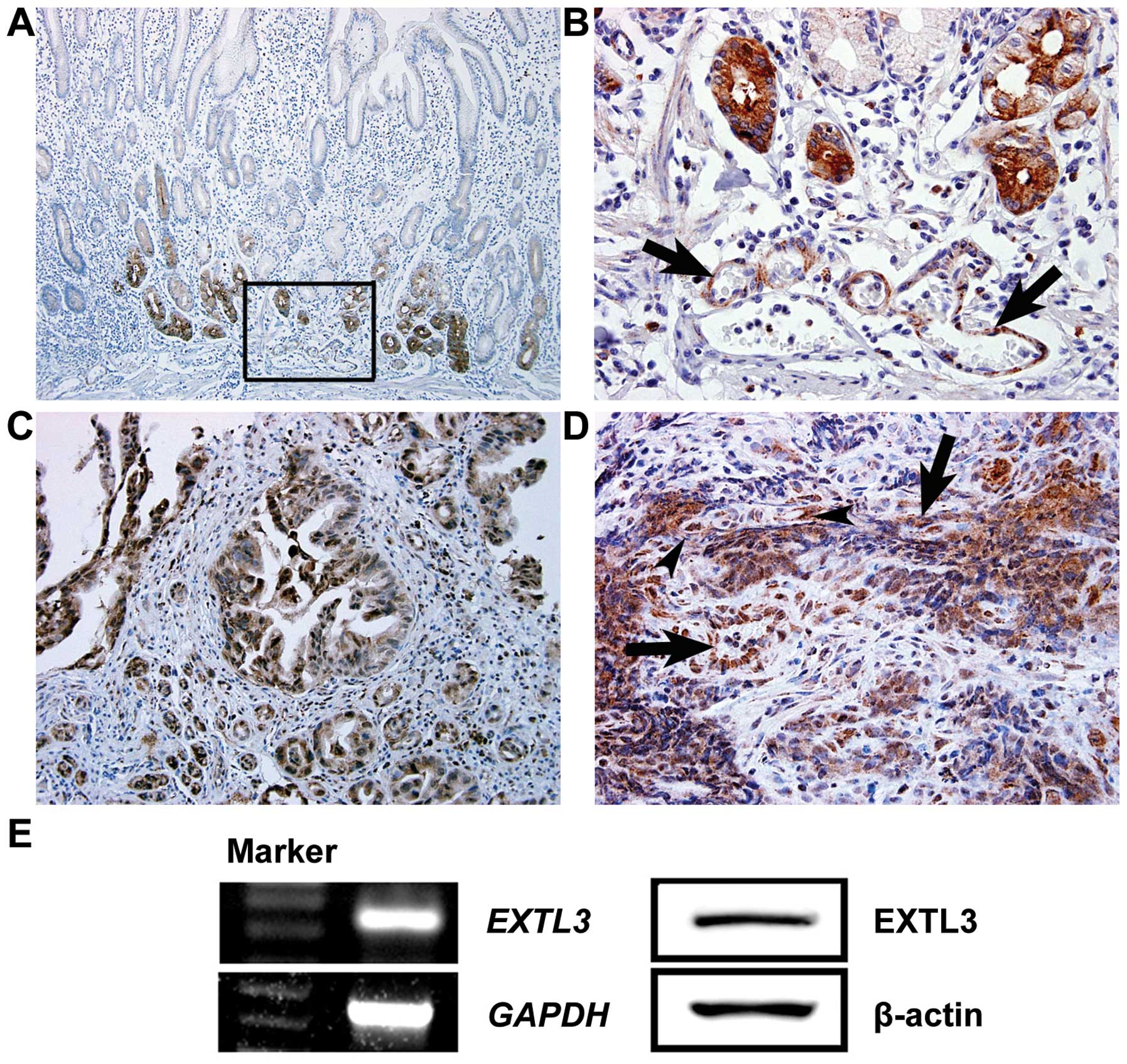
EXTL3 was ubiquitously expressed not only in the
epithelial cells, but also in the endothelial cells in the normal
gastric mucosa (Fig. 1A and B). In
gastric cancer tissues, EXTL3 was expressed in tumor vascular cells
as well as cancer cells (Fig. 1C and
D).
Before examining the effect of REG Iα protein on the
endothelial cells, we tested the expression of EXTL3 in HUVEC.
Subsequently, we confirmed that expression of EXTL3 and its
gene product was detectable in the cells by RT-PCR and western blot
analysis (Fig. 1E), suggesting that
HUVECs have the capability of responding to REG Iα stimulation.
REG Iα protein activates the
phosphorylation of ERK and Akt in HUVEC cells
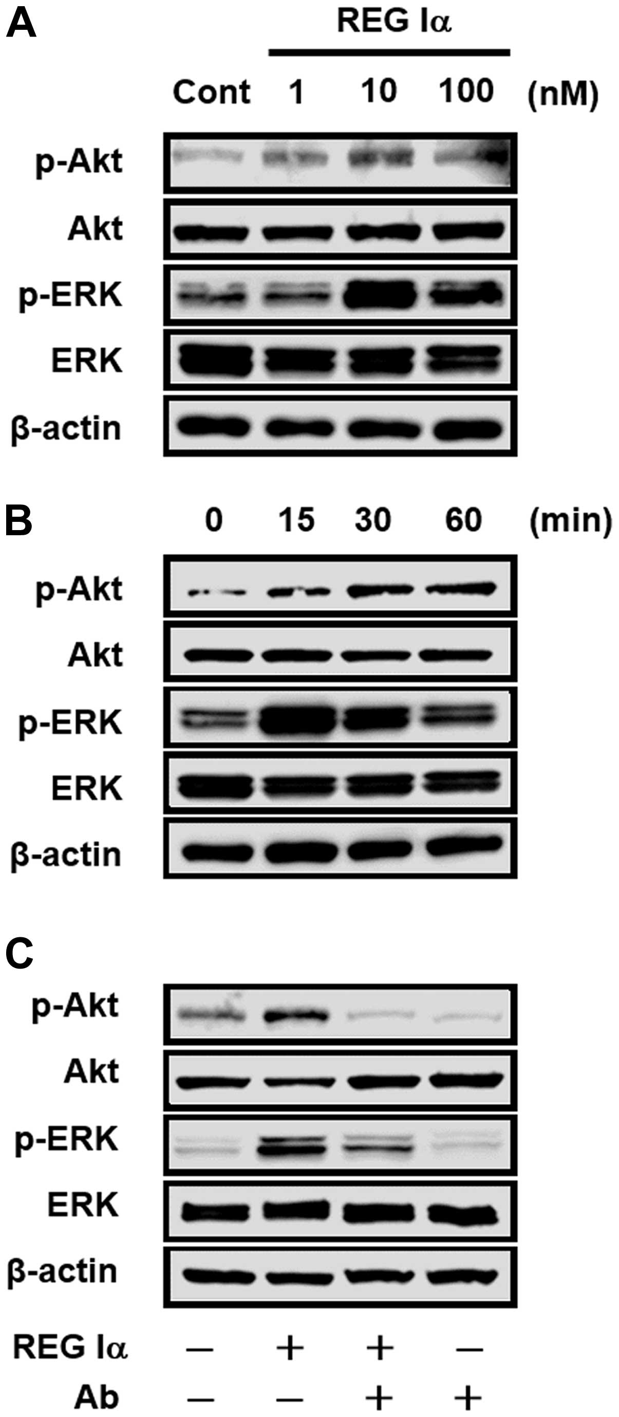
The effect of REG Iα protein on intracellular
signaling was investigated in the HUVEC cells. The expression of
p-ERK and p-Akt was enhanced by REG Iα stimulation (10–100 nM)
(Fig. 2A). The expression of p-ERK
peaked in the HUVEC cells at 15 min after REG Iα stimulation, and
that of p-Akt was enhanced from 15 min and sustained for 60 min
(Fig. 2B). We then examined whether
anti-REG Iα antibody inhibits the REG Iα-induced signaling in HUVEC
cells. As shown in Fig. 2C, the
basal level of p-ERK and p-Akt expression was decreased by
treatment with REG Iα antibody. Moreover, the increased expression
of p-ERK and p-Akt in REG Iα-treated HUVEC cells was attenuated by
concomitant administration of anti-REG Iα antibody.
REG Iα protein promotes HUVEC cell growth
and anti-apoptosis
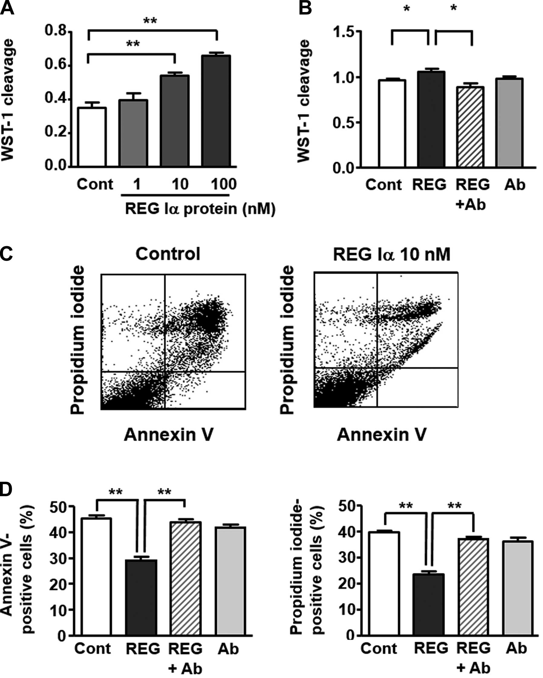
To clarify the role of REG Iα protein in
angiogenesis, we examined the growth and anti-apoptosis effects of
REG Iα protein on HUVEC cells in vitro. The rate of WST-1
cleavage was significantly and dose-dependently increased in REG
Iα-treated HUVEC cells (Fig. 3A).
Conversely, the increase in the level of WST-1 cleavage in REG
Iα-treated cells was significantly reduced to almost the control
level by addition of anti-REG Iα antibody (Fig. 3B).
In control preparations, deprivation of growth
factors in complete culture medium induced cell apoptosis and
death. However, HUVEC cells treated with REG Iα protein (10 nM)
showed significantly lower Annexin V positivity, than the control
cells (Fig. 3C and D). Similarly,
the percentage of PI-positive cells was significantly lower in the
REG Iα-treated preparations than in the controls (Fig. 3C and D). On the other hand, the
decrease of Annexin V or PI positivity in REG Iα-treated HUVEC
cells was restored to the control level by concomitant
administration of anti-REG Iα antibody (Fig. 3D).
Relationship between REG Iα expression
and MVD in gastric cancer tissues
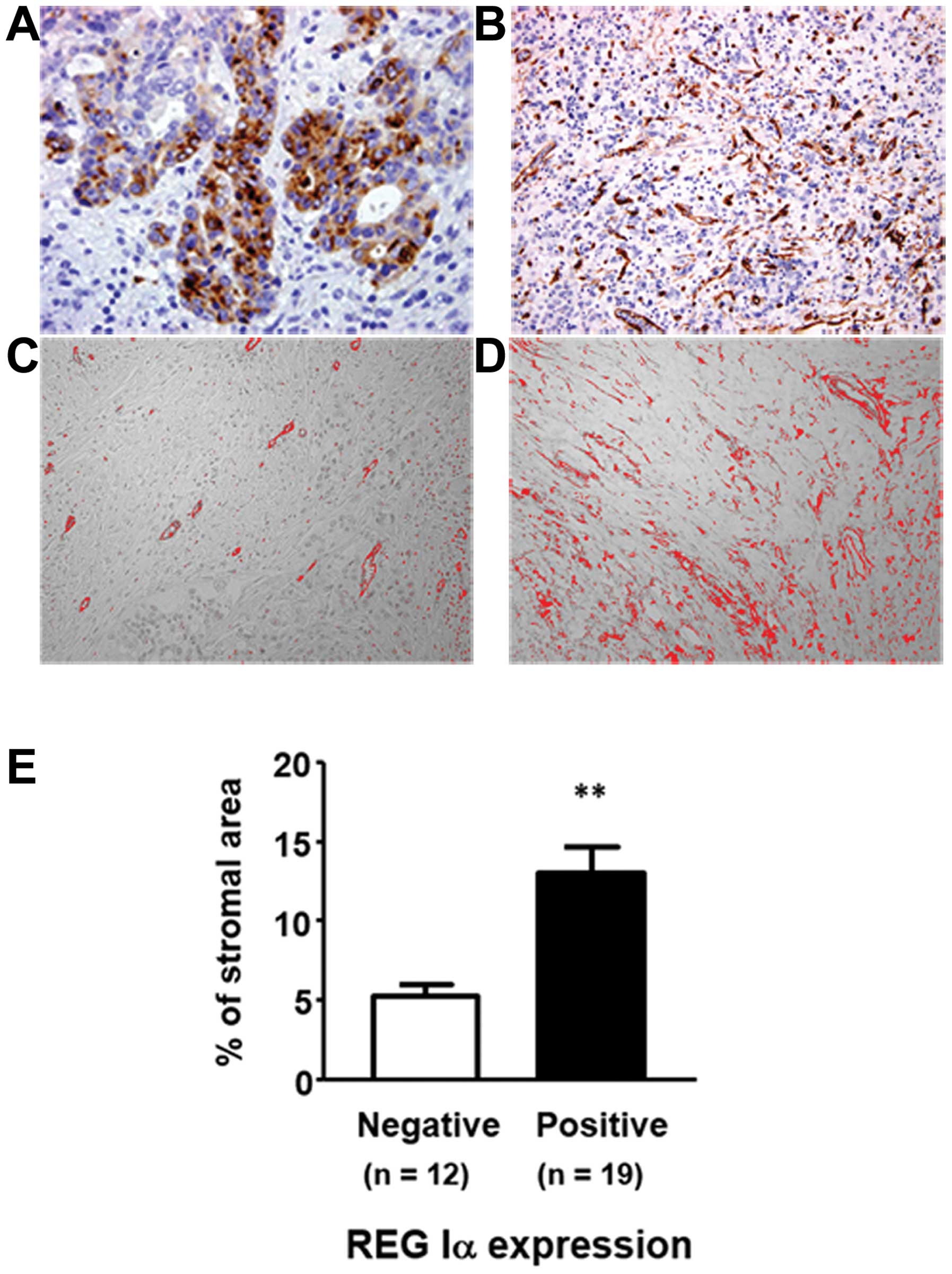
Among 31 samples of gastric cancer tissues, 19
(61.3%) were positive for REG Iα expression. Expression of REG Iα
was significantly associated with the prevalence of lymph node
metastasis and tended to correlate with the tumor stage (Table II). MVD was significantly higher in
gastric cancers at an advanced stage. In addition, MVD tended to be
higher in gastric cancers with lymph node metastasis. Furthermore,
we investigated the relationship between REG Iα expression and MVD
and observed that MVD was significantly higher in REG Iα-positive
gastric cancers (Fig. 4).
| Table IIRelationship between
clinicopathological features and REG Iα expression or MVD in
patients with gastric cancer. |
Table II
Relationship between
clinicopathological features and REG Iα expression or MVD in
patients with gastric cancer.
| Number of REG
Iα-positive/total number of patients | P-value | MVD | P-value |
|---|
| Tumor location | | 0.0575 | | NS |
| Lower | 7/7 (100%) | | 9.44±2.39 | |
| Mid | 6/12 (50.0%) | | 8.87±2.03 | |
| Upper | 6/12 (50.0%) | | 12.08±2.26 | |
| Lauren’s
classification | | 0.2181 | | 0.5414 |
| Intestinal
type | 4/9 (44.4%) | | 8.99±2.46 | |
| Diffuse type | 15/22 (68.2%) | | 10.75±1.52 | |
| Stage | | 0.1088 | | 0.0459 |
| I/II | 3/8 (37.5%) | | 5.94±1.39 | |
| III/IV | 16/23 (69.6%) | | 11.74±1.55 | |
| Lymphatic
invasion | | 0.2009 | | NS |
| None | 0/1 (0.0%) | | 9.78 | |
| Present | 19/30 (63.3%) | | 10.26±1.33 | |
| Venous
invasion | | 0.6194 | | 0.730 |
| None | 2/4 (50.0%) | | 9.06±2.56 | |
| Present | 17/27 (63.0%) | | 10.42±1.43 | |
| Lymph node
metastasis | | 0.0385 | | 0.0614 |
| None | 1/5 (20.0%) | | 4.80±1.32 | |
| Present | 18/26 (69.2%) | | 11.29±1.42 | |
Discussion
It has been reported that REG Iα is overexpressed in
various malignancies including cancers of the stomach (3,6,9),
colorectum (15), bile duct
(16) and pancreas (17). Furthermore, microarray analyses have
revealed that REG Iα expression is markedly enhanced in gastric
cancer tissues (18) and in fact we
have previously shown that REG Iα protein acts on gastric cancer
cells as a growth and/or anti-apoptotic factor (5). Although the receptor for REG Iα
protein, which is identical to EXTL3, has been discovered fairly
recently (19), its
pathophysiological roles are poorly understood. In the present
study using immunohistochemistry, we have demonstrated that EXTL3
is expressed in gastric cancer cells, in accordance with a previous
study indicating that EXTL3 is ubiquitously expressed in
gastric cancer cells in vitro (9), which would account for the observed
effects of REG Iα protein on gastric cancer cells. Interestingly,
our immunohistochemical analysis also revealed that EXTL3 was
expressed in tumor vessel cells and we confirmed the expression of
EXTL3 in HUVEC cells in vitro. Thus, the present study
indicates for the first time that REG Iα protein may act on not
only gastric cancer cells but also tumor vessel cells, which are an
important component associated with tumor progression.
In a series of in vitro studies, we have
investigated the possible role of REG Iα on the human endothelial
cells and have shown that REG Iα protein promotes the proliferation
of the endothelial cells. Furthermore, in the present study, we
clarified that REG Iα protein has an anti-apoptotic effect on the
endothelial cells. Thus, REG Iα protein appears to act on not only
the gastric cancer cells (5), but
also the endothelial cells as a growth and/or anti-apoptotic
factor. In addition, to clarify how REG Iα protein exerts its
effects on endothelial cells, we examined the signaling pathways
activated by REG Iα protein in HUVEC cells. As shown in Fig. 2, REG Iα stimulation enhanced the
phosphorylation of ERK and Akt in HUVEC cells, similarly to
stimulatory effect of REG Iα protein on gastric cancer cells
(5,20). Conversely, treatment with anti-REG
Iα antibody attenuated the enhancement of ERK and Akt
phosphorylation and simultaneously suppressed the growth-promoting
and anti-apoptotic effects of REG Iα on HUVEC cells. These findings
suggest that REG Iα protein acts on endothelial cells as a growth
and/or anti-apoptotic factor via the ERK and Akt signaling
pathways.
Angiogenesis is an important process associated with
tumor progression. In this context, REG Iα protein may promote
tumor progression through its growth-promoting and/or
anti-apoptotic effect on the endothelial cells. To address this
issue, we investigated the expression of REG Iα and microvessel
density in gastric cancer tissues. Clinicopathological analyses
revealed that expression of REG Iα was significantly associated
with the prevalence of lymph node metastasis. Moreover, gastric
cancers that were REG Iα-positive showed a significantly higher MVD
than those that were negative. Although confirmation of these
clinicopathological data may be necessary in a larger study, the
present findings suggest that REG Iα protein is indeed involved in
gastric cancer angiogenesis. In addition, since receptors for REG
Iα protein are ubiquitously expressed not only in gastric cancer,
but also in its endothelial cells, REG Iα protein may contribute at
least in part to tumor progression in REG Iα-positive gastric
cancer.
In summary, we have shown that receptors for REG Iα
are expressed not only in tumor cells, but also tumor vessel cells
in gastric cancer, and that angiogenesis is significantly promoted
in gastric cancers that are REG Iα-positive. Moreover, we have
clarified that REG Iα protein exerts growth-promoting and
anti-apoptotic effects on endothelial cells via ERK and Akt
signaling. These findings suggest that REG Iα protein plays an
important role in angiogenesis during progression of gastric
cancer.
Acknowledgments
We would like to thank Noriko Kamiya (Hyogo College
of Medicine), Chiaki Matsuyama and Ayako Shimizu (Dokkyo University
School of Medicine) for technical assistance. The present study was
supported in part by Grants-in-aid for Scientific Research 26460953
from the Ministry of Education, Culture, Sports, Science and
Technology, Japan.
Abbreviations:
|
REG
|
regenerating gene
|
|
EXTL3
|
exostoses like-3
|
|
MVD
|
microvessel density
|
|
HUVEC
|
human umbilical vein endothelial
cell
|
|
ERK
|
extracellular signal-regulated protein
kinase
|
|
PI
|
propidium iodide
|
|
FITC
|
fluorescein isothiocyanate
|
|
FACS
|
fluorescence activated cell
sorting
|
|
CD
|
cluster of differentiation
|
References
|
1
|
Terazono K, Yamamoto H, Takasawa S, Shiga
K, Yonemura Y, Tochino Y and Okamoto H: A novel gene activated in
regenerating islets. J Biol Chem. 263:2111–2114. 1988.PubMed/NCBI
|
|
2
|
Fukui H, Franceschi F, Penland RL, Sakai
T, Sepulveda AR, Fujimori T, Terano A, Chiba T and Genta RM:
Effects of Helicobacter pylori infection on the link between
regenerating gene expression and serum gastrin levels in Mongolian
gerbils. Lab Invest. 83:1777–1786. 2003. View Article : Google Scholar : PubMed/NCBI
|
|
3
|
Yamagishi H, Fukui H, Sekikawa A, et al:
Expression profile of REG family proteins REG Ialpha and REG IV in
advanced gastric cancer: comparison with mucin phenotype and
prognostic markers. Modern Pathol. 22:906–913. 2009. View Article : Google Scholar
|
|
4
|
Sekikawa A, Fukui H, Fujii S, et al:
Possible role of REG Iα protein in ulcerative colitis and colitic
cancer. Gut. 54:1437–1444. 2005. View Article : Google Scholar : PubMed/NCBI
|
|
5
|
Sekikawa A, Fukui H, Fujii S, et al: REG
Iα protein may function as a trophic and/or anti-apoptotic factor
in the development of gastric cancer. Gastroenterology.
128:642–653. 2005. View Article : Google Scholar : PubMed/NCBI
|
|
6
|
Yonemura Y, Sakurai S, Yamamoto H, et al:
REG gene expression is associated with the infiltrating growth of
gastric carcinoma. Cancer. 98:1394–1400. 2003. View Article : Google Scholar : PubMed/NCBI
|
|
7
|
Dhar DK, Udagawa J, Ishihara S, et al:
Expression of regenerating gene I in gastric adenocarcinomas:
correlation with tumor differentiation status and patient survival.
Cancer. 100:1130–1136. 2004. View Article : Google Scholar : PubMed/NCBI
|
|
8
|
Sekikawa A, Fukui H, Zhang X, et al: REG
Iα is a biomarker for predicting response to chemotherapy with S-1
plus cisplatin in patients with unresectable stage IV gastric
cancer. Br J Cancer. 108:395–401. 2013. View Article : Google Scholar : PubMed/NCBI
|
|
9
|
Fukui H, Fujii S, Takeda J, et al:
Expression of reg I alpha protein in human gastric cancers.
Digestion. 69:177–184. 2004. View Article : Google Scholar : PubMed/NCBI
|
|
10
|
Papetti M and Herman IM: Mechanism of
normal and tumor-derived angiogenesis. Am J Physiol Cell Physiol.
282:C947–C970. 2002. View Article : Google Scholar : PubMed/NCBI
|
|
11
|
Karibe T, Fukui H, Sekikawa A, Shiratori K
and Fujimori T: EXTL3 promoter methylation down-regulates EXTL3 and
heparin sulphate expression in mucinous colorectal cancers. J
Pathol. 216:32–42. 2008. View Article : Google Scholar : PubMed/NCBI
|
|
12
|
Sekikawa A, Fukui H, Fujii S, Ichikawa K,
Tomita S, Imura J, Chiba T and Fujimori T: REG Ialpha protein
mediates an anti-apoptotic effect of STAT3 signaling in gastric
cancer cells. Carcinogenesis. 29:76–83. 2008. View Article : Google Scholar
|
|
13
|
Abe A, Fukui H, Fujii S, et al:
Involvement of cyclooxygenase-2 and vascular endothelial growth
factor in vascularization and lymph node metastasis of colorectal
cancers with submucosal invasion. J Gastroenterol Hepatol.
22:1071–1077. 2007. View Article : Google Scholar : PubMed/NCBI
|
|
14
|
Ozerdem U, Wojcik EM, Duan X, Erşahin Ç
and Barkan GA: Prognostic utility of quantitative image analysis of
microvascular density in prostate cancer. Pathol Int. 63:277–282.
2013. View Article : Google Scholar : PubMed/NCBI
|
|
15
|
Macadam RC, Sareka AI, Farmery SM,
Robinson PA, Markham AF and Guillou PJ: Death form early colorectal
cancer is predicted by the presence of transcripts of the REG gene
family. Br J Cancer. 83:188–195. 2000.PubMed/NCBI
|
|
16
|
Harada K, Zen Y, Kanemori Y, et al: Human
REG I gene is up-regulated in intrahepatic cholangiocarcinoma and
its precursor lesions. Hepatology. 33:1036–1042. 2001. View Article : Google Scholar : PubMed/NCBI
|
|
17
|
Zhou L, Zhang R, Wang L, et al:
Upregulation of REG Ialpha accelerates tumor progression in
pancreatic cancer with diabetes. Int J Cancer. 127:1795–1803. 2010.
View Article : Google Scholar : PubMed/NCBI
|
|
18
|
Takaishi S and Wang TC: Gene expression
profiling in a mouse model of Helicobacter pylori-induced gastric
cancer. Cancer Sci. 98:284–293. 2007. View Article : Google Scholar : PubMed/NCBI
|
|
19
|
Kobayashi S, Akiyama T, Nata K, et al:
Identification of a receptor for reg (regenerating gene) protein, a
pancreatic beta-cell regeneration factor. J Biol Chem.
275:10723–10726. 2000. View Article : Google Scholar
|
|
20
|
Kadowaki Y, Ishihara S, Miyaoka Y, et al:
Reg protein is overexpressed in gastric cancer cells, where it
activates a signal transduction pathway that converges on ERK1/2 to
stimulate growth. FEBS Lett. 530:59–64. 2002. View Article : Google Scholar : PubMed/NCBI
|