Introduction
Cervical cancer of the uterus remains one of the
major causes of cancer-related death in women worldwide. The global
cervical cancer incidence has increased 0.6% annually from 1980 to
2010, and the disease resulted in the death of ~200,000 women in
2010 in developing countries (1).
Despite the comparatively favorable survival for early-stage
patients, numerous patients develop localized recurrence or distant
metastases after initial treatment. As the potential targets for
diagnosis and treatment of the cancer, molecules involved in cancer
development, metastasis and outcome should be identified and
functionally characterized.
Cluster of differentiation (CD) 24 is a small,
heavily glycosylated mucin-like cell surface protein (27 amino
acids in length) that binds to the membrane via a
glycosylphosphatidylinositol anchor (2). Under physiological conditions, CD24
was initially identified as a B cell marker (3). Later, it was found to be expressed not
only in developing or regenerating tissue, but also in
granulocytes, pre-B cells, keratinocytes and renal tubules
(4). CD24 has also been reported to
be a ligand for P-selectin, an adhesion receptor on activated
endothelial cells and platelets (5,6), thus
suggesting that the molecule functionally enhances the metastatic
potential of cancer cells. Under pathological conditions, CD24
plays an important role in the carcinogenesis of various human
malignancies. Its expression has been detected not only in
hematologic malignancies (7), but
also in various solid tumors, including retinoblastoma, glioma,
laryngeal squamous cell and nasopharyngeal carcinoma, small cell
lung and breast cancer, renal cell, hepatocellular and gallbladder
carcinoma, pancreatic adenocarcinoma, colorectal and epithelial
ovarian cancer, and bladder carcinoma (8–14).
These studies demonstrated that CD24 overexpression was markedly
associated with a more aggressive course of the disease. However,
the correlation of CD24 expression with uterine cervical cancer,
the underlying mechanisms and its prognostic significance still
remain unknown.
The present study was performed to evaluate the
expression of CD24 clinicopathologically and to biologically
analyze its functional behavior in uterine cervical cancer.
Materials and methods
Patients and tissue samples
The present study included 117 patients with stage
Ib-IIb cervical cancer who were treated at the Osaka Medical
College between 2002 and 2012. Patients were eligible for inclusion
in the present study when they met the following criteria: i) they
had undergone radical or modified radical hysterectomy and pelvic
lymphadenectomy as an initial treatment; and ii) they had
sufficient clinical data regarding the oncologic outcome including
the date of recurrence. All patients were staged according to the
International Federation of Gynecology and Obstetrics (FIGO)
criteria. The histological subtype was assigned according to the
criteria of the World Health Organization classification. The
patients with FIGO stage IB and lymph node metastasis or
lymphovascular space involvement, or deep half cervical stromal
invasion, and all patients with FIGO stage II received
postoperative adjuvant therapy involving entire pelvic irradiation
and/or chemotherapy. Patients receiving primary
radiotherapy/concurrent chemoradiation therapy without surgery or
receiving any preoperative treatment were excluded from the present
study. The Institutional Review Board approved the present study
and informed consent was obtained from all patients for the use of
their tissue samples.
Immunohistochemistry
Specimens were fixed in 10% formalin and embedded in
paraffin. Serial sections were cut from paraffin-embedded blocks
and were used for routine histopathology. A 4-µm section was
cut from a tissue microarray block and immunohistochemically
analyzed for the expression of CD24. Deparaffinized and rehydrated
sections (4-µm) were autoclaved in 0.01 M citrate buffer (pH
6.0) for 15 min at 121°C for antigen retrieval. The endogenous
peroxidase activity was blocked with 0.3% solution hydrogen
peroxide in methanol for 30 min. Tumor sections were incubated at
4°C for 12 h with the CD24-specific antibodies (clone SN3b; 1:50
dilution; Thermo Fisher Scientific, Waltham, MA, USA). The sections
were washed with 1X phosphate-buffered saline (PBS) and incubated
with Histofine Simple Stain MAX-PO (Multi; Nichirei) for 30 min at
room temperature. Finally, the sections were washed with 1X PBS and
then were visualized by incubation with
H2O2/diaminobenzidine substrate solution for
5 min. The sections were counterstained with hematoxylin prior to
dehydration and mounting. The evaluation of the immunohistochemical
data was performed by two independent pathologists who were blinded
to the clinicopathological data. The expression of CD24 was
assessed using a semi-quantitative system that was defined as
described by Blechschmidt et al (15). Briefly, CD24 expression was scored
as: 0 (no stain), 1+ (weak immunoreactivity in >10% of tumor
cells), 2+ (moderate immunoreactivity of >10% of tumor cells),
and 3+ (strong immunoreactivity of >10% of tumor cells). These
data were summarized into two groups; low CD24 expression (0, 1+,
and 2+) and high CD24 expression (3+). Scoring was performed three
times per slide for three distinct fields, and the three scores
were then averaged.
Cell culture
The human cervical cancer CaSki cell line was
obtained from the American Type Culture Collection (ATCC;
Rockville, MD, USA) and grown in phenol red-free Dulbecco's
modified Eagle's medium (DMEM) (Gibco) containing 10%
dextran-coated, charcoal-treated fetal calf serum, 100 U/ml
penicillin and 100 µg/ml streptomycin in a humidified
atmosphere of 5% CO2 with 95% air at 37°C.
Expression plasmids and cDNA
transfection
To create the pcDNA3.1-CD24 expression construct,
the cDNA of the full length CD24 was amplified by PCR using a human
mammary gland cDNA library as the template. For the transfection of
each sample, oligomer-Lipofectamine Plus complexes were prepared as
follows: 100 pmol of cDNA oligomer were diluted in 250 µl of
Opti-MEM (Invitrogen). The Lipofectamine Plus was mixed gently
before use, and then a 5 µl aliquot was diluted in 250
µl of Opti-MEM, were gently mixed and were incubated for 5
min at room temperature. After the 5 min incubation, the diluted
oligomer was combined with the diluted Lipofectamine Plus, mixed
gently and incubated for another 20 min at room temperature. The
oligomer-Lipofectamine Plus complexes were added to each well
containing cells and medium and mixed gently by rocking the plate
back and forth. The cells were incubated at 37°C in a
CO2 incubator for 24 h, and then the cells were prepared
for each assay.
RNA extraction and semi-quantitative
reverse transcription-polymerase chain reaction (RT-PCR)
We isolated total RNA from 1×10⁶
transfected CaSki cells using a commercially available kit
(Qiagen). We exposed the RNA samples to DNase digestion before the
cDNA synthesis. For gene-specific PCR, 1 µl of first-strand
cDNA product was amplified with Platinum Taq Polymerase
(Invitrogen) according to the manufacturer's instructions. We
designed primers specific for CD24 (forward,
5′-ACCCACGCAGATTTATTCCA-3′ and reverse,
5′-ACCACGAAGAGACTGGCTGT-3′); for Snail (forward,
5′-GCCTTCAACTGCAAATACTGC-3′ and reverse,
5′-CTTCTTGACATCTGAGTGGGTC-3′); for Slug (forward,
5′-AGCTACCCAATGGCCTCTCT-3′ and reverse,
5′-CCAGCCCAGAAAAAGTTGAA-3′); and for β-actin (forward,
5′-TGAGCGCGGC TACAGCTT-3′ and reverse, 5′-TCCTTAATGTCACGCACGA
TTT-3′) and performed a 27-cycle, three-step PCR (denaturation at
94°C for 30 sec, annealing at 54°C for 30 sec, extension at 72°C
for 30 sec) with an initial temperature of 94°C for 3 min.
Western blot analysis
After transfection, cells were washed twice with
ice-cold PBS, lysed and separated to cytoplasmic and nuclear
fractions using the Nuclear Extract kit according to the
manufacturer's instructions (Active Motif, Carlsbad, CA, USA). To
detect Akt, phosphorylated Akt, ERK, phosphorylated ERK, NF-κB,
phosphorylated NF-κB and MMP-9 proteins we separated these by SDS
polyacrylamide gel electrophoresis and electrotransferred to
nitrocellulose membranes and transferred proteins to a membrane.
Western blot analyses were performed with various specific primary
antibodies including Akt (9272), phospho-Akt (Ser473; 9271), NF-κB
(3034), phospho-NF-κB (Ser536; 3033), p44/42 MAP kinase (Erk;
4695), phospho-p44/42 MAP kinase (p-Erk; 9101), MMP-9 (2270),
β-actin (4970), E-cadherin (3195) (all from Cell Signaling,
Danvers, MA, USA) and N-Cadherin (ab18203; Abcam, Cambridge, MA,
USA). The immunoreactive bands in the immunoblots were visualized
with horseradish peroxidase-coupled goat anti-rabbit immunoglobulin
using an enhanced chemiluminescence western blotting system (ECL
Plus; GE Healthcare Life Sciences, Pittsburgh, PA, USA).
Non-specific antigen sites were blocked with 10% bovine serum
albumin in 1X Tris-buffered saline.
Migration and invasion assays
We examined the effects of CD24 on the invasive
potential of CaSki cells by an invasion assay. CaSki cells
(5×10⁵ cells) were cultured at 37°C in a humidified, 5%
CO2 atmosphere incubator in serum-free DMEM for 24 h,
seeded into the upper wells and coated with a thin layer of
Matrigel. The lower chamber contained 600 µl of DMEM.
Following a 24-h incubation at 37°C, non-invading cells on the
surface of the Matrigel-coated membrane were removed by scraping
with a cotton swab. The cells that migrated through the Matrigel
were stained with hematoxylin. Following several washes with PBS,
the stained cells were manually counted for three independent
experiments. Each point represents the mean ± SD of four
replicates.
Statistical analysis
Statistical analyses in the present study were
performed with the JMP statistical software package (version.
9.0.2). Fisher's exact probability test was used for evaluating
correlations between the immunohistochemical and clinical data. The
end points investigated were the progression-free (PFS) and overall
survival (OS) rates, respectively). The PFS was defined as the time
from the first day of treatment until either death from any cause
or disease progression. OS was defined as the time from the first
day of treatment to death from any cause. Univariate analyses of
the PFS and OS were determined with the Kaplan-Meier method using a
log-rank test. The Mann-Whitney U test was used for the comparison
of continuous variables. The continuous variables ae expressed as
the means ± SD by an analysis of the variance (ANOVA) when the
variance of the samples showed a normal standard distribution. They
were expressed as the median with interquartile ranges when the
variance did not show a normal standard distribution. Statistical
significance was considered to be present at p-values of
<0.05.
Results
Clinicopathological characteristics
A total of 117 patients with uterine cervical cancer
were enrolled in the present study. Table I shows the characteristics of the
patients. The mean (± SD) age of the patients was 51.0±11.5 years.
Eighty-nine (76.1%) patients were of FIGO stage Ib and 28 (23.9%)
were stage II. Histologically, 76 (68.1%) patients had squamous
cell carcinoma and 41 (31.9%) patients had adenocarcinoma.
Twenty-two (18.8%) patients had lymph node metastasis and 38
(32.5%) had lymphovascular involvement. Cervical cancer recurrence
developed in 19 (16.2%) of the 117 patients. The median follow-up
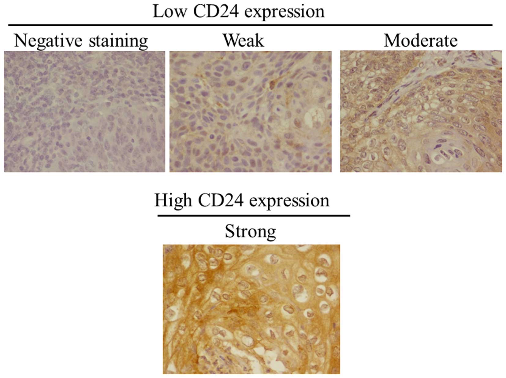
was 41.5±27.3 months. CD24 was mainly localized in the cytoplasm
and on the membranes of tumor cells (Fig. 1). A high CD24 expression was
observed in the tumors from 26 (22.2%) patients. The staining
pattern [low (negative, weak or moderate) or high (strong)] is
shown in Fig. 1.
 | Table IClinicopathological characteristics of
the patients with cervical cancer. |
Table I
Clinicopathological characteristics of
the patients with cervical cancer.
| Expression of CD24
protein Factor | Value | Reduced (%) | Preserved (%) | P-value |
|---|
| No. of patients | 117 | 91 (77.8) | 26 (22.2) | |
| Mean age (years) | 51.0±11.5 | | | |
| Stage
classification |
| Ib | 89 (76.1) | 76 (83.5) | 13 (50.0) | |
| II | 28 (23.9) | 15 (16.5) | 13 (50.0) | <0.01 |
| Histological
type |
| Squamous cell
carcinoma | 76 (68.1) | 61 (67.0) | 15 (57.7) | |
|
Adenocarcinoma | 41 (31.9) | 30 (33.0) | 11 (42.3) | 0.5 |
| Lymph node
metastasis |
| Negative | 95 (81.2) | 78 (85.7) | 17 (65.4) | |
| Positive | 22 (18.8) | 13 (14.3) | 9 (34.6) | 0.03 |
| Lymphovascular
involvement |
| Negative | 79 (67.5) | 70 (79.6) | 9 (34.6) | |
| Positive | 38 (32.5) | 21 (20.4) | 17 (65.4) | <0.01 |
| Recurrence |
| Negative | 98 (83.8) | 81 (89.0) | 17 (65.5) | |
| Positive | 19 (16.2) | 10 (11.0) | 9 (34.6) | <0.01 |
| Mean follow-up
(months) | 41.5±27.3 | | | |
Association of CD24 expression with
clinicopathological parameters of cervical cancer
Immunohistochemically, 28 (23.9%) patients had
tumors with high CD24 expression. Among 91 (77.8%) patients with
low CD24 expression tumors, 35 (29.9%) had negative, 30 (25.6%) had
weak and 26 (22.2%) had moderate staining. The patients with tumors
that expressed high CD24 levels had a higher rate of advanced
clinical stage (50 vs. 16.5%, p<0.01), lymph node metastasis
(34.6 vs. 14.3%), lymphovascular involvement (65.4 vs. 20.4%,
p=0.01) and recurrence rate of cervical cancer (34.6 vs. 11.0%,
p<0.05) (Table I).
Correlation between CD24 expression and
prognosis
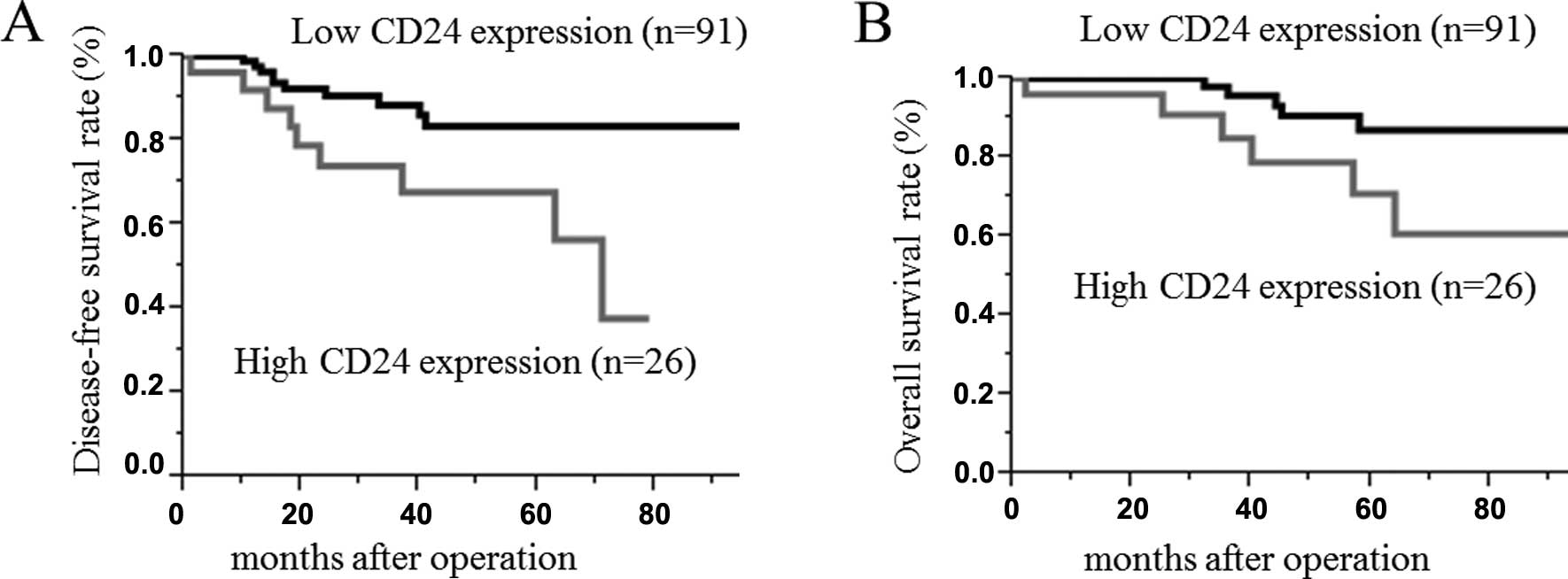
The survival rate and disease-free survival rate of
the cervical cancer following surgical treatment were analyzed
using the Kaplan-Meier method. The median follow-up time for all
patients was 41.5±27.3 months. The survival rate was determined
from the day of the initial surgery to the time of the death or the
last follow-up. The disease-free survival rate was determined from
the day of the initial surgery to the time of the detection of
cancer recurrence, death or the last follow-up. The 5-year
disease-free survival rates of cervical cancer in patients with
high and low CD24 expression tumors were 62 and 86%, respectively.
The 5-year OS rates of cervical cancer in patients with high and
low CD24 expression tumors were 62 and 86%, respectively. The
log-rank test analyses revealed a statistically significant
difference between the two groups (p<0.01) (Fig. 2).
CD24 specifically enhances the cell
invasion through Matrigel in human squamous cervical cancer
cells
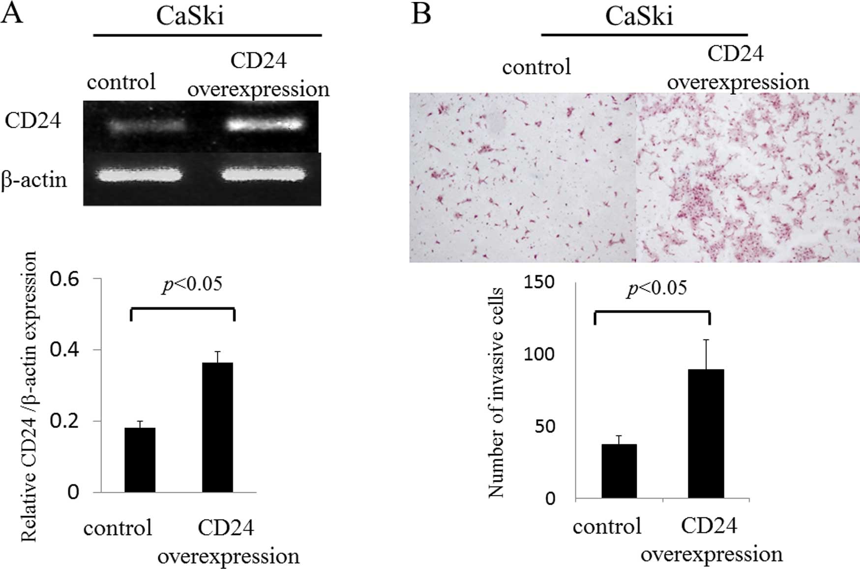
In recent studies, CD24 was shown to play an
important role in the development and progression of malignant
tumors (16). We examined whether
CD24 regulates invasive activity in uterine cervical cancer. To
assess this possibility, we transiently transfected the CD24 gene
into a cervical cancer cell line (CaSki cells). The mRNA expression
of CD24 in CaSki cells was evaluated by semi-quantitative RT-PCR
(Fig. 3). The mRNA expression level
of CD24 in CaSki cells that were transfected with the CD24
gene was found to be upregulated by 2.0-fold when compared to the
control group (0.36±0.03 vs. 0.18±0.02; p<0.05) (Fig. 3A). The sensitivity of the invasive
activity of CD24 in the CaSki cells was examined using an invasion
assay which monitors cell migration Matrigel. The overexpression of
CD24 significantly increased the migration and invasion of CaSki
cells by 2.0-fold through the Matrigel (89.2±20.8 vs. 37.1±6.5,
p<0.05) (Fig. 3B).
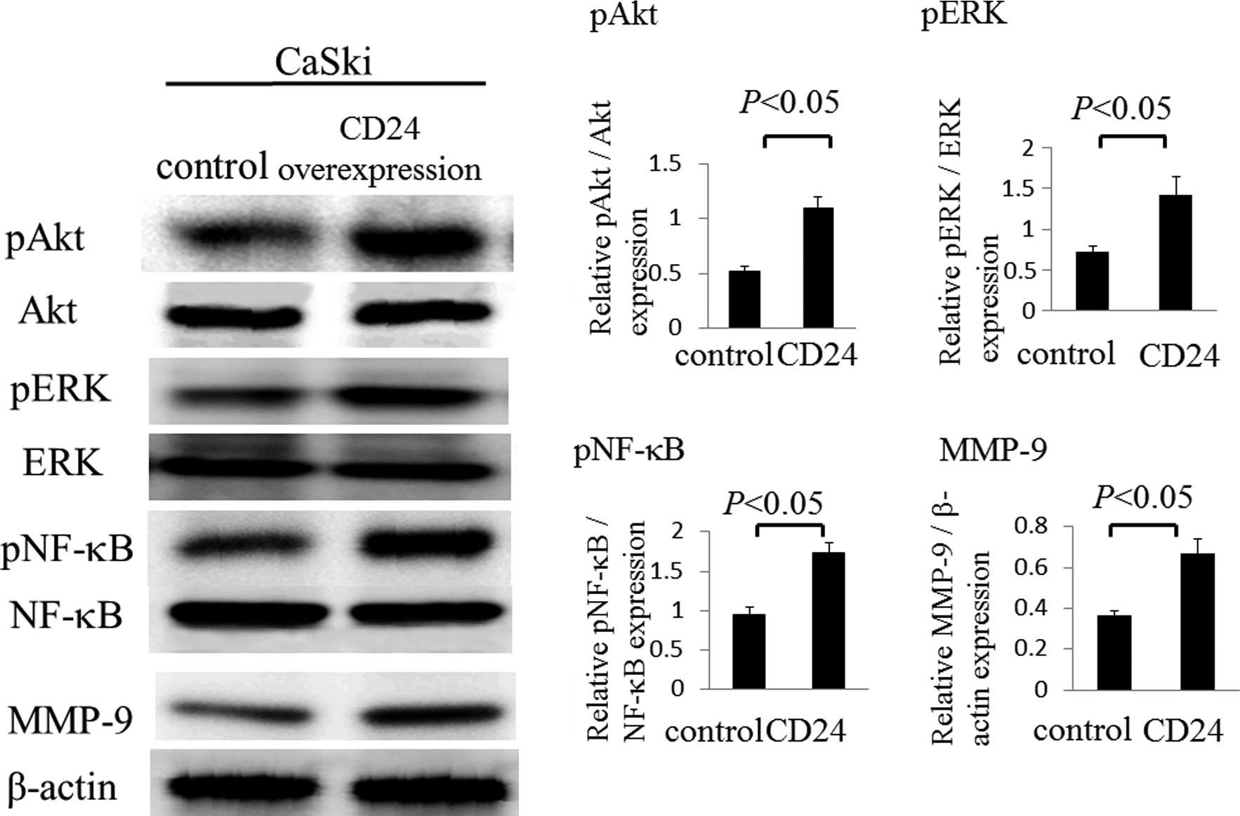
CD24 signaling enhances Akt and
MAPK-ERK1/2 signaling in human squamous cervical cancer cells
We established that in CaSki cells, phosphorylation
of Akt and MAPK-ERK1/2 were induced by CD24 through western blot
analysis. The phosphorylation of Akt was found to be upregulated by
2.1-fold in the overexpression of CD24 group when compared to the
control group (1.09±0.10 vs. 0.52±0.05; p<0.05) (Fig. 4). The phosphorylation of ERK was
also found to be upregulated by 2.0-fold in the overexpression of
CD24 group compared to be the control group (1.42±0.23 vs.
0.72±0.08; p<0.05). Next, we examined the phosphorylation of
NF-κB and the MMP-9 activity following the overexpression of CD24,
as the overexpression of CD24 significantly increased the migration
and invasion of CaSki cells through the Matrigel. The
phosphorylation of NF-κB was found to be upregulated by 1.8-fold in
the overexpression of CD24 in CaSki cells compared to the control
group (1.74±0.13 vs. 0.95±0.09; p<0.05) (Fig. 4). In addition, the activation of
MMP-9 was found to be upregulated by 1.8-fold in the overexpression
of CD24 group compared to the control group (0.67±0.07 vs.
0.37±0.02; p<0.05) (Fig. 4).
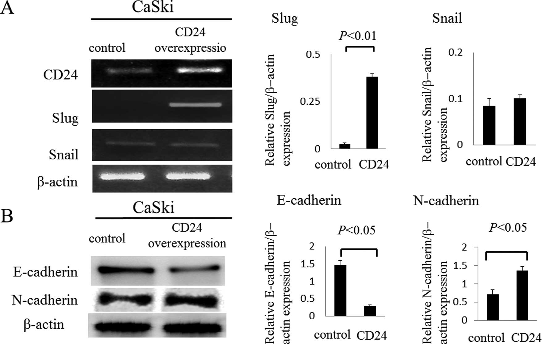
CD24 associates with epithelial to
mesenchymal transition (EMT) signaling via Slug in human squamous
cervical cancer cells
In recent studies, CD24-positive cells were shown to
be susceptible to the induction of EMT, a phenotype important for
cancer metastasis in ovarian, breast, and pancreas carcinomas
(4,17–20).
We examined whether CD24 regulates the expression of Slug or Snail
in uterine cervical cancer. To assess this possibility, we
transiently transfected the CD24 gene into a cervical cancer cell
line (CaSki cells). The mRNA expression of Slug in CaSki
cells that were transfected with the CD24 gene was found to be
upregulated by 16.5-fold according to semi-quantitative RT-PCR
compared to the control group (0.38±0.01 vs. 0.02±0.007; p<0.05)
(Fig. 5A), although the effects on
the expression of Snail were not significant. Moreover, the
expression of E-cadherin in the CD24 overexpression cells was found
to be downregulated by 0.2-fold by a western blot analysis compared
to the control group (0.28±0.05 vs. 1.47±0.14; p<0.05), and the
expression of N-cadherin in the CD24 overexpression cells was found
to be upregulated by 2.0-fold by a western blot analysis compared
to the control group (1.37±0.1 vs. 0.71±0.13; p<0.05) (Fig. 5B).
Discussion
We concluded that the present results indicate that
enhanced CD24 expression in cervical cancer was significantly
correlated with aggressive clinicopathological features.
Furthermore, CD24 overexpression appears to results in an enhanced
invasive potential of uterine cervical cancer cells through the
activation of both Akt and ERK1/2 signaling cascades. Additionally,
we showed that CD24-induced invasive potential correlated with the
EMT phenomenon in uterine cervical cancer.
CD24 has been considered to be involved in the
process of tumor metastasis and invasion as the ligand of
P-selectin and the adhesion receptor in activated endothelial cells
and platelets (21). Many cancer
cells can bind to platelets via CD24-mediated binding to
P-selectin, which facilitates the exit of cancer cells from the
bloodstream and enhance the metastatic potential of cancer cells
(22). CD24 has been identified as
a marker of cancer stem cells in various human malignancies, such
as pancreatic, breast, colorectal, gastrointestinal tract and liver
cancers (23–26). Several immunohistochemical studies
previously demonstrated that CD24 overexpression is associated with
clinicopathological features, such as the pathological grade, lymph
node metastasis and subsequent poor prognosis of various types of
cancers (11,13,14,27,28).
Furthermore, various studies showed that CD24 overexpression
contributes to cell proliferation and invasiveness in vitro
(11). In line with these previous
studies, our data demonstrated that enhanced CD24 was associated
with advanced clinical stage, lymph node metastasis,
lympho-vascular involvement and poor prognosis in patients with
uterine cervical cancer. Additionally, CD24 overexpression in a
cervical cancer cell line (CaSki) led to increased invasiveness.
Our data further demonstrated that CD24 contributes to the
invasiveness through the phosphorylation of Akt, NF-κB and ERK and
the activation of MMP-9.
Conversely, during the progression of epithelial
cancer, cells typically lose the characteristic features of
epithelial cells and gain a mesenchymal phenotype. EMT has been
implicated in the metastasis of primary tumors and provides a
molecular mechanism (29). One of
the most well-defined features of EMT is the loss of E-cadherin
expression (30). A group of
transcription factors, including Snail and Slug, have been
implicated in the control of EMT (31). Cadherin switch, which indicates a
decrease of E-cadherin and increase of N-cadherin, is also
essential for increased motility, yet is not always required for
the morphological changes that accompany EMT (32). Previous studies have reported that
CD24 promotes cell invasion and induce significant amounts of
EMT-associated markers in ovarian and breast cancers (4,21) In
the present study, CD24 overexpression in CaSki cells led to the
activation of Slug, a decrease of E-cadherin protein levels and an
increase of N-cadherin protein levels; thus, our data suggests that
CD24 contributes to EMT through the activation of Slug in uterine
cervical cancer, although we could not clarify whether Slug, via
CD24, directly regulates the activity of Akt, NF-κB or ERK.
In conclusion, CD24 expression in cervical cancer
was significantly correlated with aggressive clinicopathological
features. Furthermore, CD24 overexpression led to enhanced invasive
potential of uterine cervical cancer cells through the activation
of both Akt and ERK1/2 signaling cascades. CD24 overexpression
cancer cells additionally had enhanced invasive potential through
the activation of both Akt and ERK1/2 signaling cascades and
EMT-inducing pathways. CD24 expression may therefore not only be a
potential prognostic marker in uterine cervical cancer, but also a
target for the development of new therapeutic approaches.
Acknowledgments
The present study was supported by a JSPS KAKENHI,
grant no. 25462621 (to Y.T.).
Abbreviations:
|
CD24
|
cluster of differentiation 24
|
|
FIGO
|
International Federation of Gynecology
and Obstetrics
|
|
RT-PCR
|
reverse transcription-polymerase chain
reaction
|
|
EMT
|
epithelial to mesenchymal
transition
|
|
MAPK
|
mitogen-activated protein kinase
|
|
NF-κB
|
nuclear factor-κB
|
|
ERK
|
extracellular signal-regulated
kinase
|
|
MMP
|
matrix metalloproteinase
|
References
|
1
|
Forouzanfar MH, Foreman KJ, Delossantos
AM, Lozano R, Lopez AD, Murray CJ and Naghavi M: Breast and
cervical cancer in 187 countries between 1980 and 2010: A
systematic analysis. Lancet. 378:1461–1484. 2011. View Article : Google Scholar : PubMed/NCBI
|
|
2
|
Kay R, Rosten PM and Humphries RK: CD24, a
signal transducer modulating B cell activation responses, is a very
short peptide with a glycosyl phosphatidylinositol membrane anchor.
J Immunol. 147:1412–1416. 1991.PubMed/NCBI
|
|
3
|
Aigner S, Ruppert M, Hubbe M, Sammar M,
Sthoeger Z, Butcher EC, Vestweber D and Altevogt P: Heat stable
antigen (mouse CD24) supports myeloid cell binding to endothelial
and platelet P-selectin. Int Immunol. 7:1557–1565. 1995. View Article : Google Scholar : PubMed/NCBI
|
|
4
|
Lee KM, Ju JH, Jang K, Yang W, Yi JY, Noh
DY and Shin I: CD24 regulates cell proliferation and transforming
growth factor β-induced epithelial to mesenchymal transition
through modulation of integrin β1 stability. Cell Signal.
24:2132–2142. 2012. View Article : Google Scholar : PubMed/NCBI
|
|
5
|
Aigner S, Sthoeger ZM, Fogel M, Weber E,
Zarn J, Ruppert M, Zeller Y, Vestweber D, Stahel R, Sammar M, et
al: CD24, a mucin-type glycoprotein, is a ligand for P-selectin on
human tumor cells. Blood. 89:3385–3395. 1997.PubMed/NCBI
|
|
6
|
Sammar M, Aigner S and Altevogt P:
Heat-stable antigen (mouse CD24) in the brain: Dual but distinct
interaction with P-selectin and L1. Biochim Biophys Acta.
1337:287–294. 1997. View Article : Google Scholar : PubMed/NCBI
|
|
7
|
King JB, von Furstenberg RJ, Smith BJ,
McNaughton KK, Galanko JA and Henning SJ: CD24 can be used to
isolate Lgr5+ putative colonic epithelial stem cells in
mice. Am J Physiol Gastrointest Liver Physiol. 303:G443–G452. 2012.
View Article : Google Scholar : PubMed/NCBI
|
|
8
|
Li J, Li C, Yuan H and Gong F: Clinical
value of CD24 expression in retinoblastoma. J Biomed Biotechnol.
2012:1580842012. View Article : Google Scholar : PubMed/NCBI
|
|
9
|
Deng J, Gao G, Wang L, Wang T, Yu J and
Zhao Z: CD24 expression as a marker for predicting clinical outcome
in human gliomas. J Biomed Biotechnol. 2012:5171722012. View Article : Google Scholar : PubMed/NCBI
|
|
10
|
Hira SK and Manna PP: Down regulation of
CD24 and HER-2/neu in breast carcinoma cells by activated human
dendritic cell. Role of STAT3. Cell Immunol. 275:69–79. 2012.
View Article : Google Scholar : PubMed/NCBI
|
|
11
|
Shi Y, Gong HL, Zhou L, Tian J and Wang Y:
CD24: A novel cancer biomarker in laryngeal squamous cell
carcinoma. ORL. J Otorhinolaryngol Relat Spec. 74:78–85. 2012.
View Article : Google Scholar
|
|
12
|
Su N, Peng L, Xia B, Zhao Y, Xu A, Wang J,
Wang X and Jiang B: Lyn is involved in CD24-induced ERK1/2
activation in colorectal cancer. Mol Cancer. 11:432012. View Article : Google Scholar : PubMed/NCBI
|
|
13
|
Zhu J, Zhang G and Lu H: CD24, COX-2, and
p53 in epithelial ovarian cancer and its clinical significance.
Front Biosci. 4:2745–2751. 2012. View
Article : Google Scholar
|
|
14
|
Liu C, Zheng S, Shen H, Xu K, Chen J, Li
H, Xu Y, Xu A, Chen B, Kaku H, et al: Clinical significance of CD24
as a predictor of bladder cancer recurrence. Oncol Lett. 6:96–100.
2013.PubMed/NCBI
|
|
15
|
Blechschmidt K, Sassen S, Schmalfeldt B,
Schuster T, Höfler H and Becker KF: The E-cadherin repressor Snail
is associated with lower overall survival of ovarian cancer
patients. Br J Cancer. 98:489–495. 2008. View Article : Google Scholar
|
|
16
|
Wang W, Wang X, Peng L, Deng Q, Liang Y,
Qing H and Jiang B: CD24-dependent MAPK pathway activation is
required for colorectal cancer cell proliferation. Cancer Sci.
101:112–119. 2010. View Article : Google Scholar
|
|
17
|
Kang KS, Choi YP, Gao MQ, Kang S, Kim BG,
Lee JH, Kwon MJ, Shin YK and Cho NH: CD24+ ovary cancer
cells exhibit an invasive mesenchymal phenotype. Biochem Biophys
Res Commun. 432:333–338. 2013. View Article : Google Scholar : PubMed/NCBI
|
|
18
|
Lee SH, Kim H, Hwang JH, Shin E, Lee HS,
Hwang DW, Cho JY, Yoon YS, Han HS and Cha BH: CD24 and S100A4
expression in resectable pancreatic cancers with earlier disease
recurrence and poor survival. Pancreas. 43:380–388. 2014.
View Article : Google Scholar : PubMed/NCBI
|
|
19
|
Zhang Y, Wei J, Wang H, Xue X, An Y, Tang
D, Yuan Z, Wang F, Wu J, Zhang J, et al: Epithelial mesenchymal
transition correlates with CD24+CD44+ and
CD133+ cells in pancreatic cancer. Oncol Rep.
27:1599–1605. 2012.PubMed/NCBI
|
|
20
|
Lim J, Lee KM, Shim J and Shin I: CD24
regulates stemness and the epithelial to mesenchymal transition
through modulation of Notch1 mRNA stability by p38MAPK. Arch
Biochem Biophys. 558:120–126. 2014. View Article : Google Scholar : PubMed/NCBI
|
|
21
|
Jaggupilli A and Elkord E: Significance of
CD44 and CD24 as cancer stem cell markers: An enduring ambiguity.
Clin Dev Immunol. 2012:7080362012. View Article : Google Scholar : PubMed/NCBI
|
|
22
|
Newman H, Shapira S, Spierer O, Kraus S,
Rosner M, Pri-Chen S, Loewenstein A, Arber N and Barak A:
Involvement of CD24 in angiogenesis in a mouse model of
oxygen-induced retinopathy. Curr Eye Res. 37:532–539. 2012.
View Article : Google Scholar : PubMed/NCBI
|
|
23
|
Rasheed ZA and Matsui W: Biological and
clinical relevance of stem cells in pancreatic adenocarcinoma. J
Gastroenterol Hepatol. 27(Suppl 2): S15–S18. 2012. View Article : Google Scholar
|
|
24
|
Keysar SB and Jimeno A: More than markers:
Biological significance of cancer stem cell-defining molecules. Mol
Cancer Ther. 9:2450–2457. 2010. View Article : Google Scholar : PubMed/NCBI
|
|
25
|
Kemper K, Grandela C and Medema JP:
Molecular identification and targeting of colorectal cancer stem
cells. Oncotarget. 1:387–395. 2010. View Article : Google Scholar
|
|
26
|
Zou GM: Cancer initiating cells or cancer
stem cells in the gastrointestinal tract and liver. J Cell Physiol.
217:598–604. 2008. View Article : Google Scholar : PubMed/NCBI
|
|
27
|
Tang J, Cai H, Lin L, Xie P, Zhong W and
Tang M: Increased expression of CD24 is associated with tumor
progression and prognosis in patients suffering osteosarcoma. Clin
Transl Oncol. 15:541–547. 2013. View Article : Google Scholar
|
|
28
|
Wu JX, Zhao YY, Wu X and An HX:
Clinicopathological and prognostic significance of CD24
overexpression in patients with gastric cancer: A meta-analysis.
PLoS One. 9:e1147462014. View Article : Google Scholar : PubMed/NCBI
|
|
29
|
Lee JM, Dedhar S, Kalluri R and Thompson
EW: The epithelial-mesenchymal transition: New insights in
signaling, development, and disease. J Cell Biol. 172:973–981.
2006. View Article : Google Scholar : PubMed/NCBI
|
|
30
|
Thiery JP, Acloque H, Huang RY and Nieto
MA: Epithelial-mesenchymal transitions in development and disease.
Cell. 139:871–890. 2009. View Article : Google Scholar : PubMed/NCBI
|
|
31
|
Peinado H, Olmeda D and Cano A: Snail, Zeb
and bHLH factors in tumour progression: An alliance against the
epithelial phenotype? Nat Rev Cancer. 7:415–428. 2007. View Article : Google Scholar : PubMed/NCBI
|
|
32
|
Maeda M, Johnson KR and Wheelock MJ:
Cadherin switching: essential for behavioral but not morphological
changes during an epithelium-to-mesenchyme transition. J Cell Sci.
118:873–887. 2005. View Article : Google Scholar : PubMed/NCBI
|