Introduction
Ovarian cancer is a common malignant tumor in
females and the leading cause of mortality among gynecologic
cancers (1). The main therapeutic
strategy of ovarian cancer is a combination of surgery and
chemotherapy, which however contributes to chemoresistance.
Chemoresistance is considered one of the main obstacles in the
clinical treatment of ovarian cancer patients (2), thus, it is meaningful to figure out
the relevant molecules and mechanisms that are involved in the
development of chemoresistance.
Caveolines, a family of ubiquitously expressed
oligomeric structural proteins in many mammalian cells, are
important proteins of caveolae, which are related to endocytosis,
transcytosis and the integration of various signaling molecules and
signaling pathways (3). Three
caveolin family members, caveolin (Cav)-1, -2 and -3, have been
identified and are widely expressed in various tissues in which
Cav-1 is essential for caveolae formation and aggregation of signal
molecules. Cav-1 has phosphorylated sites at tyrosine residue 14
and serine 80 and the phosphorylation is related to epidermal
growth factor, platelet-derived growth factor, insulin and Akt
pathways (4). Various signaling
molecules, such as G protein-coupled receptors, protein kinase C
and extracellular signal regulated protein kinase interact with
Cav-1 through its caveolin-scaffolding domain (5,6).
Studies have demonstrated the role of Cav-1 in progression and
metastasis of lung cancer (7),
renal cell carcinoma (8), rectal
(9), pancreatic (10), breast (11,12)
and ovarian cancer (13) by
regulating cell cycle, proliferation, survival and migration of
tumor cells (14). Cav-1 was
regarded as a tumor suppressor for years until overexpression of
Cav-1 was found in chemoresistant breast, colon and lung cancer
(15–17), thus, Cav-1 is both a tumor
suppressor and a promoter of multi-drug resistance (MDR). Wiechen
et al (18) proposed that
Cav-1 also acts as a candidate tumor suppressor gene in human
ovarian carcinoma, but the relationship between Cav-1 and
chemoresistance in ovarian cancer is still unknown.
Notch-1 is one of the mammalian Notch gene family
members, which plays a crucial role in cell proliferation and
apoptosis. It is reported that Notch-1 may promote cell growth and
inhibit apoptosis via activation of the Akt signaling pathway
(19). PI3K/Akt is an important
anti-apoptotic and survival pathway which plays a crucial role in
cisplatin resistance (20). NF-κB
DNA-binding activity, another major regulator of cell
proliferation, is also decreased by down-regulation of Notch-1
(21). NF-κB is also an effector of
the Akt pathway (22). Recent
studies suggested a significant association between the expression
of Notch, pAkt and NF-κB in breast cancers and the
Akt1/NF-κB/Notch1/PTEN axis is involved in chemoresistance of
gastric cancer cells (23,24). Cav-1 has been found to cross-talk
with the Notch-1, Akt and NF-κB pathway in various cancer cells
(25–27).
The present study investigated the role of Cav-1 in
regulating the chemoresistance of ovarian cancer cells and we
hypothesized that Cav-1 is overexpressed in chemoresistant ovarian
cancer cells. We transfected Cav-1 siRNA to knockdown Cav-1 and
confirm the connection between Cav-1 and chemoresistance of ovarian
cancer. The role of MDR protein P-gp and apoptosis in the Cav-1
mediated cisplatin resistance was assessed. Subsequently, the
involvement of the Notch-1/Akt/NF-κB signaling pathway was
investigated. As the first study addressing the role of Cav-1 in
the chemoresistance of ovarian cancer, our purpose was to provide a
new direction to improve the chemosensitivity of ovarian cancer and
thus, provide possibilities for the cure of ovarian cancer.
Materials and methods
Cell culture
Human ovarian cancer cell lines SKOV3 and A2780 and
human cisplatin-resistance ovarian cancer cell lines SKOV3/DDP and
A2780/DDP were obtained from the American Type Culture Collection
(ATCC, Manassas, VA, USA). Cells were cultured in RPMI-1640 (Gibco
BRL, Gaithersburg, MD, USA) supplemented with 5% fetal bovine serum
(Gibco) and 1% penicillin and streptomycin (Gibco, Life
Technologies, Lofer, Austria) at 37°C in a humidified incubator
with 5% CO2.
Analysis of the 50% inhibitory
concentration (IC50)
For the determination of IC50, cells were
seeded in 96-well plates and 24 h later, the media were replaced
with 4, 8, 12, 16, 20, 24, 28, 32, 36 and 40 µg/ml cisplatin
medium and cultured for 48 h. MTT was then added and incubated for
4 h. The supernatant was removed and DMSO was added. The absorbance
at 490 nm was read using a microplate reader (Bio-Rad Laboratories,
Hercules, CA, USA). Three independent experiments were performed
for each experiment.
Real-time PCR analysis
Total RNA was extracted from cells using TRIzol
reagent (Invitrogen, Carlsbad, CA, USA) according to the
manufacturer's instructions. After the purity and concentration
were determined, total RNA was reverse transcribed using
High-Capacity RNA-to-cDNA kit (Applied Biosystems, Foster City, CA,
USA) according to the manufacturer's instructions. RT-PCR was
performed using a SYBR-Green supermix (Invitrogen) on the ABI PRISM
7700 Sequence Detection system (Perkin-Elmer Biosystems, Waltham,
MA, USA) and β-actin was used as a reference. The primers for Cav-1
and β-actin were designed as follows: Cav-1 forward,
5′-AACACGTAGCTAGCTGCCCTTCAG-3′ and reverse,
5′-GGATGGGAACGGTGTTAGAGAT-3′ and β-actin forward,
5′-AGGCCAACCGTGAAAAGATG-3′ and reverse, 5′-TGGCGTGAGGGAGAGCATAG-3′.
The expression levels of the relative genes were calculated using
β-actin mRNA as a control using the 2−ΔΔCT method
(28).
Western blot analysis
Total protein was extracted from cells using TIPA
lysis buffer (Beyotime, Jiangsu, China) and the concentration was
measured using the BCA kit (Beyotime). The proteins were separated
on 10% SDS-PAGE and then transferred onto polyvinylidene difluoride
membrane. After blocking for 4 h in 5% skim milk, the membrane was
immunoblotted with primary antibodies for rabbit anti-caveolin-1,
rabbit anti-p-Akt, rabbit anti p-NF-κB p65, goat anti P-gp and
β-actin (Santa Cruz Biotechnology, Dallas, TX, USA) overnight at
4°C. Membranes were washed three times and incubated with the
HRP-conjugated goat anti-rabbit IgG or HRP-conjugated mouse
anti-goat IgG (Santa Cruz Biotechnology) for 1 h. Chemiluminescent
detection was performed using the ECL kit (Pierce Chemical,
Rockford, IL, USA).
Plasmids and siRNA transfection
For overexpression of Cav-1, the plasmid pcDNA
3.1(+)-cav-1 (Addgene, Cambridge, MA, USA) or its empty vector
pcDNA 3.1 (Invitrogen) was transfected into SKOV3/DDP and A2780/DDP
cells using Lipofectamine 2000 (Invitrogen) according to the
manufacturer's instructions. After transfection for 24 h, the
culture medium was changed to a selection medium that contained 1.5
mg/ml of geneticin and cultured for 48 h to select the transfected
cells.
For knockdown of Cav-1 and Notch-1, the cells were
transfected with Cav-1, Notch-1 or the control siRNA using
Lipofectamine 2000 (Invitrogen) according to the manufacturer's
instructions and incubated for 24 h. Then, the transfected cells
were harvested for further analysis. The Cav-1 siRNA target
sequence: 5′-UCUGUGAUCCACUCU UUGAUU-3′, Notch-1-siRNA target
sequence: 5′-AAGTGG GACCTGCCTGAATGG-3′.
Annexin V-FITC analyses
The apoptotic ratio was detected by Annexin
V-FITC/PI assay. Forty-eight hours after transfecting with siRNA,
cells were exposed to 20 µM cisplatin for 48 h and then the
cells were collected and washed 3 times with PBS. Cells were
suspended in PBS to 1×10⁵/ml, then 5 µl of FITC
Annexin V and 5 µl PI was added to stain the cells. Cells
were incubated for 15 min in the dark and the apoptotic cells were
analyzed by flow cytometry.
Statistical analysis
All data were presented as mean ± the standard error
of the mean (SEM). The difference between treatments was compared
by the Student's t-test or one-way ANOVA. A value of P<0.05 was
considered statistically significant. All experiments were repeated
at least three times.
Results
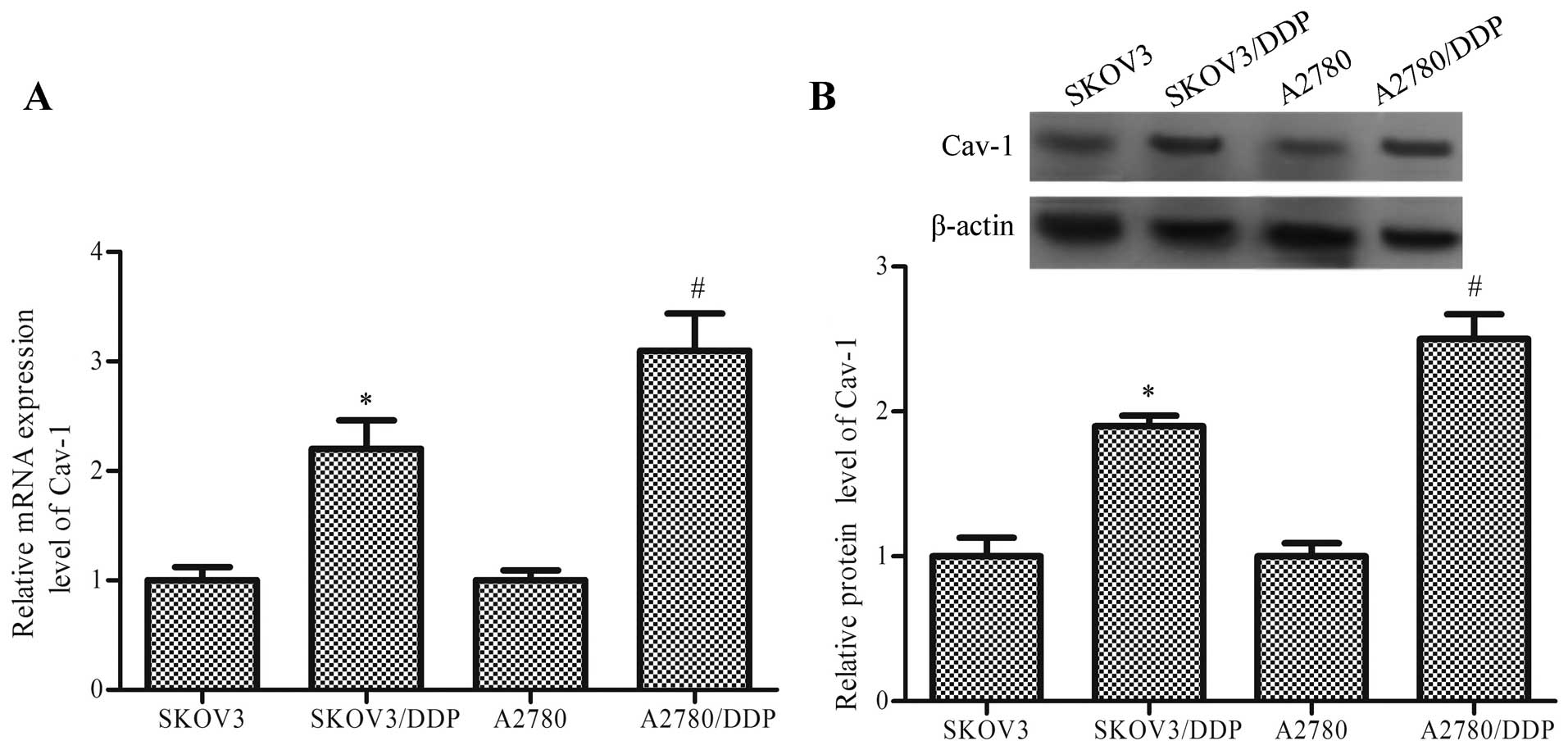
Cav-1 expression in cisplatin-resistant
ovarian cancer cells
To evaluate the expression level of Cav-1 between
normal ovarian cancer cells and cisplatin-resistant cancer cells,
SKOV3 and A2780 (normal ovarian cancer cells) and SKOV3/DDP and
A2780/DDP (cisplatin-resistant ovarian cancer cells) were cultured.
The expression level of Cav-1 was measured using RT-PCR and western
blot analysis. The results showed that both the mRNA and protein
expression level of Cav-1 in SKOV3/DDP and A2780/DDP cells was
significantly higher than SKOV3 and A2780 cells, respectively
(P<0.05, Fig. 1). Thus, we
predicted Cav-1 may be an important factor involved in the
chemoresistance in ovarian cancer cells.
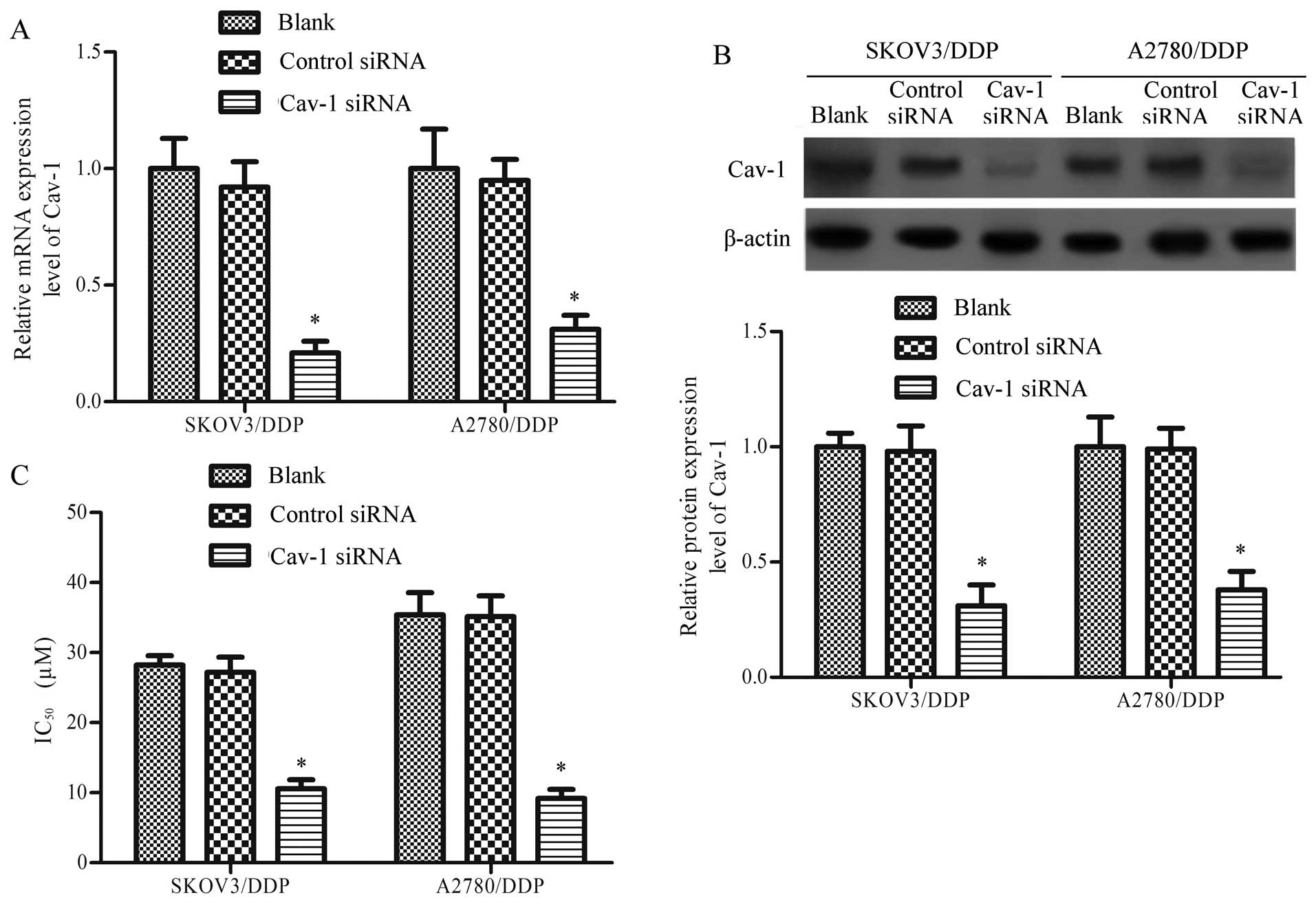
Cav-1 silencing promotes chemosensitivity
in cisplatin-resistant ovarian cancer cells
To explore the correlation between Cav-1 and the
cisplatin resistance in ovarian cancer cells, we used Cav-1 siRNA
to knock down Cav-1 and performed chemosensitivity analysis on
cisplatin-resistant cells. The results showed that Cav-1 siRNA
significantly inhibited the mRNA and protein expression level of
Cav-1 (P<0.05, Fig. 2) and
lowered the IC50 value of cisplatin from 28.21±1.35 to
10.56±1.28 (µM) in the SKOV3/DDP cells and from 35.39±3.18
to 9.17±1.32 (µM) in the A2780/DDP cells (Fig. 2C) and the statistical analysis was
significant for both SKOV3/DDP and A2780/DDP cells(P<0.05).
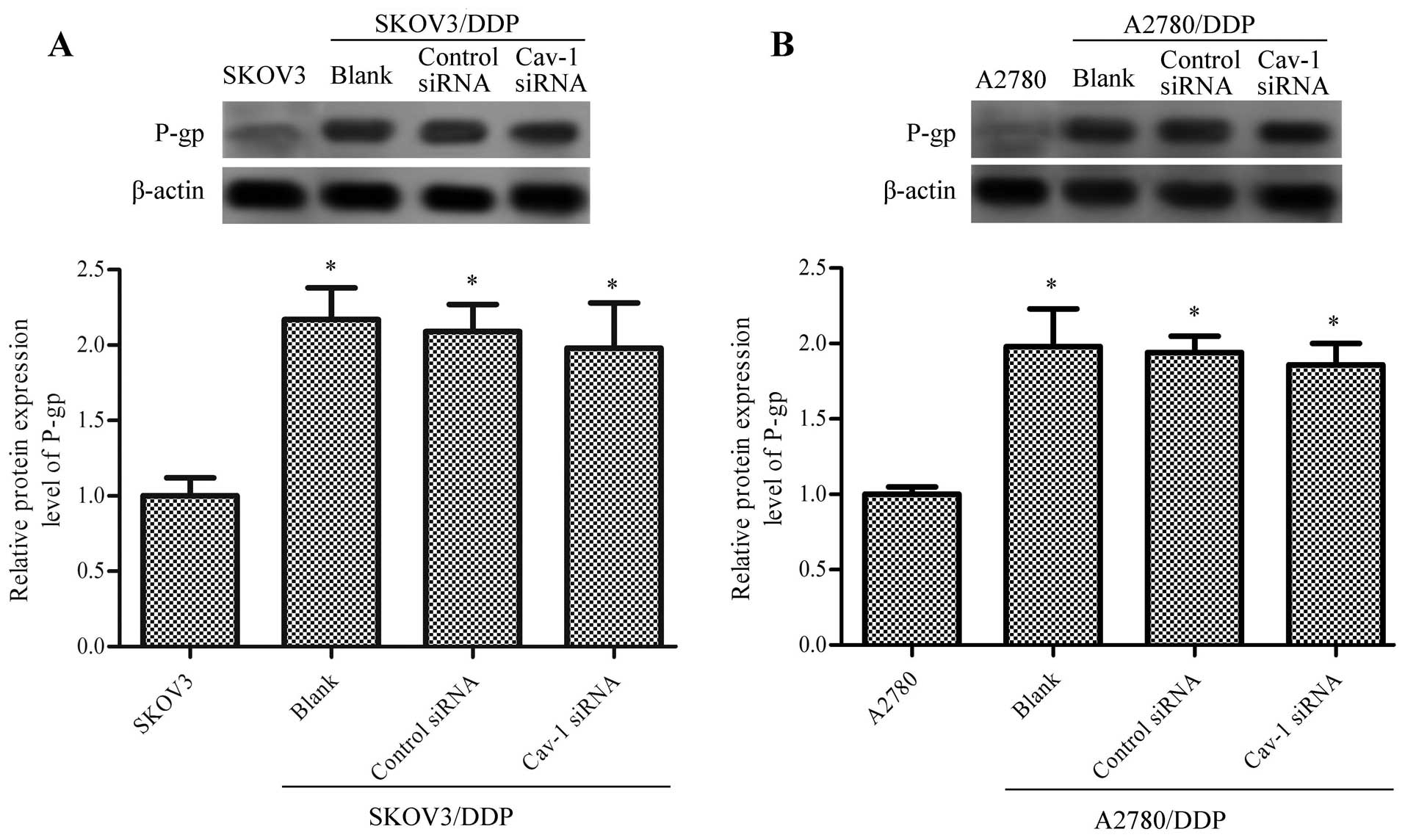
Cav-1 knockdown do not affect the protein
expression level of P-glycoprotein (P-gp)
P-gp is a protein product of the MDR1 gene, which is
one of the common genes that directly regulates drug resistance in
various types of cancer. To figure out the relationship between
Cav-1 and P-gp, we studied the P-gp expression level in
cisplatin-resistant ovarian cancer cells with Cav-1 knockdown. The
results showed that the relative protein expression level of P-gp
was significantly higher in SKOV3/DDP than SKOV3 and higher in
A280/DDP than A2780 (P<0.05, Fig.
3). There was no significant difference in the protein
expression level of P-gp in SKOV3/DDP and A2780/DDP between the
Cav-1 siRNA group and the blank or control siRNA group (P>0.05,
Fig. 3).
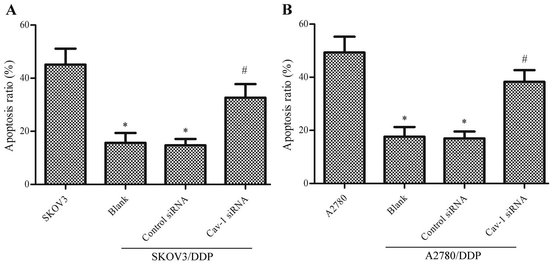
Cav-1 knockdown promotes apoptosis of
cisplatin-resistant ovarian cancer cells
Apoptosis contributes to the antitumor activity of
many chemotherapeutic drugs and plays a crucial role in
drug-induced cytotoxicity, thus the decreased apoptosis ratio is
also one of the common mechanisms that induce chemoresistance
(29). For this reason, we assessed
the apoptotic cells in Cav-1 knockdown cisplatin-resistant cells
after exposure to 20 µM cisplatin. The result showed that
the ratio of apoptosis induced by cisplatin in SKOV3 and A2780
cells was significantly higher than SKOV3/DDP or SKOV3/DDP cells
(P<0.05). Cav-1 siRNA transfection could significantly inhibited
apoptosis of SKOV3/DDP and A2780/DDP compared to the blank and
control siRNA groups (P<0.05, Fig.
4).
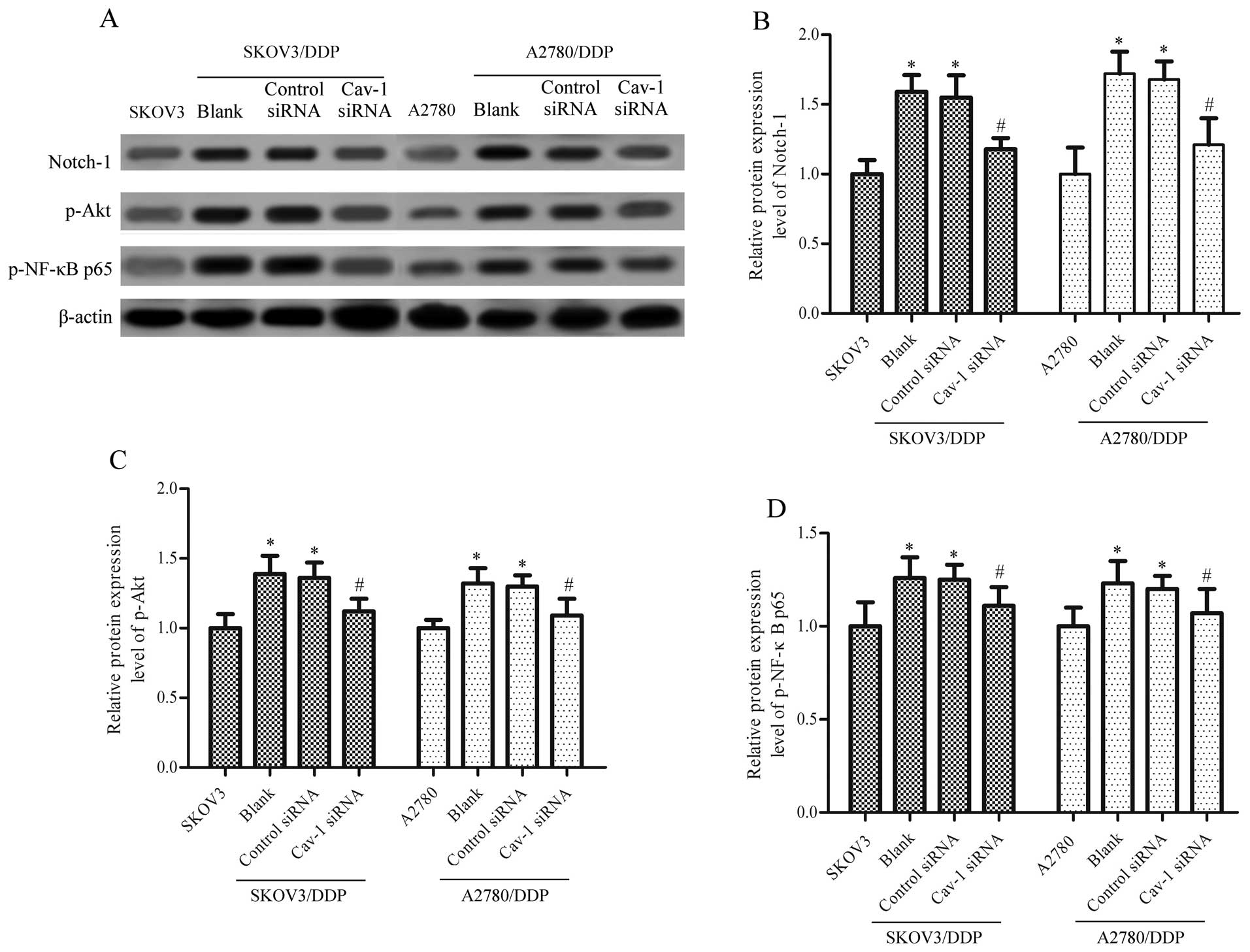
Cav-1 knockdown inhibits Notch-1, Akt and
NF-κB expression in ovarian cancer cells
Evidence has indicated that Notch-1 is an important
regulator of chemoresistance of ovarian cancer cells (30). Phosphorylated Akt is an activated
state of Akt, which is related to cancer cell survival and
cisplatin resistance of ovarian cancer cells (31). Translocation of the NF-κB p65
subunit has been reported to exert important roles in the cell
survival of cancer cells (24) and
the phosphorylation of p65 is essential for the nuclear retention
and transcriptional activity of NF-κB (32). To probe the distinctive expression
of Notch-1 in cisplatin-resistant ovarian cancer cells, we detected
the protein level of Notch-1 using western blot analysis. The
results showed that the protein level of Notch-1 was significantly
upregulated in cisplatin-resistant ovarian cancer cells (P<0.05,
Fig. 5A and B). Likewise, the
phosphorylation of Akt and NF-κB p65 was also tested using western
blot analysis and both were significantly upregulated in
cisplatin-resistant ovarian cancer cells (P<0.05, Fig. 5A, C and D).
To investigate whether the Notch-1/Akt/NF-κB pathway
was related to the expression level of Cav-1, we inhibited the
expression of Cav-1 by transfecting with Cav-1 siRNA and detected
the protein expression level of Notch-1, p-Akt and p-NF-κB p65
using western blot assay in cisplatin-resistant ovarian cancer
cells. The results showed that the knockdown of Cav-1 significantly
downregulated the protein expression level of Notch-1, p-Akt and
p-NF-κB p65 (P<0.05, Fig. 5).
These data suggested that the Notch-1/Akt/NF-κB pathway may be
involved in the Cav-1 mediated chemoresistance of ovarian cancer
cells.
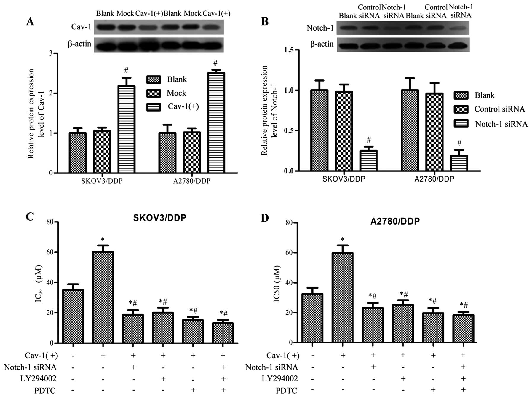
Overexpression of Cav-1 promotes
chemoresistance through Notch-1/Akt/NF-κB in cisplatin-resistant
ovarian cancer cells
To confirm the assumption that Cav-1 regulates
chemoresistance of ovarian cancer cells through the
Notch-1/Akt/NF-κB pathway, we overexpressed Cav-1 in
cisplatin-resistant ovarian cancer cells by the transfection of the
plasmid pcDNA 3.1(+)-cav-1 and inhibited the expression of Notch-1
using Notch-1 siRNA or restrained the activity of Akt and NF-κB by
treating with LY294002 and PDTC, respectively, the IC50
value of cisplatin was assessed using MTT analysis. Overexpression
of Cav-1 significantly enhanced the IC50 value of
cisplatin in both SKOV3/DDP and A2780/DDP cells (P<0.05,
Fig. 6C and D). We next detected
the effect of downregulated Notch-1, Akt and NF-κB down on the
IC50 value in Cav-1 overexpressed, cisplatin-resistant
ovarian cancer cells. The results showed that Notch-1 siRNA, Akt
siRNA and the NF-κB inhibitor PDTC may all significantly
downregulate the IC50 value in Cav-1 overexpressed
cisplatin-resistant ovarian cancer cells (P<0.05, Fig. 6C and D).
Discussion
In the past 20 years, Cav-1 has become regarded as a
tumor suppressor and pro-apoptotic protein (33–35).
The reduced expression of Cav-1 is found in several human cancer
types, including lung, colon, ovarian, breast cancer and
osteosarcomas (18,36–39).
However, in recent years, some studies have reported that Cav-1 can
exert anti-apoptotic potential, enhance tumor migration and
invasion, and have prognostic value for patient survival and cancer
recurrence (40–42). In addition, Cav-1 is an essential
modulator of cancer MDR. In the present study, we explored the
expression level in ovarian cancer cells and cisplatin-resistant
ovarian cancer cells using PCR and western blot assay. The results
showed that the expression level of Cav-1 in cisplatin-resistant
ovarian cancer cells SKOV3/DDP and A2780/DDP is significantly
higher than SKOV3 and A2780. In this case, we knocked down Cav-1 in
SKOV3/DDP and A2780/DDP by transfecting Cav-1 siRNA and assessed
the IC50 value by MTT assay. We found that knockdown of
Cav-1 downregulated the IC50 value significantly, both
in SKOV3/DDP and A2780/DDP cells. These data suggest for the first
time that: i) Cav-1 is upregulated in chemoresistant ovarian
cancer; ii) downregulation of Cav-1 may inhibit the cisplatin
resistance of ovarian cancer.
Chemoresistance is a major clinical obstacle for the
treatment of patients with ovarian cancer and a better
understanding of the molecular mechanisms involved in this cellular
protection is required to develop more successful therapies. In
cisplatin induced drug resistance, expressions of MDR proteins and
inhibitions of apoptosis are two important mechanisms that are
involved in cisplatin-resistance (43). As the protein product of MDR1 gene,
P-gp is a membrane protein belonging to the adenosine triphosphate
(ATP)-binding cassette family. The overexpression of P-gp is one of
the most common mechanisms that is associated with MDR by
inhibiting intracellular drug accumulation and catalyzing the drug
efflux (44). Several studies have
reported that the expression of P-gp is tightly related to the drug
resistance in ovarian cancer cells (45–47).
Most of the anticancer therapies, including drugs and chemotherapy,
target cancer cells mainly by contributing to apoptosis. Indeed,
when the cancer cells are chemoresistant, the apoptotic cells
induced by anticancer therapy were limited, thus apoptosis
resistance is one of the important pathway in mediating the
chemoresistance of cancer (29). In
the present study, we explored the final effector in Cav-1 mediated
cisplatin-resistance. The results showed that the protein
expression level of P-gp is much higher in cisplatin-resistant than
normal cells and there is no connection between the expression of
Cav-1 and P-gp. However, knockdown of Cav-1 significantly enhanced
the apoptosis ratio induced by cisplatin in cisplatin-resistant
ovarian cancer cells, which means that the mediation of
chemoresistance by Cav-1 is not targeting P-gp, but cell
apoptosis.
The Notch signaling pathway is crucial for the
regulation of chemoresistance in cancer by controlling the cell
survival and proliferation (48,49).
There are four Notch receptors, Notch1, Notch2, Notch3 and Notch4
in mammals, in which Notch1 signaling plays an important role in
ovarian tumor growth and is closely associated with chemoresistance
in ovarian cancer stem cells (30,50).
It is reported that Notch1 may regulate cancer cell growth via
activation of the Akt pathway (19), which plays a key regulatory role in
cell survival, proliferation, migration and apoptosis. Enhanced Akt
kinase activity is involved in resistance to chemotherapeutic
agents by regulating the activity of NF-κB in various types of
cancer. Zhou et al (23)
demonstrated that the expression of Notch1 and the activity of Akt1
and NF-κB were upregulated by doxorubicin (a chemotherapeutic
agent), and the Akt1/NF-κB/Notch-1 axis has an important role in
chemoresistance of gastric cancer cells. Thus, the present study
explored the relationship between Notch1, Akt and NF-κB and the
chemoresistance in ovarian cancer cells. We found that the protein
level of Notch1, p-Akt and p-NF-κB p65 are all significantly higher
in the cisplatin-resistant cancer cells SKOV3/DDP and A2780/DDP
than in SKOV3 and A2780 cells, which indicated a role of the
Notch1/Akt/NF-κB pathway in the chemoresistance of ovarian cancer
cells.
Recent studies indicated that the Notch1 signaling
pathway plays a role in the regulating effect of Cav-1 (51,52).
The expression of Cav-1 is associated with the phosphorylation of
Akt and the activity of NF-κB. Our results demonstrated that Cav-1
knockdown can significantly down regulate the protein expression
level of Notch-1, p-Akt and p-NF-κB p65. To further distinguish the
role of the Notch-1/Akt/NF-κB pathway in the mediation of Cav-1 in
chemoresistant ovarian cancer, we inhibited the effect of Notch-1,
Akt and NF-κB in Cav-1 overexpressed, cisplatin-resistant ovarian
cancer and detected the IC50 of cisplatin. We found that
inhibiting any one of Notch-1, Akt and NF-κB, or all of them, can
block the upregulating effect of Cav-1 overexpression on
IC50, which suggested a crucial role of
NF-κB/AKT/Notch-1 pathway in the Cav-1 mediated chemoresistance in
ovarian cancer.
In conclusion, our data have shown that Cav-1 is
aberrantly expressed and contributes to the chemoresistance in
cisplatin-resistant ovarian cancer. We also found that the
regulatory effect of Cav-1 on cisplatin-resistance is targeting
cell apoptosis rather than P-gp. Moreover, upregulation of Notch-1,
p-Akt, and p-NF-κB p65 was observed in cisplatin-resistant ovarian
cancer cells. Furthermore, we demonstrated that knockdown of Cav-1
was able to inhibit the protein expression level of Notch-1, p-Akt
and p-NF-κB p65. Besides, we found that inhibiting the
Notch-1/Akt/NF-κB pathway restrained the increased IC50
value induced by Cav-1 overexpression. Overall, the present finding
suggests that Cav-1 is a positive factor mediating chemoresistance
of ovarian cancer cells by targeting apoptosis through
Notch-1/Akt/NF-κB pathway.
Acknowledgments
The present study was supported by grants from the
National Natural Science Foundation of China (no. 81101958 and no.
81101959).
Abbreviations:
|
Cav-1
|
caveolin-1
|
|
P-gp
|
P-glycoprotein
|
|
IC50
|
50% inhibitory concentration
|
|
MDR
|
multi-drug resistance
|
References
|
1
|
Siegel RL, Miller KD and Jemal A: Cancer
statistics, 2015. CA Cancer J Clin. 65:5–29. 2015. View Article : Google Scholar : PubMed/NCBI
|
|
2
|
Kuczynski EA, Sargent DJ, Grothey A and
Kerbel RS: Drug rechallenge and treatment beyond progression -
implications for drug resistance. Nat Rev Clin Oncol. 10:571–587.
2013. View Article : Google Scholar : PubMed/NCBI
|
|
3
|
Parton RG and Simons K: The multiple faces
of caveolae. Nat Rev Mol Cell Biol. 8:185–194. 2007. View Article : Google Scholar : PubMed/NCBI
|
|
4
|
Zhang B, Peng F, Wu D, Ingram AJ, Gao B
and Krepinsky JC: Caveolin-1 phosphorylation is required for
stretch-induced EGFR and Akt activation in mesangial cells. Cell
Signal. 19:1690–1700. 2007. View Article : Google Scholar : PubMed/NCBI
|
|
5
|
Park JH and Han HJ: Caveolin-1 plays
important role in EGF-induced migration and proliferation of mouse
embryonic stem cells: involvement of PI3K/Akt and ERK. Am J Physiol
Cell Physiol. 297:C935–C944. 2009. View Article : Google Scholar : PubMed/NCBI
|
|
6
|
Quest AF, Gutierrez-Pajares JL and Torres
VA: Caveolin-1: An ambiguous partner in cell signalling and cancer.
J Cell Mol Med. 12:1130–1150. 2008. View Article : Google Scholar : PubMed/NCBI
|
|
7
|
Chen HL, Fan LF, Gao J, Ouyang JP and
Zhang YX: Differential expression and function of the caveolin-1
gene in non-small cell lung carcinoma. Oncol Rep. 25:359–366. 2011.
View Article : Google Scholar
|
|
8
|
Park J, Bae E, Lee C, Yoon SS, Chae YS,
Ahn KS and Won NH: RNA interference-directed caveolin-1 knockdown
sensitizes SN12CPM6 cells to doxorubicin-induced apoptosis and
reduces lung metastasis. Tumor Biol. 31:643–650. 2010. View Article : Google Scholar
|
|
9
|
Rödel F, Capalbo G, Rödel C and Weiss C:
Caveolin-1 as a prognostic marker for local control after
preoperative chemoradiation therapy in rectal cancer. Int J Radiat
Oncol Biol Phys. 73:846–852. 2009. View Article : Google Scholar : PubMed/NCBI
|
|
10
|
Hehlgans S, Eke I, Storch K, Haase M,
Baretton GB and Cordes N: Caveolin-1 mediated radioresistance of 3D
grown pancreatic cancer cells. Radiother Oncol. 92:362–370. 2009.
View Article : Google Scholar : PubMed/NCBI
|
|
11
|
Burrows AD, Restall C, Sloan EK and
Anderson RL: The contribution of stromal caveolin-1 to breast
cancer metastasis. Cancer Res. 72:1486. 2012. View Article : Google Scholar
|
|
12
|
Rao X, Evans J, Chae H, Pilrose J, Kim S,
Yan P, Huang RL, Lai HC, Lin H, Liu Y, et al: CpG island shore
methylation regulates caveolin-1 expression in breast cancer.
Oncogene. 32:4519–4528. 2013. View Article : Google Scholar :
|
|
13
|
Xu J, Agyemang S, Qin Y, Aysola K, Giles
M, Oprea G, O'Regan RM, Partridge EE, Harris-Hooker S, Rice VM, et
al: A novel pathway that links Caveolin-1 down-regulation to BRCA1
dysfunction in serous epithelial ovarian cancer cells. Enliven
Chall Cancer Detect Ther. 1:pii0042014.
|
|
14
|
Goetz JG, Lajoie P, Wiseman SM and Nabi
IR: Caveolin-1 in tumor progression: The good, the bad and the
ugly. Cancer Metastasis Rev. 27:715–735. 2008. View Article : Google Scholar : PubMed/NCBI
|
|
15
|
Cai C and Chen J: Overexpression of
caveolin-1 induces alteration of multidrug resistance in Hs578T
breast adenocarcinoma cells. Int J Cancer. 111:522–529. 2004.
View Article : Google Scholar : PubMed/NCBI
|
|
16
|
Selga E, Morales C, Noe V, Peinado MA and
Ciudad CJ: Role of caveolin 1, E-cadherin, Enolase 2 and PKCalpha
on resistance to methotrexate in human HT29 colon cancer cells. BMC
Med Genomics. 1:1755–8794. 2008. View Article : Google Scholar
|
|
17
|
Ho CC, Kuo SH, Huang PH, Huang HY, Yang CH
and Yang PC: Caveolin-1 expression is significantly associated with
drug resistance and poor prognosis in advanced non-small cell lung
cancer patients treated with gemcitabine-based chemotherapy. Lung
Cancer. 59:105–110. 2008. View Article : Google Scholar
|
|
18
|
Wiechen K, Diatchenko L, Agoulnik A,
Scharff KM, Schober H, Arlt K, Zhumabayeva B, Siebert PD, Dietel M,
Schäfer R, et al: Caveolin-1 is down-regulated in human ovarian
carcinoma and acts as a candidate tumor suppressor gene. Am J
Pathol. 159:1635–1643. 2001. View Article : Google Scholar : PubMed/NCBI
|
|
19
|
Liu ZJ, Xiao M, Balint K, Smalley KS,
Brafford P, Qiu R, Pinnix CC, Li X and Herlyn M: Notch1 signaling
promotes primary melanoma progression by activating
mitogen-activated protein kinase/phosphatidylinositol 3-kinase-Akt
pathways and up-regulating N-cadherin expression. Cancer Res.
66:4182–4190. 2006. View Article : Google Scholar : PubMed/NCBI
|
|
20
|
Aoki K, Ogawa T, Ito Y and Nakashima S:
Cisplatin activates survival signals in UM-SCC-23 squamous cell
carcinoma and these signal pathways are amplified in
cisplatin-resistant squamous cell carcinoma. Oncol Rep. 11:375–379.
2004.PubMed/NCBI
|
|
21
|
Wang Z, Li Y, Banerjee S, Kong D, Ahmad A,
Nogueira V, Hay N and Sarkar FH: Down-regulation of Notch-1 and
Jagged-1 inhibits prostate cancer cell growth, migration and
invasion, and induces apoptosis via inactivation of Akt, mTOR, and
NF-kappaB signaling pathways. J Cell Biochem. 109:726–736.
2010.PubMed/NCBI
|
|
22
|
Romashkova JA and Makarov SS: NF-κB is a
target of AKT in anti-apoptotic PDGF signalling. Nature. 401:86–90.
1999. View Article : Google Scholar : PubMed/NCBI
|
|
23
|
Zhou W, Fu XQ, Zhang LL, Zhang J, Huang X,
Lu XH, Shen L, Liu BN, Liu J, Luo HS, et al: The
AKT1/NF-kappaB/Notch1/PTEN axis has an important role in
chemoresistance of gastric cancer cells. Cell Death Dis.
4:e8472013. View Article : Google Scholar : PubMed/NCBI
|
|
24
|
Zhu H, Bhaijee F, Ishaq N, Pepper DJ,
Backus K, Brown AS, Zhou X and Miele L: Correlation of Notch1, pAKT
and nuclear NF-κB expression in triple negative breast cancer. Am J
Cancer Res. 3:2302013.
|
|
25
|
Wang S, Kan Q, Sun Y, Han R, Zhang G, Peng
T and Jia Y: Caveolin-1 regulates neural differentiation of rat
bone mesen-chymal stem cells into neurons by modulating Notch
signaling. Int J Dev Neurosci. 31:30–35. 2013. View Article : Google Scholar
|
|
26
|
Chanvorachote P, Chunhacha P and
Pongrakhananon V: Caveolin-1 induces lamellipodia formation via an
Akt-dependent pathway. Cancer Cell Int. 14:1475–2867. 2014.
View Article : Google Scholar
|
|
27
|
Hada N, Okayasu M, Ito J, Nakayachi M,
Hayashida C, Kaneda T, Uchida N, Muramatsu T, Koike C, Masuhara M,
et al: Receptor activator of NF-κB ligand-dependent expression of
caveolin-1 in osteoclast precursors, and high dependency of
osteoclastogenesis on exogenous lipoprotein. Bone. 50:226–236.
2012. View Article : Google Scholar
|
|
28
|
Schmittgen TD and Livak KJ: Analyzing
real-time PCR data by the comparative C(T) method. Nat Protoc.
3:1101–1108. 2008. View Article : Google Scholar : PubMed/NCBI
|
|
29
|
Rebucci M and Michiels C: Molecular
aspects of cancer cell resistance to chemotherapy. Biochem
Pharmacol. 85:1219–1226. 2013. View Article : Google Scholar : PubMed/NCBI
|
|
30
|
Groeneweg JW, Foster R, Growdon WB,
Verheijen RH and Rueda BR: Notch signaling in serous ovarian
cancer. J Ovarian Res. 7:952014. View Article : Google Scholar : PubMed/NCBI
|
|
31
|
Liu L-Z, Zhou X-D, Qian G, Shi X, Fang J
and Jiang B-H: AKT1 amplification regulates cisplatin resistance in
human lung cancer cells through the mammalian target of
rapamycin/p70S6K1 pathway. Cancer Res. 67:6325–6332. 2007.
View Article : Google Scholar : PubMed/NCBI
|
|
32
|
Zhong H, Voll RE and Ghosh S:
Phosphorylation of NF-κB p65 by PKA stimulates transcriptional
activity by promoting a novel bivalent interaction with the
coactivator CBP/p300. Mol Cell. 1:661–671. 1998. View Article : Google Scholar : PubMed/NCBI
|
|
33
|
Bélanger MM, Roussel E and Couet J:
Caveolin-1 is down-regulated in human lung carcinoma and acts as a
candidate tumor suppressor gene. Chest. 125(5 Suppl): 106S2004.
View Article : Google Scholar : PubMed/NCBI
|
|
34
|
Razani B, Schlegel A, Liu J and Lisanti M:
Caveolin-1, a putative tumour suppressor gene. Biochem Soc Trans.
29:494–499. 2001. View Article : Google Scholar : PubMed/NCBI
|
|
35
|
Huertas-Martínez J, Barrau I,
Sainz-Jaspeado M, Lagares-Tena L, Mateo-Lozano S, Mora J, Roma J,
Gallego S, Moran S, Esteller M, et al: Caveolin-1 acts as a tumor
suppressor promoting muscular differentiation in alveolar
rhabdomyosarcomas. Cancer Res. 73:38272013. View Article : Google Scholar
|
|
36
|
Wikman H, Kettunen E, Seppänen JK,
Karjalainen A, Hollmén J, Anttila S and Knuutila S: Identification
of differentially expressed genes in pulmonary adenocarcinoma by
using cDNA array. Oncogene. 21:5804–5813. 2002. View Article : Google Scholar : PubMed/NCBI
|
|
37
|
Lee SW, Reimer CL, Oh P, Campbell DB and
Schnitzer JE: Tumor cell growth inhibition by caveolin
re-expression in human breast cancer cells. Oncogene. 16:1391–1397.
1998. View Article : Google Scholar : PubMed/NCBI
|
|
38
|
Bender FC, Reymond MA, Bron C and Quest
AF: Caveolin-1 levels are down-regulated in human colon tumors, and
ectopic expression of caveolin-1 in colon carcinoma cell lines
reduces cell tumorigenicity. Cancer Res. 60:5870–5878.
2000.PubMed/NCBI
|
|
39
|
Cantiani L, Manara MC, Zucchini C, De
Sanctis P, Zuntini M, Valvassori L, Serra M, Olivero M, Di Renzo
MF, Colombo MP, et al: Caveolin-1 reduces osteosarcoma metastases
by inhibiting c-Src activity and met signaling. Cancer Res.
67:7675–7685. 2007. View Article : Google Scholar : PubMed/NCBI
|
|
40
|
Tang Y, Zeng X, He F, Liao Y, Qian N and
Toi M: Caveolin-1 is related to invasion, survival, and poor
prognosis in hepatocellular cancer. Med Oncol. 29:977–984. 2012.
View Article : Google Scholar
|
|
41
|
Wang R, He W, Li Z, Chang W, Xin Y and
Huang T: Caveolin-1 functions as a key regulator of
17β-estradiol-mediated autophagy and apoptosis in BT474 breast
cancer cells. Int J Mol Med. 34:822–827. 2014.PubMed/NCBI
|
|
42
|
Karam JA, Lotan Y, Roehrborn CG, Ashfaq R,
Karakiewicz PI and Shariat SF: Caveolin-1 overexpression is
associated with aggressive prostate cancer recurrence. Prostate.
67:614–622. 2007. View Article : Google Scholar : PubMed/NCBI
|
|
43
|
Florea A-M and Büsselberg D: Cisplatin as
an anti-tumor drug: Cellular mechanisms of activity, drug
resistance and induced side effects. Cancers (Basel). 3:1351–1371.
2011. View Article : Google Scholar
|
|
44
|
Cole SP, Bhardwaj G, Gerlach JH, Mackie
JE, Grant CE, Almquist KC, Stewart AJ, Kurz EU, Duncan AM and
Deeley RG: Overexpression of a transporter gene in a
multidrug-resistant human lung cancer cell line. Science.
258:1650–1654. 1992. View Article : Google Scholar : PubMed/NCBI
|
|
45
|
Stordal B, Hamon M, McEneaney V, Roche S,
Gillet JP, O'Leary JJ, Gottesman M and Clynes M: Resistance to
paclitaxel in a cisplatin-resistant ovarian cancer cell line is
mediated by P-glycoprotein. PLoS One. 7:e407172012. View Article : Google Scholar : PubMed/NCBI
|
|
46
|
Bourhis J, Goldstein LJ, Riou G, Pastan I,
Gottesman MM and Bénard J: Expression of a human multidrug
resistance gene in ovarian carcinomas. Cancer Res. 49:5062–5065.
1989.PubMed/NCBI
|
|
47
|
van der Zee AG, Hollema H, Suurmeijer AJ,
Krans M, Sluiter WJ, Willemse PH, Aalders JG and de Vries EG: Value
of P-glycoprotein, glutathione S-transferase pi, c-erbB-2, and p53
as prognostic factors in ovarian carcinomas. J Clin Oncol.
13:70–78. 1995.PubMed/NCBI
|
|
48
|
Bray SJ: Notch signalling: a simple
pathway becomes complex. Nat Rev Mol Cell Biol. 7:678–689. 2006.
View Article : Google Scholar : PubMed/NCBI
|
|
49
|
Perdigoto CN and Bardin AJ: Sending the
right signal: Notch and stem cells. Biochim Biophys Acta.
1830:2307–2322. 2013. View Article : Google Scholar
|
|
50
|
Hopfer O, Zwahlen D, Fey M and Aebi S: The
Notch pathway in ovarian carcinomas and adenomas. Br J Cancer.
93:709–718. 2005. View Article : Google Scholar : PubMed/NCBI
|
|
51
|
Yanjie J, Jiping S, Yan Z, Xiaofeng Z,
Boai Z and Yajun L: Effects of Notch-1 signalling pathway on
differentiation of marrow mesenchymal stem cells into neurons in
vitro. Neuroreport. 18:1443–1447. 2007. View Article : Google Scholar : PubMed/NCBI
|
|
52
|
Campos LS, Decker L, Taylor V and Skarnes
W: Notch, epidermal growth factor receptor, and beta1-integrin
pathways are coordinated in neural stem cells. J Biol Chem.
281:5300–5309. 2006. View Article : Google Scholar
|