Introduction
Head and neck squamous cell carcinoma (HNSCC) is the
sixth most common cancer in the world (1). It is characterized by phenotypical,
etiological, biological and clinical heterogeneity. Despite
surgery, radiation therapy, and chemotherapy, approximately half of
all patients die (2–4). Laryngeal squamous cell carcinoma and
hypopharyngeal squamous cell carcinoma are two common malignancies
of HNSCC that mainly occur in middle-aged men. Tobacco smoking and
alcohol abuse are predominant risk factors in HNSCC. A subset of
oropharyn-geal carcinoma cases are strongly associated with the
infection of high-risk human papilloma virus (HPV), predominantly
HPV-16 (5–7). The oncogenicity of high-risk HPV is
dependent on the constitutive expression of oncogenes, such as E6
and E7 (8–15).
The E6 and E7 genes of the HPV-16 genome encode the
oncoproteins E6 and E7, respectively (16,17).
Lentiviral vectors can be used to transfect cells with high
efficiency, allowing for the stable integration of genes into
cells. MicroRNAs are involved in almost all biological processes in
the human body, including cell proliferation, differentiation,
apoptosis, invasion and migration (18-20).
Abnormal expression of miRNAs is associated with the occurrence and
development of many types of tumors (21–23).
In the present study, we used a lentiviral vector to
transfect and integrate the HPV-16 E6-E7 genes into the
hypopharyngeal squamous cell carcinoma cell line, FaDu. We then
observed the effects of E6-E7 expression on these cells. We also
sought to determine any effects of HPV-related miRNAs on HNSCC by
examining miRNA expression levels in hypopharyngeal squamous cell
carcinoma tissues.
Materials and methods
Patients and tumor samples
Tumor samples were collected from 28 patients with
pharyngeal cancer who had undergone surgery at the Department of
Otolaryngology-Head and Neck Surgery, The First Affiliated Hospital
of Zhengzhou University (Zhengzhou, China). Patients recruited to
this study had not undergone previous chemotherapy, radiotherapy or
immunotherapy. Collected tumor samples were frozen in liquid
nitrogen and then stored at −80°C until required. This study was
approved by the Ethics Committee of Zhengzhou University, and
informed consent was obtained from each patient.
HPV DNA detection and typing
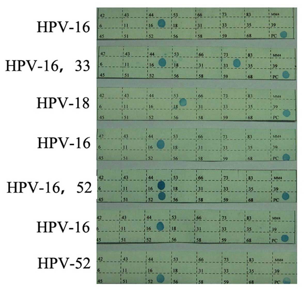
We detected the presence of HPV genes in fresh
frozen samples using polymerase chain reaction (PCR) assays
followed by reverse dot blots. Using PCR, 28 HPV gene segments were
amplified. These were then hybridized to specific probes that were
affixed to membranes. The probes we used corresponded with 5
low-risk and 18 high-risk HPV genotypes.
Cell culture
The hypopharyngeal squamous cell carcinoma cell
line, FaDu, along with the Hep-2 and 293T cell lines, were
purchased from the Type Culture Collection of the Chinese Academy
of Sciences (Shanghai, China). Cells were cultured in Dulbecco's
modified Eagle's medium (DMEM) supplemented with 10% fetal bovine
serum (FBS) (both from Gibco, USA) and grown in a 37°C, 5%
CO2 incubator.
Integration, transfection, and expression
of HPV-16 E6-E7 genes
HPV-16 E6-E7 genes were amplified and cloned into
the pLV-EGFP-C lentiviral vector, between the HindIII and
KpnI sites, to produce the recombinant lentivirus
LV-HPV-16-E6-E7. The empty pLV-EGFP-C vector was used as an empty
vector control. We co-transfected 5 µg of LV-HPV-16-E6-E7 with 3.75
µg of pH1 and 1.25 µg of pH2 into 293T packaging cells using
PolyFect-V (Invitrogen, USA). After incubation at 37°C/5%
CO2 for 48 h, the culture medium was harvested and
concentrated 100- to 200-fold by ultrafiltration. Virus titers in
the concentrated supernatants were determined on 293T cells based
on the expression level of enhanced green fluorescent protein
(EGFP). Cells were cultured in DMEM containing 10% FBS, and
infected at a multiplicity of infection of 10-30 in the presence of
6 µg/ml Polybrene (Sigma-Aldrich, St. Louis, MO, USA) and 1 mg/ml
puromycin. Cell culture medium was changed every 72 h. Positive
clones were identified through the expression of EGFP.
RNA isolation and quantitative reverse
transcription-PCR (qRT-PCR) assays
RNA was extracted from FaDu and Hep-2 cells using
E.Z.N.A.® Total RNA kit I (Omega Bio-Tek, Norcross, GA,
USA), according to the manufacturer's instructions. Reverse
transcription and PCR amplification were performed using a qRT-PCR
quantitation kit (Novland, China). An ABI 7500 HT Sequence
Detection system (Applied Biosystems, Foster City, CA, USA) was
used to determine the relative levels of E6 and E7 mRNAs in the
cells. Primers and probes designed for TaqMan assays were purchased
from Applied Biosystems. Amplification was conducted according to
the manufacturer's instructions. Results from the qRT-PCR assays
were analyzed by the 2−ΔΔCt method (24).
Western blot analysis
FaDu cells infected with LV-HPV-16-E6-E7, uninfected
FaDu cells, Hep-2 cells, and cells transfected with the empty
pLV-EGFP-C vector were lysed, and total proteins were isolated.
Total protein concentration was determined using a Bradford assay.
We used 30 µg of total protein per sample for sodium dodecyl
sulfate-polyacrylamide gel electrophoresis, with 12% polyacrylamide
gels. Electrophoresed proteins were transferred to nitrocellulose
membranes (GE Healthcare, USA), which were subsequently blocked
with 5% (w/v) non-fat milk and incubated overnight at 4°C with
antibodies against HPV E6 (diluted 1:800; Cell Signaling
Technology, Danvers, MA, USA) and HPV E7 (1:400; Santa Cruz
Biotechnology, Santa Cruz, CA, USA). The membranes were then
incubated with the appropriate horseradish peroxidase-conjugated
secondary antibody (1:2,000; Santa Cruz Biotechnology). The
intensity of the protein bands was evaluated using a Molecular
Dynamics densitometer (Molecular Dynamics, Sunnyvale, CA, USA). We
used glycer-aldehyde 3-phosphate dehydrogenase as an internal
reference.
Cell proliferation assays
Cell proliferation was evaluated using Cell Counting
Kit-8 reagents (CCK-8; Dojindo, Japan). Cells in the logarithmic
phase of growth were seeded in 96-well plates at a density of
1×10⁴ cells/well. We added 10 µl of CCK-8 to each well
on 5 consecutive days, at the same time each day. The optical
density at 450 nm in each well was assessed using an EL×800
microplate reader (BioTek, Winooski, VT, USA). All experiments were
conducted in triplicate.
Cell cycle analysis
Cells in the logarithmic phase of growth were
harvested by trypsinization, washed with phosphate-buffered saline
(PBS), and fixed with 75% ethanol overnight at 4°C. Cells were then
incubated with RNase at 37°C for 30 min, and stained with propidium
iodide (PI) for 30 min. We examined 106 events/sample
using a BD FACSCalibur™ (BD Biosciences, San Jose, CA, USA). All
experiments were performed in triplicate.
Apoptosis assays
The Annexin V-FITC Apoptosis Detection kit (Abcam,
USA) was used to detect and quantify apoptosis by flow cytometry.
Briefly, cells in the logarithmic phase of growth were harvested
using cold PBS and centrifuged (5 min at 1,000 × g). The cells were
resuspended in binding buffer at a density of 1×10⁶
cells/ml, stained with FITC-labeled Annexin V for 5 min, and
subjected to flow cytometry on a BD FACSCalibur™. Samples were
tested in triplicate and analyzed with CellQuest software (BD
Biosciences).
Transwell assays
Cell invasion assays were performed using Transwell
chambers with 8.0-µm pores (Costar, Cambridge, NY, USA). Basement
membrane matrix was added to the top chambers and allowed to
solidify for 30 min at 37°C. We added 500 µl of culture medium
containing chemotactic factor into the lower chamber. Cells were
then seeded into the top chambers at a density of 5×10⁵
cells/well and allowed to incubate at 37°C for 24 h. Cells were
then fixed with paraformaldehyde, stained with 0.1% crystal violet,
and quantified. Experiments were independently repeated six times,
in quadruplicate.
miRNA expression assays
We isolated miRNAs using TRIzol reagent (Invitrogen)
according to the manufacturer's instructions. For reverse
transcription and qPCR assays, we used miR-155, miR-363, miR-15A or
U6 as primers (Table I). Assays
were independently repeated three or more times.
 | Table ImiR-155, miR-363, miR-15A and U6
reverse transcription primers. |
Table I
miR-155, miR-363, miR-15A and U6
reverse transcription primers.
| Gene name | RT primers | PCR primers |
|---|
| miR-155 |
5′-GTCGTATCCAGTGCAGGGTCCGAGG | F
5′-TCCGATGGGGATAGTGCTAAT-3′ |
|
TATTCGCACTGGATACGACAATTACG-3′ | R
5′-GTGCAGGGTCCGAGGT-3′ |
| miR-363 |
5′-GTCGTATCCAGTGCAGGGTCCGAGG | F
5′-TCCGATTTAACGTAGCACTA-3′ |
|
TATTCGCACTGGATACGACGCCCACC-3′ | R
5′-GTGCAGGGTCCGAGGT-3′ |
| miR-15A |
5′-GTCGTATCCAGTGCAGGGTCCGAGG | F
5′-TCCGAGTGTTTGGTAATACA-3′ |
|
TATTCGCACTGGATACGACATCGTCG-3′ | R
5′-GTGCAGGGTCCGAGGT-3′ |
| U6 nRNA |
5′-GTCGTATCCAGTGCAGGGTCCGAGG | F
5′-TCCGATCGTGAAGCGTTC-3′ |
|
TATTCGCACTGGATACGACAAAATA-3′ | R
5′-GTGCAGGGTCCGAGGT-3′ |
Statistical analysis
All statistical analyses were performed using SPSS
17.0 (SPSS Inc., Chicago, IL, USA) software. Student's t-test was
used to compare the mean between two samples. Multiple comparisons
between parental and control vector groups were made using Tukey's
honest significant difference test. The expression levels of miRNAs
in cells and tissues were analyzed using the Wilcoxon signed-rank
test. Values are presented as the mean ± SD. A p-value <0.05 was
considered statistically significant.
Results
Presence of HPV in the specimens and
clinical features of the hypopharyngeal squamous cell carcinoma
cases
We observed indicators of HPV infection in 25%
(7/28) of the hypopharyngeal squamous cell carcinoma cases
(Table II and Fig. 1). The criteria used to define heavy
smoking were: an individual that smoked for more than 20 years; and
smoked not less than one pack per day. The criteria used to define
heavy drinking were: an individual that had regularly consumed
alcohol for more than 20 years; and drank not less than 150 g of
alcohol per day. Patients were separated into two groups for
statistical analyses: HPV-positive and HPV-negative. There was a
significant difference between the two groups when heavy smoking
was considered as a variable (P<0.05, Table III). Differences between the two
groups of patients with respect to age, gender, pathological type,
and tumor T stage were not significantly different (P>0.05,
Table III).
| Table IIHPV infection in patients (7 cases)
showing patient no. and type of HPV infection. |
Table II
HPV infection in patients (7 cases)
showing patient no. and type of HPV infection.
| HPV-positive
patient no. | Type of HPV
infection |
|---|
| 3 |
HPV-16 |
| 8 | HPV-16,
HPV-33 |
| 10 |
HPV-18 |
| 15 |
HPV-16 |
| 18 | HPV-16,
HPV-52 |
| 21 |
HPV-16 |
| 26 |
HPV-52 |
| Table IIIAnalysis of the HPV status and the
laryngopharyngeal carcinoma clinical characteristics of the
cases. |
Table III
Analysis of the HPV status and the
laryngopharyngeal carcinoma clinical characteristics of the
cases.
| Factors | HPV positive
(n) | HPV negative
(n) |
χ2-value | P-value |
|---|
| Age (years) | | | 3.111 | 0.078 |
| ≤60 | 5 | 7 | | |
| >60 | 2 | 14 | | |
| Gender | | | 0.718 | 0.397 |
| Male | 6 | 20 | | |
| Female | 1 | 1 | | |
| Heavy
drinking/smoking | | | 8.400 | 0.004a |
| Yes | 2 | 18 | | |
| No | 5 | 3 | | |
| Pathological
type | | | 1.600 | 0.449 |
| High | 2 | 2 | | |
| Moderate | 2 | 3 | | |
| Poor | 3 | 16 | | |
| Tumor T stage | | | 1.159 | 0.763 |
| T1 | 1 | 1 | | |
| T2 | 2 | 5 | | |
| T3 | 3 | 13 | | |
| T4 | 1 | 2 | | |
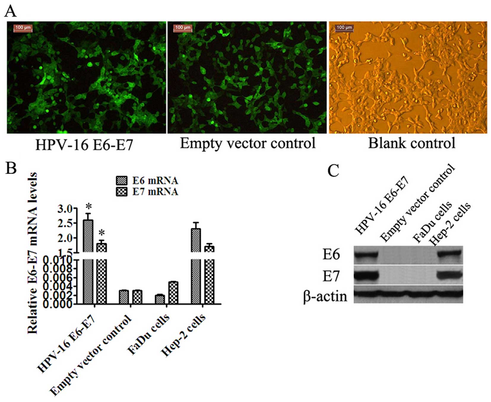
Overexpression of HPV-16 E6-E7
Positive clones were identified through the
expression of EGFP. We observed E6-E7 expression in stably
transfected FaDu cells at the mRNA and protein levels. The relative
E6-E7 mRNA levels in the HPV-16 E6-E7 FaDu cells (2.6±0.22,
1.8±0.12) were higher than these levels in the empty vector control
cells (0.003±0.0001, 0.003±0.0002, P<0.05) and blank control
cells (FaDu cells) (0.002±0.0002, 0.005±0.0001, P<0.05), while
consistent with the Hep-2 cells (Table
IV and Fig. 2).
| Table IVHPV-16 E6-E7 mRNA relative
expression. |
Table IV
HPV-16 E6-E7 mRNA relative
expression.
| Cell groups | n | E6 mRNA | E7 mRNA |
|---|
| HPV-16 E6-E7 | 5 | 2.6±0.22 | 1.8±0.12 |
| Empty vector
control | 5 | 0.003±0.0001 | 0.003±0.0002 |
| Blank control (FaDu
cells) | 5 | 0.002±0.0002 | 0.005±0.0001 |
| Hep-2 cells | 5 | 2.3±0.21 | 1.7±0.11 |
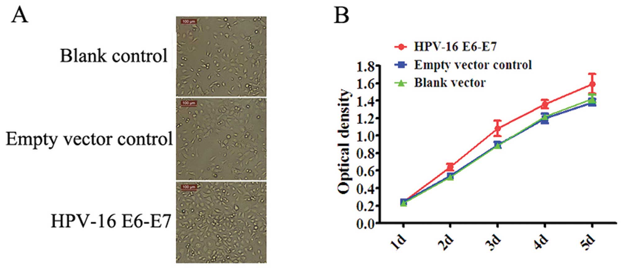
HPV-16 E6-E7 promotes FaDu cell
proliferation
We observed that HPV-16 E6-E7 promoted the
proliferation of FaDu cells in vitro (Fig. 3), and that these effects were
dependent on time. Proliferation levels were maximal after 5
days.
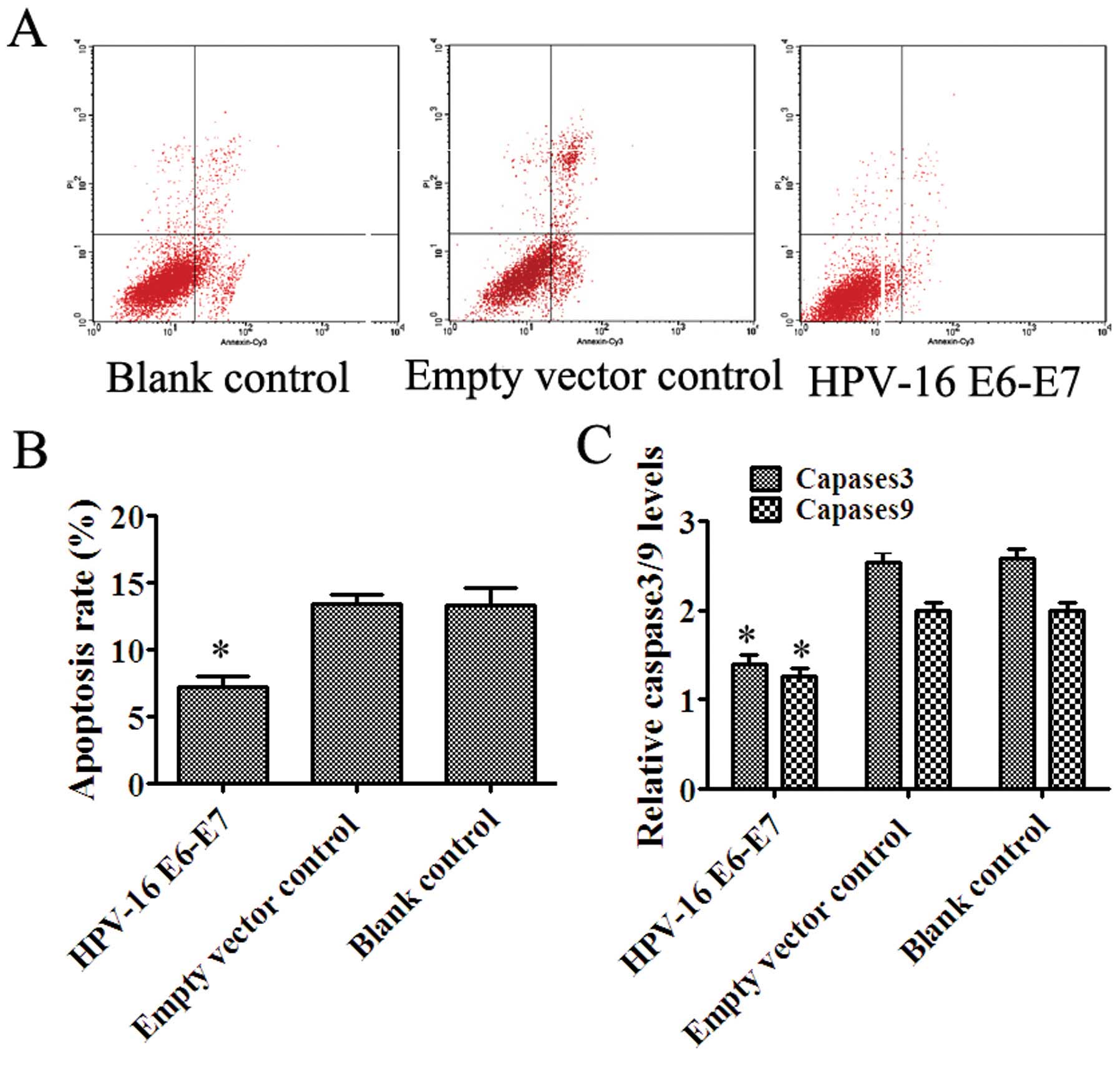
HPV-16 E6-E7 inhibits the apoptosis of
FaDu cells
Apoptosis was determined using flow cytometry and
caspase-3- and caspase-9-specific enzyme-linked immunosorbent
assays (ELISAs). We observed a significant decrease in the number
of Annexin V+ apoptotic FaDu cells that were stably
transfected compared with the numbers in the cells containing the
empty vector (7.246±0.815 vs. 13.464±0.609%; P<0.05) or blank
control (7.246±0.815 vs. 13.298±1.324%; P<0.05). According to
our ELISA results, no significant difference was noted between the
blank and empty vector control (P>0.05), while there was a
significant difference with the HPV-16 E6-E7 group (Fig. 4).
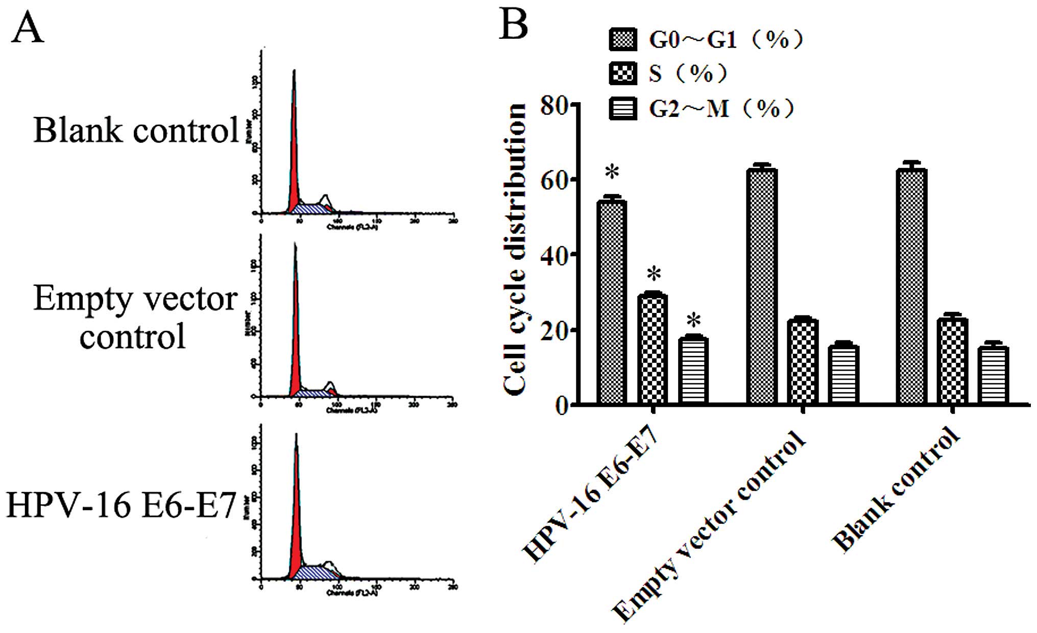
HPV-16 E6-E7 reduces G0/G1 arrest in the
FaDu cells and promotes progression of the cell cycle and cell
proliferation
The proportions of FaDu cells in the G0/G1 phase of
the cell cycle were 53.816±1.665, 62.284±1.609, and 62.262±2.139%
for those that were stably transfected, those transfected with the
empty vector, and the blank control, respectively (Fig. 5).
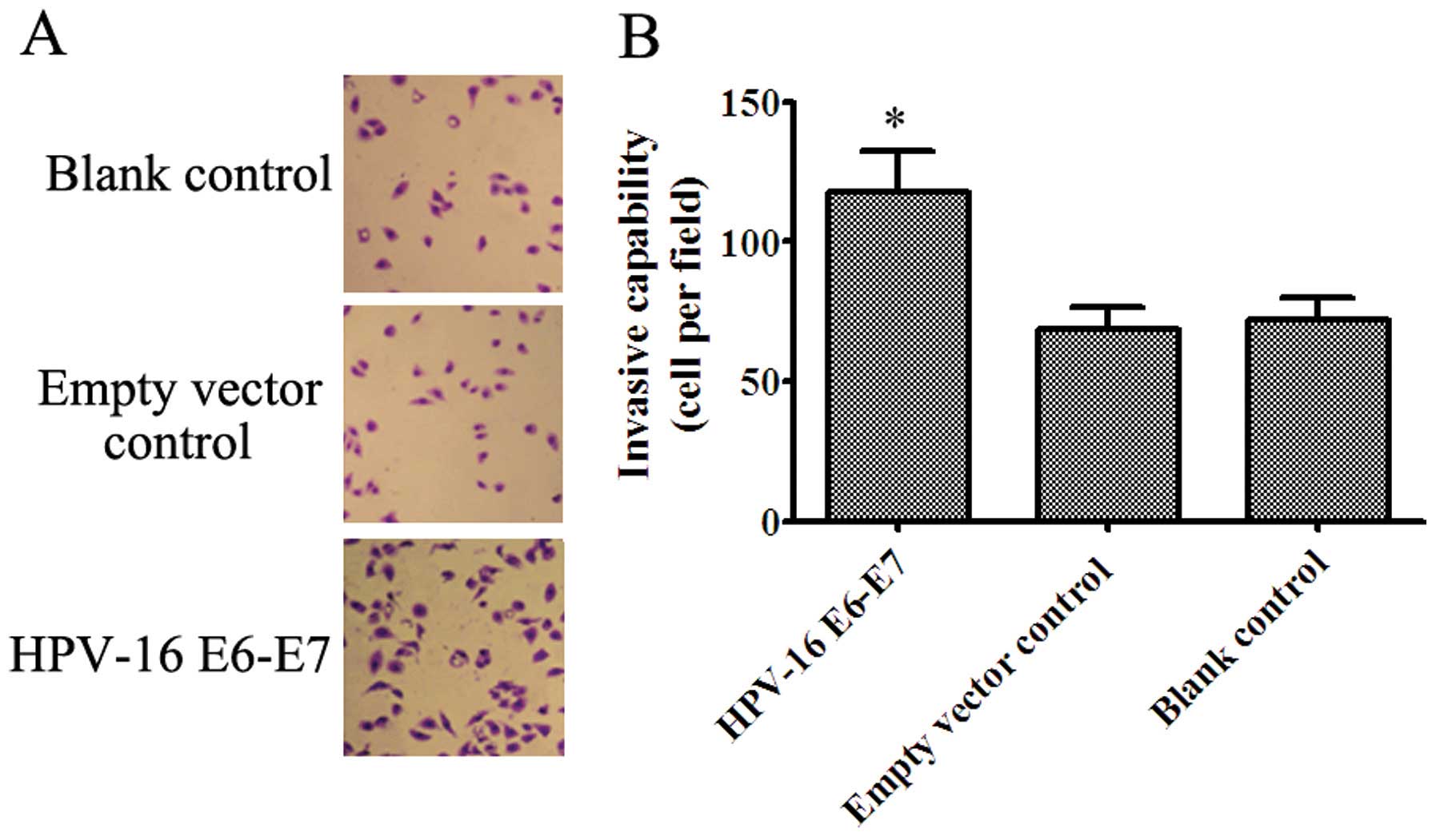
HPV-16 E6-E7 increases the invasive
ability of the FaDu cells
Our in vitro cell invasion assay results
showed that HPV-16 E6-E7 promoted the invasive ability of the FaDu
cells when compared with the control cells (Fig. 6). These results demonstrate that
HPV-16 E6-E7 promotes the invasive ability of FaDu cells in
vitro.
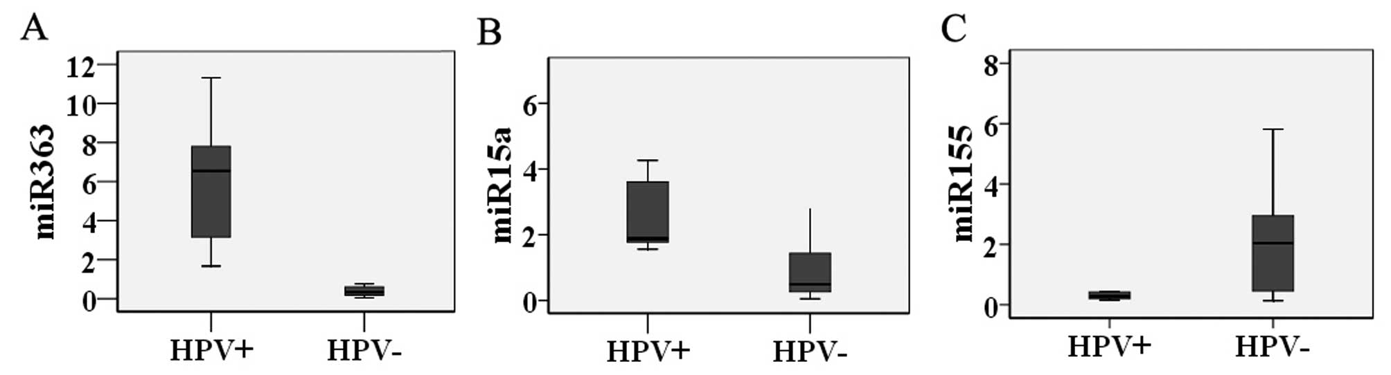
miR-363 and miR-15a are overexpressed in
the HPV-positive hypopharyngeal squamous cell carcinoma
samples
Relative expression levels of miR-363 and miR-15a
were significantly higher in the HPV-positive specimens than these
levels in the HPV-negative specimens. We did not observe a
significant difference in miR-155 levels for specimens that were
HPV-positive/negative and in FaDu cells that stably expressed
HPV-16 E6-E7 (Fig. 7).
Discussion
It is estimated that HNSCC affects 600,000
individuals per year worldwide (25). Smoking has been implicated in
the increased occurrence of HNSCC in developing countries. The role
of HPV has emerged as an important factor in the increase in the
incidence of oropharyngeal tumors affecting non-smokers in
developed countries (26). In
comparison with environment-related HNSCC, patients with
HPV-related malignancies display a better response to treatment and
a lower risk of death and tumor progression (27,2,28–30).
Therefore, we investigated the effects of HPV-16 infection on the
behavior of hypopharyngeal squamous cell carcinoma.
Of the 28 frozen hypopharyngeal squamous cell
carcinoma tissues we examined, 7 were positive for the presence of
HPV, with HPV-16 as the predominant genotype. We generated the
LV-HPV-16-E6-E7 lentivirus to establish a FaDu cell line that
stably expressed HPV-16 E6-E7. Our findings indicate that the E6-E7
proteins of HPV-16 inhibited apoptosis and increased the levels of
proliferation, invasion and metastasis in the transfected FaDu
cells.
In addition, we investigated miRNA expression levels
in hypopharyngeal squamous cell carcinoma tissues and the generated
FaDu cell line. Results from previous studies have demonstrated
that the miRNA expression profiles are altered in HNSCC and that
these changes can be attributed to HPV infection (31,32).
We found that expression levels of miR-363, miR-33 and miR-497 were
upregulated in the HPV-16-positive HNSCC cases. Expression levels
of miR-181a, miR-181b, miR-29a and miR-218 were downregulated, and
this was significant for miR-363 and miR-155.
Results from another study showed that miR-15a
expression was upregulated in HPV-positive HNSCC. In the present
study, we found that miR-15a was upregulated in the hypopharyngeal
squamous cell carcinoma tissues and in LV-HPV-16-E6-E7-infected
FaDu cells. This particular miRNA plays an important role as a
tumor suppressor, and may be associated with a favorable prognosis
in HPV-related HNSCC. It is possible that miR-15a could be used in
the development of miRNA-based therapies for hypopharyngeal
squamous cell carcinoma. We failed to observe any significant
changes in miR-155 expression levels for HPV-positive/negative
hypopharyngeal squamous cell carcinoma tissues and
LV-HPV-16-E6-E7-infected FaDu cells. Findings from previous studies
have shown that miR-155 expression was significantly downregulated
in HNSCC cells that were positive for HPV-16. We speculate that
these contrasting results may be due to inconsistencies between
tumor tissues and tumor-derived cells, and since different
detection methods were used. Future studies to assess the roles of
miR-363, miR-15a, and miR-155 in hypopharyngeal squamous cell
carcinoma are warranted.
Acknowledgments
We are grateful to Professor Guoqiang Zhao for
helpful comments and suggestions during all stages of the project.
This study was partially supported by the Scientific and
Technological Foundation of Henan Province (no. 112102310679).
References
|
1
|
Akao Y, Nakagawa Y, Kitade Y, Kinoshita T
and Naoe T: Downregulation of microRNAs-143 and -145 in B-cell
malignancies. Cancer Sci. 98:1914–1920. 2007. View Article : Google Scholar : PubMed/NCBI
|
|
2
|
Ferlay J, Shin HR, Bray F, Forman D,
Mathers C and Parkin DM: Estimates of worldwide burden of cancer in
2008: GLOBOCAN 2008. Int J Cancer. 127:2893–2917. 2010. View Article : Google Scholar
|
|
3
|
Ang KK, Harris J, Wheeler R, Weber R,
Rosenthal DI, Nguyen-Tân PF, Westra WH, Chung CH, Jordan RC, Lu C,
et al: Human papillomavirus and survival of patients with
oropharyngeal cancer. N Engl J Med. 363:24–35. 2010. View Article : Google Scholar : PubMed/NCBI
|
|
4
|
Stransky N, Egloff AM, Tward AD, Kostic
AD, Cibulskis K, Sivachenko A, Kryukov GV, Lawrence MS, Sougnez C,
McKenna A, et al: The mutational landscape of head and neck
squamous cell carcinoma. Science. 333:1157–1160. 2011. View Article : Google Scholar : PubMed/NCBI
|
|
5
|
Cancer Genome Atlas Network: Comprehensive
genomic characterization of head and neck squamous cell carcinomas.
Nature. 517:576–582. 2015. View Article : Google Scholar : PubMed/NCBI
|
|
6
|
Cancer Genome Atlas Research Network:
Comprehensive molecular characterization of clear cell renal cell
carcinoma. Nature. 499:43–49. 2013. View Article : Google Scholar : PubMed/NCBI
|
|
7
|
Miller DL, Puricelli MD and Stack MS:
Virology and molecular pathogenesis of HPV (human
papillomavirus)-associated oropharyngeal squamous cell carcinoma.
Biochem J. 443:339–353. 2012. View Article : Google Scholar : PubMed/NCBI
|
|
8
|
Funk JO, Waga S, Harry JB, Espling E,
Stillman B and Galloway DA: Inhibition of CDK activity and
PCNA-dependent DNA replication by p21 is blocked by interaction
with the HPV-16 E7 oncoprotein. Genes Dev. 11:2090–2100. 1997.
View Article : Google Scholar : PubMed/NCBI
|
|
9
|
Kehmeier E, Rühl H, Voland B, Stöppler MC,
Androphy E and Stöppler H: Cellular steady-state levels of 'high
risk' but not 'low risk' human papillomavirus (HPV) E6 proteins are
increased by inhibition of proteasome-dependent degradation
independent of their p53- and E6AP-binding capabilities. Virology.
299:72–87. 2002. View Article : Google Scholar : PubMed/NCBI
|
|
10
|
Gillison ML, D'Souza G, Westra W, Sugar E,
Xiao W, Begum S and Viscidi R: Distinct risk factor profiles for
human papillomavirus type 16-positive and human papillomavirus type
16-negative head and neck cancers. J Natl Cancer Inst. 100:407–420.
2008. View Article : Google Scholar : PubMed/NCBI
|
|
11
|
Chaturvedi AK, Engels EA, Pfeiffer RM,
Hernandez BY, Xiao W, Kim E, Jiang B, Goodman MT, Sibug-Saber M,
Cozen W, et al: Human papillomavirus and rising oropharyngeal
cancer incidence in the United States. J Clin Oncol. 29:4294–4301.
2011. View Article : Google Scholar : PubMed/NCBI
|
|
12
|
Karim R, Tummers B, Meyers C, Biryukov JL,
Alam S, Backendorf C, Jha V, Offringa R, van Ommen GJ, Melief CJ,
et al: Human papillomavirus (HPV) upregulates the cellular
deubiquitinase UCHL1 to suppress the keratinocyte's innate immune
response. PLoS Pathog. 9:e10033842013. View Article : Google Scholar : PubMed/NCBI
|
|
13
|
Hashibe M, Brennan P, Benhamou S,
Castellsague X, Chen C, Curado MP, Dal Maso L, Daudt AW, Fabianova
E, Fernandez L, et al: Alcohol drinking in never users of tobacco,
cigarette smoking in never drinkers, and the risk of head and neck
cancer: Pooled analysis in the International Head and Neck Cancer
Epidemiology Consortium. J Natl Cancer Inst. 99:777–789. 2007.
View Article : Google Scholar : PubMed/NCBI
|
|
14
|
Mork J, Lie AK, Glattre E, Hallmans G,
Jellum E, Koskela P, Møller B, Pukkala E, Schiller JT, Youngman L,
et al: Human papillomavirus infection as a risk factor for
squamous-cell carcinoma of the head and neck. N Engl J Med.
344:1125–1131. 2001. View Article : Google Scholar : PubMed/NCBI
|
|
15
|
de Villiers EM, Fauquet C, Broker TR,
Bernard HU and zur Hausen H: Classification of papillomaviruses.
Virology. 324:17–27. 2004. View Article : Google Scholar : PubMed/NCBI
|
|
16
|
Muñoz N, Bosch FX, de Sanjosé S, Herrero
R, Castellsagué X, Shah KV, Snijders PJ and Meijer CJ;
International Agency for Research on Cancer Multicenter Cervical
Cancer Study Group: Epidemiologic classification of human
papillomavirus types associated with cervical cancer. N Engl J Med.
348:518–527. 2003. View Article : Google Scholar : PubMed/NCBI
|
|
17
|
Phelps WC, Barnes JA and Lobe DC:
Molecular targets for human papillomaviruses: Prospects for
antiviral therapy. Antivir Chem Chemother. 9:359–377. 1998.
View Article : Google Scholar
|
|
18
|
Sandhu SK, Volinia S, Costinean S, Galasso
M, Neinast R, Santhanam R, Parthun MR, Perrotti D, Marcucci G,
Garzon R, et al: miR-155 targets histone deacetylase 4 (HDAC4) and
impairs transcriptional activity of B-cell lymphoma 6 (BCL6) in the
Eµ-miR-155 transgenic mouse model. Proc Natl Acad Sci USA.
109:20047–20052. 2012. View Article : Google Scholar
|
|
19
|
Lenze D, Leoncini L, Hummel M, Volinia S,
Liu CG, Amato T, De Falco G, Githanga J, Horn H, Nyagol J, et al:
The different epidemiologic subtypes of Burkitt lymphoma share a
homogenous micro RNA profile distinct from diffuse large B-cell
lymphoma. Leukemia. 25:1869–1876. 2011. View Article : Google Scholar : PubMed/NCBI
|
|
20
|
Lajer CB, Nielsen FC, Friis-Hansen L,
Norrild B, Borup R, Garnæs E, Rossing M, Specht L, Therkildsen MH,
Nauntofte B, et al: Different miRNA signatures of oral and
pharyngeal squamous cell carcinomas: A prospective translational
study. Br J Cancer. 104:830–840. 2011. View Article : Google Scholar : PubMed/NCBI
|
|
21
|
Lace MJ, Anson JR, Klingelhutz AJ, Lee JH,
Bossler AD, Haugen TH and Turek LP: Human papillomavirus (HPV) type
18 induces extended growth in primary human cervical, tonsillar, or
foreskin keratinocytes more effectively than other high-risk
mucosal HPVs. J Virol. 83:11784–11794. 2009. View Article : Google Scholar : PubMed/NCBI
|
|
22
|
Rahimy E, Kuo SZ and Ongkeko WM:
Evaluation of non-coding RNAs as potential targets in head and neck
squamous cell carcinoma cancer stem cells. Curr Drug Targets.
15:1247–1260. 2014. View Article : Google Scholar : PubMed/NCBI
|
|
23
|
Li M, Liu L, Zang W, Wang Y, Du Y, Chen X,
Li P, Li J and Zhao G: miR-365 overexpression promotes cell
proliferation and invasion by targeting ADAMTS-1 in breast cancer.
Int J Oncol. 47:296–302. 2015.PubMed/NCBI
|
|
24
|
Schmittgen TD and Livak KJ: Analyzing
real-time PCR data by the comparative C(T) method. Nat Protoc.
3:1101–1108. 2008. View Article : Google Scholar : PubMed/NCBI
|
|
25
|
Walter V, Yin X, Wilkerson MD, Cabanski
CR, Zhao N, Du Y, Ang MK, Hayward MC, Salazar AH, Hoadley KA, et
al: Molecular subtypes in head and neck cancer exhibit distinct
patterns of chromosomal gain and loss of canonical cancer genes.
PLoS One. 8:e568232013. View Article : Google Scholar : PubMed/NCBI
|
|
26
|
Lajer CB and von Buchwald C: The role of
human papillomavirus in head and neck cancer. APMIS. 118:510–519.
2010. View Article : Google Scholar : PubMed/NCBI
|
|
27
|
Scheffner M, Huibregtse JM, Vierstra RD
and Howley PM: The HPV-16 E6 and E6-AP complex functions as a
ubiquitin-protein ligase in the ubiquitination of p53. Cell.
75:495–505. 1993. View Article : Google Scholar : PubMed/NCBI
|
|
28
|
Fakhry C, Westra WH, Li S, Cmelak A, Ridge
JA, Pinto H, Forastiere A and Gillison ML: Improved survival of
patients with human papillomavirus-positive head and neck squamous
cell carcinoma in a prospective clinical trial. J Natl Cancer Inst.
100:261–269. 2008. View Article : Google Scholar : PubMed/NCBI
|
|
29
|
Flores ER, Allen-Hoffmann BL, Lee D and
Lambert PF: The human papillomavirus type 16 E7 oncogene is
required for the productive stage of the viral life cycle. J Virol.
74:6622–6631. 2000. View Article : Google Scholar : PubMed/NCBI
|
|
30
|
O'Rorke MA, Ellison MV, Murray LJ, Moran
M, James J and Anderson LA: Human papillomavirus related head and
neck cancer survival: A systematic review and meta-analysis. Oral
Oncol. 48:1191–1201. 2012. View Article : Google Scholar : PubMed/NCBI
|
|
31
|
Wald AI, Hoskins EE, Wells SI, Ferris RL
and Khan SA: Alteration of microRNA profiles in squamous cell
carcinoma of the head and neck cell lines by human papillomavirus.
Head Neck. 33:504–512. 2011. View Article : Google Scholar :
|
|
32
|
Lajer CB, Garnæs E, Friis-Hansen L,
Norrild B, Therkildsen MH, Glud M, Rossing M, Lajer H, Svane D,
Skotte L, et al: The role of miRNAs in human papilloma virus
(HPV)-associated cancers: Bridging between HPV-related head and
neck cancer and cervical cancer. Br J Cancer. 106:1526–1534. 2012.
View Article : Google Scholar : PubMed/NCBI
|