Introduction
Osteosarcoma is the most common malignant bone tumor
(1). The survival outcomes remain
unsatisfactory since recurrences are common due to the development
of invasion, distant metastasis and chemoresistance.
Chemoresistance, both intrinsic and acquired, is a main cause of
recurrence or failure of current treatment (2). Up to date, several factors contributed
to the development of chemoresistance including genetic alterations
(3), altered drug accumulation
(4), drug-target amplification
(5) and autophagy (6). However, the underlying molecular
mechanism for chemoresistance remains unclear.
MicroRNAs (miRNAs) are small (22-nucleotide)
non-coding single stranded RNAs that regulate gene expression by
binding to the 3′-untranslated region (3′UTR) of their target
mRNAs, modulating mRNA stability and protein expression at the
post-transcriptional level. The aberrant expression of miRNAs has
been implicated in the mechanism of chemo-resistance (7–9) and
several miRNAs have been identified in osteosarcoma tissues and
osteosarcoma cell lines (10,11).
miR-146b-5p was originally verified as involved in
inflammatory bowel disease and was later shown upregulated in IL-10
deficient mice (12). Subsequently,
it was found that miR-146b-5p alleviated intestinal injury in mouse
colitis via the activation of NF-κB. Recently, miR-146b-5p has been
reported to be involved in solid tumors, including prostate,
pancreatic cancer, malignant gliomas and glioblastoma. Moreover,
two studies demonstrated that its involvement in chemoresistance in
thyroid cancer (13) and lymphoma
(14). However, the role of
miR-146b-5p in osteosarcoma chemoresistance remains elusive.
Activation of Wnt/β-catenin is a crucial step in the
process of tumorigenesis and chemoresistance (15). Zinc and ring finger 3 (ZNRF3), an
cell-surface transmembrane E3 ubiquitin ligase, negatively
regulates Wnt/β-catenin signalling by promoting the turnover of
frizzled and LRP6. Originally, ZNRF3 was found to regulate the
growth and survival of embryos stem cells and two previous studies
demonstrated that ZNRF3-deficient embryos died around birth
(16,17). Moreover, ZNRF3 is also involved in
the process of tumorigenesis, migration and invasiveness. Deng
et al (13) reported that
ZNRF3 suppressed the effect of miR-146b-5p on migration,
invasiveness and proliferation of papillary thyroid cancer cells.
ZNRF3 has also been confirmed to be linked to pancreatic ductal
adenocarcinoma and mucinous ovarian tumors (18,19).
To date, however, the exact role of miR-146b in
osteosarcoma is not well understood. In the present study, we
investigated the expression of miR-146b-5p in osteosarcoma tissues.
We demonstrated osteosarcoma samples frequently featured highly
expressed miR-146b-5p comparing to the normal tissue counterparts.
An elevated expression of miR-146b-5p was observed in human
osteosarcoma tissues after chemotherapy. Further study showed that
overexpression of miR-146b-5p promoted invasion, metastasis and
chemo-resistance of osteosarcoma cells. In addition, ZNRF3 was
identified as the target gene of miR-146b-5p in osteosarcoma. All
these results indicate miR-146b-5p and its downstream target gene
ZNRF3 can be used to treat osteosarcoma chemoresistance in the
future.
Materials and methods
Tissue samples and papillary carcinoma
cell lines
All research involving human tissue samples was
approved by the Ethics Review Committee of Second Military Medical
University Shanghai and written informed consent was obtained from
all participating patients. Tumor specimens and the adjacent
non-cancer tissues (ANCT) were obtained from 35 osteosarcoma
patients who underwent surgery in Shanghai Changzheng Hospital
between January 2010 and January 2013. Among them, 13 patients had
received chemotherapy at the time of the operation. Ten patients
were confirmed to have metastasis.
Cell culture
Human osteosarcoma hFOB1.19, MG-63 and U-2 OS cell
lines were purchased from ATCC and reserved in our laboratory as
previously described (20). Human
MG-63 and U-2 OS cell lines were maintained in high glucose DMEM
medium supplemented with 10% fetal bovine serum (FBS; HyClone), 100
U/ml penicillin, and 100 µg/ml streptomycin, and were
cultured in an incubator maintained at 5% CO2 and
37°C.
Quantitative real-time RT-PCR
Total RNA containing miRNA was extracted from
surgical specimens or cell lines with TRIZol reagent (Invitrogen,
Carlsbad, CA, USA) according to the manufacturer's instructions.
For miRNA expression analysis, the miRNA was transcribed into cDNA
using a TaqMan MicroRNA Reverse Transcription kit (Applied
Biosystems) and RT primers provided with the miR-146b-5p TaqMan
miRNA assay (Applied Biosystems) according to the manufacturer's
instructions. miR-146b-5p and U6 small nuclear 2 (U6) expression
was detected from the cDNA product, using TaqMan miRNA
sequence-specific probes using SYBR Premix Ex Taq (Takara, Shiga,
Japan) and ABI Prism 7500 sequence detection system (Applied
Biosystems). U6 was used as a control. The fold-change in gene
expression was calculated by the 2−ΔΔCT method.
Overexpression and knockdown of
miR-146b-5p
Recombinant lentiviruses containing overexpression
of miR-146b-5p, or knocked down miR-146b-5p and miRNA control were
purchased from Shanghai GeneChem Co., Ltd. (Shanghai, China). To
generate the stable cell line, human MG-63 osteosarcoma cells were
transfected with lentiviruses containing overexpression of
miR-146b-5p (OE), knocked down miR-146b-5p or a blank lentivirus
expression vectors (blank). After incubation for 72 h, the
supernatant containing lentivirus was removed and replaced with
complete culture medium. Infection efficiency was confirmed by
RT-PCR 96 h after transfection and the cells were selected with 2
µg/ml to establish stable cell lines.
miRNA target prediction
Candidate targets of miR-146b-5p and ZNRF3 were
predicted by miRBase (http://www.mirbase.org/) and TargetScan (http://www.Targetscan.org/).
Luciferase reporter assay
For validation of ZNRF3 as a target gene of
miR-146b-5p in osteosarcoma cells, luciferase assay was performed
as previously described (21).
Briefly, the target sequence was amplified by PCR using the
following primers: forward, 5′-ACTAGTGTATAGCAGCACATTTCATTT-3′ and
the reverse primer 5′-AAGCTTCAGGGTCTTGTGTTAGTC-3′ and cloned into
pGL3-control vector through KpnI and XhoI sites.
Mimic was synthesized according to the sequence of miR-146b-5p. A
ZNRF3 3′-UTR segment (516 bp) containing the predicted miR-146b-5p
binding site was cloned into the pGL3-control vector (Promega
Corp., Madison, WI, USA) downstream of the firefly luciferase gene,
after which the 3′-UTR luciferase reporter was obtained. The
miR-146b-5p mimic and miR-146b-5p inhibitor used in the present
study were synthesized and provided by the GenePharma.
Western blot analysis
Protein extracts from cells were prepared through a
modified lysis buffer and then performed as previously described
(20). The immunoreactive bands
were visualized using ECL-PLUS/kit. The relative protein level was
normalized to β-actin concentration. Antibodies against matrix
metalloproteinase-2 (MMP-2), MMP-9, MMP-16, cleaved caspase-3,
caspase-3, cleaved PARP PARP, ZNRF3 and β-actin were purchased from
Cell Signaling Technology, Inc. (Danvers, MA, USA).
Cell proliferation assay
Cell growth was determined by the MTT assay
according to the manufacturer's instructions. Briefly, cells in OE,
KD and control groups were incubated in 96-well plates at a density
of 1×10⁴ cells/well. Cells were treated with 10
µl MTT reagents at 0, 1, 2, 3, 4, 5, 6 and 7 days and then
measured at 450 nm with enzyme immunoassay analyzer (Bio-Rad
Laboratories, Hercules, CA, USA).
Migration and invasion assays
The invasive potential of cells was measured in 6.5
mm Transwell with 8.0 mm pore polycarbonate membrane insert
(Corning, NY, USA). The filter of the top chamber was
matrigel-coated with 50 µl of diluted matrigel and the lower
chamber was filled with 500 µl of DMEM containing 10% FBS.
Cells were resuspended in migration medium (serum-free medium) and
added into each top chamber. After the cells were incubated for 16
h, the non-invading cells on the upper surface were removed by
using a cotton swab. The invasive cells on the lower surface of the
membrane insert were fixed with 4% paraformaldehyde for 30 min, and
then stained with hematoxylin-eosin. The number of cells on the
lower surface, which had invaded through the membrane were counted.
The procedure for Transwell migration assays were the same as the
Transwell invasion assay except that the filter of top chamber was
not coated with Matrigel. The number of cells were counted in five
random fields of each chamber under the microscope.
Wound healing assay
When MG-63 cells in OE, KD and control groups were
grown to confluence, a scratch was made with a micropipette tip.
Following incubation for 24 and 48 h, the images were captured. The
migration potential was estimated by counting the cells that
migrated from the wound edge. The cell migration rate was obtained
by counting three fields per area and presented as the average of
six independent evaluations.
Measurement of apoptosis by flow
cytometry
The incidence of apoptosis after chemotherapy was
assessed by using the Annexin V-FITC/PI apoptosis detection kit (BD
Pharmingen, San Diego, CA, USA) according to our previous method
(20). Apoptotic cells, including
those staining positive for Annexin V-FITC and negative for PI and
those that were double positive, were counted as a percentage of
the total cell count.
Statistical analysis
SPSS 13.0 was used for the statistical analysis.
One-way analysis of variance (ANOVA) was used to analyze the
differences between groups. The LSD method of multiple comparisons
was employed when the probability for ANOVA was statistically
significant. Statistical significance was P<0.05.
Results
miR-146b-5p is upregulated in human
osteosarcoma tissues after chemotherapy and correlates with
multiple clinical features
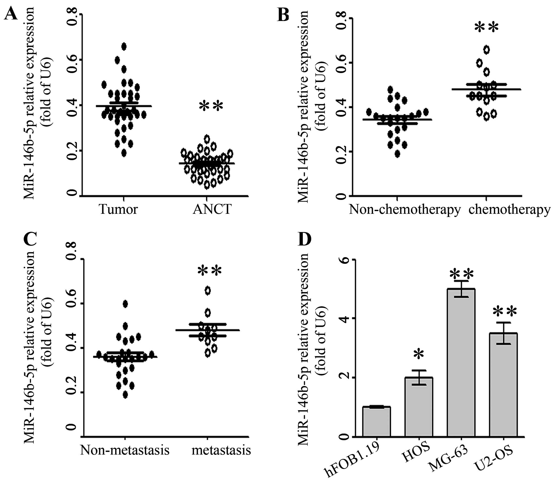
First, we tested the expression of miR-146b-5p by
qRT-PCR and normalized against an endogenous control (U6 RNA) in 35
pairs of tumor tissues and matched adjacent non-tumor tissues
(ANCT) from osteosarcoma patients who received tumor resection. The
expression level of miR-146b-5p in osteosarcoma tissues was
significantly higher than that in matched ANCT (P<0.05; Fig. 1A). Next, osteosarcoma tissues sample
were divided into patients that received chemotherapy (chemotherapy
group) and those that did not (non-chemotherapy group).
As compared with non-chemotherapy group, miR-146b-5p
levels were prominently upregulated in chemotherapy group
(P<0.05; Fig. 1B), indicating
that miR-146b-5p was involved in chemoresistence. Moreover,
miR-146b-5p levels were obviously increased in tumor tissues for
patients with tumor recurrence and lung metastasis as compared with
those in tumor tissues arising from patients without tumor
recurrence or metastasis (P<0.05, P<0.05; Fig. 1C and D), suggesting that miR-146b-5p
was correlated with metastasis and recurrence of osteosarcoma. In
addition, we analyzed miR-146b-5p expression in osteosarcoma cell
lines (HOS, MG-63 and U2-OS) and human fetal osteoblastic 1.19
(hFOB1.19) cells. The miR-146b-5 expression was significantly
upregulated in all osteosarcoma cell lines as compared with that in
hFOB1.19 (P<0.05; Fig. 1D).
These data indicate that elevated miR-146b-5p expression
contributed to chemoresistence and metastatic potential of
osteosarcoma cells.
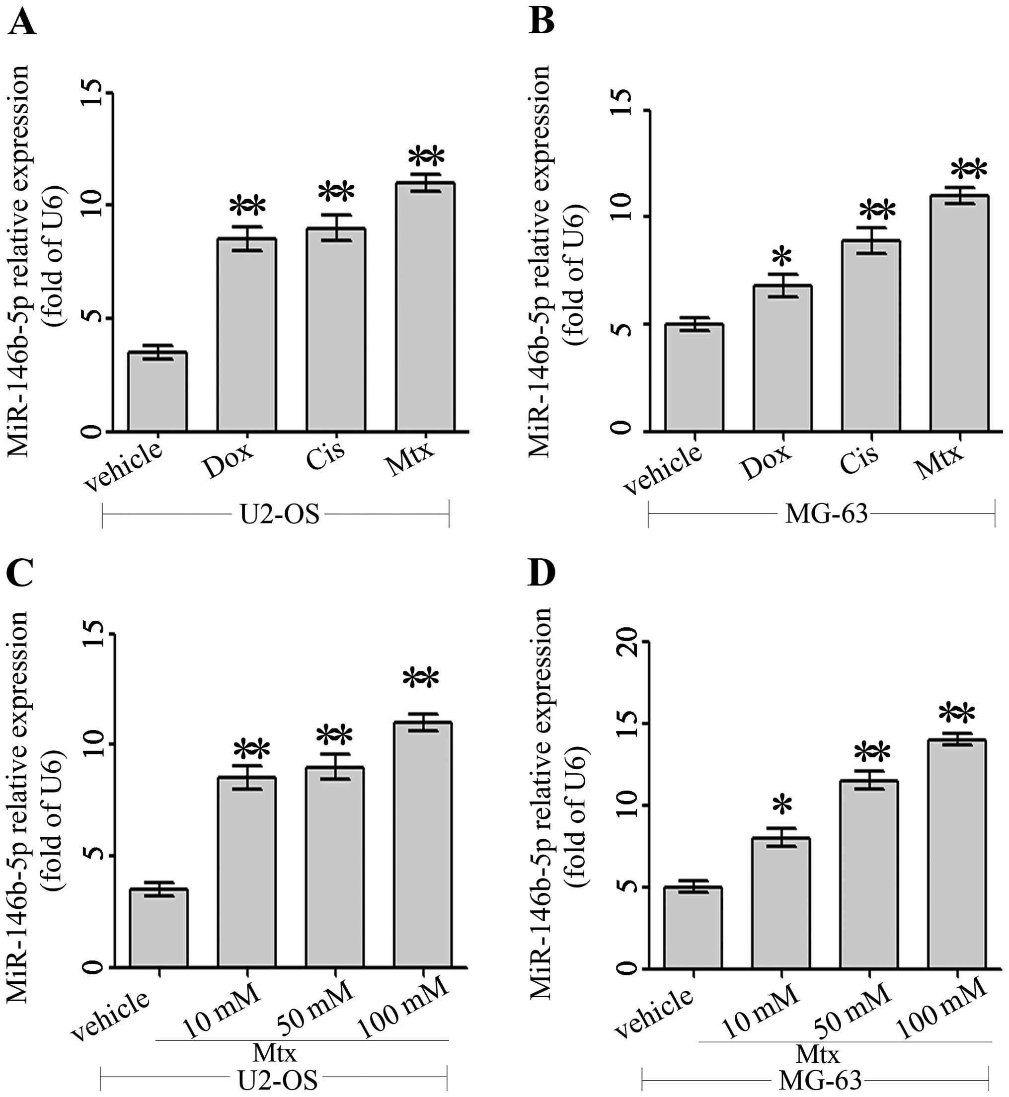
Anticancer agents promote miR-146b-5p
expression in osteosarcoma cells
The role of miR-146b-5p in different cancer cells is
controversial. The exact role of miR-146b-5p in osteosarcoma
remains to be clarified. To address this question, the expression
of miR-146b-5p in human osteosarcoma MG-63 and U-2 OS cell lines
after anticancer agents was evaluated using qRT-PCR. As doxorubicin
(Dox), cisplatin (Cis), and methotrexate (Mtx) are commonly used
anticancer agents in osteosarcoma, we assayed the effects of the
anticancer agents on the expression of miR-146b-5p. As shown in
Fig. 2A and B, anticancer agents
significantly enhanced expression of miR-146b-5p in the human
osteosarcoma cell lines MG-63, and U-2 OS. Moreover, this effect
was dose-dependent in the case of Mtx in MG-63 and U-2 OS cell
lines (Fig. 2C and D). These
findings show that miR-146b-5p expression was upregulated during
chemotherapy in osteosarcoma cells.
miR-146b-5p promotes migration and
invasion of osteosarcoma in vitro
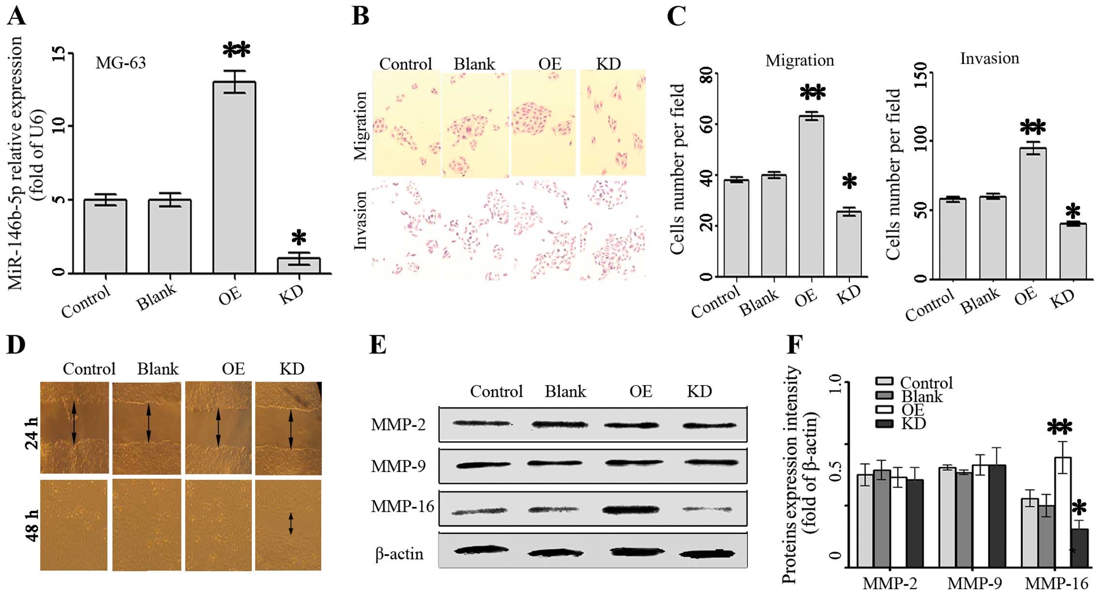
Next, we applied a lentivirus system to make stable
cell lines to knock down and overexpress miR-146b-5p based on the
osteosarcoma cell line MG-63 including a blank group (untransfected
cells), a control group (cells transfected with the control
lentivirus), an OE group (overexpressing miR-146b-5p) and a KD
group (knocked down miR-146b-5p). The miR-146b-5p expression levels
in these groups were evaluated using qRT-PCR. As expected, the
expression of miR-146b-5p was dramatically suppressed in KD group
cells (P<0.05) while increased in the OE group (P<0.01;
Fig. 3A).
We then performed Transwell migration and invasion
assay to investigate the effects of miR-146b-5p on the migratory
and invasive behavior of osteosarcoma cells in vitro. In the
migration assay, the number of OE MG-63 cells (63.0±5.9, P<0.01)
passing through the Matrigel were significantly higher than control
cells (40.0±3.8) (Fig. 3C).
Whereas, the number of KD MG-63 cells (25.5±6.0, P<0.05) were
significantly less than that in control cells (40.0±3.8). No
significant difference was observed between the blank and control
cells (P=0.68) (Fig. 3C). Invasion
assay showed that the number of OE cells (95.0±15.0, P<0.01)
passing through the Matrigel were significantly higher than control
cells (58.6±6.5) (Fig. 3C).
Similarly, KD MG-63 cells were significantly less than that in
control cells (P<0.05) (Fig.
3D). These results strongly indicated that miR-146b-5p played a
role in the migratory and invasive potential of osteosarcoma in
vitro. In addition, wound healing assay was also used to
examine the effects of miR-146b-5p on the migration ability. Cells
in OE groups exhibited an obvious increase in migration rate as
compared to the other three groups. KD MG-63 cells slightly failed
to close the wound at 72 h after incubation, whereas the other
three groups were able to close the wound at the same time-point
(Fig. 3D).
In addition, western blot assay was performed to
investigate the effect of miR-146b-5p on the endogenous expression
of MMP-2, MMP-9 and MMP-16 protein since they were involved in
tumor invasion and metastasis based on previous research (22–25).
As shown in Fig. 3C, a significant
increase of MMP-16 expression in the OE group while a reduction in
the KD group as compared with control groups was observed
(P<0.05). These data demonstrated that miR-146b-5p might promote
osteosarcoma invasion and metastasis through regulation of MMP-16
expression.
miR-146b-5p increased chemoresistance in
osteosarcoma MG-63 cells
To confirm the role of miR-146b-5p in osteosarcoma,
the cell proliferation were assessed in the MG-63 cells with
overexpressed or knocked down miR-146b-5p. As a result, neither the
knockdown nor overexpression of miR-146b-5p could alter cell growth
in MG-63 cells (Fig. 4A).
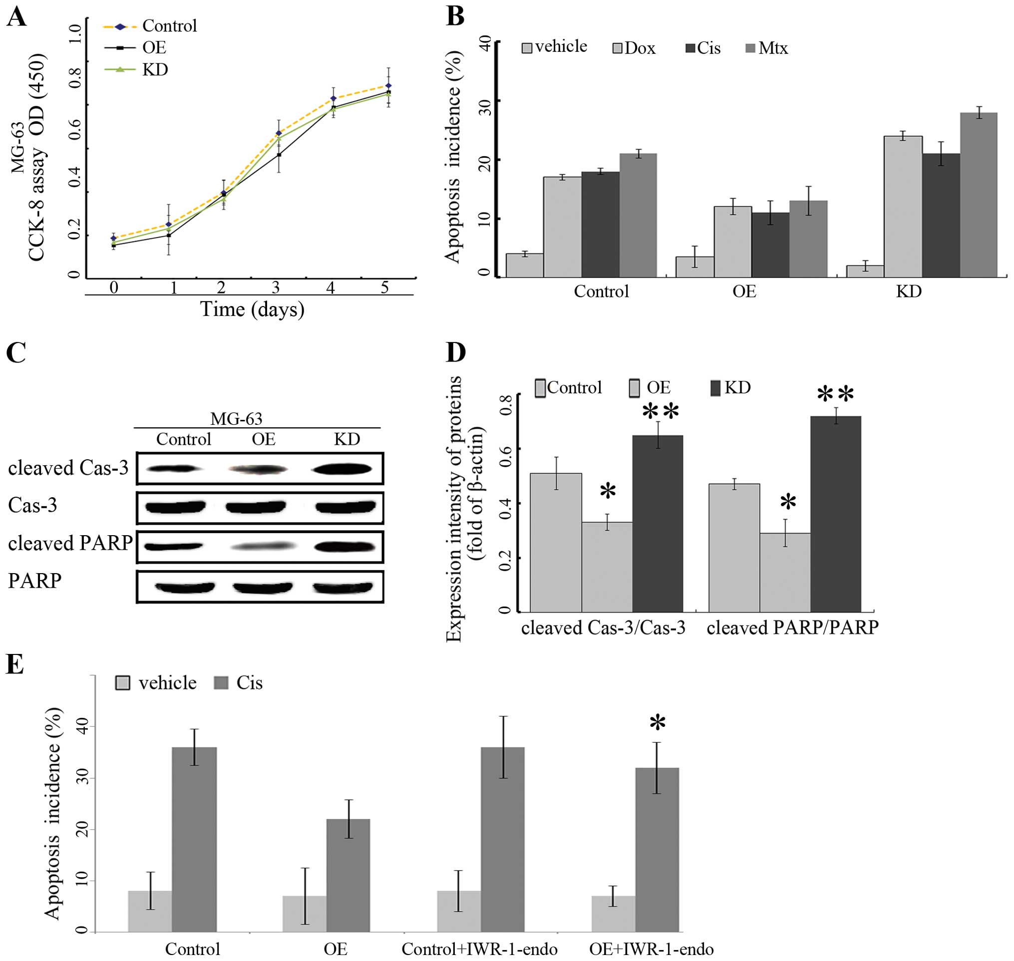 | Figure 4Overexpression of miR-146b-5p
increases resistance to chemotherapy in vitro. (A) CCK-8
assay for miR-146b-5p overexpression (OE) knockdown (KD) and
control. No difference was found among OE, KD and control groups.
(B) Annexin V-FITC/PI staining for apoptotic cells. MG63 cells in
OE, KD and control groups were treated with vehicle (normal
saline), Dox (0.2 mg/ml), Cis (20 mmol/l), and Mtx (50 mmol/l) for
24 h and then the apoptosis incidence was quantified by flow
cytometer. Overexpression of miR-146b-5p reduced the incidence of
apoptosis induced by Dox, Cis and Mtx. (*P<0.05,
**P<0.01 vs. vehicle group). Representative blots (C)
and quantitative results (D) of western blot analysis for cleaved
caspase-3, caspase-3, cleaved PARP, and PARP protein expression.
Both the ratio of cleaved caspase-3 to caspase-3 expression and the
ratio of cleaved PARP to PARP were significantly reduced in OE
group compared with KD and control groups. (E) Annexin V-FITC/PI
staining for apoptotic cells. MG63 cells in OE and control groups
were treated with vehicle (normal saline) or Cis (20 mmol/l) in
combination with 300 nM IWR-1-endo. The apoptosis was assessed by
flow cytometer. *P<0.05 OE+ IWR-1-endo vs. OE. |
Next, to explore the potential role for miR-146b-5p
in chemoresistance, apoptosis in MG-63 cells was induced by vehicle
(veh), doxorubicin (Dox), cisplatin (Cis) and methotrexate (Mtx).
Overexpression of miR-146b-5p increased chemoresistance, while
knockdown of miR-146b-5p in these cells rendered them significantly
more sensitive to Dox-, Cis-, and Mtx-induced cell apoptosis
(Fig. 4B). In the case of
cisplatin, the apoptosis markers, cleaved caspase-3 and cleaved
PARP proteins were assessed in KD, OE and control group, cleaved
caspase-3 and cleaved PARP proteins were suppressed in OE group.
However, cleaved caspase-3 and cleaved PARP were increased in KD
group (Fig. 4C and D). These data
suggested that miR-146b-5p increased chemoresistance possibly
through suppressing the apoptotic pathway.
We investigated whether the Wnt/β-catenin signaling
was involved in the miR-146b-5p mediated chemoresistance as
previous studies demonstrated that Wnt/β-catenin played an
important role in chemoresistance (26,27).
The cell apoptosis was assessed in osteosarcoma overexpressing
miR-146b-5p (OE group) in combination with or without the Wnt
inhibitor (IWR-1-endo). Our results showed that the decrease in
apoptosis of OE cells caused by miR-146b-5p overexpression was
significantly suppressed by IWR-1-endo (Fig. 4E). These results suggested that the
effect of miR-146b-5p on increasing the chemoresistance of
osteosarcoma cells was mediated by Wnt/β-catenin signaling.
ZNRF3 is a target of miR-146b-5p, and
highly expressed in osteosarcoma after chemotherapy in vivo and in
vitro
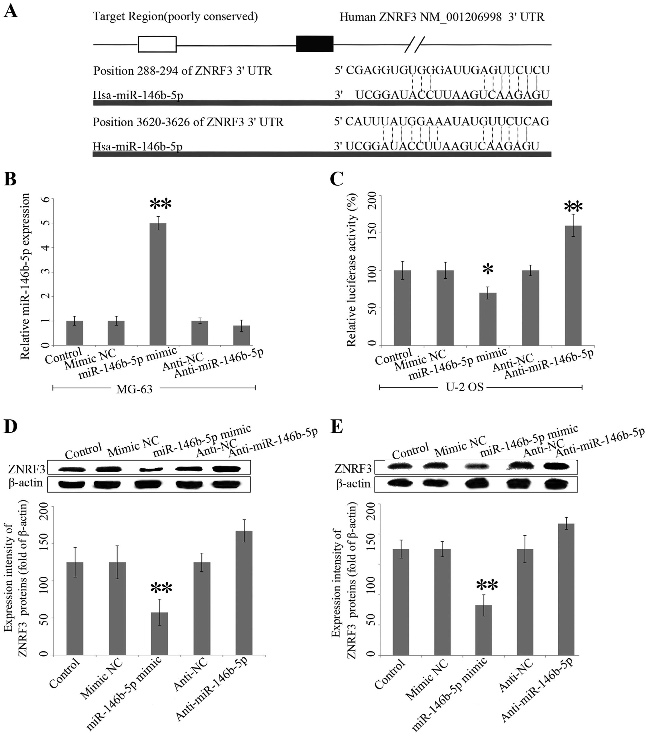
To investigate the mechanism of miR-146b-5p in
chemoresistance, bioinformatics analysis demonstrated that the
3′-UTR region of ZNRF3 were identified as the binding sites for
miR-146b-5p (Fig. 5A). To further
verify that ZNRF3 was a direct target of miR-146b-5p, luciferase
assays were performed and mimic NC, miR-146b-5p mimics, anti-NC or
anti-miR-146b-5p were transfected into the MG-63 cell lines. Our
results demonstrated that miR-146b-5p expression was high in
miR-146b-5p mimics group (Fig. 5B).
Luciferase reporter assays showed that miR-146b-5p mimics
significantly inhibited the luciferase activity of ZNRF3 3′-UTR by
~30% in MG-63 cells relative to the control, whereas
co-transfection with anti-miR-146b-5p significantly promoted
luciferase activity by 160% (P<0.05) (Fig. 5C).
To further confirm that miR-146b-5p modulates the
expression of ZNRF3, MG-63 and U-2 OS cells were transfected with
miR-146b-5p mimics, anti-miR-146b mimics or the respective
controls. Subsequently, the protein expression of ZNRF3 was
analyzed by western blot analysis. Transfection of the miR-146b-5p
mimics significantly decreased the protein level of ZNRF3, whereas
anti-miR-146b-5p had the opposite effects (Fig. 5D and E).
Discussion
Osteosarcoma is one of the predominant tumors in
children and chemoresistance is a leading cause of mortality for
osteosarcoma patients. The molecular mechanism for chemoresistance
remains unclear. Previously, we identified several genes and
proteins regarding chemoresistance ability, including, high
mobility group nucleosome binding domain 5 (20,28),
tumor necrosis factor-α-inducible protein-1 (29) and nucleosome-binding protein
(30). However, knockdown of these
genes only partly suppress the chemoresistance, suggesting that
these genes exert only a part of their biological effects as
chemoresistance. MicroRNAs are important regulators in
tumorigenesis as well as chemoresistance. Increasing evidence
demonstrated that miR-146b-5p played an oncogenic role in papillary
thyroid carcinoma (31), pancreatic
cancer (21) and lung cancer cells
(32). However, to date, the role
of miR-146b-5p in osteosarcoma is unclear.
To confirm the biological role of miR-146b-5p in
osteosarcoma, we first assessed the miR-146b-5p expression in human
osteosarcoma tissues before and after chemotherapy, tumor and ANCT,
tumor with metastasis or without. The expression of miR-146b-5p was
also evaluated in hFOB1.19 osteoblast cell line, HOS, MG-63 and
U2-OS osteosarcoma cell lines. As expected, these results suggested
that miR-146b-5p was highly expressed in osteosarcoma tissues,
especially after chemotherapy. Furthermore, the expression of
miR-146b-5p was upregulated during chemotherapy in osteosarcoma
cells in vitro. These data suggested that miR-146b-5p was
possibly involved in chemoresistence and metastatic potential of
osteosarcoma cells. To confirm our hypothesis, we applied
lentivirus system to make stable cell lines to knock down and
overexpress miR-146b-5p and our results showed that miR-146b-5p
promoted osteosarcoma invasion and metastasis through regulation of
MMP-16 expression. In the present study, gain-of-function studies
demonstrated that miR-146b-5p overexpression reduced the apoptosis
incidence and apoptosis marker expression, suggesting at
miR-146b-5p plays a crucial role in the chemoresistance of
osteosarcoma.
The Wnt/β-catenin pathway plays a crucial role in
the chemoresistance of osteosarcoma (33) and we also investigated whether
Wnt/β-catenin pathway was involved in chemoresistance of
osteosarcoma. As expected, IWR-1-endo, an inhibitor of
Wnt/β-catenin signaling, could restore the decrease in apoptosis of
OE cells caused by miR-146b-5p overexpression, suggested that the
effect of miR-146b-5p on increasing the chemoresistance is mediated
by Wnt/β-catenin signaling.
To investigate the mechanism of miR-146b-5p in
regulating Wnt/β-catenin signaling, Bioinformatics analysis was
performed which showed that the 3′-UTR region of ZNRF3 were the
binding sites for miR-146b-5p. A similar role for ZNRF3 was
demonstrated in human gastric adenocarcinoma and papillary thyroid
carcinoma, showing that ZNRF3 promoted apoptosis through the
modulation of the Wnt/β-catenin signaling pathway (13,34).
To the best of our knowledge, this is the first
report of miR-146b-5p in the chemoresistance of osteosarcoma.
Although our research demonstrated that miR-146b-5p participated in
migration, invasion and chemoresistance in osteosarcoma, the use of
osteosarcoma cells from two cell lines provide very limited
evidence. Further research using more cell lines and primary tumors
in animal studies is necessary to confirm the findings of the
present study. In conclusion, our investigation revealed that the
miR-146b-5p affects migration, invasion and chemoresistance in
osteosarcoma possibly via downregulation of ZNRF3.
References
|
1
|
Jemal A, Bray F, Center MM, Ferlay J, Ward
E and Forman D: Global cancer statistics. CA Cancer J Clin.
61:69–90. 2011. View Article : Google Scholar : PubMed/NCBI
|
|
2
|
Scotlandi K, Picci P and Kovar H: Targeted
therapies in bone sarcomas. Curr Cancer Drug Targets. 9:843–853.
2009. View Article : Google Scholar : PubMed/NCBI
|
|
3
|
Lønning PE and Knappskog S: Mapping
genetic alterations causing chemoresistance in cancer: Identifying
the roads by tracking the drivers. Oncogene. 32:5315–5330. 2013.
View Article : Google Scholar : PubMed/NCBI
|
|
4
|
Derdak Z, Mark NM, Beldi G, Robson SC,
Wands JR and Baffy G: The mitochondrial uncoupling protein-2
promotes chemoresistance in cancer cells. Cancer Res. 68:2813–2819.
2008. View Article : Google Scholar : PubMed/NCBI
|
|
5
|
Yamamoto S, Tsuda H, Honda K, Onozato K,
Takano M, Tamai S, Imoto I, Inazawa J, Yamada T and Matsubara O:
Actinin-4 gene amplification in ovarian cancer: A candidate
oncogene associated with poor patient prognosis and tumor
chemoresistance. Mod Pathol. 22:499–507. 2009. View Article : Google Scholar : PubMed/NCBI
|
|
6
|
Zhang W, Li Q, Song C and Lao L: Knockdown
of autophagy- related protein 6, Beclin-1, decreases cell growth,
invasion, and metastasis and has a positive effect on
chemotherapy-induced cytotoxicity in osteosarcoma cells. Tumour
Biol. 36:2531–2539. 2014. View Article : Google Scholar
|
|
7
|
Zhang Y, Duan G and Feng S: MicroRNA-301a
modulates doxorubicin resistance in osteosarcoma cells by targeting
AMP-activated protein kinase alpha 1. Biochem Biophys Res Commun.
459:367–373. 2015. View Article : Google Scholar : PubMed/NCBI
|
|
8
|
Zhao Z, Zhang L, Yao Q and Tao Z: miR-15b
regulates cisplatin resistance and metastasis by targeting PEBP4 in
human lung adenocarcinoma cells. Cancer Gene Ther. 22:108–114.
2015. View Article : Google Scholar : PubMed/NCBI
|
|
9
|
Zhao Y, Zhao L, Ischenko I, Bao Q, Schwarz
B, Nieß H, Wang Y, Renner A, Mysliwietz J, Jauch KW, et al:
Antisense inhibition of microRNA-21 and microRNA-221 in
tumor-initiating stem-like cells modulates tumorigenesis,
metastasis, and chemotherapy resistance in pancreatic cancer.
Target Oncol. Feb 3–2015.Epub ahead of print. View Article : Google Scholar : PubMed/NCBI
|
|
10
|
Lian F, Cui Y, Zhou C, Gao K and Wu L:
Identification of a plasma four-microRNA panel as potential
noninvasive biomarker for osteosarcoma. PLoS One. 10:e01214992015.
View Article : Google Scholar : PubMed/NCBI
|
|
11
|
Luo XJ, Tang DG, Gao TL, Zhang YL, Wang M,
Quan ZX and Chen J: MicroRNA-212 inhibits osteosarcoma cells
proliferation and invasion by down-regulation of Sox4. Cell Physiol
Biochem. 34:2180–2188. 2014. View Article : Google Scholar
|
|
12
|
Nata T, Fujiya M, Ueno N, Moriichi K,
Konishi H, Tanabe H, Ohtake T, Ikuta K and Kohgo Y: MicroRNA-146b
improves intestinal injury in mouse colitis by activating nuclear
factor-κB and improving epithelial barrier function. J Gene Med.
15:249–260. 2013. View
Article : Google Scholar : PubMed/NCBI
|
|
13
|
Deng X, Wu B, Xiao K, Kang J, Xie J, Zhang
X and Fan Y: MiR-146b-5p promotes metastasis and induces
epithelial-mesenchymal transition in thyroid cancer by targeting
ZNRF3. Cell Physiol Biochem. 35:71–82. 2015. View Article : Google Scholar
|
|
14
|
Wu PY, Zhang XD, Zhu J, Guo XY and Wang
JF: Low expression of microRNA-146b-5p and microRNA-320d predicts
poor outcome of large B-cell lymphoma treated with
cyclophospha-mide, doxorubicin, vincristine, and prednisone. Hum
Pathol. 45:1664–1673. 2014. View Article : Google Scholar : PubMed/NCBI
|
|
15
|
Dieudonné FX, Marion A, Marie PJ and
Modrowski D: Targeted inhibition of T-cell factor activity promotes
syndecan-2 expression and sensitization to doxorubicin in
osteosarcoma cells and bone tumors in mice. J Bone Miner Res.
27:2118–2129. 2012. View Article : Google Scholar : PubMed/NCBI
|
|
16
|
Kreslova J, Machon O, Ruzickova J, Lachova
J, Wawrousek EF, Kemler R, Krauss S, Piatigorsky J and Kozmik Z:
Abnormal lens morphogenesis and ectopic lens formation in the
absence of beta-catenin function. Genesis. 45:157–168. 2007.
View Article : Google Scholar : PubMed/NCBI
|
|
17
|
Machon O, Kreslova J, Ruzickova J, Vacik
T, Klimova L, Fujimura N, Lachova J and Kozmik Z: Lens
morphogenesis is dependent on Pax6-mediated inhibition of the
canonical Wnt/beta-catenin signaling in the lens surface ectoderm.
Genesis. 48:86–95. 2010.
|
|
18
|
Jiang X, Hao HX, Growney JD, Woolfenden S,
Bottiglio C, Ng N, Lu B, Hsieh MH, Bagdasarian L, Meyer R, et al:
Inactivating mutations of RNF43 confer Wnt dependency in pancreatic
ductal adenocarcinoma. Proc Natl Acad Sci USA. 110:12649–12654.
2013. View Article : Google Scholar : PubMed/NCBI
|
|
19
|
Ryland GL, Hunter SM, Doyle MA, Rowley SM,
Christie M, Allan PE, Bowtell DD and Gorringe KL; Campbell IG;
Australian Ovarian Cancer Study Group: RNF43 is a tumour suppressor
gene mutated in mucinous tumours of the ovary. J Pathol.
229:469–476. 2013. View Article : Google Scholar
|
|
20
|
Zhou X, Yuan B, Yuan W, Wang C, Gao R and
Wang J: The expression and clinical significance of high mobility
group nucleosome binding domain 5 in human osteosarcoma. Tumour
Biol. 35:6539–6547. 2014. View Article : Google Scholar : PubMed/NCBI
|
|
21
|
Lin F, Wang X, Jie Z, Hong X, Li X, Wang M
and Yu Y: Inhibitory effects of miR-146b-5p on cell migration and
invasion of pancreatic cancer by targeting MMP16. J Huazhong Univ
Sci Technolog Med Sci. 31:509–514. 2011. View Article : Google Scholar : PubMed/NCBI
|
|
22
|
Zhao Z, Tao L, Shen C, Liu B, Yang Z and
Tao H: Silencing of Barkor/ATG14 sensitizes osteosarcoma cells to
cisplatin-induced apoptosis. Int J Mol Med. 33:271–276. 2014.
|
|
23
|
Wang J, Shi Q, Yuan TX, Song QL, Zhang Y,
Wei Q, Zhou L, Luo J, Zuo G, Tang M, et al: Matrix
metalloproteinase 9 (MMP-9) in osteosarcoma: Review and
meta-analysis. Clin Chim Acta. 433:225–231. 2014. View Article : Google Scholar : PubMed/NCBI
|
|
24
|
Shang HS, Chang JB, Lin JH, Lin JP, Hsu
SC, Liu CM, Liu JY, Wu PP, Lu HF, Au MK, et al: Deguelin inhibits
the migration and invasion of U-2 OS human osteosarcoma cells via
the inhibition of matrix metalloproteinase-2/-9 in vitro.
Molecules. 19:16588–16608. 2014. View Article : Google Scholar : PubMed/NCBI
|
|
25
|
Li Y, Wang Y, Yu L, Sun C, Cheng D, Yu S,
Wang Q, Yan Y, Kang C, Jin S, et al: miR-146b-5p inhibits glioma
migration and invasion by targeting MMP16. Cancer Lett.
339:260–269. 2013. View Article : Google Scholar : PubMed/NCBI
|
|
26
|
Aguilera Ó, González-Sancho JM, Zazo S,
Rincón R, Fernández AF, Tapia O, Canals F, Morte B, Calvanese V,
Orgaz JL, et al: Nuclear DICKKOPF-1 as a biomarker of
chemoresistance and poor clinical outcome in colorectal cancer.
Oncotarget. 6:5903–5917. 2015. View Article : Google Scholar : PubMed/NCBI
|
|
27
|
Scholten DJ II, Timmer CM, Peacock JD,
Pelle DW, Williams BO and Steensma MR: Down regulation of Wnt
signaling mitigates hypoxia-induced chemoresistance in human
osteosarcoma cells. PLoS One. 9:e1114312014. View Article : Google Scholar : PubMed/NCBI
|
|
28
|
Yang C, Gao R, Wang J, Yuan W, Wang C and
Zhou X: High-mobility group nucleosome-binding domain 5 increases
drug resistance in osteosarcoma through upregulating autophagy.
Tumour Biol. 35:6357–6363. 2014. View Article : Google Scholar : PubMed/NCBI
|
|
29
|
Zhang CL, Wang C, Yan WJ, Gao R, Li YH and
Zhou XH: Knockdown of TNFAIP1 inhibits growth and induces apoptosis
in osteosarcoma cells through inhibition of the nuclear factor-κB
pathway. Oncol Rep. 32:1149–1155. 2014.PubMed/NCBI
|
|
30
|
Liang G, Xu E, Yang C, Zhang C, Sheng X
and Zhou X: Nucleosome-binding protein HMGN2 exhibits antitumor
activity in human SaO2 and U2-OS osteosarcoma cell lines. Oncol
Rep. 33:1300–1306. 2015.
|
|
31
|
He H, Jazdzewski K, Li W, Liyanarachchi S,
Nagy R, Volinia S, Calin GA, Liu CG, Franssila K, Suster S, et al:
The role of microRNA genes in papillary thyroid carcinoma. Proc
Natl Acad Sci USA. 102:19075–19080. 2005. View Article : Google Scholar : PubMed/NCBI
|
|
32
|
Patnaik SK, Kannisto E, Mallick R and
Yendamuri S: Overexpression of the lung cancer-prognostic miR-146b
microRNAs has a minimal and negative effect on the malignant
phenotype of A549 lung cancer cells. PLoS One. 6:e223792011.
View Article : Google Scholar : PubMed/NCBI
|
|
33
|
Lin CH, Ji T, Chen CF and Hoang BH: Wnt
signaling in osteosar-coma. Adv Exp Med Biol. 804:33–45. 2014.
View Article : Google Scholar
|
|
34
|
Zhou Y, Lan J, Wang W, Shi Q, Lan Y, Cheng
Z and Guan H: ZNRF3 acts as a tumour suppressor by the Wnt
signalling pathway in human gastric adenocarcinoma. J Mol Histol.
44:555–563. 2013. View Article : Google Scholar : PubMed/NCBI
|