A transcriptional miRNA-gene network associated with lung adenocarcinoma metastasis based on the TCGA database

  • Authors:
    • Yubo Wang
    • Rui Han
    • Zhaojun Chen
    • Ming Fu
    • Jun Kang
    • Kunlin Li
    • Li Li
    • Hengyi Chen
    • Yong He
  • View Affiliations

  • Published online on: January 14, 2016     https://doi.org/10.3892/or.2016.4560
  • Pages: 2257-2269
Metrics: Total Views: 0 (Spandidos Publications: | PMC Statistics: )
Total PDF Downloads: 0 (Spandidos Publications: | PMC Statistics: )


Abstract

Lung adenocarcinoma is the most common subtype of non-small cell lung cancer (NSCLC), leading to the largest number of cancer-related deaths worldwide. The high mortality rate may be attributed to the delay of detection. Therefore, it is of great importance to explore the mechanism of lung adenocarcinoma metastasis and the strategy to block metastasis of the disease. We searched and downloaded mRNA and miRNA expression data and clinical data from The Cancer Genome Atlas (TCGA) database to identify differences in mRNA and miRNA expression of primary tumor tissues from lung adenocarcinoma that did and did not metastasize. In addition, combined with bioinformatic prediction, we constructed an miRNA-target gene regulatory network. Finally, we employed RT-qPCR to validate the bioinformatic approach by determining the expression of 10 significantly differentially expressed genes which were also putative targets of several dysregulated miRNAs. RT-qPCR results indicated that the bioinformatic approach in our study was acceptable. Our data suggested that some of the genes including PKM2, STRAP and FLT3, may participate in the pathology of lung adenocarcinoma metastasis and could be applied as potential markers or therapeutic targets for lung adenocarcinoma.

Introduction

Lung adenocarcinoma, the most common subtype of non-small cell lung cancer (NSCLC), leads to one million deaths each year, affecting an increasing percentage of the population over the past few years (1). Despite advances in suitable therapies, the 5-year survival of lung adenocarcinoma patients remains low. The high mortality rate may be attributed to the delay of detection, since patients are asymptomatic in early stages. Local and distant metastases occur in most cases by the time symptoms are obvious, resulting in treatment failure in advanced NSCLC (2). Therefore, it is of great importance to explore the mechanism of NSCLC metastasis and strategies to block metastasis of the disease.

As post-transcriptional modulators, microRNAs (miRNAs) are a class of endogenous, non-coding, single-stranded RNAs with 21–24 nucleotides (3). Dysregulation of miRNAs contributes to many pathological conditions, such as the initiation and progression of lung cancer. A number of studies have assessed the potential role of miRNA signatures to classify histological subtypes (4) or to predict diagnosis (5), metastasis, recurrence or survival of NSCLC patients (69). Wang et al revealed a set of 24 differentially expressed miRNAs between NSCLC metastatic primary loci and non-metastatic primary loci in a mouse model (9). Larzabal et al showed that miR-205 overexpression inhibited metastasis of NSCLC by targeting integrin α5 (a pro-invasive protein) upon TMPRSS4 blockade, which was confirmed by in vivo and in vitro experiments (10).

High throughput data, such as gene expression data from RNA sequencing or microarrays, could be widely used in the exploration of molecular mechanisms that drive tumor behavior. The Cancer Genome Atlas (TCGA) database, a publically available database, offers a multilayered view of genomic and epigenomic data of approximately 10,000 patient samples together with clinicopathological information across more than 30 human cancer types, which is a rich resource for data mining and biological discovery.

Based on the fact that the collection of lung adenocarcinoma specimens that have metastasized to other organs is difficult, investigation of the early molecular events underlying lung adenocarcinoma metastasis is difficult. Toward this end, we analyzed mRNA and miRNA expression data and clinical data derived from TCGA to identify differences in mRNA and miRNA expression in primary tumor tissues from lung adenocarcinoma that did and did not metastasize. In addition, combined with bioinformatic prediction, we constructed an miRNA-target gene regulatory network, suggesting the regulation of the beginning of lung adenocarcinoma metastasis.

Materials and methods

TCGA gene expression profiles

Lung adenocarcinoma level 3 mRNA and miRNA expression data, and the corresponding clinical information, were downloaded from the TCGA data portal (https://tcga-data.nci.nih.gov/tcga/). The tumor staging information for all patient samples was derived from TCGA clinical information. Gene expression data were available for 291 lung adenocarcinoma samples without metastases, and 248 lung adenocarcinoma samples with lymph node metastasis or distant metastases. The miRNA expression data were available for 186 lung adenocarcinoma samples without metastases, and 144 lung adenocarcinoma with metastases.

Ranking of differentially expressed genes and miRNAs

The raw expression data of all lung adenocarcinoma patients was downloaded, and transformed into log2 scale. Z-score normalization was also employed. The Limma (Linear Models for Microarray Data) package in R was used to identify the differentially expressed probe sets between the metastatic primary loci and non-metastatic primary loci by two-tailed Student's t-test, and p-values from the same platform were obtained. MetaMA package in R was used to combine p-values from different platforms, and the false discovery rate (FDR) was calculated for multiple comparisons using the Benjamini and Hochberg method. We selected differentially expressed mRNAs and miRNAs with criterion of FDR <0.05. Hierarchical clustering of differentially expressed genes was performed using the 'heatmap.2' function of the R/Bioconductor package 'gplots' (11).

Target gene prediction of differentially expressed miRNAs

To understand the potential association between differentially expressed mRNAs and miRNAs obtained in the study, we predicted the transcriptional targets of the identified miRNAs using the online tools of miRWalk (http://www.umm.uni-heidelberg.de/apps/zmf/mirwalk/) (12) based on six bioinformatic algorithms (DIANAmT, miRanda, miRDB, miRWalk, PicTar and TargetScan). Target genes that were commonly predicted by ≥4 algorithms or experimentally validated based on miRWalk database, were considered as putative targets.

Construction of the regulatory network of miRNA-target genes

Given that miRNAs tend to decrease the expression of their target genes, we matched putative target genes with the list of differentially expressed genes between the metastatic primary loci and non-metastatic primary loci to increase the accuracy of target prediction. We selected miRNA-target pairs whose expression was inversely correlated, to subject to further investigation (1315). We conducted miRNA-target gene interaction networks with miRNA-target gene interacting pairs, whose expression levels are inversely correlated, and the miRNA regulation networks were visualized by Cytoscape (16).

Functional annotation

Functional enrichment analysis is essential to uncover biological functions of miRNA target genes. To gain insight into the biological function of the miRNA target genes, we performed Gene Ontology (GO) classification and Kyoto Encyclopedia of Genes and Genomes (KEGG) pathway enrichment analysis based on the online software GENECODIS (17). GO which includes three categories such as biological process, molecular function and cellular component, provides a common descriptive framework of gene annotation and classification for analyzing gene set data. The KEGG pathway enrichment analysis was also performed to detect the potential pathway of miRNA target genes based on the KEGG pathway database, which is a recognized and comprehensive database including all types of biochemistry pathways (18). FDR <0.05 was set as the cut-off for selecting significantly enriched functional GO terms and KEGG pathways.

Validating the expression of lung adenocarcinoma metastasis-associated miRNA target genes

Based on the published studies, hsa-mir-133a-1 and hsa-let-7d were closely linked to the metastatic ability of lung adenocarcinoma (19,20). In our study, SurvMicro (http://bioinformatica.mty.itesm.mx:8080/Biomatec/Survmicro.jsp), an online validation tool, was used to evaluate the association of miRNA expression with survival (21) in human cancer datasets.

Furthermore, the expression of putative targets of hsa-mir-133a-1 (PKM2, PTBP1, FSCN1 and STRAP) and hsa-let-7d (KIT, BCL2, CNTN3, CISH, FLT3 and ESR2) was detected in primary tumor tissues from 10 lung adenocarcinoma patients by RT-qPCR, to validate the differential expression observed in the bioinformatic analysis. The 2−ΔΔCt method was used to analyze the data of RT-qPCR. SPSS version 13.0 was used to perform all statistical analyses. The sequences of the primers used are provided in Table I.

Table I

List of primers designed for the RT-qPCR experiments.

Table I

List of primers designed for the RT-qPCR experiments.

PrimerPrimer sequence (5′ to 3′) size (bp)Product
PKM2
 Forward AAGTCTGGCAGGTCTGCTCAC241
 Reverse TCAGCACAATGACCACATCTCC
PTBP1
 Forward TCCTTCTCCAAGTCCACCATCT128
 Reverse AAAATCTCTGGTCTGCTAAGGTCAC
FSCN1
 Forward CGTCCAATGGCAAGTTTGTG241
 Reverse GTGGAGTCTTTGATGTTGTAGGC
STRAP
 Forward GCAAACTGTGGTAGGAAAAACG203
 Reverse ACTAACTGCAACATATGATTGACGC
CISH
 Forward TATTGGGGTTCCATTACGGC235
 Reverse GCACAAGGCTGACCACATCC
ESR2
 Forward ATCACATCTGTATGCGGAACCTC158
 Reverse AGTGAGCATCCCTCTTTGAACCT
FLT3
 Forward TATGTGACTTTGGATTGGCTCG175
 Reverse CCAAGTGAGAAGATTTCCCACAGTA
KIT
 Forward TGAAGTGGATGGCACCTGAAAG82
 Reverse CAAAGAAAAATCCCATAGGACCAGAC
CNTN3
 Forward TGAGCAATGGACATTTACTGGG219
 Reverse GAGGCGTTTTCTTGGTGGTT
BCL2
 Forward GCCTTCTTTGAGTTCGGTGGG107
 Reverse TGCCGGTTCAGGTACTCAGTCATC
ACTIN
 Forward ACTTAGTTGCGTTACACCCTT156
 Reverse GTCACCTTCACCGTTCCA

Among the 10 patients included, 5 presented with metastasis and 5 were without metastasis. The clinical specimens were provided by Daping Hospital. Written informed consent forms were received from the patients or legal guardians of the patients. All protocols and documents were approved by the Medical Ethics Committee of Daping Hospital. Frozen sections were prepared from the tumor tissues for the cytological or histological diagnosis. The resected tumor tissues were stored in liquid nitrogen until RNA extraction.

Results

Differences in miRNA and mRNA expression in lung adenocarcinoma with or without metastasis

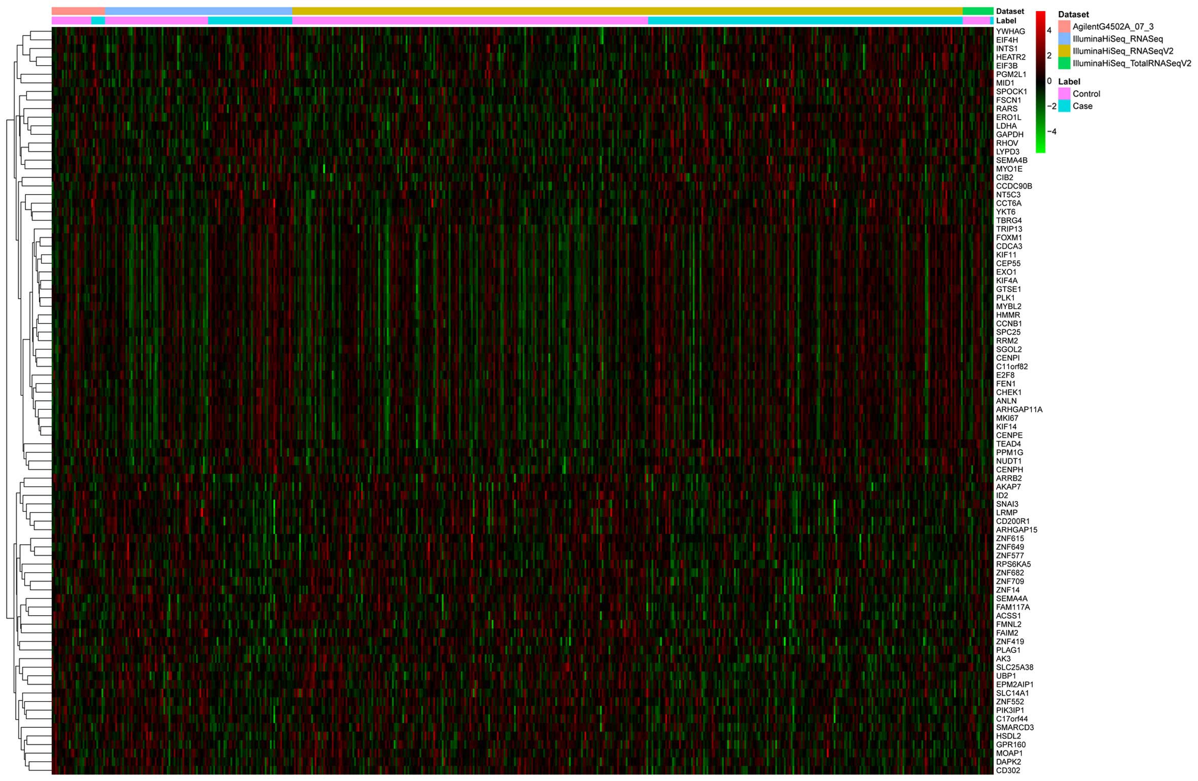
Based on clinical information 'AJCC pathologic tumor stage' in the TCGA lung adenocarcinoma, we classified these data into lung adenocarcinoma with or without metastasis. After the gene expression data were downloaded, we performed differential expression analysis of the genes and miRNAs between the metastatic primary loci and non-metastatic primary loci. Genes (1,257) were identified to be differentially expressed under a threshold of FDR <0.05, with 585 upregulated and 672 downregulated genes in the lung adenocarcinoma with metastasis. The hierarchical clustering of differentially expressed genes is shown in Fig. 1. Fifty-eight miRNAs were identified as differentially expressed under the threshold of FDR <0.05, with 44 upregulated and 14 downregulated miRNAs in the lung adenocarcinoma samples with metastasis (Table II).

Table II

Summary of the differentially expressed miRNAs.

Table II

Summary of the differentially expressed miRNAs.

miRNAsP-valuemiRNAsP-valuemiRNAsP-value
Upregulated miRNAs
hsa-mir-12936.62E-04hsa-mir-3811.17E-02hsa-mir-147b3.00E-02
hsa-mir-105-21.87E-03hsa-mir-365-11.50E-02hsa-mir-1285-13.13E-02
hsa-mir-365-22.82E-03hsa-mir-550a-11.52E-02hsa-mir-105-13.25E-02
hsa-mir-193a3.22E-03hsa-mir-5391.70E-02hsa-mir-4323.43E-02
hsa-mir-196b3.64E-03hsa-mir-4331.76E-02hsa-mir-9-33.68E-02
hsa-mir-1185-13.86E-03hsa-mir-509-21.88E-02hsa-mir-376c3.92E-02
hsa-mir-9448.07E-03hsa-mir-31912.09E-02hsa-mir-21164.35E-02
hsa-mir-7678.36E-03hsa-mir-3822.15E-02hsa-mir-3794.40E-02
hsa-mir-193b8.48E-03hsa-mir-6652.27E-02hsa-mir-513a-14.53E-02
hsa-mir-39428.49E-03hsa-mir-5822.31E-02hsa-mir-12764.76E-02
hsa-mir-9-18.65E-03hsa-mir-8892.40E-02hsa-mir-2124.89E-02
hsa-mir-9-29.43E-03hsa-mir-4212.48E-02hsa-mir-516a-24.92E-02
hsa-mir-6551.14E-02hsa-mir-7442.72E-02hsa-mir-984.96E-02
hsa-mir-4091.15E-02hsa-mir-6532.86E-02hsa-let-7d4.98E-02
hsa-mir-4961.16E-02hsa-mir-1342.96E-02
Downregulated miRNAs
hsa-mir-133a-11.07E-02hsa-mir-518a-12.77E-02hsa-mir-3383.21E-02
hsa-mir-30651.11E-02hsa-mir-5522.94E-02hsa-mir-5773.24E-02
hsa-mir-31861.42E-02hsa-mir-30b3.02E-02hsa-mir-6223.95E-02
hsa-mir-135a-22.08E-02hsa-mir-6603.03E-02hsa-mir-3202-24.16E-02
hsa-mir-12502.35E-02hsa-mir-302a3.17E-02
The regulatory network of differentially expressed mRNAs and miRNAs associated with lung adenocarcinoma metastasis

To find the potential link between the differentially expressed mRNAs and miRNAs observed in the bioinformatic analysis, we predicted the putative targets of the identified miRNAs in the miRWalk database. Matching predicted putative targets with those found to be disregulated genes in metastatic primary tumors, miRNA-target gene pairs whose expression levels were inversely correlated were selected. As a result, we identified 1,154 miRNA-target gene pairs for the upregulated miRNAs with 61 pairs validated by previous experiments, and 474 miRNA-target gene pairs for the downregulated miRNAs with 57 pairs validated by previous experiments (Table III).

Table III

miRNA-target gene pairs whose expression levels are inversely correlated.

Table III

miRNA-target gene pairs whose expression levels are inversely correlated.

miRNAStatus (miRNAs)No. of miRNA target genesTarget mRNAs
hsa-mir-133a-1Downregulated7PTBP1, PKM2, NKX2-3, STRAP, FSCN1, TGFBI, PA2G4
hsa-mir-135a-2Downregulated2SLC27A4, FOXM1
hsa-mir-302aDownregulated67MCM7, SLC27A4, ELOVL6, RPE, CKAP4, KPNA2, SEMA3C, POLQ, KCND2, SLC45A2, MCM10, ASF1B, PBK, ST8SIA2, USP42, E2F7, CCT7, ERLIN1, BLCAP, PDAP1, CD109, KDELC2, ESCO2, DDX10, DHCR7, DSG3, E2F2, TMEM184A, C11orf82, GGA2, PSD4, NUP62, PANX1, PGM2L1, RACGAP1, HOXA11, CYP27C1, ACADVL, KRT14, MCM4, MCM6, NEDD4, MRPS16, NIP7, CYCS, PON2, RNF216, CDCA8, CEP55, OGFOD1, SYT13, KIAA1609, GNPNAT1, BRCA1, UPK1B, C1orf135, SHCBP1, FBXL18, MFAP5, CDT1, C10orf58, BARX2, STC2, EXO1, NCAPD2, CDC25A, ECT2
hsa-mir-30bDownregulated100MRPS24, DPP7, NKX2-3, F2, TRIP13, AP2A1, MYO1E, GARS, PLK1, UTP15, ZNF384, KDELC2, DSG2, DSP, AURKB, ABCF2, POLD2, TXNRD1, NCL, MYBL2, CARHSP1, SFXN1, CHORDC1, NEDD4, PTBP1, RARS, GAPDH, PTGFRN, IQGAP3, GNPNAT1, LMNB2, POLA2, NT5C3, DLGAP4, GALNT2, CYP24A1, LASS3, MESDC2, DPY19L1, GRM5, INCENP, KIF11, PELO, ERRFI1, PON2, AVEN, HECW2, RRM2, YWHAG, SLC7A5, ARHGAP11A, LPGAT1, CHST1, PSME3, CIB2, CCT4, ERLIN1, C1QL1, PDAP1, CBX3, SLC5A11, CPD, TICAM1, ESCO2, DLX1, TTLL12, RNASEH1, C16orf73, ABCA12, GPR110, PGM2L1, HDLBP, HNRNPA2B1, HOXA11, KCND2, MID1, MTHFD1, NEFM, ORC2L, CYCS, ANLN, CDCA8, PGM2, DEPDC1B, KIF15, SH3GL1, CENPH, STC1, TFAP2A, UPK1B, ZIC2, C1orf135, CALB2, MLF1IP, CALU, FAM136A, PRC1, HN1L, KIAA0101, E2F7
hsa-mir-338Downregulated1CCT4
hsa-mir-552Downregulated52CYP24A1, FOXC1, CYCS, UBFD1, C10orf58, EIF2S2, ABCF2, CARM1, CENPF, YKT6, CDCA5, WBSCR22, CD109, CPD, AP2A2, ENO1, TMEM184A, EXTL3, TTLL12, NCAPH, FOSL2, GNG4, PGM2L1, HOXA10, BIRC5, CYP27C1, CHST6, MCM4, MKI67, MRPS16, PKM2, ERRFI1, PNPLA2, PTBP1, SYT13, NKX3-2, RFC2, BCAN, GNPNAT1, FSCN1, BRCA1, TGM4, TULP3, GLT25D1, MAFK, USP5, ST8SIA2, CDT1, LMNB2, CDK5R2, HN1L, DDX21
hsa-mir-1276Upregulated15CNOT6L, CHIC2, PIK3CG, C1orf190, BDH2, BCL2, SLC14A1, HSDL2, PSTPIP2, CD5, PKHD1L1, ITM2A, LPIN2, MTSS1, EPM2AIP1
hsa-mir-1293Upregulated5NMUR1, ITIH4, ROBO2, DNALI1, FCRLA
hsa-mir-134Upregulated74KIT, KLHDC1, SEC14L3, IL16, PCMTD2, SLC14A1, FCRL5, RCL1, ATP8A1, RRH, CHKA, WIF1, ERMAP, FMNL2, CYP2R1, CNR1, CSF3R, PRICKLE2, ZNF25, LNX2, DENND3, MMRN1, P2RX2, PDZD2, GPD1L, ISCU, CBX7, CNOT6L, SLC41A1, GCET2, PDE7B, ZNF776, ZNF615, SLC25A42, S100A7A, FAM19A2, ID2, RSPO2, C3orf62, KCNJ8, CLEC12B, SMAD6, MOCS1, UNC13C, MTHFR, GIMAP6, CDON, CECR1, PLCB2, GNG2, RSBN1, TNFRSF19, PRKCQ, MOSPD1, SLC24A3, BCL2, SLAMF1, UBP1, C18orf1, ZFP161, C21orf2, ZNF20, DNALI1, BTG2, ZNF552, C1orf21, PIGY, MPDZ, LIMD1, ZNF468, IL33, RPS6KA5, TP53INP1, LPIN2
hsa-mir-147bUpregulated3MACROD2, LY9, NFIX
hsa-mir-193bUpregulated42BCL2, KIT, UPRT, CAMTA1, CBX7, MMP19, UBP1, RAMP3, UNC13B, MGAT4A, UNC45B, PDIK1L, PRICKLE2, TAPT1, FAIM2, CNOT6L, PPP1R16B, GATA6, CRB2, C3orf62, RHOH, MAP3K3, CD244, PLAG1, C21orf29, POU2AF1, SNRK, TRIM68, NXF3, WDR48, SLC4A5, C18orf1, EVI5, C1orf21, CAMKK1, KBTBD8, ATOH8, VAMP8, NMT2, TP53INP1, AATK, SPNS3
hsa-mir-196bUpregulated53FLT3, BCL2, GATA6, AQP4, ZMYND11, EPS15, WDR37, CAPN7, ATRNL1, PDE7B, SMAD6, MGAT4A, CYP2U1, UNC45B, PRICKLE2, KLHDC8B, GPD1L, SLC41A1, PPP1R16B, SERP1, HLF, SNAI3, RSPO2, C3orf62, MAOA, MTHFR, CDON, PLAG1, PLCB2, BCL11A, BEST2, TNFRSF19, MRPS25, COL14A1, WNT2B, TMEM50B, SLC25A20, EEA1, ZNF577, ATOH8, PRSS12, FAM125B, LIMD1, CREB3L1, ACVR2A, VAPA, MS4A1, TP53INP1, FHL5, AATK, LPIN2, WSCD2, LPPR4
hsa-mir-212Upregulated92RPS6KA5, REM1, ANKRD29, PRICKLE2, SOSTDC1, PDE7B, SERP1, MAOA, SLC24A3, TMEM50B, BTG2, SLC25A20, USP38, KLRG1, CXCR6, WASF3, FGL2, MGAT4A, CYP2U1, PIK3IP1, FMNL2, CNR1, C1orf88, MAP3K8, CPT1B, UPRT, GAB3, GPR155, BTLA, ZNF483, TAPT1, ENPP4, KLRK1, CLASP2, GLT25D2, TBC1D9, GPD1L, CAMTA1, FOSB, CNOT6L, PPP1R16B, NAP1L5, GPR160, SESN1, ZNF615, FAM19A2, IKBKB, IL12B, IL16, IMPG1, B3GNT8, MAP3K3, MLLT3, UNC13C, AK3, CDON, EMCN, PLAG1, C21orf29, GFOD1, LAX1, PCMTD2, FBXW7, PNRC2, MOSPD1, LHX9, GALNTL1, WDR48, STIM2, PTPN4, SLC4A5, RSU1, SDF2, C18orf1, ZFP161, ZNF708, ZNF80, C1orf21, TCF7L1, SYT15, HSDL2, KBTBD8, PDE8B, LIMD1, LARGE, VAPA, NMT2, TP53INP1, CHST10, TOX, ELMO1, SEMA4A
hsa-mir-365-1Upregulated1BCL2
hsa-mir-365-2Upregulated1BCL2
hsa-mir-376cUpregulated51PDIK1L, SMYD1, ALCAM, GNG2, KLHL9, ARID4A, VLDLR, ACYP2, TESK2, FBLN5, RCBTB2, SLAMF6, KLHDC1, UNC45B, ZNF483, NLRC3, DAPK2, PLA2G4F, C1orf101, GCET2, D4S234E, SERP1, SNAI3, FAM19A2, FAM19A1, MAP3K3, GIMAP6, AK3, C3orf18, PLAG1, P2RY13, RCOR3, MOSPD1, ST6GAL1, ZNF649, C18orf1, ZFP161, ZNF10, ZNF708, ZNF614, NDFIP1, C1orf21, IL33, CABLES1, GPR55, MS4A1, WSCD2, TOX, MTSS1, FLT3, RORB
hsa-mir-379Upregulated52ATP8A2, CYP2U1, KLHL9, C7, KLRG1, NMUR1, UNC13B, ZNF211, INMT, SCGB3A2, C1orf88, GAB3, ANKRD29, PRICKLE2, ALCAM, DENND3, FAIM2, SLC41A1, C1orf101, HSPB7, ZNF776, C13orf15, GZMK, HLF, S100A7A, ID4, SLCO4C1, IL16, KIT, ARRB1, UNC13C, GIMAP6, CDON, C9orf68, SLC25A36, SPTLC3, BEX4, WNT2B, C18orf1, ZNF10, DNALI1, C1orf21, ACSS1, IL18RAP, CD5, LARGE, VAPA, GPR55, MS4A1, CD40LG, WSCD2, LPPR4
hsa-mir-381Upregulated102KIT, DLC1, WDR37, MMRN1, ID2, CNTN3, SLC25A36, SLC24A3, PCDH20, ACVR2A, MERTK, FBLN5, CXCR6, WASF3, MGAT4A, CYP2U1, FMNL2, KLHDC1, CNR1, CNTFR, C1orf88, CD200R1, MAP3K8, UPRT, PDIK1L, SMYD1, BTLA, ZNF483, PRICKLE2, SUSD3, F11, ENPP4, SACM1L, SORCS3, SWAP70, CLASP2, TBC1D9, GPD1L, ANKRD12, CAMTA1, CAPN7, CNOT6L, SLC41A1, GCET2, NAP1L5, PDE7B, SERP1, ZNF776, ZNF615, GPR34, GRIA1, C13orf15, ZC3H7A, GZMK, HTR2A, ID4, RSPO2, KCNT2, RBPJ, SLCO4C1, IKBKB, IL12B, KCNA4, RHOH, MLLT3, NINJ1, PLAG1, BCL11A, PMM1, P2RY13, SETD4, FAM105A, SNRK, LAX1, TRIM68, PCMTD2, TNFRSF19, KLHL9, BEX4, WDR48, SLC4A5, ARID4A, PRDM16, SLAMF1, BTG1, VLDLR, C18orf1, ZNF10, ZNF136, DNALI1, LRRC27, FAM117A, EEA1, HSDL2, KBTBD8, CD1D, VAPA, RCSD1, TP53INP1, KIAA0408, MTSS1, NFIB
hsa-mir-382Upregulated59DLC1, ATP8A1, MAP3K8, CRHBP, CAPN7, GPR160, ZNF615, ID4, SLCO4C1, CABC1, ROBO2, CD96, MGAT4A, CISH, CNR1, PPM1M, MACROD2, GPR155, BTLA, PRICKLE2, TAPT1, EPS15, ALCAM, SORCS3, CAMTA1, CNOT6L, PPP1R16B, RSPO2, IL10RA, IL12B, ITGAD, B3GNT8, MAOA, LRP2BP, KLHL9, MOSPD1, SLC24A3, SLC4A5, PRDM16, PCDH20, C18orf1, ZNF708, TMEM50B, CA3, ZNF614, C1orf21, SYT15, CLDN2, IL33, CD1D, RCSD1, HMGN3, GRAP2, ITM2A, AKAP7, CHST10, TOX, NFIB, PPP3CC
hsa-mir-421Upregulated79CBX7, TESK2, CLASP2, SOSTDC1, RHOH, SLC24A3, NDFIP1, KBTBD8, B3GALT2, VAPA, TSPAN32, CXCR6, WASF3, FRS3, FGL2, MGAT4A, ERMAP, CNR1, GAB3, PDIK1L, SMYD1, DBH, ZNF540, PRICKLE2, NLRC3, ZNF25, ENPP4, ANKRD12, CAMTA1, CNOT6L, SLC41A1, PDE7B, SERP1, SLC46A3, NKIRAS1, ZC3H7A, FAM19A2, RSPO2, SLCO4C1, IL10RA, IL12B, AQP4, C3orf62, NR3C2, MTHFR, GIMAP6, CDON, BCL11A, FBXO42, RSBN1, LAX1, SLC25A36, PCMTD2, LRP2BP, GALNTL1, WDR48, STIM2, PTPRB, RSU1, GZF1, MRPS25, C18orf1, BTG2, ZNF614, LRRC27, FCRL5, SYT15, ZNF577, LIMD1, CLDN2, ACVR2A, GPR55, NMT2, AKAP7, USP6NL, TOX, LPPR4, RORB, ACTN2
hsa-mir-432Upregulated79CXCR6, SLCO4C1, GNG2, CA3, C1orf21, HSDL2, PPAP2A, RCL1, CD96, DLC1, WASF3, CHKA, MGAT4A, ADH1B, CNR1, MAP3K8, CPT1B, MACROD2, UNC45B, ANKRD29, GPR155, NLRC3, TAPT1, F11, TMEM130, KLRK1, SORCS3, GPD1L, CAMTA1, PPP1R13B, FOS, SLC41A1, MYRIP, SETBP1, NAP1L5, GPR160, HSPB7, GNG7, SNAI3, ID4, RSPO2, IL16, LY9, MAP3K3, MLLT3, GIMAP6, P2RX1, C3orf18, REV1, HMGCLL1, RSBN1, PPP1R1A, SLC25A36, TMEM57, FBXW7, SPTLC3, GIMAP5, LRP2BP, C1orf183, BCL2, PRDM16, GZF1, C5, UBP1, C18orf1, ZNF80, EVI5, DENND1C, ATOH8, FAM125B, BZRAP1, GPR55, MS4A1, ENTPD3, CD40LG, WSCD2, USP6NL, TOX, PDZD2
hsa-mir-433Upregulated55NR0B2, ZNF211, RAB40B, TAGAP, NLRC3, ZNF615, NINJ1, RSBN1, PTPN4, PRDM16, MRPS25, ZNF649, ZNF91, BTG2, ACVR2A, RAMP3, ATP8A1, UNC13B, FGL2, FCRL3, CNR1, CD200R1, GAB3, PDIK1L, BTLA, DMP1, GPC5, SACM1L, SWAP70, ANKRD12, CAMTA1, CNOT6L, NAP1L5, ZNF776, IL16, UNC13C, C9orf68, TRIM68, FBXW7, TNFRSF19, LRP2BP, SLC4A5, PTPRB, BCL2, ZNF708, EVI5, LRRC27, C1orf21, KBTBD8, ZNF439, VAPA, NMT2, AKAP7, KIAA0408, LPPR4
hsa-mir-496Upregulated49KIT, ANKRD12, FAM55D, ZNF20, ZNF136, MGAT4A, CNR1, ADHFE1, CR2, CRHBP, GPR155, ZNF781, P2RX2, TBC1D9, C20orf194, SETBP1, GATA6, CLUL1, HSPB7, ANGPT1, ZNF776, GPR34, S100A7A, SLCO4C1, C3orf62, CDON, EMCN, P2RY13, HMGCLL1, LAX1, SUSD2, BEX4, BDH2, BCL2, MRPS25, SPATA20, SLC14A1, ZNF708, ZNF614, C1orf21, EEA1, ZNF160, IL33, VAPA, BZRAP1, ITM2A, LPPR4, NFIB, PAQR8
hsa-mir-513a-1Upregulated1DNAH8
hsa-mir-539Upregulated101KIT, KLRG1, WASF3, PIK3IP1, MACROD2, BTLA, CLASP2, PDE7B, ZNF776, ID4, RSPO2, PNRC2, C7, NDFIP1, FCRL5, SYT15, PRSS12, ZNF266, RCBTB2, ZPBP2, CNR1, CNTFR, MAP3K8, UNC45B, SMYD1, GPR155, ZNF483, DLG4, DMP1, FAIM2, PDZD2, GLT25D2, ANKRD12, PPP1R13B, FOS, PIK3R5, FOSB, CCNDBP1, SLC41A1, GCET2, ATRNL1, SEC14L3, ANGPT1, S100A7A, SLCO4C1, C3orf62, CLEC12B, MAOA, NR3C2, MTHFR, GIMAP6, P2RX1, CNTN3, CDON, PHF7, CECR1, PIK3CG, BCL11A, PNOC, C21orf29, C1orf190, GNG2, PALMD, SLC25A36, SPTLC3, GIMAP5, TNFRSF19, MOSPD1, CPA6, TNFRSF17, RSU1, NAPB, USP4, C18orf1, DNALI1, EVI5, BTG2, DENND1C, ZNF614, LRRC27, C1orf21, CDADC1, EEA1, C15orf48, KBTBD8, EMR3, ST3GAL5, FAM125B, LIMD1, ZNF439, CLDN2, CD1D, C22orf32, CD5, VAPA, MS4A1, NMT2, TP53INP1, AKAP7, CD69, USP6NL
hsa-mir-582Upregulated1SMAD6
hsa-mir-9-1Upregulated9CD19, CEBPA, BCL2, RASSF1, CBX7, ATP8A2, CISH, GRIA1, IGFALS
hsa-mir-9-2Upregulated8ATP8A2, CD19, RASSF1, BCL2, GRIA1, CEBPA, IGFALS, CISH
hsa-mir-9-3Upregulated7ATP8A2, CD19, BCL2, IGFALS, GRIA1, CEBPA, CISH
hsa-mir-98Upregulated3ESR2, CISH, CNTN3
hsa-let-7dUpregulated61ACVR2A, BCL2, FOSB, FLT3, PGC, KIT, CNTN3, ESR2, ATP8A2, CXCR4, CEBPA, FBXW7, CACNA2D2, CISH, MTSS1, RASSF1, MGAT4A, USP38, ADAMTS8, WDR37, RUFY3, RSPO2, COL14A1, WASF3, CYP2U1, SLAMF6, CNR1, CD200R1, GAB3, GPR155, DAPK1, ZNF540, NLRC3, KLHDC8B, EPS15, ENPP4, CLASP2, DAPK2, PPP1R16B, HSPB7, CRB2, HLF, SNAI3, TMEM110, P2RX1, PLCB2, GFOD1, BEST2, SFTPB, MRPS25, C18orf1, ZNF10, ZNF614, C1orf21, TCF7L1, SYT15, EEA1, ZNF577, MPDZ, FAM125B, TP53INP1

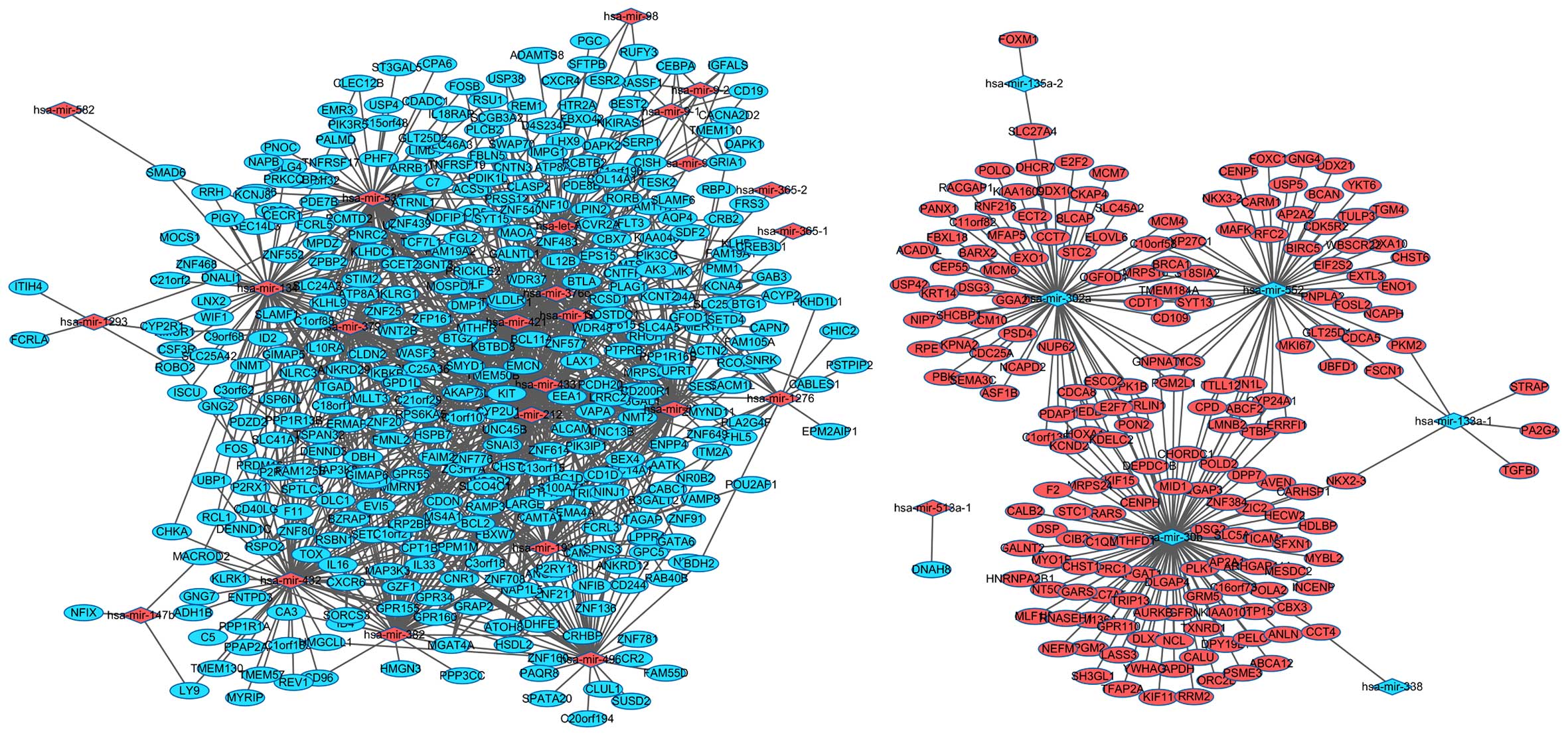
Using the 770 miRNA-target gene pairs, an miRNA-target gene regulatory network was constructed (Fig. 2). In the miRNA-target gene regulatory network, we identified the top 10 miRNAs which regulated the most target genes, such as hsa-miR-7, hsa-miR-182, hsa-miR-324-3p, hsa-miR-139-5p, hsa-miR-130b, hsa-let-7f, hsa-miR-18a, hsa-miR-188-5p, hsa-let-7d, and hsa-miR-590-5p, and the target genes such as RPS6KA3, TSC1, AIM1, GAS7, GFOD1, GGA2, IGF1, IL28RA, and INSR were regulated by the most miRNAs.

GO classification and KEGG pathways of the miRNA target genes

We performed the GO classification and KEGG pathway enrichment analysis for miRNA target genes whose expression was differentially expressed. We found that lymphoid progenitor cell differentiation (GO:0002320, FDR=4.75E-04) and hematopoietic progenitor cell differentiation (GO:0002244, FDR=2.08E-03) were significantly enriched for biological processes. While for molecular functions, signaling receptor activity (GO:0038023, FDR=9.24E-04) and signal transducer activity (GO:0004871, FDR=8.77E-04) were significantly enriched (Table IV). The most significant pathway in our KEGG analysis was DNA replication (FDR=1.78E-04). Furthermore, T cell receptor signaling pathway (FDR=1.12E-03) and endocytosis (FDR=1.17E-03) were also highly enriched (Table V).

Table IV

GO functional annotation of the predicted miRNA target genes.

Table IV

GO functional annotation of the predicted miRNA target genes.

GO IDGO termFDR
Biological process
 GO:0002320Lymphoid progenitor cell differentiation4.75E-04
 GO:0002244Hematopoietic progenitor cell differentiation2.08E-03
 GO:0070661Leukocyte proliferation1.44E-02
 GO:0002521Leukocyte differentiation2.32E-02
 GO:0046425Regulation of JAK-STAT cascade1.94E-02
 GO:0048639Positive regulation of developmental growth1.65E-02
 GO:0007166Cell surface receptor signaling pathway1.88E-02
 GO:0043552Positive regulation of phosphatidylinositol 3-kinase activity2.20E-02
 GO:0002318Myeloid progenitor cell differentiation1.96E-02
 GO:0048070Regulation of developmental pigmentation1.76E-02
 GO:0030318Melanocyte differentiation1.60E-02
 GO:0050931Pigment cell differentiation1.47E-02
 GO:0090218Positive regulation of lipid kinase activity1.36E-02
 GO:0060563Neuroepithelial cell differentiation1.26E-02
 GO:0048534Hematopoietic or lymphoid organ development1.23E-02
 GO:0001932Regulation of protein phosphorylation1.20E-02
 GO:0042113B cell activation1.53E-02
 GO:0031399Regulation of protein modification process1.67E-02
 GO:0002065Columnar/cuboidal epithelial cell differentiation1.73E-02
 GO:0042509Regulation of tyrosine phosphorylation of STAT protein1.64E-02
 GO:0042531Positive regulation of tyrosine phosphorylation of STAT protein1.57E-02
 GO:1903727Positive regulation of phospholipid metabolic process1.50E-02
 GO:0046427Positive regulation of JAK-STAT cascade1.43E-02
 GO:0071310Cellular response to organic substance1.38E-02
 GO:0050853B cell receptor signaling pathway1.41E-02
 GO:0050769Positive regulation of neurogenesis1.64E-02
 GO:0051094Positive regulation of developmental process1.67E-02
Molecular function
 GO:0038023Signaling receptor activity9.24E-04
 GO:0004871Signal transducer activity8.77E-04
 GO:0004888Transmembrane signaling receptor activity8.24E-04
 GO:0004872Receptor activity1.40E-03
 GO:0060089Molecular transducer activity1.57E-03
 GO:0005057Receptor signaling protein activity8.40E-03
 GO:0002020Protease binding1.02E-02
 GO:0004714Transmembrane receptor protein tyrosine kinase activity8.93E-03
 GO:0042803Protein homodimerization activity1.36E-02
 GO:0019199Transmembrane receptor protein kinase activity3.21E-02
 GO:0042802Identical protein binding3.54E-02
 GO:0046983Protein dimerization activity3.64E-02
 GO:0044389Ubiquitin-like protein ligase binding4.93E-02
 GO:0031625Ubiquitin protein ligase binding4.58E-02
 GO:0004713Protein tyrosine kinase activity4.60E-02

Table V

KEGG pathway enrichment analysis of the predicted miRNA target genes (top 15).

Table V

KEGG pathway enrichment analysis of the predicted miRNA target genes (top 15).

IDItemsCountFDRGenes
hsa03030DNA replication71.78E-04MCM4, RNASEH1, MCM7, POLD2, RFC2, MCM6, POLA2
hsa04660T cell receptor signaling pathway91.12E-03IKBKB, FOS, PIK3R5, PPP3CC, PRKCQ, CD40LG, GRAP2, PIK3CG, MAP3K8
hsa04144Endocytosis121.17E-03CXCR4, KIT, AP2A2, NEDD4, SH3GL1, PSD4, SMAD6, ARRB1, EEA1, EPS15, FAM125B, AP2A1
hsa04380Osteoclast differentiation51.19E-03IKBKB, FOS, PIK3R5, PPP3CC, PIK3CG
hsa05145Toxoplasmosis51.23E-03IKBKB, CYCS, PIK3R5, BCL2, PIK3CG
hsa04210Apoptosis51.23E-03IKBKB, CYCS, PIK3R5, BCL2, PIK3CG
hsa04110Cell cycle31.24E-03MCM4, MCM7, MCM6
hsa05222Small cell lung cancer51.26E-03IKBKB, PIK3R5, E2F2, BCL2, PIK3CG
hsa05210Colorectal cancer71.27E-03CYCS, TCF7L1, FOS, PIK3R5, BIRC5, BCL2, PIK3CG
hsa04620Toll-like receptor signaling pathway51.27E-03IKBKB, FOS, PIK3R5, PIK3CG, MAP3K8
hsa05142Chagas disease (American trypanosomiasis)61.28E-03IKBKB, FOS, TICAM1, PIK3R5, IL12B, PIK3CG
hsa04722Neurotrophin signaling pathway41.29E-03IKBKB, PIK3R5, BCL2, PIK3CG
hsa05200Pathways in cancer171.31E-03IKBKB, CYCS, TCF7L1, FOS, PIK3R5, KIT, BIRC5, WNT2B, E2F2,CSF3R, FLT3, RASSF1, BCL2, DAPK1, CEBPA, PIK3CG, DAPK2
hsa05221Acute myeloid leukemia71.39E-03IKBKB, TCF7L1, PIK3R5, KIT, FLT3, CEBPA, PIK3CG
hsa04640Hematopoietic cell lineage81.43E-03KIT, CSF3R, CD1D, FLT3, CR2, CD19, CD5, MS4A1
Validating the expression of lung adenocarcinoma metastasis-associated miRNA target genes

By the online tool of SurvMicro, survival analysis was performed to evaluate the correlation between miRNA expression level (hsa-miR-133a-1 and hsa-let-7d) and the overall survival time of the lung adenocarcinoma patients. With the data from TCGA database, Kaplan-Meier curves indicated that the expression of hsa-mir-133a-1 was significantly correlated with the overall survival time of lung adenocarcinoma patients (P=0.03654), while the expression of hsa-let-7d was not significantly correlated with the overall survival time of the lung adenocarcinoma patients (P=0.114) (Fig. 3).

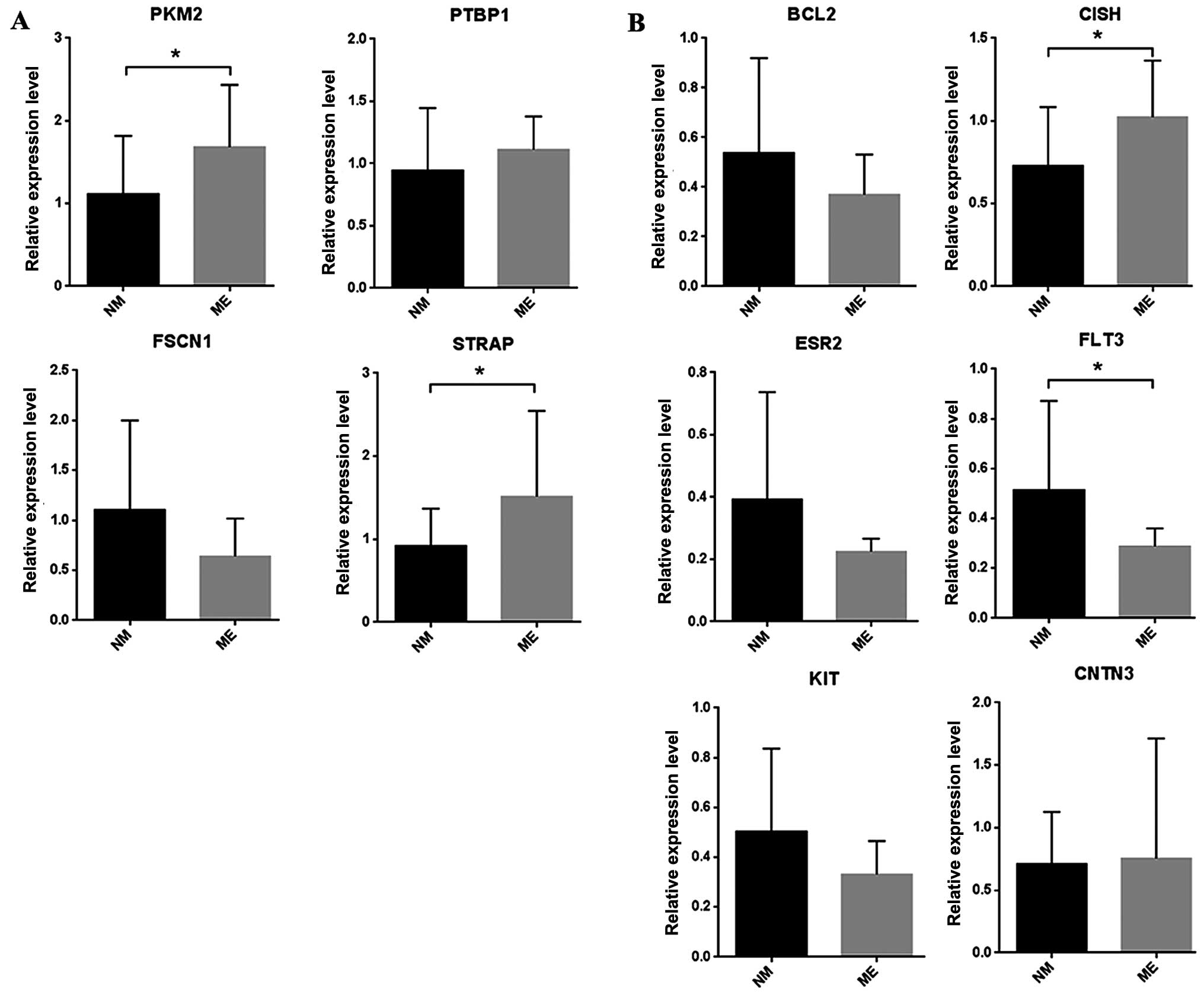
We performed RT-qPCR for the putative target genes of hsa-mir-133a-1 and hsa-let-7d, which exhibited significantly differential expression between the primary tumor tissues from lung adenocarcinoma with metastasis and that from the lung adenocarcinoma patiemts without metastasis in the bioinformatic analysis. In the RT-qPCR assay, most of the target genes showed similar expression patterns to what was observed in the bioinformatic analysis. The expression levels of the selected upregulated and downregulated genes are shown in Fig. 4. Among the upregulated genes in the lung adenocarcinoma with metastasis, PKM2 and STRAP mRNA expression was significantly increased (P=0.04, P=0.05), and PTBP1 mRNA expression was also increased, but not obviously, while FSCN1 mRNA expression was decreased. Among the downregulated genes in the lung adenocarcinoma with metastasis, FLT3 mRNA expression was significantly decreased (P=0.023), and BCL2, ESR2, and KIT mRNA expression was also mildly decreased. In contrast, CISH mRNA expression was significantly increased (P=0.027). CNTN3 mRNA expression appeared unchanged between the metastatic primary loci and the non-metastatic primary loci.

Discussion

Due to a substantially lower cost, high-throughput gene expression data are becoming widely available. A key question in the field is how to use the data to identify both biological drivers and strong metastatic markers. In this study, we compared miRNA and mRNA expression data differences between lung adenocarcinoma with metastasis and lung adenocarcinoma without metastasis from the TCGA data portal with the aim to screen the miRNAs and genes associated with lung adenocarcinoma metastasis.

In line with previous findings, some of the identified differentially expressed miRNAs were found to be implicated in lung adenocarcinoma metastasis, such as hsa-mir-98, hsa-let-7d, hsa-mir-134, hsa-mir-196b, hsa-mir-381, hsa-mir-133a, hsa-mir-552, hsa-mir-944, hsa-mir-550, and hsa-mir-655. miR-98 was also found to be overexpressed in lung cancer cell lines, and it binds the 3′UTR of Fus1, a tumor suppressor, to inhibit protein expression (22). Despite no reports related to lung adenocarcinoma metastasis, a recent study uncovered the regulatory roles of miR-98 in melanoma metastasis (23). miR-134 and miR-655, belonging to the same cluster on chromosome 14q32, were demonstrated to be involved in TGF-β1-induced EMT which is recognized as a key element of cell invasion, migration, and metastasis, by directly targeting MAGI2, a scaffold protein required for PTEN (24). Directly targeting oncogenic receptors, such as IGF-1R, TGFBR1, and EGFR, miR-133a inhibits cell invasiveness and cell growth in lung cancer cells. Furthermore, an in vivo animal model showed that miR-133a markedly suppressed the metastatic ability of lung cancer cells (19). An miRNA microarray revealed that the expression level of miR-552 in colorectal cancer metastases in the lung was 39 times higher than that of primary lung adenocarcinomas, suggesting its possible roles in cancer metastases (25). miR-944 affected NSCLC cell growth, proliferation, and invasion by targeting a tumor suppressor, SOCS4. miR-944 was found to be associated with lymph node metastasis by determining its expression in 52 formalin-fixed paraffin-embedded SCC tissues (26).

More importantly, two miRNAs, hsa-mir-133a and hsa-let-7d, were found to be involved in lung adenocarcinoma metastasis by previous studies. Consequently, we performed survival analysis to evaluate the correlation between miRNA expression level and the overall survival time of lung adenocarcinoma patients, and we found that the expression of hsa-mir-133a was significantly correlated with the overall survival of lung adenocarcinoma, while the expression of hsa-let-7d was not significantly correlated with the survival of lung adenocarcinoma.

miR-133a was identified to function as a tumor suppressor in NSCLCs directly targeting several membrane receptors including IGF-1R, TGFBR1 and EGFR. In addition, by using the in vivo animal model, ectopically expressing miR-133a markedly suppressed the metastatic ability of the lung cancer cells, which may be applied in the clinical therapy of lung cancer in the future. Accordingly, NSCLC patients with higher expression levels of miR-133a had longer survival rates compared with those with lower miR-133a expression levels (19).

Let-7d, one of the members of the let-7 family, is deregulated in many types of cancers (2730). Let-7d is involved in cellular differentiation, epithelial-to-mesenchymal transition (EMT) as a switch between EMT and MET (mesenchymal-epithelial transition), and regulates tumor-initiating cell (TIC) formation (31). Mairinger et al showed that let-7d expression was significantly associated with overall survival in pulmonary neuroendocrine tumors, suggesting its important role in metastasis and tumor progression (20).

The putative targets of hsa-mir-133a and hsa-let-7d were subjected to RT-qPCR validation in primary tumor tissues from 5 lung adenocarcinoma patients with metastasis and 5 lung adenocarcinoma patients without metastasis. The result of the RT-qPCR assay revealed that most of the genes showed similar expression patterns to what were observed in the bioinformatic analysis, suggesting that the findings in the bioinformatic analysis of the differential gene expression data were credible. PKM2, as a common putative target of both hsa-mir-133a and hsa-mir-552, was significantly upregulated at the mRNA level in the lung adenocarcinoma with metastasis. Interesting, the protein level of PKM2 was upregulated in the lung adenocarcinoma tissues compared with the paired surrounding normal tissue, which was also correlated with chemotherapy resistance, the severity of epithelial dysplasia, as well as a relatively poor prognosis (32,33), indicating that PKM2 could be used as a tumor marker for diagnosis and, in particular, as a potential target for cancer therapy.

STRAP, a putative target of hsa-mir-133a, was significantly increased at the mRNA level in lung adenocarcinoma with metastasis. As a WD domain-containing protein, STRAP inhibits TGF-β signaling through interaction with receptors and Smad7, and promotes growth and enhances tumorigenicity. Similarly, strong upregulation of STRAP was also observed in lung tumors by immunoblot analyses (34).

FLT3, a common putative target of hsa-let-7d, hsa-mir-196b and hsa-mir-376c, was significantly upregulated at the mRNA level among the downregulated genes in the lung adenocarcinoma with metastasis. It was found that in a mouse model of lung metastases treatment with the Flt3 ligand significantly reduced the number of lung metastases after laparotomy or radiation therapy, and thus prolonged survival (35,36). Unlike the findings in the bioinformatic analysis, CISH mRNA expression was significantly increased. As negative feedback regulators of JAK-STAT and several other signalling pathways, CISH, a putative common target of hsa-let-7d, hsa-mir-382, hsa-mir-9-3, hsa-mir-9-2 and hsa-mir-9-1, was recently found to be involved in the development of many solid organ and haematological malignancies (37). Prostate cancer LNCaP-S17 cells were resistant to exogenous IL-6-induced neuroendocrine differentiation due to increased levels of CISH and SOCS7 that blocked activation of the JAK2-STAT3 pathways (38).

Functional enrichment analysis of miRNA target genes showed that those genes were highly correlated with carcinogenesis. The most significantly enriched pathway based on the KEGG database was DNA replication. Based on the fact that cancer cells are characterized by uncontrolled cell proliferation properties, deregulation of DNA replication may promote the process of carcinogenesis. In addition, in cancer cells several DNA replication-initiation proteins, such as CDC6 and minichromosome maintenance (MCM) proteins, were overexpressed (39,40).

There are two limitations in this study. Firstly, the expression levels of the miRNAs were not determined in the metastatic primary loci compared with non-metastatic primary loci of lung adenocarcinomas. Secondly, the small size of the clinical samples for RT-qPCR validation was also a limitation of our study, which may be the reason that the RT-qPCR results were not significant. Given that surgery is not recommended for the traditional stage IIIB-IV patients with lung adenocarcinoma classified according to the taxonomy of cancer staging (TNM) system, it is difficult to obtain specimens of metastatic lung adenocarcinoma. Thus, detailed further studies are needed to investigate the selected miRNAs and the corresponding target genes in lung adenocarcinomas metastasis.

In summary, by comparing the difference in the miRNA and mRNA expression profiling in lung adenocarcinomas with and without metastases, an miRNA-regulated network involved in lung adenocarcinoma metastasis was identified combined with the bioinformatic analysis. RT-qPCR results indicated that the bioinformatic approach in our study was acceptable. Our results may contribute to the identification of markers which could be used to detect lung adenocarcinoma metastasis and assess prognosis.

References

1 

Alberg AJ, Ford JG and Samet JM; American College of Chest Physicians: Epidemiology of lung cancer: ACCP evidence-based clinical practice guidelines (2nd edition). Chest. 132(Suppl 3): 29S–55S. 2007. View Article : Google Scholar : PubMed/NCBI

2 

Xie L, Yang Z, Li G, Shen L, Xiang X, Liu X, Xu D, Xu L, Chen Y, Tian Z, et al: Genome-wide identification of bone metastasis-related microRNAs in lung adenocarcinoma by high-throughput sequencing. PLoS One. 8:e612122013. View Article : Google Scholar : PubMed/NCBI

3 

Bartel DP: MicroRNAs: Genomics, biogenesis, mechanism, and function. Cell. 116:281–297. 2004. View Article : Google Scholar : PubMed/NCBI

4 

Zhang YK, Zhu WY, He JY, Chen DD, Huang YY, Le HB and Liu XG: miRNAs expression profiling to distinguish lung squamous-cell carcinoma from adenocarcinoma subtypes. J Cancer Res Clin Oncol. 138:1641–1650. 2012. View Article : Google Scholar : PubMed/NCBI

5 

Zheng D, Haddadin S, Wang Y, Gu LQ, Perry MC, Freter CE and Wang MX: Plasma microRNAs as novel biomarkers for early detection of lung cancer. Int J Clin Exp Pathol. 4:575–586. 2011.PubMed/NCBI

6 

Landi MT, Zhao Y, Rotunno M, Koshiol J, Liu H, Bergen AW, Rubagotti M, Goldstein AM, Linnoila I, Marincola FM, et al: MicroRNA expression differentiates histology and predicts survival of lung cancer. Clin Cancer Res. 16:430–441. 2010. View Article : Google Scholar : PubMed/NCBI

7 

Yanaihara N, Caplen N, Bowman E, Seike M, Kumamoto K, Yi M, Stephens RM, Okamoto A, Yokota J, Tanaka T, et al: Unique microRNA molecular profiles in lung cancer diagnosis and prognosis. Cancer Cell. 9:189–198. 2006. View Article : Google Scholar : PubMed/NCBI

8 

Yu SL, Chen HY, Chang GC, Chen CY, Chen HW, Singh S, Cheng CL, Yu CJ, Lee YC, Chen HS, et al: MicroRNA signature predicts survival and relapse in lung cancer. Cancer Cell. 13:48–57. 2008. View Article : Google Scholar : PubMed/NCBI

9 

Wang R, Chen XF and Shu YQ: Prediction of non-small cell lung cancer metastasis-associated microRNAs using bioinformatics. Am J Cancer Res. 5:32–51. 2015.PubMed/NCBI

10 

Larzabal L, de Aberasturi AL, Redrado M, Rueda P, Rodriguez MJ, Bodegas ME, Montuenga LM and Calvo A: TMPRSS4 regulates levels of integrin α5 in NSCLC through miR-205 activity to promote metastasis. Br J Cancer. 110:764–774. 2014. View Article : Google Scholar : PubMed/NCBI

11 

Reimers M and Carey VJ: Bioconductor: An open source framework for bioinformatics and computational biology. Methods Enzymol. 411:119–134. 2006. View Article : Google Scholar : PubMed/NCBI

12 

Dweep H, Sticht C, Pandey P and Gretz N: miRWalk - database: Prediction of possible miRNA binding sites by 'walking' the genes of three genomes. J Biomed Inform. 44:839–847. 2011. View Article : Google Scholar : PubMed/NCBI

13 

Luo Z, Zhang L, Li Z, Li X, Li G, Yu H, Jiang C, Dai Y, Guo X, Xiang J, et al: An in silico analysis of dynamic changes in microRNA expression profiles in stepwise development of naso-pharyngeal carcinoma. BMC Med Genomics. 5:32012. View Article : Google Scholar

14 

Lionetti M, Biasiolo M, Agnelli L, Todoerti K, Mosca L, Fabris S, Sales G, Deliliers GL, Bicciato S, Lombardi L, et al: Identification of microRNA expression patterns and definition of a microRNA/mRNA regulatory network in distinct molecular groups of multiple myeloma. Blood. 114:e20–e26. 2009. View Article : Google Scholar : PubMed/NCBI

15 

Enerly E, Steinfeld I, Kleivi K, Leivonen SK, Aure MR, Russnes HG, Rønneberg JA, Johnsen H, Navon R, Rødland E, et al: miRNA-mRNA integrated analysis reveals roles for miRNAs in primary breast tumors. PLoS One. 6:e169152011. View Article : Google Scholar : PubMed/NCBI

16 

Shannon P, Markiel A, Ozier O, Baliga NS, Wang JT, Ramage D, Amin N, Schwikowski B and Ideker T: Cytoscape: A software environment for integrated models of biomolecular interaction networks. Genome Res. 13:2498–2504. 2003. View Article : Google Scholar : PubMed/NCBI

17 

Tabas-Madrid D, Nogales-Cadenas R and Pascual-Montano A: GeneCodis3: A non-redundant and modular enrichment analysis tool for functional genomics. Nucleic Acids Res. 40:W478–W483. 2012. View Article : Google Scholar : PubMed/NCBI

18 

Altermann E and Klaenhammer TR: PathwayVoyager: Pathway mapping using the Kyoto Encyclopedia of Genes and Genomes (KEGG) database. BMC Genomics. 6:602005. View Article : Google Scholar : PubMed/NCBI

19 

Wang LK, Hsiao TH, Hong TM, Chen HY, Kao SH, Wang WL, Yu SL, Lin CW and Yang PC: MicroRNA-133a suppresses multiple oncogenic membrane receptors and cell invasion in non-small cell lung carcinoma. PLoS One. 9:e967652014. View Article : Google Scholar : PubMed/NCBI

20 

Mairinger FD, Ting S, Werner R, Walter RF, Hager T, Vollbrecht C, Christoph D, Worm K, Mairinger T, Sheu-Grabellus SY, et al: Different micro-RNA expression profiles distinguish subtypes of neuroendocrine tumors of the lung: results of a profiling study. Mod Pathol. 27:1632–1640. 2014. View Article : Google Scholar : PubMed/NCBI

21 

Aguirre-Gamboa R, Gomez-Rueda H, Martínez-Ledesma E, Martínez-Torteya A, Chacolla-Huaringa R, Rodriguez-Barrientos A, Tamez-Peña JG and Treviño V: SurvExpress: An online biomarker validation tool and database for cancer gene expression data using survival analysis. PLoS One. 8:e742502013. View Article : Google Scholar : PubMed/NCBI

22 

Du L, Schageman JJ, Subauste MC, Saber B, Hammond SM, Prudkin L, Wistuba II, Ji L, Roth JA, Minna JD, et al: miR-93, miR-98, and miR-197 regulate expression of tumor suppressor gene FUS1. Mol Cancer Res. 7:1234–1243. 2009. View Article : Google Scholar : PubMed/NCBI

23 

Li F, Li XJ, Qiao L, Shi F and Liu W, Li Y, Dang YP, Gu WJ, Wang XG and Liu W: miR-98 suppresses melanoma metastasis through a negative feedback loop with its target gene IL-6. Exp Mol Med. 46:e1162014. View Article : Google Scholar : PubMed/NCBI

24 

Kitamura K, Seike M, Okano T, Matsuda K, Miyanaga A, Mizutani H, Noro R, Minegishi Y, Kubota K and Gemma A: miR-134/487b/655 cluster regulates TGF-β-induced epithelial-mesenchymal transition and drug resistance to gefitinib by targeting MAGI2 in lung adenocarcinoma cells. Mol Cancer Ther. 13:444–453. 2014. View Article : Google Scholar

25 

Kim J, Lim NJ, Jang SG, Kim HK and Lee GK: miR-592 and miR-552 can distinguish between primary lung adenocarcinoma and colorectal cancer metastases in the lung. Anticancer Res. 34:2297–2302. 2014.PubMed/NCBI

26 

Ma J, Mannoor K, Gao L, Tan A, Guarnera MA, Zhan M, Shetty A, Stass SA, Xing L and Jiang F: Characterization of microRNA transcriptome in lung cancer by next-generation deep sequencing. Mol Oncol. 8:1208–1219. 2014. View Article : Google Scholar : PubMed/NCBI

27 

Boyerinas B, Park SM, Hau A, Murmann AE and Peter ME: The role of let-7 in cell differentiation and cancer. Endocr Relat Cancer. 17:F19–F36. 2010. View Article : Google Scholar

28 

Ali S, Saleh H, Sethi S, Sarkar FH and Philip PA: MicroRNA profiling of diagnostic needle aspirates from patients with pancreatic cancer. Br J Cancer. 107:1354–1360. 2012. View Article : Google Scholar : PubMed/NCBI

29 

Nagadia R, Pandit P, Coman WB, Cooper-White J and Punyadeera C: miRNAs in head and neck cancer revisited. Cell Oncol (Dordr). 36:1–7. 2013. View Article : Google Scholar

30 

Liu X, Chen Z, Yu J, Xia J and Zhou X: MicroRNA profiling and head and neck cancer. Comp Funct Genomics. 2009:8375142009. View Article : Google Scholar :

31 

Kolenda T, Przybyła W, Teresiak A, Mackiewicz A and Lamperska KM: The mystery of let-7d - a small RNA with great power. Contemp Oncol (Pozn). 18:293–301. 2014.

32 

Peng XC, Gong FM, Zhao YW, Zhou LX, Xie YW, Liao HL, Lin HJ, Li ZY, Tang MH and Tong AP: Comparative proteomic approach identifies PKM2 and cofilin-1 as potential diagnostic, prognostic and therapeutic targets for pulmonary adenocarcinoma. PLoS One. 6:e273092011. View Article : Google Scholar : PubMed/NCBI

33 

Shi HS, Li D, Zhang J, Wang YS, Yang L, Zhang HL, Wang XH, Mu B, Wang W, Ma Y, et al: Silencing of pkm2 increases the efficacy of docetaxel in human lung cancer xenografts in mice. Cancer Sci. 101:1447–1453. 2010. View Article : Google Scholar : PubMed/NCBI

34 

Anumanthan G, Halder SK, Friedman DB and Datta PK: Oncogenic serine-threonine kinase receptor-associated protein modulates the function of Ewing sarcoma protein through a novel mechanism. Cancer Res. 66:10824–10832. 2006. View Article : Google Scholar : PubMed/NCBI

35 

Carter JJ, Feingold DL, Oh A, Kirman I, Wildbrett P, Stapleton G, Asi Z, Fowler R, Bhagat G, Huang EH, et al: Perioperative immunomodulation with Flt3 kinase ligand or a whole tumor cell vaccine is associated with a reduction in lung metastasis formation after laparotomy in mice. Surg Innov. 13:41–47. 2006. View Article : Google Scholar : PubMed/NCBI

36 

Chakravarty PK, Alfieri A, Thomas EK, Beri V, Tanaka KE, Vikram B and Guha C: Flt3-ligand administration after radiation therapy prolongs survival in a murine model of metastatic lung cancer. Cancer Res. 59:6028–6032. 1999.

37 

Sasi W, Sharma AK and Mokbel K: The role of suppressors of cytokine signalling in human neoplasms. Mol Biol Int. 2014:6307972014. View Article : Google Scholar : PubMed/NCBI

38 

Ge D, Gao AC, Zhang Q, Liu S, Xue Y and You Z: LNCaP prostate cancer cells with autocrine interleukin-6 expression are resistant to IL-6-induced neuroendocrine differentiation due to increased expression of suppressors of cytokine signaling. Prostate. 72:1306–1316. 2012. View Article : Google Scholar : PubMed/NCBI

39 

Karakaidos P, Taraviras S, Vassiliou LV, Zacharatos P, Kastrinakis NG, Kougiou D, Kouloukoussa M, Nishitani H, Papavassiliou AG, Lygerou Z, et al: Overexpression of the replication licensing regulators hCdt1 and hCdc6 characterizes a subset of non-small-cell lung carcinomas: Synergistic effect with mutant p53 on tumor growth and chromosomal instability - evidence of E2F-1 transcriptional control over hCdt1. Am J Pathol. 165:1351–1365. 2004. View Article : Google Scholar : PubMed/NCBI

40 

Li JN, Feng CJ, Lu YJ, Li HJ, Tu Z, Liao GQ and Liang C: mRNA expression of the DNA replication-initiation proteins in epithelial dysplasia and squamous cell carcinoma of the tongue. BMC Cancer. 8:3952008. View Article : Google Scholar

Related Articles

Journal Cover

April-2016
Volume 35 Issue 4

Print ISSN: 1021-335X
Online ISSN:1791-2431

Sign up for eToc alerts

Recommend to Library

Copy and paste a formatted citation
x
Spandidos Publications style
Wang Y, Han R, Chen Z, Fu M, Kang J, Li K, Li L, Chen H and He Y: A transcriptional miRNA-gene network associated with lung adenocarcinoma metastasis based on the TCGA database. Oncol Rep 35: 2257-2269, 2016
APA
Wang, Y., Han, R., Chen, Z., Fu, M., Kang, J., Li, K. ... He, Y. (2016). A transcriptional miRNA-gene network associated with lung adenocarcinoma metastasis based on the TCGA database. Oncology Reports, 35, 2257-2269. https://doi.org/10.3892/or.2016.4560
MLA
Wang, Y., Han, R., Chen, Z., Fu, M., Kang, J., Li, K., Li, L., Chen, H., He, Y."A transcriptional miRNA-gene network associated with lung adenocarcinoma metastasis based on the TCGA database". Oncology Reports 35.4 (2016): 2257-2269.
Chicago
Wang, Y., Han, R., Chen, Z., Fu, M., Kang, J., Li, K., Li, L., Chen, H., He, Y."A transcriptional miRNA-gene network associated with lung adenocarcinoma metastasis based on the TCGA database". Oncology Reports 35, no. 4 (2016): 2257-2269. https://doi.org/10.3892/or.2016.4560