Introduction
As one of gynecologic malignant tumors that do harm
to women's health, ovarian malignant carcinoma can occur at any age
and seriously threaten women's health (1,2).
Although there are many types of ovarian malignant carcinoma,
epithelial ovarian carcinoma is more common. At present, there is
no effective and thorough method to treat epithelial ovarian
carcinoma (3). The occurrence of
epithelial ovarian carcinoma is a process with many factors and
steps and is related to the activation of oncogenes and the
inactivation of tumor suppressor genes. Many investigations have
been conducted on its pathogenesis on molecular biology level in
order to find a specific and effective method to treat ovarian
carcinoma (4,5). E74-like factor 5 (ELF5) is a member
with epithelial specificity in E-twenty-six (Ets) gene family and
is also known as ESE2. ELF5 plays a key role in the process of cell
development, differentiation and apoptosis and regulates and
controls cell proliferation and the occurrence of tumors (6,7). The
relationship between ELF5 and epithelial ovarian carcinoma has not
been reported yet. Therefore, the present study applied RT-PCR
technology to detect the expression of ELF5 mRNA in epithelial
ovarian carcinoma tissues and analyzed its correlation with
clinicopathological indexes. We constructed recombinant plasmid
pcDNA3.1-ELF5+EGFP containing ELF5, applied gene transfection
technology in vivo to transfect eukaryotic expression vector
containing ELF5 gene into human ovarian carcinoma SKOV3 cells, and
observed its effects on the cell cycle, apoptosis and invasive
capacity of ovarian carcinoma SKOV3 cells; and report the effects
of this gene on the occurrence and development of epithelial
ovarian carcinoma.
Subjects and methods
Research subjects
The patients signed an informed consent form as
required by the Ethics Committee of our hospital. Patients who
underwent operative treatment in the Department of Obstetrics and
Gynecology of the Affiliated Hospital of Xuzhou Medical University
from February 2013 to October 2015 were studied, including 49 cases
of epithelial ovarian carcinoma, 19 cases of borderline ovarian
epithelial tumor, 31 cases of benign ovarian epithelial tumor and
40 cases of normal ovarian tissues. Patients with epithelial
ovarian carcinoma had no history of preoperative radiotherapy and
chemotherapy, aged from 26 to 73 years and an average of 49.1
years. Pathological types: 32 cases of serous cystadenocarcinoma
and 17 cases of mucinous cystadenocarcinoma. Clinical stages:
Staging criteria of International Federation of Gynecology and
Obstetrics (FIGO) in 2013 (8) was
applied and there were 9 cases of phase I, 14 cases of phase II, 23
cases of phase III and 3 cases of phase IV; pathological grading:
20 cases of G1, 17 cases of G2 and 12 cases of G3; 21 cases with
lymph node metastases and 28 cases without lymph node metastases;
16 cases with ascites and 33 cases without ascites. After the
specimens were excised during the operation, tumor tissues were
isolated from primary ovarian lesion immediately and rapidly frozen
in liquid nitrogen, and then stored at −70°C until use. The rest of
the sample was sent for pathological examination. Normal ovarian
tissues were from older patients with uterine fibroids, who
required unilateral or bilateral adnexectomy.
Sources of materials
TRIzol reagent was purchased from Gibco (Grand
Island, NY, USA), RT-PCR kit was purchased from Promega Corp.
(Madison, WI, USA), heat-resistant DNA polymerase and ELF5 gene
primer was compounded by Sangon Biotech Co., Ltd. (Shanghai,
China). DNA endonuclease, DNA ligase and DNA marker were purchased
from Fermentas (Glen Burnie, MD, USA). DNA gel extraction kit and
agarose were purchased from Tiangen Biotech (Beijing) Co., Ltd.
(Beijing, China). Medium extraction kit was purchased from Axygen
Biosciences Co., Ltd. (Hangzhou, China). Absolute ethyl alcohol,
isopropanol, glycerol and anhydrous calcium chloride were purchased
from Sinopharm Chemical Reagent Co., Ltd. (Beijing, China). Human
ovarian carcinoma SKOV3 cells were purchased and stored by the
Laboratory of Obstetrics and Gynecology Department of the
Affiliated Hospital of Xuzhou Medical University. Methyl thiazolyl
tetrazolium (MTT) was the product of Sigma-Aldrich (St. Louis, MO,
USA). Liptapfector lipofectin transfection reagent was provided by
Beyotime Biotech (Jiangsu, China). Transwell chamber and ELF5
primary antibody (dilution, 1:1,000; cat. no. AM254515) was
provided by Chemicon (Temecula, CA, USA). Escherichia coli
DH5α was purchased and stored by the Laboratory of Obstetrics and
Gynecology Department of the Affiliated Hospital of Xuzhou Medical
University.
Methods
Tissue RNA was extracted by TRIzol reagent
Reverse transcription reaction was conducted by
applying the standard conditions provided by reverse transcription
reagent (Promega Corp.), and the obtained cDNA served as PCR
reactive template.
Primers were compounded according to reference
(9): ELF5 upstream primer,
5′CAAGACTGTCACAGTCATAG3′ and downstream primer, 5′GTCAACCCGCTCCAAAA
TTC3′; amplified fragment was 260 bp. Serving as the internal
reference, β-actin upstream primer, 5′CGGGAAGCTTGTG ATCAT3′ and
downstream primer, 5′GGCAGTGATGGCAT GAC3′; amplified fragment was
157 bp. The reaction conditions were 94°C for 45 sec, 55°C for 60
sec and 72°C for 60 sec. After 30 cycles, extension was performed
at 72°C for 10 min.
Detection of PCR products: The fragments amplified
by PCR were inspected by 2% agarose gel electrophoresis and
observed under ultraviolet detector; images were taken and records
were made: 260 bp amplified bands occurred in patients with the
expression of ELF5 gene, while specific amplified bands did not
occur in patients with the deletion of ELF5 gene. Chemi Imager 5500
software of automatic electrophoresis gel imaging analyzer was
applied to analyze the contents of products of all amplified bands
and the relative expression level of ELF5 mRNA was expressed by the
ratio of ELF5/β-actin content.
Construction and identification of
pcDNA3.1-ELF5+EGFP eukaryotic expression vector
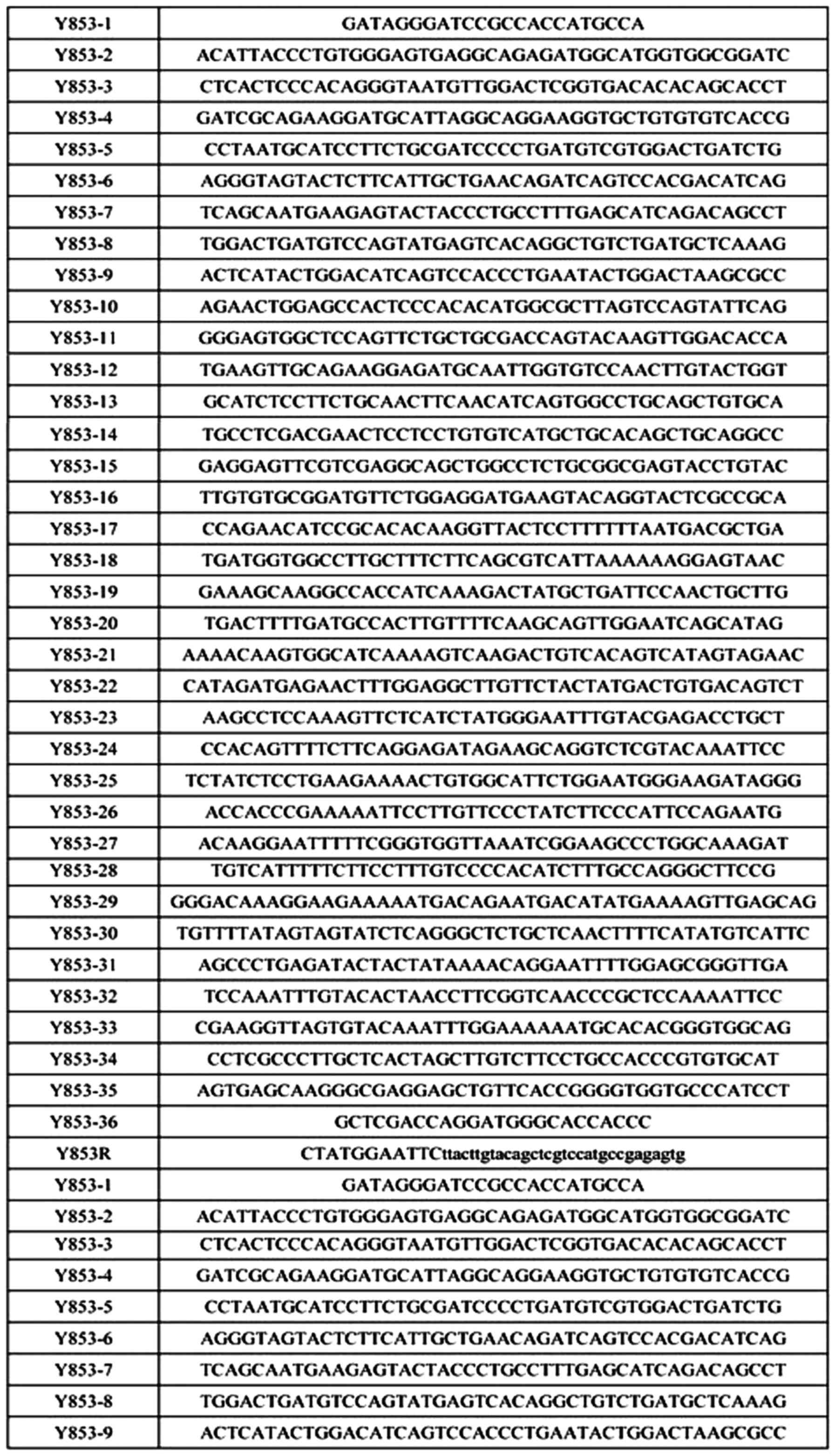
Oligo design is used for vector subcloning by adding
BamHI, EcoRI and protective base to upstream and
downstream primers of target gene. See Fig. 1 for oligo sequences.
The above primers were compounded by Gene Pharma
(Shanghai, China). The oligo was dissolved to 50 µM and the same
volume of oligo was taken and put into a 1.5 ml centrifuge tube,
mixed well to prepare the oligo mix used in the first round of PCR
reaction. The reaction system was as follows: Oligo mix 6 µl, 10X
Pfu buffer (+Mg2+) 5 µl, dNTP 1 µl, oligo-1 1 µl,
oligo-34 1 µl, ddH2O 36 µl, Pfu DNA polymerase 0.3 µl.
Cycling conditions: 95°C for 3 min 1 cycle, 94°C for 30 sec 30
cycles, 55°C for 30 sec, 72°C for 30 sec, 72°C 5 min 1 cycle. The
oligo-1 and oligo-34 were used for the second round of PCR
reaction, with the products of the first PCR reaction serving as
template. Reaction conditions remained the same as those of the
first round. The first round of PCR obtain non-single band PCR
product with the target gene band. With the PCR products of the
first round as template, single target gene band was obtained
through the second round of PCR. After the completion of PCR
reaction, agarose electrophoresis and gel extraction was used to
recycle ELF5+EGFP gene fragments.
ELF5+EGFP gene fragments were conducted with enzyme
digestion for 2 h at 37°C by BamHI and EcoRI and
vector pcDNA3.1 was conducted with enzyme digestion for 2 h at 37°C
by BamHI and EcoRI. ELF5+EGFP gene fragments and
vector pcDNA3.1 were recycled by applying DNA gel extraction kit.
ELF5+EGFP gene fragments and linear vector were obtained by
applying T4 DNA ligase to connect double enzyme digestion at 22°C
for 2 h. Competent cells were prepared by calcium chloride method
and a single colony was selected from the fresh plate cultured at
37°C for 16 h to transfer into a one-liter flask containing 100 ml
LB medium, cultured by shaking at 37°C for 3 h (rotary shaker, 300
rpm). Under aseptic conditions, cells were transferred into a
disposable ice-precooling 50 ml polypropylene tube, placed on ice
for 10 min and culture was cooled to 0°C. Cells were recycled by
centrifuging at 1,200 × g at 4°C for 10 min. Culture solution was
poured out and the tube was drained for 1 min. The connection
products were transferred to competent cells, colonies were
selected, and mini preparation plasmids were identified to select
positive clones. Four single colonies were selected and placed in
tubes containing 5 ml (containing 50 µg/ml ampicillin) LB medium.
The above tubes were placed in a shaker to cultivate at 37°C and
250 rpm for 16 h. Plasmids were extracted with plasmid
minipreparation kit from cultured bacterial liquid and enzyme
digestion identification was performed for the extracted
plasmids.
Bacterial liquid (200 µl) corresponding to positive
clone was sequenced and the rest was stored with glycerin.
Sequencing results were compared with target genes. After ensuring
that they were corrected and LB medium was inoculated with stored
glycerin bacterial liquid, a large amount of plasmids were
extracted for sufficient recombinant plasmid.
Cell culture
Human ovarian carcinoma SKOV3 cells were subcultured
at 37°C, 5% CO2, in saturated humidity constant
temperature incubator by applying routine RPMI-1640 medium
(containing penicillin and streptomycin) added with 10% fetal calf
serum.
Gene transfection and grouping
Eukaryotic expression vectors with ELF5 gene were
transfected in vitro into human ovarian carcinoma SKOV3
cells (recombinant plasmid group) by lipofectin method. The
operation was performed according to the instructions of
liptapfector lipofectin transfection kit. Stably transfected cells
were screened to implement amplification and culture with
pcDNA3.1-EGFP empty vector plasmid (empty plasmid group) and
untransfected human ovarian carcinoma SKOV3 cells (blank control
group) as control. After transfection, screening and passage were
conducted by applying selective medium containing neomycin
(G418).
Detection of the expression of ELF5 protein by
western blot analysis
Three groups of cells in logarithmic growth phase
were harvested in 200 µl cell lysis buffer on ice. The
concentration of protein was measured by bicinchoninic acid (BCA)
method and 10% sodium dodecyl sulfate-polyacrylamide gel
electrophoresis (SDS-PAGE), respectively. Then, electrotransferred
onto nitrocellulose membrane, sealed with 5% skim milk powder for
60 min, and ELF5 primary antibody (rabbit anti-human) (dilution,
1:1,000; cat. no. AM254515) was added at 4°C overnight. After 2-h
reaction with goat anti-rabbit secondary antibody (1:10,000; cat.
no. MAB4310) marked with horseradish peroxidase at room
temperature. Strengthened electrochemical luminescence (ECL)
reagent was added for exposure and development with highly
sensitive X-ray film.
Detection of proliferation capacity of ovarian
carcinoma SKOV3 cells by MTT method
Three groups of cells were inoculated on 96-well
plates at 1.5×10⁵/well and 20 µl MTT working solution
was added after cultivation for 1, 2, 3, 4, 5 and 6 days. After
cells were cultivated in CO2 incubator at 37°C for 4 h,
dimethyl sulfoxide was added to terminate the reaction. Absorbance
(A) values of all wells at 490 nm were determined on enzyme-linked
immunoassay and the cell growth curve was drawn.
Detection of invasive capacity of ovarian
carcinoma SKOV3 cells by in vitro invasion assay
Transwell chamber in vitro invasion assay was
applied. Membrane was prepared with Matrigel between upper and
lower chambers. Single cell suspension of ovarian carcinoma SKOV3
cells was inoculated at 200 µl/well (containing about
105 cells). After cultivating at 37°C and 5%
CO2 for 12 h, the cells and Matrigel on membrane were
wiped off and fixed and H&E stained. The number of cells was
calculated under a microscope. Three wells were set for each group
of cells and the experiment was repeated 3 times.
Detection of cell cycle of ovarian carcinoma
SKOV3 cells by flow cytometry
Ovarian carcinoma SKOV3 cells that stably expressed
ELF5 protein were selected. The cells were digested with 0.25%
pancreatin, routinely washed with normal saline, centrifuged at
1.200 × g at 4°C for 8 min and fixed with 70% ethyl alcohol
overnight. Single-cell suspension was prepared by adding 100 µl
phosphate-buffered saline (PBS) and the cell cycle was analyzed by
flow cytometry.
Detection of apoptosis rate of ovarian carcinoma
SKOV3 cells by flow cytometry
Ovarian carcinoma SKOV3 cells that stably expressed
ELF5 protein after transfection were selected. The cells were
digested with 0.25% pancreatin, routinely washed with normal
saline, centrifuged 1.200 × g at 4°C for 8 min and fixed with 70%
ethyl alcohol overnight. Single-cell suspension was prepared by
adding 100 µl PBS. Operation was performed according to the
instructions of Annexin V-PI cell apoptosis detection kit. The cell
apoptotic rate was analyzed by flow cytometry.
Statistical analysis
SPSS 13.0 statistical software (SPSS, Inc., Chicago,
IL, USA) was used for analysis. Comparison of rates among groups
was tested by χ2 test and Fisher's exact probability
calculation method. Measurement data were expressed as mean ± SD;
differences among groups were analyzed by variance, comparison of
data among groups was tested by q-test; and P<0.05 was
considered to indicate a statistically significant difference.
Results
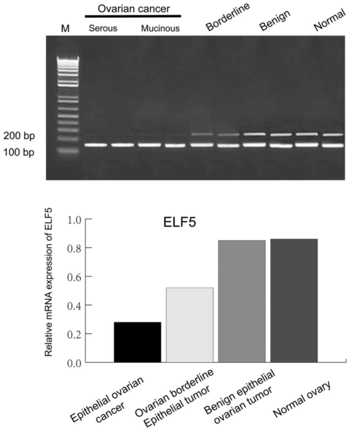
Expression of ELF5 mRNA in different
ovarian tissues
The rate of positive expression and the level of
expression of ELF5 mRNA in epithelial ovarian carcinoma and
borderline ovarian epithelial tumor tissues were significantly
lower (P<0.05) than those in benign ovarian epithelial tumor and
normal ovarian tissues. The rate of positive expression and the
level of expression of ELF5 mRNA in epithelial ovarian carcinoma
tissues were significantly lower (P<0.05) than those in
borderline ovarian epithelial tumor. The rate of positive
expression and the level of expression in benign ovarian epithelial
tumor and normal ovarian tissues had no significant differences
(Fig. 2 and Table I).
 | Table I.ELF5 mRNA expression in various
ovarian tissues. |
Table I.
ELF5 mRNA expression in various
ovarian tissues.
|
| Total cases | Positive cases | Percentage (%) |
|---|
| Epithelial ovarian
cancer | 49 | 9 | 18.4 |
| Ovarian borderline
epithelial tumor | 19 | 7 | 36.8 |
| Benign epithelial
ovarian tumor | 31 | 21 | 67.7 |
| Normal ovary | 40 | 29 | 72.5 |
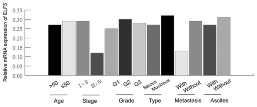
Expression of ELF5 mRNA in epithelial
ovarian carcinoma tissues at different clinicopathological
indexes
The rate of positive expression and the level of
expression of ELF5 mRNA in early phase (phase I and II) in
epithelial ovarian carcinoma tissues were significantly higher than
(P<0.05) those in late phase (phase III and IV). The positive
expression rate and expression level of ELF5 mRNA in epithelial
ovarian carcinoma tissues with lymph node metastases were
significantly lower (P<0.05) than those in epithelial ovarian
carcinoma tissues without lymph node metastases. The rate of
positive expression and the level of expression of ELF5 mRNA showed
no correlation with age, pathological grading, pathological types
and ascites formation (Table II
and Fig. 3).
| Table II.ELF5 mRNA expression in epithelial
ovarian cancer tissues of clinicopathological indexes. |
Table II.
ELF5 mRNA expression in epithelial
ovarian cancer tissues of clinicopathological indexes.
| Parameters | Total | Positive | Percentage (%) | P-value |
|---|
| Age (years) |
|
|
|
|
|
>50 | 27 | 5 | 18.5 | 00.817 |
| ≤50 | 22 | 4 | 18.2 |
|
| Pathological
staging |
|
|
|
|
| I–II | 23 | 6 | 26.1 | 0.015 |
|
III–IV | 26 | 3 | 11.5 |
|
| Pathological
grading |
|
|
|
|
| G1 | 20 | 4 | 20.0 | 0.797 |
| G2 | 17 | 3 | 17.6 |
|
| G3 | 12 | 2 | 16.7 |
|
| Pathological
type |
|
|
|
|
| Serous
cystadenocarcinoma | 32 | 6 | 18.8 | 0.812 |
| Mucinous
cystadenocarcinoma | 17 | 3 | 17.6 |
|
| Lymphatic
metastasis |
|
|
|
|
| With | 21 | 2 | 9.5 | 0.019 |
|
Without | 28 | 7 | 25.0 |
|
| Ascites |
|
|
|
|
|
With | 16 | 3 | 18.8 | 0.896 |
| Without | 33 | 6 | 18.2 |
|
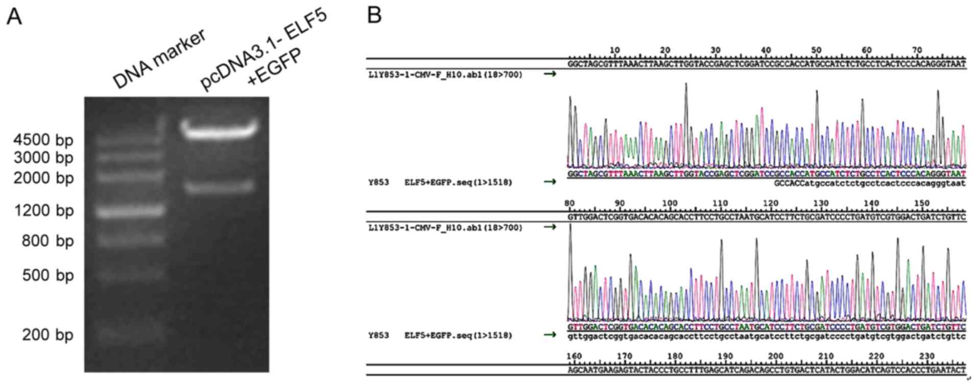
Successfully constructed
pcDNA3.1-ELF5+EGFP eukaryotic expression vector
We utilized continuous PCR method to obtained
complete gene sequence fragments of ELF5+EGFP, cloned target gene
ELF5+EGFP into vector pcDNA3.1. After enzyme digestion, it showed
significant bands at the position of 1,518 bp, which was in line
with the size of fragment expected to be amplified (Fig. 4A).
The results of DNA sequence analysis showed that the
sequence of target gene was identical to that of the gene sequence
in GeneBank. The insert direction of target gene was correct and
the reading frame remained unchanged, which proved that recombinant
plasmid pcDNA3.1-ELF5+EGFP was constructed successfully (Fig. 4B).
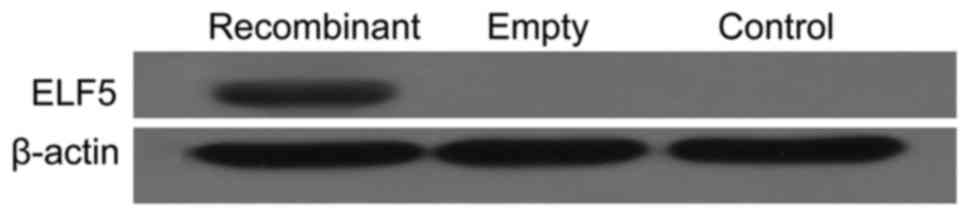
Changes in the expression of ELF5 protein in ovarian
carcinoma SKOV3 cells after the transfection of ELF5 gene. The
western blot results of the detection of the expression of ELF5 in
recombinant plasmid group was high, while the expression of ELF5
was not detected in empty plasmid group or blank control group
(Fig. 5).
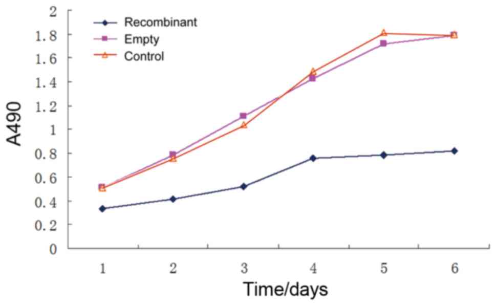
Changes in the reproductive capacity
of ovarian carcinoma SKOV3 cells after the transfection of ELF5
gene
The results of the detection of MTT method showed
that after being cultivated for 1, 2, 3, 4, 5 and 6 days, the A
values of recombinant plasmid group were 0.337±0.002, 0.413±0.012,
0.522±0.007, 0.761±0.002, 0.787±0.021 and 0.821±0.019,
respectively. These decreased obviously compared with blank control
group and empty plasmid group at each time-point (P<0.05) while
the comparison of blank control group and empty plasmid group
showed the differences were not statistically significant (Fig. 6).
Changes in the invasive capacity of
ovarian carcinoma SKOV3 cells after the transfection of ELF5
gene
Transwell chamber in vitro invasion assay
showed that the number of cells permeating in recombinant plasmid
group was 92.1±2.7, compared with empty plasmid group (149.6±4.6)
and blank control group (147.4±5.3), and the differences were
statistically significant (P<0.05).
Changes in the cell cycle of ovarian
carcinoma SKOV3 cells after the transfection of ELF5 gene
Flow cytometry detection showed that 67.03% of cells
in recombinant plasmid group was blocked in
G0/G1 phase, compared with empty plasmid
group (37.17%) and blank control group (38.24%), and the
differences were statistically significant (P<0.05) (Table III).
| Table III.Changes of ovarian cancer SKOV3 cell
cycle after transfection by ELF5 gene (mean ± SD, %). |
Table III.
Changes of ovarian cancer SKOV3 cell
cycle after transfection by ELF5 gene (mean ± SD, %).
|
| Stage
G0/G1 | Stage S | Stage
G2/M |
|---|
| Recombinant
plasmid | 67.03±0.14 | 18.21±1.02 | 14.37±0.13 |
| Empty plasmid | 39.23±1.21 | 39.41±0.11 | 21.62±0.33 |
| Control | 40.22±0.38 | 40.28±0.31 | 19.22±0.15 |
Changes in the apoptotic rate of
ovarian carcinoma SKOV3 cells after the transfection of ELF5
gene
Flow cytometry showed that the apoptotic rate of
recombinant plasmid group was 31.4±1.9%, compared with that of
empty plasmid group (9.1±2.2%) and blank control group (8.7±1.5%),
and the differences were statistically significant (P<0.05).
Discussion
Ets family members have very important regulating
effects on normal physiological activities, including cell
development, differentiation, proliferation and apoptosis (10). As a member of Ets family, ELF5
locates in 13–15 region of short arm of chromosome 11, generating
loss of heterozygosity in cancer. ELF5 contains two identifiable
domains, one is the sequence motif that is contained by all ETS
transcription factors and can combine with TTCC core sequence, the
other is the point domain that takes part in the interaction
between proteins (6). ELF5 is
expressed in hair follicle, epithelial exocrine glands, placental
trophoblastic cells, kidney, mammary gland and other organs
(11–15).
Studies on ELF5 are mainly focused on mammary gland
cells and breast carcinoma. In the breast tissues, ELF5 is secreted
by the mammary duct precursor cells and ELF5 is an important
regulatory factor for development of mammary acinar cells. On one
hand, it can decide the fate of acinar cells (16), and on the other hand, it can affect
embryogenesis and hyperplasia of breast acinus through regulating
breast lactation acinus structure during pregnancy. Studies of
Chakrabarti et al (17)
showed that during the process of normal mammogenesis, the loss of
ELF5 could cause the increase of mammary adult stem cells, thus to
increase the risk of forming breast carcinoma. It has been shown
that ELF5 was a potential tumor-inhibiting factor and played a role
of tumor suppressor gene during the process of occurrence and
development of breast carcinoma. Due to the expression loss of ELF5
in human breast carcinoma tissues and cell lines, regulating the
transcriptional level of ELF5 may provide a new method for the
treatment of breast carcinoma (18–20).
Inspired by above studies, we considered the
following: How does ELF5 express in epithelial ovarian carcinoma
tissues, and does ELF5 gene have effects on biological behavior of
human ovarian carcinoma SKOV3 cells?
This study found that the positive expression rate
and expression level of ELF5 mRNA in epithelial ovarian carcinoma
and borderline ovarian epithelial tumor tissues were significantly
lower than those in benign ovarian epithelial tumor and normal
ovarian tissues. Our study also showed that the positive expression
rate and expression level of ELF5 mRNA in epithelial ovarian
carcinoma tissues showed no correlation with its pathological
grading, pathological types and ascites formation. The positive
expression rate and expression level of ELF5 mRNA in epithelial
ovarian carcinoma tissues in patients at early clinical stage
without lymph node metastasis were apparently higher than those in
patients at late clinical stage with lymph node metastasis. Thus,
we could further speculated that the ELF5 gene, not only as a tumor
suppressor gene, but also an inhibiting gene for tumor metastasis,
plays an important role in the process of inhibiting infiltration
and metastasis for ovarian carcinoma cells, and the infiltration
and metastasis is an important index for judging prognosis of
patients with epithelial ovarian carcinoma.
In order to further implement deep and extensive
studies on ELF5, we successfully constructed recombinant plasmid
pcDNA3.1-ELF5+EGFP, transfected recombinant plasmid
pcDNA3.1-ELF5+EGFP into human ovarian carcinoma SKOV3 cells and
conducted screening. The cultivation and identification for
positive clone showed successful transfection of recombinant
plasmid pcDNA3.1-ELF5+EGFP at the protein level, and then studied
the effects of ELF5 gene after transfection on in vitro
biological behavior of ovarian carcinoma SKOV3 cells. The cell
growth curve experiment showed that ovarian carcinoma SKOV3 cells
grew slower after the transfection of ELF5 gene, suggesting that
the ELF5 gene delayed the proliferation of ovarian carcinoma SKOV3
cells. The analysis of flow cytometry showed, the high expression
of ELF5 gene could delay cells in phase G0/G1
from entering into phase S and phase G2/M; and thus, it
could be speculated that ELF5 gene could inhibit the proliferation
of ovarian carcinoma SKOV3 cells through intervening in the cell
cycle. The apoptosis rate of ovarian carcinoma SKOV3 cells after
the transfection of ELF5 gene increased significantly and showed
that ELF5 gene could promote apoptosis of ovarian carcinoma SKOV3
cells.
The in vitro invasive capacity is important
to measure tumor metastasis potential. Transwell chamber in
vitro invasion assay showed that the invasive capacity of
ovarian carcinoma SKOV3 cells after the transfection of ELF5 gene
decreased significantly. This showed that the ELF5 gene had obvious
inhibiting effects on invasive capacity to host and motor ability
of ovarian carcinoma SKOV3 cells. Our experiments proved that on
one hand, ELF5 mRNA had low expression in epithelial ovarian
carcinoma tissues and its expression loss at different levels was
related to the occurrence and development of epithelial ovarian
carcinoma. ELF5 gene disturbed the cell cycle of human ovarian
carcinoma SKOV3 cells, promoting apoptosis of human ovarian
carcinoma SKOV3 cells and inhibited its growth and invasive
capacity. The results of our study provide some experimental
foundation for conducting gene treatment of ovarian carcinoma, but
the following problems are still not cleared: i) What is the
mechanism of the expression loss of ELF5 mRNA for causing the
occurrence of ovarian carcinoma. The expression of ELF5 mRNA in
ovarian carcinoma is generally low. Moreover, it shows abnormal
expression in borderline ovarian epithelial tumor tissues. The
effects on the occurrence and development of epithelial ovarian
carcinoma and whether it can serve as a new therapeutic target of
epithelial ovarian carcinoma are worth further research. ii) The
specific mechanism of ELF5 gene that affects the biological
behavior of ovarian carcinoma SKOV3 cells is not clear, and thus
ELF5 gene therapy needs further investigation to improve the
survival rate of ovarian carcinoma.
References
|
1
|
Jemal A, Bray F, Center MM, Ferlay J, Ward
E and Forman D: Global cancer statistics. CA Cancer J Clin.
61:69–90. 2011. View Article : Google Scholar : PubMed/NCBI
|
|
2
|
Lee JY, Kim HS, Suh DH, Kim MK, Chung HH
and Song YS: Ovarian cancer biomarker discovery based on genomic
approaches. J Cancer Prev. 18:298–312. 2013. View Article : Google Scholar : PubMed/NCBI
|
|
3
|
Suh DH, Kim MK, Kim HS, Chung HH and Song
YS: Epigenetic therapies as a promising strategy for overcoming
chemoresistance in epithelial ovarian cancer. J Cancer Prev.
18:227–234. 2013. View Article : Google Scholar : PubMed/NCBI
|
|
4
|
Zheng H, Liu JY, Song FJ and Chen KX:
Advances in circulating microRNAs as diagnostic and prognostic
markers for ovarian cancer. Cancer Biol Med. 10:123–130.
2013.PubMed/NCBI
|
|
5
|
Lin JY, Qin JB, Li XY, Dong P and Yin BD:
Diagnostic value of human epididymis protein 4 compared with
mesothelin for ovarian cancer: a systematic review and
meta-analysis. Asian Pac J Cancer Prev. 13:5427–5432. 2012.
View Article : Google Scholar : PubMed/NCBI
|
|
6
|
Zhou J, Ng AY, Tymms MJ, Jermiin LS, Seth
AK, Thomas RS and Kola I: A novel transcription factor, ELF5,
belongs to the ELF subfamily of ETS genes and maps to human
chromosome 11p13-15, a region subject to LOH and rearrangement in
human carcinoma cell lines. Oncogene. 17:2719–2732. 1998.
View Article : Google Scholar : PubMed/NCBI
|
|
7
|
Choi YS and Sinha S: Determination of the
consensus DNA-binding sequence and a transcriptional activation
domain for ESE-2. Biochem J. 398:497–507. 2006. View Article : Google Scholar : PubMed/NCBI
|
|
8
|
Prat J: FIGO Committee on Gynecologic
Oncology: Staging classification for cancer of the ovary, fallopian
tube, and peritoneum. Int J Gynaecol Obstet. 124:1–5. 2014.
View Article : Google Scholar : PubMed/NCBI
|
|
9
|
He J, Pan Y, Hu J, Albarracin C, Wu Y and
Dai JL: Profile of Ets gene expression in human breast carcinoma.
Cancer Biol Ther. 6:76–82. 2007. View Article : Google Scholar : PubMed/NCBI
|
|
10
|
Lee HJ and Ormandy CJ: Elf5, hormones and
cell fate. Trends Endocrinol Metab. 23:292–298. 2012. View Article : Google Scholar : PubMed/NCBI
|
|
11
|
Lapinskas EJ, Palmer J, Ricardo S, Hertzog
PJ, Hammacher A and Pritchard MA: A major site of expression of the
ets transcription factor Elf5 is epithelia of exocrine glands.
Histochem Cell Biol. 122:521–526. 2004. View Article : Google Scholar : PubMed/NCBI
|
|
12
|
Hemberger M, Udayashankar R, Tesar P,
Moore H and Burton GJ: ELF5-enforced transcriptional networks
define an epigenetically regulated trophoblast stem cell
compartment in the human placenta. Hum Mol Genet. 19:2456–2467.
2010. View Article : Google Scholar : PubMed/NCBI
|
|
13
|
Lee HJ, Gallego-Ortega D, Ledger A,
Schramek D, Joshi P, Szwarc MM, Cho C, Lydon JP, Khokha R,
Penninger JM, et al: Progesterone drives mammary secretory
differentiation via RankL-mediated induction of Elf5 in luminal
progenitor cells. Development. 140:1397–1401. 2013. View Article : Google Scholar : PubMed/NCBI
|
|
14
|
Lapinskas EJ, Svobodova S, Davis ID, Cebon
J, Hertzog PJ and Pritchard MA: The Ets transcription factor ELF5
functions as a tumor suppressor in the kidney. Twin Res Hum Genet.
14:316–322. 2011. View Article : Google Scholar : PubMed/NCBI
|
|
15
|
Pearton DJ, Smith CS, Redgate E, van
Leeuwen J, Donnison M and Pfeffer PL: Elf5 counteracts precocious
trophoblast differentiation by maintaining Sox2 and 3 and
inhibiting Hand1 expression. Dev Biol. 392:344–357. 2014.
View Article : Google Scholar : PubMed/NCBI
|
|
16
|
Lee HJ, Hinshelwood RA, Bouras T,
Gallego-Ortega D, Valdés-Mora F, Blazek K, Visvader JE, Clark SJ
and Ormandy CJ: Lineage specific methylation of the Elf5 promoter
in mammary epithelial cells. Stem Cells. 29:1611–1619. 2011.
View Article : Google Scholar : PubMed/NCBI
|
|
17
|
Chakrabarti R, Wei Y, Romano RA, DeCoste
C, Kang Y and Sinha S: Elf5 regulates mammary gland stem/progenitor
cell fate by influencing notch signaling. Stem Cells. 30:1496–1508.
2012. View Article : Google Scholar : PubMed/NCBI
|
|
18
|
Chakrabarti R, Hwang J, Blanco M Andres,
Wei Y, Lukačišin M, Romano RA, Smalley K, Liu S, Yang Q, Ibrahim T,
et al: Elf5 inhibits the epithelial-mesenchymal transition in
mammary gland development and breast cancer metastasis by
transcriptionally repressing Snail2. Nat Cell Biol. 14:1212–1222.
2012. View
Article : Google Scholar : PubMed/NCBI
|
|
19
|
Kalyuga M, Gallego-Ortega D, Lee HJ, Roden
DL, Cowley MJ, Caldon CE, Stone A, Allerdice SL, Valdes-Mora F,
Launchbury R, et al: ELF5 suppresses estrogen sensitivity and
underpins the acquisition of antiestrogen resistance in luminal
breast cancer. PLoS Biol. 10:e10014612012. View Article : Google Scholar : PubMed/NCBI
|
|
20
|
Frend HT and Watson CJ: Elf5 - breast
cancer's little helper. Breast Cancer Res. 15:3072013. View Article : Google Scholar : PubMed/NCBI
|