Introduction
Cancer is known to be a key contributor to mortality
around the world, of which ovarian cancer is recognized as one of
the most prevalent malignancies among women and is ranking the
fifth leading cause of cancer death in women (1,2). The
histologic subtypes of ovarian cancer are composed of clear cell,
endometrioid, and mucinous adenocarcinomas and serous carcinoma
(3). Surgery and chemoradiotherapy
are mostly developed for the treatment of ovarian cancer, and had
improved the quality of life in patients with ovarian cancer, but
the clinical prognosis in these patients is still poor (4). It is reported that a 5-year survival
rate is 93% in patients with ovarian cancer patients at stage I,
but only 25% at the stage IV due to the high recurrence rate,
strong metastatic capacity and deficiency of specific symptoms
(5). Most of ovarian carcinoma
patients are commonly diagnosed at stage III or IV, only 15% of
ovarian cancer patients are diagnosed at early stage (6). Transvaginal ultrasound, manual pelvic
examination, and measurement of cancer antigen (CA)-125 are primary
tools for the screening of ovarian cancer, but these methods have
lowe specificity and sensitivity (7). It has been verified that pathological
grade, histological subtype and cancer stage may be critical
prognostic factors for ovarian cancer (8). The development of ovarian cancer may
be strongly associated with many molecular signals, the ideal
molecular biomarkers may be useful for predication of early
diagnosis and prognosis of ovarian cancer.
Salusins are bioactive peptides originally
identified by bioinformatic analysis of full-length complementary
DNA library (9). Salusins are
divided into two related bioactive peptides of 28 and 20 amino
acids designated salusin-α and salusin-β. It has been demonstrated
that salusins exist in human plasma and vascular system (10). Salusin-β is a novel modulator of
systemic hemodynamics. It may be critical for aggravation of
inflammation and oxidative stress in vasculature (11). Inflammation and oxidative stress are
strongly involved in the development of ovarian cancer (12). Moreover, women with polycystic
ovarian syndrome have higher salusin-β in comparison with the
control group (13). It has been
confirmed that plasma salusin-β level is upregulated in patients
with endometrioma and positively associated with endometrioma size
(14). Existing evidence suggests
that salusin-β may play an important role in the reproductive
system. However, the roles of salusin-β in tumorigenesis,
metastasis, and the prognostic significance of ovarian cancer has
not yet been clearly established.
Patients and methods
Patients and specimens
The present study was reviewed and approved by the
Research Ethics Committee of the First Affiliated Hospital of
Wenzhou Medical University. All experiments were conformed to the
Code of Ethics of the World Medical Association (Declaration of
Helsinki). A signed written informed consent was provided by the
patients. A total of 57 ovarian cancer patients who underwent a
curative resection were included from the Department of
Gynaecology, the First Affiliated Hospital of Wenzhou Medical
University from February 2005 to May 2009. The diagnosis of ovarian
cancer was based on the clinical presentations, immunohistochemical
staining and morphological criteria. The patients who had history
of radiation, chemoradiation, or hormonal therapy were excluded
from this study. Fifty-seven cases with ovarian cancer were
enrolled in accordance with the inclusion and exclusion criteria.
The clinicopathological and demographic data pre- and
post-operation were preserved in medical records. The
characteristics of patients including age, tumor grade, Federation
of Gynecology and Obstetrics (FIGO) stage, histological subtype,
tumor size, serum CA-125 level and lymphatic invasion were
summarized in Table I. The survival
information of each patient was collected by letter, e-mail or
phone calls. The overall survival time was considered as the time
from the date of surgery to the date of death or the date of last
follow-up. The paired tumor tissues and adjacent normal ovarian
tissues were immediately frozen in liquid nitrogen and kept at
−80°C prior to use.
 | Table I.Correlation of salusin-β expression
with the patient clinicopathological features. |
Table I.
Correlation of salusin-β expression
with the patient clinicopathological features.
|
|
| Salusin-β
expression |
|
|
|---|
|
|
|
|
|
|
|---|
| Variables | All cases (%) | Low (%) | High (%) | χ2 | P-value |
|---|
| Age (years) |
| ≤60 | 35 (61.4) | 18 (51.4) | 17 (48.6) | 0.193 | 0.661 |
|
>60 | 22 (38.6) | 10 (45.5) | 12 (54.5) |
| Pathological
grade |
| G1 +
G2 | 38 (66.7) | 18 (47.4) | 20 (52.6) | 0.141 | 0.707 |
| G3 | 19 (33.3) | 8 (42.1) | 11 (57.9) |
| FIGO stage |
| I +
II | 21 (36.8) | 16 (76.2) | 5 (23.8) | 7.402 | 0.007 |
| III +
IV | 36 (63.2) | 14 (38.9) | 22 (61.1) |
| Tumor size
(cm) |
| ≤2 | 37 (64.9) | 20 (54.1) | 17 (45.9) | 1.891 | 0.169 |
|
>2 | 20 (35.1) | 7 (35.0) | 13 (65.0) |
| Histological
type |
|
Serous | 25 (43.9) | 11 (44.0) | 14 (56.0) | 0.189 | 0.979 |
|
Endometrioid | 14 (24.6) | 6 (42.9) | 8 (57.1) |
|
Mucinous | 8 (14.0) | 4 (50.0) | 4 (50.0) |
| Clear
cell | 10 (17.5) | 4 (40.0) | 6 (60.0) |
| Lymph node
involvement |
|
Negative | 21 v(36.8) | 14 (66.7) | 7 (33.3) | 9.567 | 0.002 |
|
Positive | 36 (63.2) | 9 (25.0) | 27 (75.0) |
| Serum CA125 level
(U/l) |
|
≤900 | 26 (45.6) | 16 (61.5) | 10 (38.5) | 1.521 | 0.217 |
|
>900 | 31 (54.4) | 14 (45.2) | 17 (54.8) |
RNA isolation and quantitative
real-time PCR
Total RNA in each sample was isolated from frozen
specimen by the TRIzol (Invitrogen, Carlsbad, CA, USA) protocol
according to the manufacturers instructions (15). The concentration and purity of RNA
was determined by measuring the absorbance at 260 and 280 nm with
the aid of NanoDrop 1000 spectrophotometer (Thermo Fisher
Scientific, Wilmington, DE, USA). The reverse transcription of RNA
was then performed using the PrimeScript RT-PCR kit (Takara, Shiga,
Japan). The quantitative real-time polymerase chain reaction
(RT-PCR) was performed using the SYBR Premix Ex Taq™ (Takara) on
ABI 7500 Real-Time PCR system (Applied Biosystems, Foster City, CA,
USA). The relative proportion of target gene expression was
quantified by normalizing the targeted gene level to that of
internal control by the ΔΔCt method. Primer sequences used are as
follows: salusin-β: forward, 5-GAACTTCCACCAA GGGTTCTG-3 and
reverse, 5-GTAAAACGACGGCCAGT AGGAGCGTCGGGAGTTGTAG-3;
glyceraldehyde-3-phosphate dehydrogenase (GAPDH): forward,
5-GCTCTCTGC TCCTCCTGTTC-3 and reverse, 5-ACGACCAAATCCGTTG
ACTC-3.
Enzyme linked immunosorbent assay
(ELISA) for salusin-β
The frozen paired ovarian tissues and adjacent
normal ovarian tissues were homogenized and centrifuged in lysis
buffer. Total protein concentration in the homogenate was measured
with the Bradford assay (BCA; Pierce, Santa Cruz, CA, USA). The
level of salusin-β was assayed by using a commercially available
ELISA kit (USCN Life Science, Inc., Houston, TX, USA) according to
the manufacturers protocols as previously reported (15). The values were averaged and
expressed as mean picograms per minute per milligram of
protein.
Cell culture
The ovarian cancer cell lines including A2780,
IGROV1, SKOV3 and OVCAR3 and a control human ovarian surface
epithelial cell line (HOSE 6.3) were purchased from the Cell Bank
of Chinese Academy of Sciences (Shanghai, China). OVCAR-8 and
SKOV-3 were cultured in Dulbeccos modified Eagles medium (DMEM),
and A2780 and IGROV-1 were cultured in RPMI-1640 medium,
supplemented with 10% fetal bovine serum (FBS; Gibco, Grand Island,
NY, USA) coupled with 100 U/ml penicillin sodium, and 100 mg/ml
streptomycin sulfate at 37°C in a humidified incubator with 5%
CO2. HOSE 6.3 cells were cultured in MCDB Medium
supplemented with 10% FBS. In addition, SKOV3 cells were incubated
with different dose of salusin-β at the indicated time, the
proliferation and epithelial mesenchymal transition of SKOV3 cells,
and the possible signaling pathways in response to salusin-β were
investigated.
Cell proliferation assay
SKOV3 cell proliferation was assessed with the Cell
Counting kit-8 (CCK-8) assay (Dojindo Molecular Technologies, Inc.,
Kumamoto, Japan) in accordance with the manufacturers suggestions.
Cells were seeded at an initial density of 2×10⁴
cells/ml in 96-well plates. The OD450 absorbance was determined to
measure cell viability (16).
Furthermore, the cell proliferation markers proliferating cell
nuclear antigen (PCNA) and phosphorylated histone H3 (P-H3) were
also detected.
Western blot analysis
The cell lysates were collected by using
radioimmunoprecipitation assay (RIPA) buffer (Beyotime Institute of
Biotechnology, Haimen, China). The total protein concentration in
the supernatant was quantified with the Bradford assay (BCA;
Pierce). Total cellular proteins were loaded to sodium dodecyl
sulfate-polyacrylamide gel electrophoresis (Bio-Rad Laboratories,
Inc., Hercules, CA, USA) and transferred to immobilon
polyvinylidene difluoride (PVDF) membranes (Millipore, Billerica,
MA, USA). The membranes were blocked with 5% non-fat milk at room
temperature and incubated with designed primary antibodies
overnight at 4°C. The positive signals from HRP-coupled secondary
antibodies (Pierce) were visualized. The densitometric analysis of
the band intensities was measured and normalized to the band
intensities of GAPDH using ImageJ software (NIH, Bethesda, MD,
USA). The primary antibodies against proliferating cell nuclear
antigen (PCNA), phosphorylated histone H3 (P-H3) and GAPDH were
obtained from Santa Cruz Biotechnology (Santa Cruz, CA, USA).
N-cadherin, vimentin, GSK-3β, p-GSK-3β, Snail cyclin D1 and C-myc
antibodies were purchased from Cell Signaling Technology, Inc.
(Beverly, MA, USA) (17).
Statistical analysis
Data on continuous variables are presented as means
± standard deviation (SD), and the categorical data are expressed
as the percentage. SPSS 19.0 software (SPSS, Inc., Chicago, IL,
USA) was used for statistical analysis. The Chi-square or Fishers
exact tests, or t-test were used to evaluate the relationship
between the expression of salusin-β and other clinical parameters.
One-way or two-way ANOVA followed by post hoc Bonferroni test was
used when multiple comparisons were made. Survival analysis was
estimated with the Kaplan-Meier method. Differences between
survival curves were analyzed using the log-rank test. The Cox
proportional hazards model was applied for the multivariate
survival analysis. The diagnostic efficacy of salusin-β was
evaluated by receiver operating characteristic (ROC) curve. A
two-sided P-value of <0.05 was considered statistically
significant.
Results
Overexpression of salusin-β expression
in prostate cancer tissues
We determined salusin-β level in 57 ovarian cancer
tissues and matched normal adjacent tissue specimens. RT-PCR
results showed that salusin-β mRNA level was significantly higher
in ovarian cancer tissues than paired adjacent tissue specimens
(Fig. 1A). The expression of
salusin-β at the protein level was obviously upregulated in the
ovarian cancer tissues (Fig. 1B).
In addition, the levels of salusin-β mRNA were dramatically
increased in ovarian cancer cell lines including SKOV3, IGROV1,
A2780 and OVCAR3 in comparison with normal human ovarian surface
epithelial HOSE 6.3 cells (Fig.
1C). As the SKOV3 cell line exhibited the highest salusin-β
mRNA level, the SKOV3 cell line was selected for the in
vitro experiments.
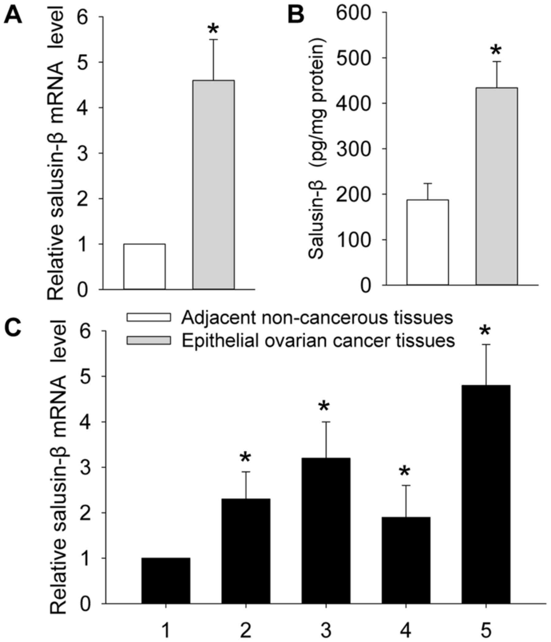 | Figure 1.Expressions of salusin-β in paired
adjacent non-cancerous tissues and epithelial ovarian cancer
tissues detected by real-time quantitative RT-PCR assay (A) or
enzyme linked immunosorbent assay (ELISA) kit (B). (C) Higher
expression levels of salusin-β were detected in 4 ovarian cancer
cell lines (2, A2780), (3, OVCAR3), (4, IGROV1), (5, SKOV3),
compared with values obtained for the normal human ovarian surface
epithelial cell line (1, HOSE 6.3) determined with RT-PCR. The
results showed that salusin-β expression level was significantly
increased in epithelial ovarian cancer tissues and cell lines.
*P<0.05 vs. adjacent non-cancerous tissues or 1, HOSE 6.3.
Values are mean ± SD. *P<0.05 vs. control. |
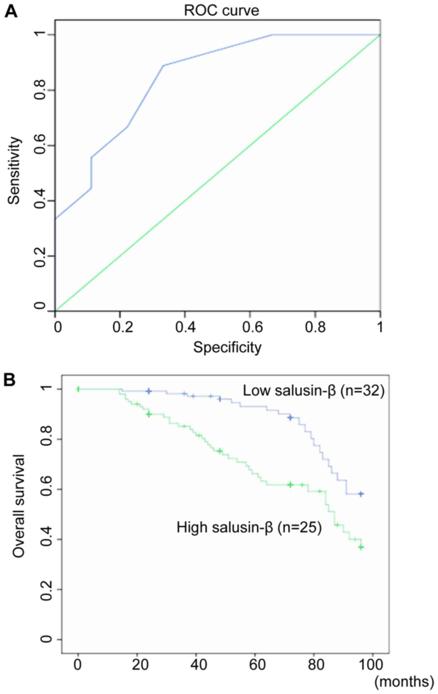
Diagnostic efficacy of miR-129 in
prostate cancer patients
The assessment of diagnostic efficacy of salusin-β
in ovarian cancer patients was performed by calculating the area
under the receiver operating characteristic curve. The ROC curve
analysis revealed that AUC was 0.154 (P=0.026). At the cut-off
value 339.8, the diagnostic sensitivity (70.2%) and specificity
(89.4%) reached their peak values (P<0.05; Fig. 2). Thus, the salusin-β expression was
further classified into the low expression group (n=salusin-β
expression <339.8, n=32) and high expression group (salusin-β
expression ≥339.8, n=25) as the threshold ROC curve value of 339.8
(Fig. 2A).
Correlation of salusin-β expression
with clinical parameters of ovarian cancer patients
As shown in Table I,
the high salusin-β expression is closely associated with the FIGO
stage (P=0.007) and lymph node involvement (P=0.002) in ovarian
cancer patients. However, we found no significant correlations
between salusin-β expression and age (P=0.661), pathological grade
(P=0.707), tumor size (P=0.169), histological type (P=0.979) or
serum CA125 level (P=0.217) in patients with ovarian cancer.
Relationship between salusin-β
expression and overall survival
The possible prognostic value of salusin-β in
overall survival in patients with ovarian cancer was performed by
calculating the cumulative survival curves with the Kaplan-Meier
method. The Kaplan-Meier curves between high or low salusin-β
expression and overall survival demonstrated that the ovarian
cancer patients with high salusin-β expression had an obviously
shorter overall survival (P=0.000; Fig.
2B). Multivariate Cox proportional hazards regression analysis
revealed that FIGO stage, lymph node involvement, salusin-β
expression were closely related with overall survival rate in
ovarian cancer patients, and these parameters may be employed as
independent prognostic indicators for overall survival of ovarian
cancer patients (P<0.05; Table
II).
| Table II.Cox multivariate analysis of the
clinicopathological parameters for overall survival. |
Table II.
Cox multivariate analysis of the
clinicopathological parameters for overall survival.
| Variables | Hazard ratio | 95% CI | P-value |
|---|
| Age (≤60 vs. >60
years) | 1.271 | 0.436–3.702 | 0.868 |
| Pathological grade
(G1 + G2 vs. G3) | 1.238 | 0.407–3.760 | 0.926 |
| FIGO stage (I + II
vs. III + IV) | 5.029 | 1.504–16.816 | 0.015 |
| Tumor size (≤2 vs.
>2 cm) | 2.185 | 0.710–6.720 | 0.277 |
| Serum CA125 (≤900
vs. >900 U/l) | 1.943 | 0.673–5.610 | 0.338 |
| Lymph node
involvement (negative vs. positive) | 7.286 | 2.286–23.223 | 0.001 |
| Salusin-β
expression (high vs. low) | 4.675 | 1.518–14.395 | 0.006 |
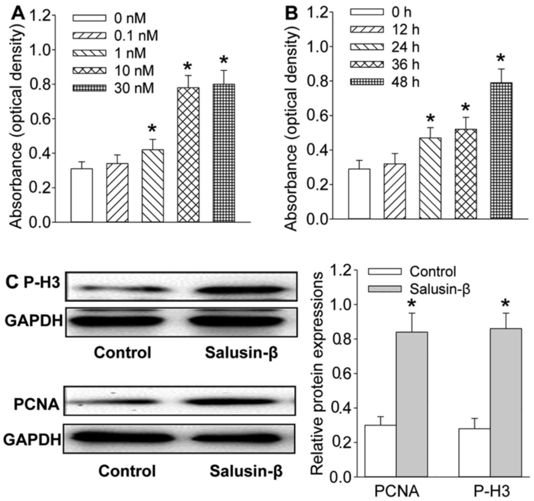
Salusin-β promotes the proliferation
of SKOV3 cells
The excessive proliferation of cancer cells is
critical for the development and progression of ovarian cancer
patients (18). The SKOV3 cells
were challenged by salusin-β in the dedicated time and
concentration for evaluation of the effect of salusin-β on the
proliferation of ovarian cancer cell with CCK8 assay in
vitro. Salusin-β stimulated the SKOV3 cell proliferation in a
time-dependent manner (Fig. 3A) and
dose-related fashion (Fig. 3B).
Furthermore, the proliferating markers including PCNA and
phosphorylated histone H3 (P-H3) were also obviously increased in
SKOV3 cells in response to salusin-β incubation at dose of 10 nM
for 48 h (Fig. 3C).
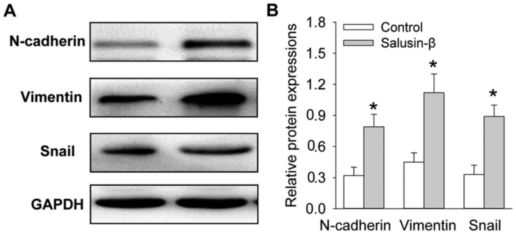
Salusin-β stimulated the epithelial
mesenchymal transition of SKOV3 cells
Epithelial mesenchymal transition is a key event in
the acceleration of migration and invasion of cancer cells
(19). Western blot analysis
illustrated that salusin-β significantly upregulated the protein
expressions of mesenchymal markers including N-cadherin, vimentin
and Snail in SKOV3 cells (Fig.
4).
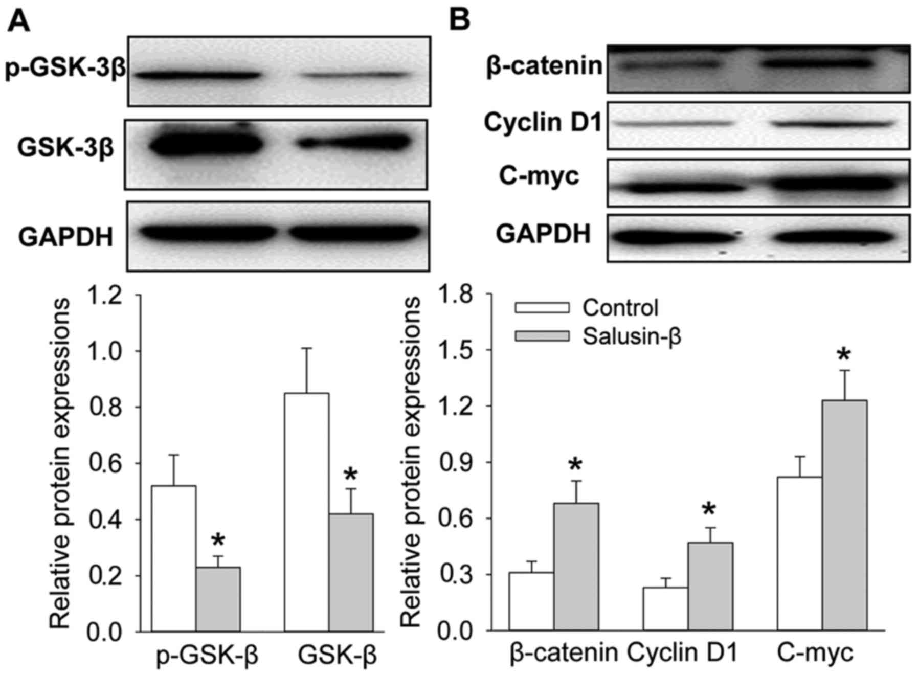
Salusin-β modulated Wnt/β-catenin
signaling pathway in SKOV3 cells
Wnt/β-catenin signaling pathway is one of the
crucial pathways, and majorly contributes to the proliferation and
EMT and epithelial mesenchymal transition in ovarian cancer
(20,21). Incubation of SKOV3 cells with
salusin-β substantially inhibited expressions of p-GSK-3β and
GSK-3β, and remarkably augmented the β-catenin levels and increased
the transcription of the targeted genes of Wnt/β-catenin, such as
cyclin D1 and C-myc (Fig. 5).
Discussion
Ovarian cancer patients suffer from high disease
recurrence and mortality (1). Early
diagnosis and treatment may be helpful for prevention of
progression of ovarian cancer (4).
The absence of useful biomarkers has disappointed the clinical
outcome of ovarian cancer therapy. It has been disclosed that
various molecules are identified to be biomarkers for prognosis in
cancer. The utility of novel biomarkers may provide a better
understanding of cancer biological behavior, thus, useful
biomarkers for cancer are continuously characterized in recent
years (2). The present study
established an intimate relationship of salusin-β with aggressive
clinicopathological parameters including FIGO stage and lymph node
involvement in ovarian cancer patients. Salusin-β may be a crucial
component in the pathogenesis of ovarian cancer associated with the
deteriorated proliferation and epithelial-mesenchymal transition of
ovarian cancer cells via modulation of Wnt/β-catenin signaling
pathway.
Deficiency of diagnostic and prognostic factors may
be largely responsible for the poor outcome of ovarian cancer. It
has been recommended that early and accurate diagnosis of ovarian
cancer is beneficial for the appropriate therapy. It was recently
reported that the microRNA (miRNA) miR-498 is significantly
decreased in ovarian cancer tissues, and may be taken as a valuable
factor in predicting the prognosis in patients with ovarian cancer
(22). It is also demonstrated that
downregulated miR-22 was closely related with the overall survival
of epithelial ovarian cancer, and is an efficient prognostic factor
for patients with epithelial ovarian cancer (23). The secretory small guanosine
5′-triphosphate binding enzyme, Rab27B was also recently identified
to be an independent prognostic factor for the survival of ovarian
cancer patients (24). Salusin-β is
a bioactive peptide with hypotensive and bradycardiac effects
(25). Salusin-β is found to
accelerate inflammation responses, oxidative stress and tube
formation in human in human endothelial cells, and promotes
monocyte-endothelial adhesion (26). Salusin-β is a stimulator for the
migration of vascular smooth muscle cells (VSMCs) with regard to
vascular injury (15). Salusin-β
may be used as a biomarker for atherosclerotic cardiovascular
diseases (27). The serum salusin-β
level is increased in patients with endometrioma and positively
associated with endometrioma size (14). A series of studies in salusin-β led
us to explore whether or how salusin-β affected the progression of
ovarian cancer. In this study, we showed that the ovarian cancer
tissues exhibited an obvious increase in salusin-β expression. The
upregulation of salusin-β was positively associated with the FIGO
stage and lymph node metastasis in ovarian cancer patients. The
ovarian cancer patients with high expression of salusin-β had
shorter overall survival. Multivariate analyses further proved that
salusin-β was a potential independent prognostic factor for overall
survival of patients with ovarian cancer. To the best of our
knowledge, our results are the first identifying the prognostic
significance of salusin-β in ovarian cancer. These results hinted
that salusin-β may be a new prognostic biomarker for ovarian
cancer.
The abnormal growth of cancer cells is one of the
most important events in tumorigenesis and tumor progression
(28,29). Salusin-β induces potent mitogenic
effects on human VSMCs and fibroblasts (9). In the present study, we showed that
salusin-β stimulated the SKOV3 cell proliferation time- and
dose-dependently. Furthermore, the proliferating markers including
PCNA and phosphorylated histone H3 (P-H3) were also obviously
increased in SKOV3 cells in response to salusin-β. These results
suggested that salusin-β may function as an accelerator in the
proliferation of ovarian cancer cells.
Epithelial mesenchymal transition is defined as a
process of cell remodeling in embryonic development and
organogenesis, and it functionally contributes to malignant tumor
progression (30). N-cadherin,
vimentin and Snail are key components in the process of
epithelial-mesenchymal transition (31). Herein, we showed that salusin-β
significantly upregulated the protein expression of mesenchymal
markers including N-cadherin, vimentin and Snail in SKOV3 cells.
These results indicated that salusin-β may be a new player in the
regulation of epithelial-mesenchymal transition for ovarian cancer
metastasis.
Wnt/β-catenin pathway is essential for the
proliferation and epithelial-mesenchymal transition in ovarian
cancer (32). Activation of
Wnt/β-catenin pathway is known to promote the proliferation,
epithelial-mesenchymal transition of ovarian cancer cells (33,34).
Application of the Wnt/β-catenin pathway inhibitor WNT974, induces
cell cycle arrest and inhibits the proliferation of primary ovarian
cancer cells (35). Cyclin Y exerts
promotion on the proliferation, migration, and invasion of ovarian
cancer cells associated with increased expression of C-myc, cyclin
D1 and β-catenin protein (36).
In vitro studies demonstrate cyclin G2 retards ovarian
cancer cell proliferation, migration, invasion and
epithelial-mesenchymal transition through disruption of
Wnt/β-catenin pathway (37). In the
present study, we displayed that incubation of SKOV3 cells with
salusin-β substantially inhibited expression of p-GSK-3β and
GSK-3β, and remarkably augmented the β-catenin levels and increased
the transcription of the targeted genes of Wnt/β-catenin, such as
cyclin D1 and C-myc. These results implied that salusin-β may
activate the Wnt/β-catenin signaling pathway, which may be involved
in proliferation and epithelial-mesenchymal transition in ovarian
cancer.
Collectively, our results showed that overexpression
of salusin-β may participate in the progression of ovarian cancer.
We also highlighted the potential usefulness of salusin-β for
prognosis in patients with ovarian cancer. Salusin-β may activate
the Wnt/β-catenin signaling pathway to promote the proliferation
and epithelial mesenchymal transition of ovarian cancer cells.
Salusin-β might be considered as a novel molecular target for the
diagnosis and treatment of prostate cancer.
References
|
1
|
Siegel R, Naishadham D and Jemal A: Cancer
statistics, 2013. CA Cancer J Clin. 63:11–30. 2013. View Article : Google Scholar : PubMed/NCBI
|
|
2
|
Jemal A, Bray F, Center MM, Ferlay J, Ward
E and Forman D: Global cancer statistics. CA Cancer J Clin.
61:69–90. 2011. View Article : Google Scholar : PubMed/NCBI
|
|
3
|
Ma M and Yu N: Ubiquitin-specific protease
7 expression is a prognostic factor in epithelial ovarian cancer
and correlates with lymph node metastasis. Onco Targets Ther.
9:1559–1569. 2016.PubMed/NCBI
|
|
4
|
Zhao GY, Lin ZW, Lu CL, Gu J, Yuan YF, Xu
FK, Liu RH, Ge D and Ding JY: USP7 overexpression predicts a poor
prognosis in lung squamous cell carcinoma and large cell carcinoma.
Tumour Biol. 36:1721–1729. 2015. View Article : Google Scholar : PubMed/NCBI
|
|
5
|
Holschneider CH and Berek JS: Ovarian
cancer: Epidemiology, biology, and prognostic factors. Semin Surg
Oncol. 19:3–10. 2000. View Article : Google Scholar : PubMed/NCBI
|
|
6
|
Nathan JA, Sengupta S, Wood SA, Admon A,
Markson G, Sanderson C and Lehner PJ: The ubiquitin E3 ligase
MARCH7 is differentially regulated by the deubiquitylating enzymes
USP7 and USP9X. Traffic. 9:1130–1145. 2008. View Article : Google Scholar : PubMed/NCBI
|
|
7
|
Jelovac D and Armstrong DK: Recent
progress in the diagnosis and treatment of ovarian cancer. CA
Cancer J Clin. 61:183–203. 2011. View Article : Google Scholar : PubMed/NCBI
|
|
8
|
Zhang X and Zhang H: Diminished miR-613
expression as a novel prognostic biomarker for human ovarian
cancer. Eur Rev Med Pharmacol Sci. 20:837–841. 2016.PubMed/NCBI
|
|
9
|
Shichiri M, Ishimaru S, Ota T, Nishikawa
T, Isogai T and Hirata Y: Salusins: Newly identified bioactive
peptides with hemodynamic and mitogenic activities. Nat Med.
9:1166–1172. 2003. View
Article : Google Scholar : PubMed/NCBI
|
|
10
|
Celik E, Celik O, Yilmaz E, Turkcuoglu I,
Karaer A, Turhan U and Aydin S: Association of low maternal levels
of salusins with gestational diabetes mellitus and with
small-for-gestational-age fetuses. Eur J Obstet Gynecol Reprod
Biol. 167:29–33. 2013. View Article : Google Scholar : PubMed/NCBI
|
|
11
|
Sato K, Watanabe R, Itoh F, Shichiri M and
Watanabe T: Salusins: Potential use as a biomarker for
atherosclerotic cardiovascular diseases. Int J Hypertens.
2013:9651402013. View Article : Google Scholar : PubMed/NCBI
|
|
12
|
Olson SH, Carlson MD, Ostrer H, Harlap S,
Stone A, Winters M and Ambrosone CB: Genetic variants in SOD2, MPO,
and NQO1, and risk of ovarian cancer. Gynecol Oncol. 93:615–620.
2004. View Article : Google Scholar : PubMed/NCBI
|
|
13
|
Celik Ö, Yılmaz E, Celik N, Minareci Y,
Turkcuoglu I, Simsek Y, Celik E, Karaer A and Aydin S: Salusins,
newly identified regulators of hemodynamics and mitogenesis,
increase in polycystic ovarian syndrome. Gynecol Endocrinol.
29:83–86. 2013. View Article : Google Scholar : PubMed/NCBI
|
|
14
|
Sahin L, Bozkurt M, Celik O, Celik N,
Aydin S and Gencdal S: Serum salusins levels are increased and
correlated positively with cyst size in ovarian endometrioma.
Gynecol Endocrinol. 31:639–642. 2015. View Article : Google Scholar : PubMed/NCBI
|
|
15
|
Sun HJ, Zhao MX, Ren XS, Liu TY, Chen Q,
Li YH, Kang YM, Wang JJ and Zhu GQ: Salusin-beta promotes vascular
smooth muscle cell migration and intimal hyperplasia after vascular
injury via ROS/NFkappaB/MMP-9 pathway. Antioxid Redox Signa.
24:1045–1057. 2016. View Article : Google Scholar
|
|
16
|
Sun HJ, Liu TY, Zhang F, Xiong XQ, Wang
JJ, Chen Q, Li YH, Kang YM, Zhou YB, Han Y, et al: Salusin-β
contributes to vascular remodeling associated with hypertension via
promoting vascular smooth muscle cell proliferation and vascular
fibrosis. Biochim Biophys Acta. 1852:1709–1718. 2015. View Article : Google Scholar : PubMed/NCBI
|
|
17
|
Sun HJ, Zhang LL, Fan ZD, Chen D, Zhang L,
Gao XY, Kang YM and Zhu GQ: Superoxide anions involved in
sympathoexcitation and pressor effects of salusin-β in
paraventricular nucleus in hypertensive rats. Acta Physiol (Oxf).
210:534–545. 2014. View Article : Google Scholar : PubMed/NCBI
|
|
18
|
Wang S and Liu W: Paeoniflorin inhibits
proliferation and promotes apoptosis of multiple myeloma cells via
its effects on microRNA-29b and matrix metalloproteinase-2. Mol Med
Rep. 14:2143–2149. 2016.PubMed/NCBI
|
|
19
|
Chen J, Wang S, Su J, Chu G, You H, Chen
Z, Sun H, Chen B and Zhou M: Interleukin-32α inactivates JAK2/STAT3
signaling and reverses interleukin-6-induced epithelial-mesenchymal
transition, invasion, and metastasis in pancreatic cancer cells.
Onco Targets Ther. 9:4225–4237. 2016. View Article : Google Scholar : PubMed/NCBI
|
|
20
|
Shan S, Lv Q, Zhao Y, Liu C, Sun Y, Xi K,
Xiao J and Li C: Wnt/β-catenin pathway is required for epithelial
to mesenchymal transition in CXCL12 over expressed breast cancer
cells. Int J Clin Exp Pathol. 8:12357–12367. 2015.PubMed/NCBI
|
|
21
|
Xiao C, Wu CH and Hu HZ: LncRNA UCA1
promotes epithelial-mesenchymal transition (EMT) of breast cancer
cells via enhancing Wnt/beta-catenin signaling pathway. Eur Rev Med
Pharmacol Sci. 20:2819–2824. 2016.PubMed/NCBI
|
|
22
|
Cong J, Liu R, Wang X, Wang J, Wang H and
Hou J: Low miR-498 expression levels are associated with poor
prognosis in ovarian cancer. Eur Rev Med Pharmacol Sci.
19:4762–4765. 2015.PubMed/NCBI
|
|
23
|
Wan WN, Zhang YQ, Wang XM, Liu YJ, Zhang
YX, Que YH, Zhao WJ and Li P: Down-regulated miR-22 as predictive
biomarkers for prognosis of epithelial ovarian cancer. Diagn
Pathol. 9:1782014. View Article : Google Scholar : PubMed/NCBI
|
|
24
|
Ren P, Yang XQ, Zhai XL, Zhang YQ and
Huang JF: Overexpression of Rab27B is correlated with distant
metastasis and poor prognosis in ovarian cancer. Oncol Lett.
12:1539–1545. 2016.PubMed/NCBI
|
|
25
|
Niepolski L and Grzegorzewska AE: Salusins
and adropin: New peptides potentially involved in lipid metabolism
and atherosclerosis. Adv Med Sci. 61:282–287. 2016. View Article : Google Scholar : PubMed/NCBI
|
|
26
|
Xu T, Zhang Z, Liu T, Zhang W, Liu J, Wang
W and Wang J: Salusin-β contributes to vascular inflammation
associated with pulmonary arterial hypertension in rats. J Thorac
Cardiovasc Surg. 152:1177–1187. 2016. View Article : Google Scholar : PubMed/NCBI
|
|
27
|
Liu J, Ren YG, Zhang LH, Tong YW and Kang
L: Serum salusin-beta levels are associated with the presence and
severity of coronary artery disease. J Investig Med. 63:632–635.
2015. View Article : Google Scholar : PubMed/NCBI
|
|
28
|
Hu X and Sun S: RAD51 Gene 135G/C
polymorphism and ovarian cancer risk: A meta-analysis. Int J Clin
Exp Med. 8:22365–22370. 2015.PubMed/NCBI
|
|
29
|
Liu X and Li G: MicroRNA-133b inhibits
proliferation and invasion of ovarian cancer cells through Akt and
Erk1/2 inactivation by targeting epidermal growth factor receptor.
Int J Clin Exp Pathol. 8:10605–10614. 2015.PubMed/NCBI
|
|
30
|
Diepenbruck M and Christofori G:
Epithelial-mesenchymal transition (EMT) and metastasis: Yes, no,
maybe? Curr Opin Cell Biol. 43:7–13. 2016. View Article : Google Scholar : PubMed/NCBI
|
|
31
|
Xu Q, Deng F, Qin Y, Zhao Z, Wu Z, Xing Z,
Ji A and Wang QJ: Long non-coding RNA regulation of
epithelial-mesenchymal transition in cancer metastasis. Cell Death
Dis. 7:e22542016. View Article : Google Scholar : PubMed/NCBI
|
|
32
|
Kypta RM and Waxman J: Wnt/β-catenin
signalling in prostate cancer. Nat Rev Urol. 9:418–428. 2012.
View Article : Google Scholar : PubMed/NCBI
|
|
33
|
Yoshida S, Furukawa N, Haruta S, Tanase Y,
Kanayama S, Noguchi T, Sakata M, Yamada Y, Oi H and Kobayashi H:
Expression profiles of genes involved in poor prognosis of
epithelial ovarian carcinoma: a review. Int J Gynecol Cancer.
19:992–997. 2009. View Article : Google Scholar : PubMed/NCBI
|
|
34
|
Arend RC, Londoño-Joshi AI, Straughn JM Jr
and Buchsbaum DJ: The Wnt/β-catenin pathway in ovarian cancer: A
review. Gynecol Oncol. 131:772–779. 2013. View Article : Google Scholar : PubMed/NCBI
|
|
35
|
Boone JD, Arend RC, Johnston BE, Cooper
SJ, Gilchrist SA, Oelschlager DK, Grizzle WE, McGwin G Jr, Gangrade
A, Straughn JM Jr, et al: Targeting the Wnt/β-catenin pathway in
primary ovarian cancer with the porcupine inhibitor WNT974. Lab
Invest. 96:249–259. 2016. View Article : Google Scholar : PubMed/NCBI
|
|
36
|
Liu H, Shi H, Fan Q and Sun X: Cyclin Y
regulates the proliferation, migration, and invasion of ovarian
cancer cells via Wnt signaling pathway. Tumour Biol.
37:10161–10175. 2016. View Article : Google Scholar : PubMed/NCBI
|
|
37
|
Bernaudo S, Salem M, Qi X, Zhou W, Zhang
C, Yang W, Rosman D, Deng Z, Ye G, Yang B, et al: Cyclin G2
inhibits epithelial-to-mesenchymal transition by disrupting
Wnt/β-catenin signaling. Oncogene. 35:4816–4827. 2016. View Article : Google Scholar : PubMed/NCBI
|