Introduction
Melanoma is described as a malignant tumor that
originates from melanocytes, the melanin-producing cells. The
tumors can be diagnosed on the skin, ocular membranes,
retroperitoneal space, parenchymatous organs and different mucosa
(1–3); however, 95% of all melanoma cases are
located on the skin (4). Even if
these tumors originate from the same cell, melanomas should be
considered as a family of diseases instead of a single tumor, with
a different etiology, pathogenesis, behavior and evolution,
depending on the site of occurrence.
The genetic signature of melanomas comprises
different profiles of somatic mutations engaged in tumorigenesis
(5), and also multiple risk
factors, including environmental, genetic and phenotypic factors
(6), features that convert this
pathology into an aggressive and a disorder with a poor prognosis;
melanoma is considered one of the most lethal types of cancer,
accounting for 75% of skin cancer-related mortalities (4). The high rate of mortality, which is
characteristic of this aggressive disease is associated with a
heterogeneous molecular pattern caused by multiple progressive
mutations. The recent advances in the molecular therapy of mutant
melanoma are considerable; however, this type of therapy is still
confronted with secondary effects and is resistant to treatment
(7–9). Despite considerable progress in the
understanding of the mechanisms responsible for melanoma
development and progression, mechanistic insights and in the
discovery of alternative therapeutic approaches (10), the approved anti-melanoma agents
have failed to enhance the survival rate of patients with melanoma,
and there are still some gaps to fill concerning melanoma biology
(11).
The resolution for the aforementioned issues can be
achieved by using in vivo models that recreate the exact
biological features of melanoma.
An alternative in vivo experimental model
suitable for cancer studies is the chick embryo chorioallantoic
membrane (CAM) assay (12), due to
its capillary plexus characteristics, and to the fact that it is
considered favorable to tumor grafting, enabling neovascularization
and invasiveness. Hence the assay is applied in order to
investigate the multiple steps of tumor progression, metastatic
behavior and molecular deregulated pathways for a large number of
cancer types, including sarcoma, lymphoma, ovarian cancer, breast
cancer, hepatocellular carcinoma and melanoma (13–18).
Another important tool for understanding the
fundamental molecular mechanisms involved in the development of
cancer, in general, and melanoma in particular, and to ascertain
new or improved procedures for the prevention, diagnosis and
treatment of cancer is considered to be the use of animal models
(19). Mouse models are by far the
most widely employed animal models for melanoma studies due to the
extensive genetic knowledge available at present, and due to their
easy manipulation and availability (3,20).
These models provide important data about the function of specific
proteins in melanoma progression. Moreover, these types of assays
play a major role in the evaluation of novel anticancer agents
(21).
The A375 cell line is derived from a skin primary
melanoma of a 54-year-old female and has an epithelioid morphology
carrying two mutant genes, B-RAF and CDKN2. Both
mutations are associated with melanoma of sun-damaged skin
(6).
The present study was carried out with the aim: i)
to present a well-established protocol that includes the steps for
obtaining in vivo xenograft models of human melanoma using
the A375 cell line on two hosts, namely CAM and immunocompromised
Balb/c nude mice; ii) to offer a complete macroscopic and
histological characterization; and iii) to highlight the
inconveniences associated with model development.
Materials and methods
Cell lines
The A375 achromic human melanoma cells were obtained
from ECACC (European Collection of Cell Cultures, Wiltshire, UK).
In order to obtain the xenografts, the A375 cells were cultured in
Dulbecco's modified Eagle's medium (DMEM) with high glucose (4.5
g/l), L-glutamine and sodium bicarbonate, supplemented with 100
U/ml penicillin, 100 µg/ml streptomycin and 10% fetal calf serum
(FCS). The cells were kept in a humidified atmosphere with 5%
CO2 at 37°C and were passaged every 2 days. The culture
media and all the other supplements and reagents required for cell
culture were purchased from Sigma-Aldrich Chemie GmbH (Steinheim,
Germany).
CAM model of A375 human melanoma
Fertilized eggs were horizontally incubated, 7 days
prior to use, at 37°C, in a controlled wet atmosphere. On the 3rd
day of incubation, 3 ml of albumen were aspired through a
perforation at the more pointed end of the eggs, in order to detach
the chorioallantoic membrane from the inner shell. The perforated
hole was resealed and the eggs were placed into an incubator. The
following day, a window was cut and resealed at the superior side
of the shell, as previously described (22).
The A375 achromic human melanoma cell suspension in
culture medium (104 cells/2 µl) was inoculated inside
plastic rings on the 10th embryonic developmental day (EDD;
experimental day 0). The control samples were treated with the same
volume of culture medium administered inside plastic rings on the
same day as the cell grafts. In ovo stereomicroscopic
monitoring was performed daily for 7 days in order to register the
development and progression of the tumor and metastasis formation,
as well as the changes in the vascular response surrounding the
tumor area. Significant images were further used for morphometric
analysis. On day 8, the final day of the experiment, after
sacrificing the embryos, the chorioallantoic membrane, the formed
tumors and metastasis were also analyzed ex ovo and ex
vivo, by dissecting the areas of interest of the CAM and
observing them in a petri dish under a Zeiss Axio V16
Stereomicroscope, using the Axio CAM digital camera and Zeiss ZEN
software (Carl Zeiss, Jena, Germany) to record and process
representative images.
Vessel density was evaluated by applying an
arbitrary 0–5 scale that estimates the number of capillaries that
are convergent towards the tumor area (12). The same scale was applied for the
specimens that were treated with cell medium. The evaluated scores
are expressed as mean values ± standard deviation as follows: 0
indicates an unaltered vascular network comparable to the blank
sample; 1 indicates a slight increase in vessel density; while 2,
3, 4 and 5 indicate a progressive increase in the number of
capillaries converging to the tumor area. Low values are indicative
of a normal degree of vascularization, while high values (>3)
correlate with an ongoing angiogenic process. The evaluation was
performed on the 4th day after the inoculation of cells, or the
cell medium, respectively.
Mouse model of A375 human
melanoma
Animals
Balb/c nude male mice (8 weeks old) were purchased
from Charles River Laboratories (Budapest, Hungary) and allowed to
acclimatize for 2 weeks prior to the experiment. The standard
conditions for animal housing were in agreement with the European
Directive 2010/63/EU and the national law 43/2014: a 12-h
light/dark cycle, at a normal (20-24°C) temperature, humidity
between 45–65%, food ad libitum with free access to water.
The protocol applied respected the 3R concept (replacement,
reduction and refinement) and the number of animals used for tumor
development was n=15. The control group consisted of 3 healthy mice
that were not inoculated with melanoma cells.
All experimental procedures and protocols were in
agreement with the European Directive 2010/63/EU and the American
Veterinary Medical Association (AVMA) Guidelines for the Euthanasia
of Animals (2013 Edition) regarding the protection of animals used
for scientific purposes. The experiments were approved by the
Bioethical Committee of ‘Victor Babes’ University of Medicine and
Pharmacy Timisoara, Romania.
Mouse model experimental design
For the ectopic inoculation, the A375 cells were
prepared as follows: the cells were cultured in DMEM, grown until
appropriate confluence (80–90%), numbered in the presence of trypan
blue and re-suspended in phosphate-buffered saline (PBS). Each
Balb/c nude male mouse was inoculated subcutaneously with
1×10⁷ A375 cells in 100 µl PBS (23) into the left flank under isoflurane
anesthesia, the degree of pain and distress being reduced at
minimum.
For the macroscopic evaluation of mouse evolution,
images were acquired starting with the inoculation day and
continued every 3rd day/daily following tumor onset until the end
of the experiment. Body weights were recorded every 3rd day. Tumor
volume was measured with calipers and was calculated using the
formula V=0.5ab2, where ‘a’ and ‘b’ are the long and the
short diameters of the tumor, respectively, as previously described
(23).
Histopathological evaluation
The mice were sacrificed when the tumor reached the
parameters that confine mouse pain and distress, as postulated by
the Institutional Animal Care and Use Committee (IACUC) Guidelines
regarding tumor production in rats and mice: tumor size must not
exceed 20 mm at the largest diameter and a volume of 1,700
mm3. Therefore, the mice were euthanized on days 30, 45
and 60 post-inoculation, using isoflurane anesthesia followed by
cervical dislocation, a procedure conforming with the AVMA
Guidelines. The same procedure was performed when the healthy mice
from the control group were euthanized, one mouse at each time
point aforementioned.
For histological analysis, samples of the tumor
developed at the inoculation site and apparently normal,
non-involved surrounding skin, liver, lung, kidney and spleen were
harvested. The specimens were fixed in 10% buffered formalin,
processed using a classical histological technique and embedded in
paraffin. Four micrometer-thick sections were cut using a Leica
Rotary Microtome (Leica Biosystems Nussloch GmbH, Nussloch,
Germany) and attached on microscopic slides. For diagnostic
purposes, the slides were stained with hematoxylin and eosin
(H&E).
Results
CAM model of human melanoma
Following the inoculation of A375 achromic human
melanoma cells on chick chorioallantoic membrane, a relatively good
survival rate of the embryos was recorded, approximately 65% on day
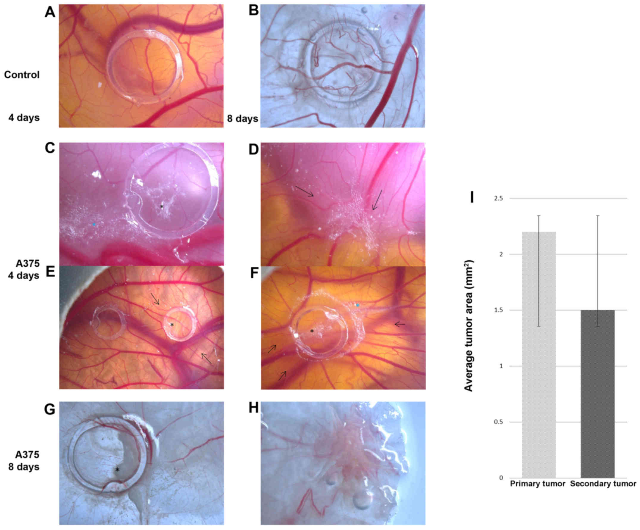
4 and 50% on day 8 (data not shown). On the final day of the
experiment, sections were dissected representing control CAM areas,
those presenting the primary tumors inside the ring, as well as
areas with secondary formed tumors, as recorded in the ex
vivo images [Fig. 1B (normal
CAM), G (primary tumor site) and H (secondary tumor site)].
On day 2 after inoculation, tumor cells were still
scattered inside the ring without forming compact masses, but the
onset of tumor establishment was already observed by the 3rd day.
On this day, areas of A375 cells were found outside the ring
perimeter, indicating their tendency to form secondary tumors.
Tumor masses were more compact from the 4th day on the primary site
(Fig. 1C), and compact secondary
tumor masses were firstly observed at some distance from the ring
(Fig. 1D).
By day 7 the areas reached approximately one fifth
of the surface inside the ring. On day 8, the final day of the
experiment, tumors and metastases were evaluated ex vivo,
and dimensions were estimated as the average surface area of
2.2±0.4 mm2 for primary tumors (Fig. 1G) and 1.5±0.3 mm2 for
secondary tumors (Fig. 1H).
From day 4 of the experiment, primary and secondary
perfused tumor masses were formed, showing strong angiogenic
reactions, with a capillary mesh with a tortuous aspect and some
signs of hemorrhagic foci (Fig. 1D and
F).
The vascular reaction to the presence of tumor cells
was progressively stronger from day 4 to the final stage of the
experiment. The number of capillaries converging toward the ring
area was significantly higher in the specimens with A375 cells
[Fig. 1C (day 4) and E (day 4,
right ring), and G (day 8)] as compared with the control treated
only with cell medium [Fig. 1A (day
4), B (day 8) and E (left ring)]. The characteristic spoke wheel
pattern of vessel arrangement indicates the strong angiogenic
reaction noticeable from the 3rd day after cell inoculation and
which was prominent on day 4 [Fig.
1E (left ring) and F]. The same type of strong vascular
reaction was present around the metastatic sites (Fig. 1D). On day 8, tumors were intensely
vascularized and exhibited perfused intratumoral vessels and at the
periphery of tumor areas as well, which were confirmed by
microscopic evaluation (data not shown).
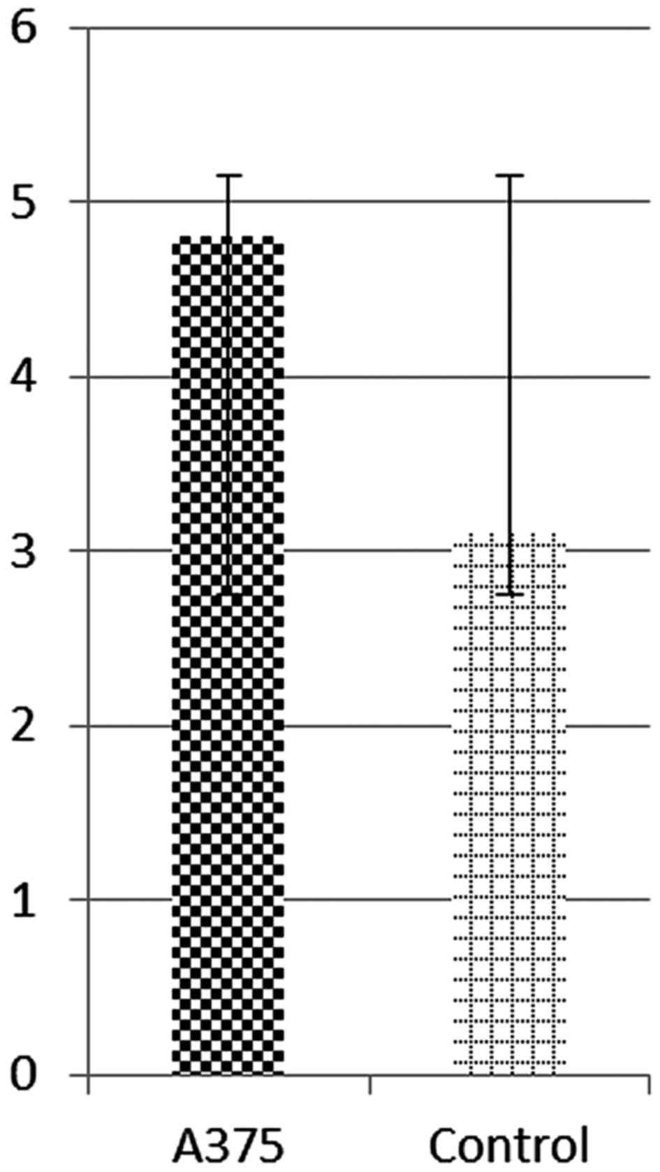
Vessel density was estimated by applying an
arbitrary scale and represents the degree of vascularization
convergent towards the tumor area. The average vascular density was
significantly higher in areas where tumor masses were present
(4.8±0.2), compared to the control specimens (3.1±0.2), as
presented in Fig. 2.
Mouse model of A375 human
melanoma
Macroscopic evaluation
The inoculation of melanoma cells was well-tolerated
by all animals and no signs of clinical deterioration (significant
weight loss, loss of appetite or reduced mobility) were detected
post-inoculation. A liquid bubble was formed during inoculation
that retracted on the first day post-inoculation (data not
shown).
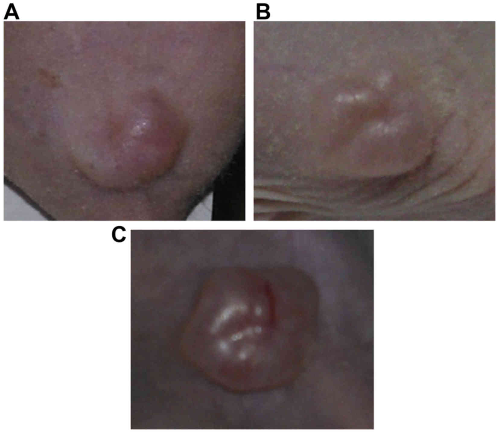
Following the resorption of the inoculation bubble,
in 14 mice, the first signs of tumor onset were observed on day
10–12 post-injection, with slight individual differences (Fig. 3A). Only one mouse did not develop a
primary tumor at the site of inoculation.
On day 20 post-inoculation, the initial tumors
became well-defined, as solid polypoid tumors with a lobulated
surface (Fig. 3B). Moreover, by day
30 post-inoculation, the tumor dimensions grew larger and newly
formed blood vessels within the primary tumors were prominent
(Fig. 3C). No sign of ulceration
was observed on the overlying skin during the entire
experiment.
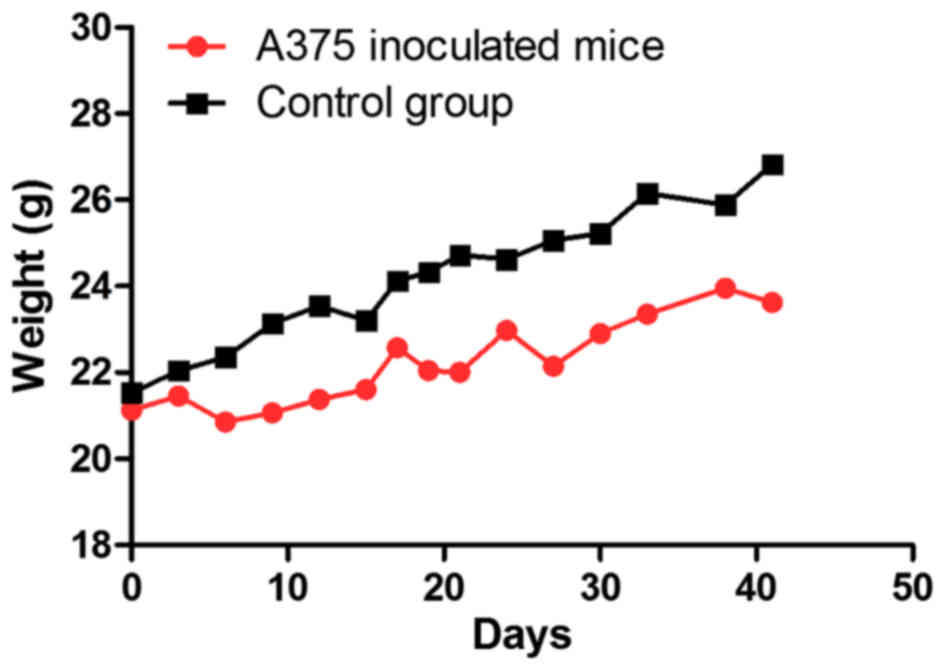
The mice inoculated with A375 xenografts presented
some weight variations during the experimental period and the
values recorded were lower than the ones measured for the control
group, the differences not being statistically significant
(Fig. 4). Some of the inoculated
mice presented a greater weight due to the developed tumor.
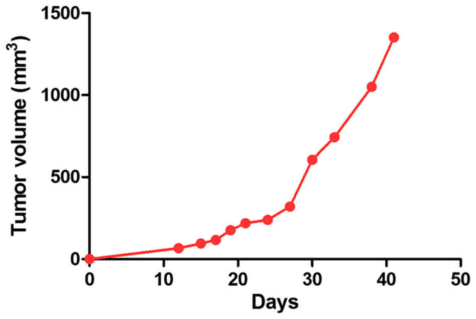
The tumor volumes are presented in Fig. 5, calculated to chart the tumor
growth curve. It was observed that the tumors presented a linear
growth until day 30 post-inoculation, whereas after this time
point, the growth became accelerated, and an increase was also
detected in tumor volume values (Fig.
5).
Histopathological evaluation of human melanoma
mouse model
Due to the tumor size, 3 mice were sacrificed at 30
days post-inoculation, 3 mice at 45 days and the remaining 8 mice
at 60 days post-inoculation.
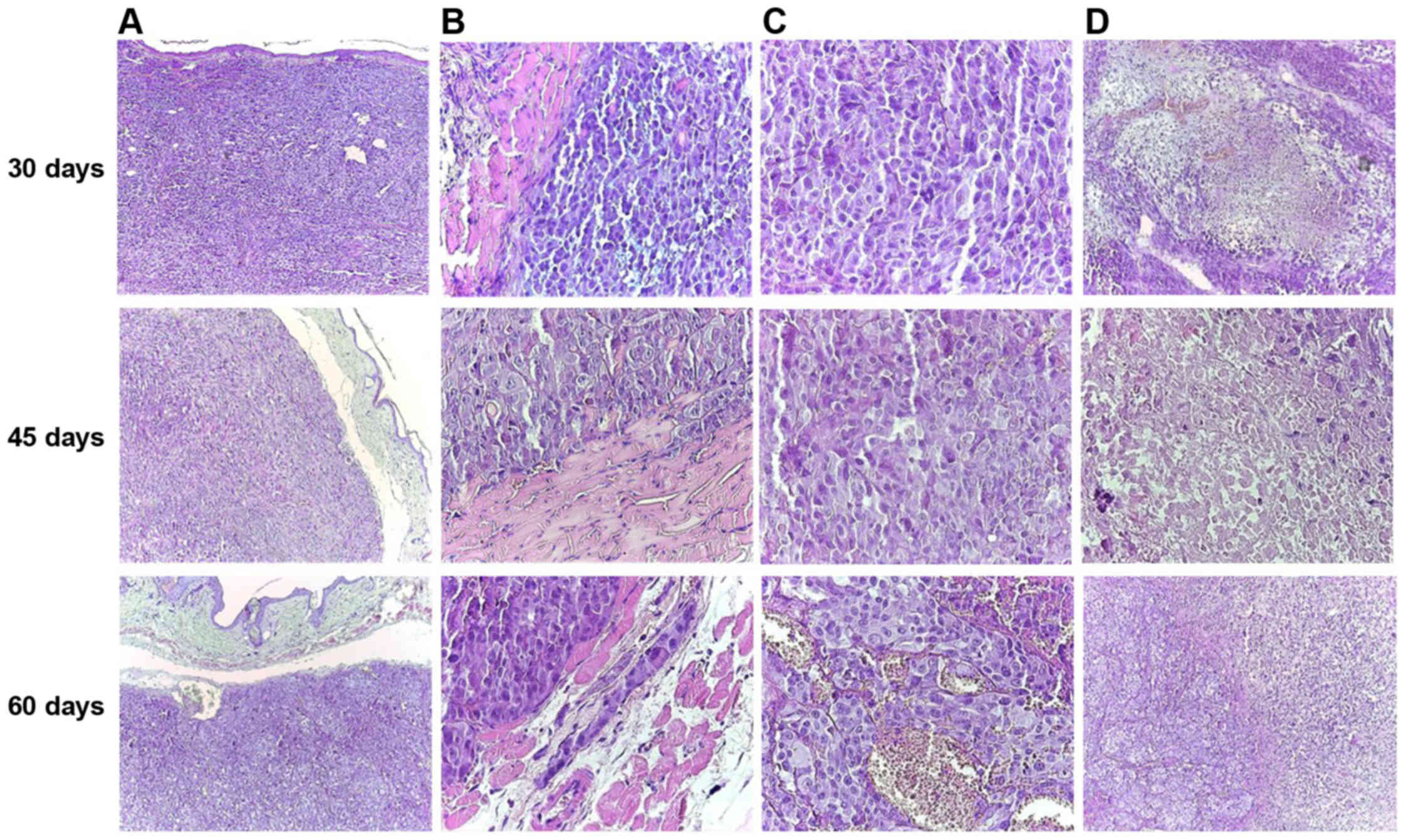
In all 14 cases, nodular tumors composed of islands
of achromic epithelioid cells were observed, with a large
cytoplasm, vesiculous nuclei and prominent macronucleoli. The tumor
stroma was represented by few collagen fibers. The tumor occupied
the entire dermis and the subcutis, with the grenz zone to the
epidermis extended to the subcutis and invading the adjacent
striated muscle cells. Large areas of necrosis were noted. The
epidermis was not involved, and was not ulcerated by the tumor
(Fig. 6). These features were
consistent with achromic nodular melanoma.
The volume of the tumor and the areas of necrosis
increased from 30 to 60 days (Fig.
6D). Moreover, at 60 days post-inoculation, a number of
hyperemiated vessels were identified between the tumor cells
(Fig. 6C).
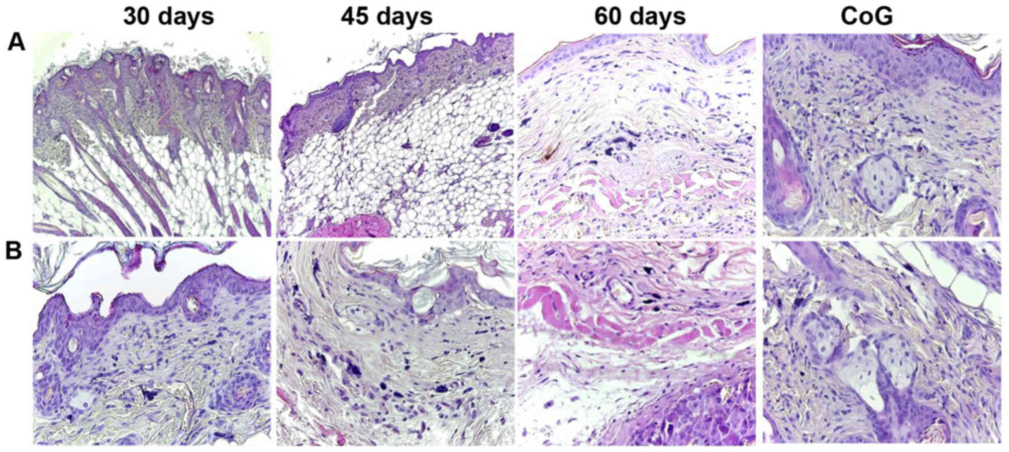
In the skin around the tumor, an increased number of
mast cells was observed compared to the group control. Moreover,
the number of mast cells increased from the mice sacrificed at 30
days compared to those sacrificed at 60 days. The distribution of
mast cells varied between groups. In the control group, few mast
cells filled with granules were observed in the superficial dermis,
while on the 60th day of the experiment, large numbers of mast
cells were observed in all levels of the dermis and in the subcutis
in the close vicinity of the tumor (Fig. 7).
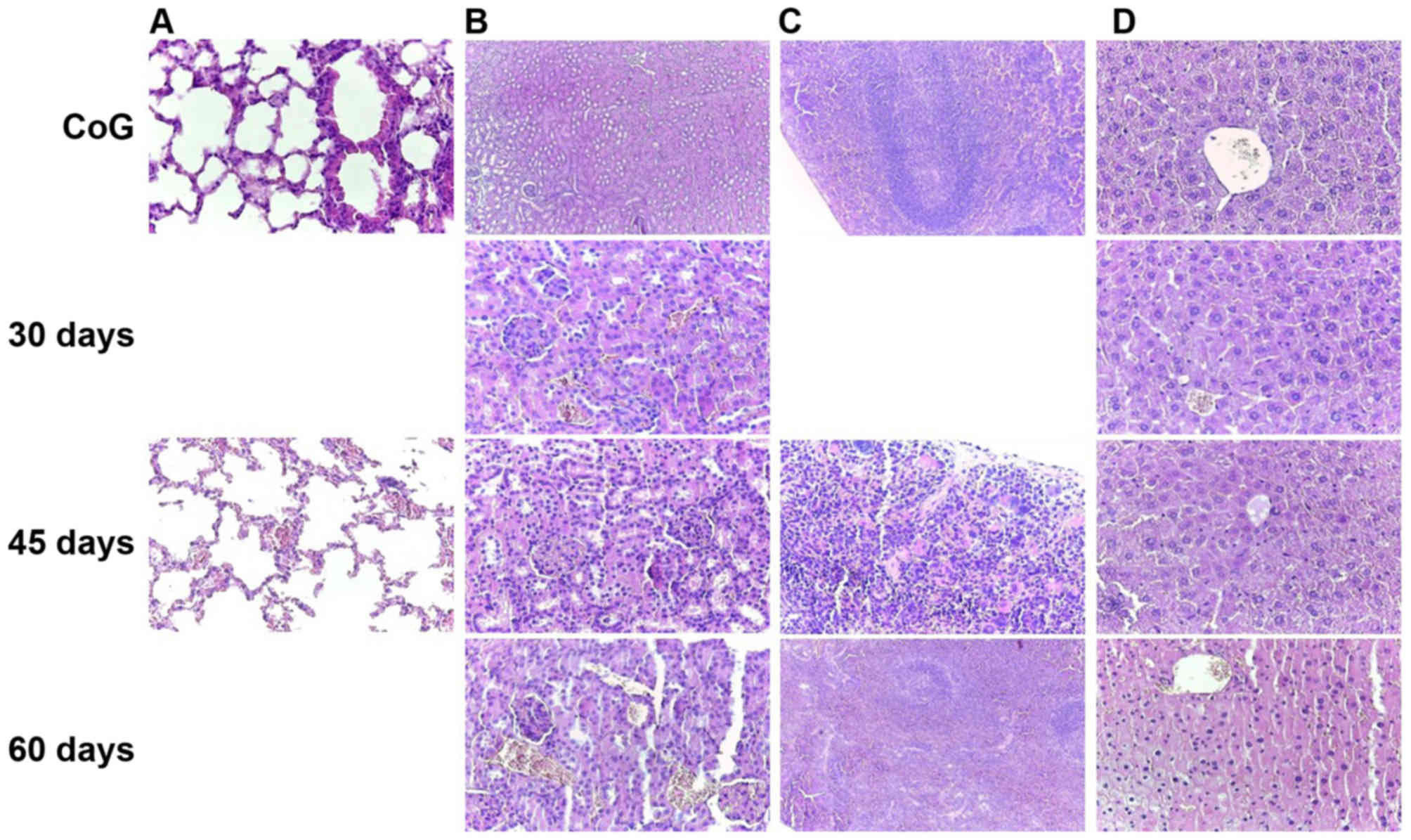
The ability to develop metastasis was decreased from
the first group to the last. At 30 days post-inoculation, one mouse
presented lung metastasis (Fig. 8A and
B, 30 days) and one spleen metastasis (Fig. 8C, 30 days). In a third mouse
sacrificed at that time, no metastasis was observed, only large
tumor thrombi in the large-sized arterial blood vessel (Fig. 8D, 30 days). In the second group,
only one mouse presented kidney metastasis (Fig. 8, 45 days), while in the third group,
the mice developed any metastasis. In this group, the most affected
organ in response to the tumor status was the lung, exhibiting
hemorrhagic alveolitis with many erythrocytes extravasated in the
alveolar space and hyperemia of the large vessels (Fig. 8, 60 days). All the metastases were
microscopic.
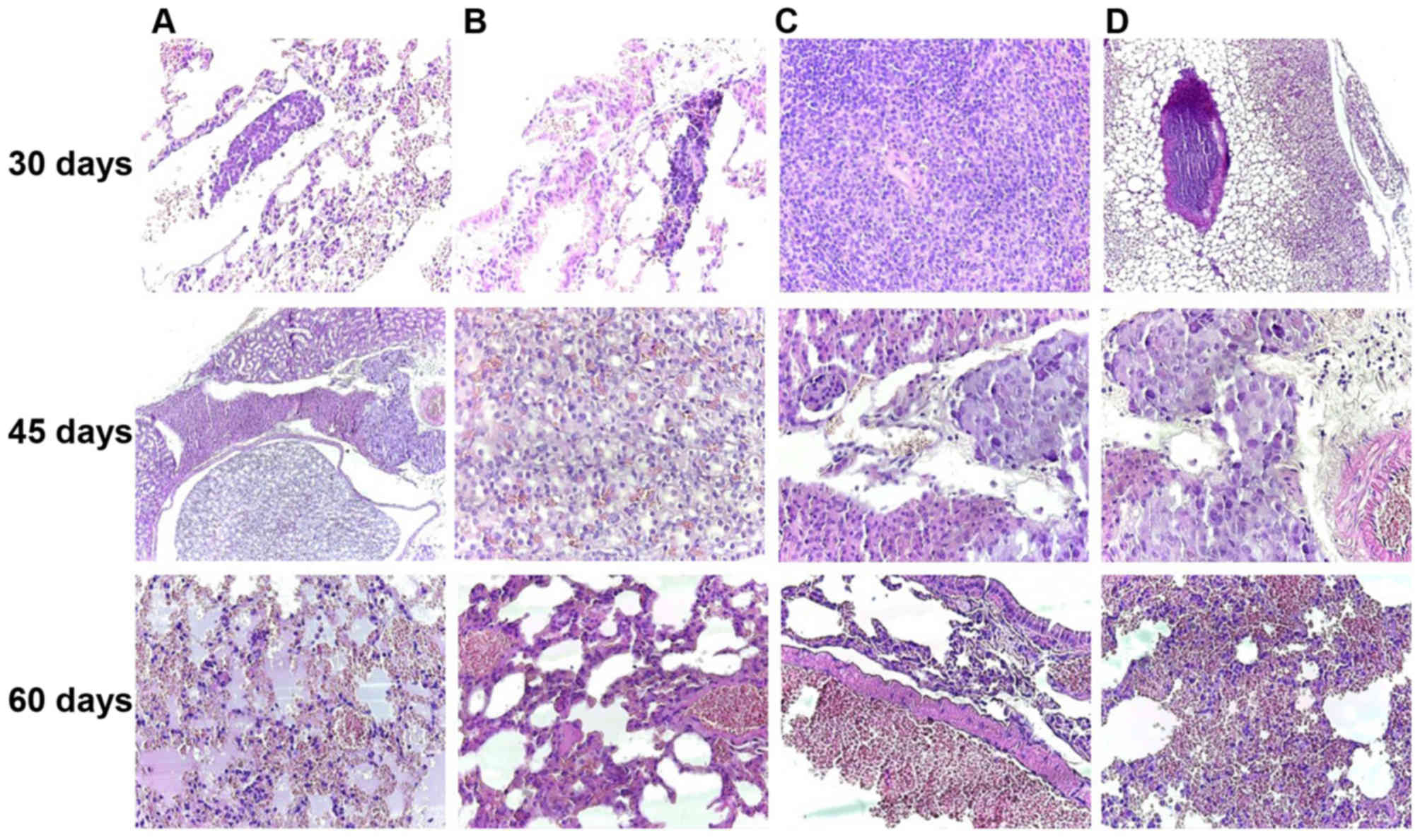 | Figure 8.Melanoma achromic metastasis at 30
days post-inoculation in the lung (A and B, original magnification,
×40) and spleen (C, original magnification, ×100) and tumor
thrombus in the big arterial blood vessel (D, original
magnification, ×40); Kidney metastases was noted in the 45th day
after inoculation, disposed between the renal tubules and nearby
medium-sized arterial vessels of the renal medulla (A, original
magnification, ×40) and composed of achromic epithelioid cells
similar to those observed in the primary tumor (B, original
magnification, ×100; C and D, original magnification, ×40); On the
60th day of the experiment, the most affected internal organ was
the lung, with hemorrhagic alveolitis (A, original magnification,
×40), hyperemia of small- and medium-sized blood vessels (B and C,
original magnification, ×100) and many extravasated erythrocytes in
the alveolar spaces (D, original magnification, ×40), H&E
staining. |
The other harvested internal organs exhibited only
hyperemia of small vessels (Fig.
9), with increasing intensity from the first group to the last
(30–60 days).
Discussion
The incidence of cutaneous melanoma has increased at
an alarming rate worldwide over the past years. According to the
World Cancer Research Fund International, GLOBOCAN 2012, there were
an estimated 230,000 new cases of melanoma worldwide in 2012
(24), and the statistics offered
by the WHO indicate that almost 132,000 cases of melanoma are being
diagnosed each year globally (25).
On the other hand, the incidence of mucosal and ocular melanomas
seems to remain stable worldwide (1,2,26),
with some racial and regional differences.
Even if the presence of skin melanoma in Asiatic
individuals seems to be uncommon, the oral cavity melanomas are
more often diagnosed in Japanese and Ugandan African populations,
accounting 35% of all mucosal melanomas, compared to 3.6% in
caucasions. Even so, a recent study demonstrated a higher
prevalence of oral cavity melanomas, accounting 25% of all mucosal
melanomas diagnosed during a period of 10 years, due to the
habitual particularity of Romania's population (1,2).
There are also differences between ages at the time
of diagnosis. Cutaneous melanomas are diagnosed at a young age, but
the extra-cutaneous counterpart seems to be a disease affecting the
elderly, with a peak of incidence in the 7th decade of life
compared to the 3rd decade for the former. Moreover, the age at the
time of diagnosis appears to decrease over the past years of life
for skin melanomas (27), a fact
that is not been observed for the extra-cutaneous counterpart.
Over the past years, an entire cascade of genetic
abnormalities has been identified which is associated with
melanoma. Studies have reported that 25 molecules/genes are
associated with melanoma biology, namely progression, metastasis
and prognosis, such as CDKN2A (cell cycle-related gene),
PI3K and PTEN, N-RAS (15–20% of all cutaneous
melanomas), B-RAF (50% of all cutaneous melanomas) and
MITF (28–30). The B-RAF gene encodes a
serine/threonine protein kinase that acts as a regulator of cell
division, differentiation and secretion through the
RAS-RAF-MEK-ERK-MAP kinase signaling pathway. The B-RAF
mutation is represented by the substitution of valine with glutamic
acid at codon 600, also known as V600E, and is responsible for the
constitutive activation of the protein (30,31).
Moreover, it has been demonstrated that the
B-RAFV600E pathway promotes vascular development
by activating the autocrine secretion of VEGF (31). Mutations in the B-RAF gene
have been diagnosed in up to 82% melanocytic nevi, concluding that
B-RAFV600 mutation cannot lead to cancer itself,
and requires the presence of other mutated genes (32). Another study on B16 mouse melanomas,
demonstrated that the inhibition of MITF reduces the
proliferation of malignant melanocytes (28). Therefore, it can be concluded that
MITF and B-RAF activation are necessary for
melanocytes to acquire their malignant potential (28).
In the present study, we used the A375 melanoma cell
line that maintains the characteristics of the human genitor and
harbors B-RAF and CDKN2 mutations, typical of
cutaneous melanoma, but with a morphology similar to that of
mucosal melanomas. Even so, it represents an eligible candidate for
the development of in vivo models.
It is well-known that melanoma progression occurs in
several steps which include the formation of a primary tumor that
develops horizontally through the epidermis, followed by the
vertical growth phase initiated in the primary tumor that extends
to dermis and the last phase, the invasion of tumor cells and
formation of secondary tumors (33). Metastasis is a complex multi-stage
process and a lethal feature of melanoma, causing also resistance
to the existent treatment. The process of angiogenesis is a
prominent feature in melanoma development, progression and
metastasis, the newly formed vessels being the suppliers of oxygen
and nutrients for cancer cells. Moreover, it is considered that the
onset of angiogenesis is related to inflammation and the
development of the melanoma vertical growth phase (33,34).
On this basis, it is mandatory to develop proper in vitro
and in vivo models that mimic the human pathogenesis
concerning metastasis development, in order to fully elucidate the
processes involved (35). However,
even though multiple models have been employed, these processes are
not yet fully elucidated.
The in vivo model using melanoma cells
grafted on chick chorioallantoic membrane presented in this study
demonstrated a 100% rate of tumor formation, and it could be
applied as a pre-screening tool for the evaluation of the antitumor
and anti-angiogenic potential of various agents, the results being
obtained in a short period of time (4–8 days). The easily
accessible, highly vascularized extraembryonic membrane of the
chicken embryo is often used to assess tumor angiogenesis, since
the discovery of tumor angiogenesis by Folkman and Cotran 4 decades
ago (36). Several advantages of
this technique recommend it as a pre-screening assay to the murine
models. The procedure assets are in terms of costs, time,
simplicity and reproducibility. The model does not require
sophisticated technical equipment, and qualified surgical skills
are not compulsory. The experimental setting takes up to 17 days,
which implies a relatively short interval for tumor growth and
metastasis, thus facilitating multiple assessments. Human tumor
xenografts are applicable on the CAM surface due to immunological
immaturity up to embryonic development day 18 (37,38).
Moreover, the number of specimens that can be included in an
experimental set is higher than in mouse studies, making it
possible to investigate a greater number of therapeutic agents.
Another feature that can be investigated by
assessing the melanoma CAM assay is the degree of angiogenesis by
morphometric measurements, applying an arbitrary scale in
correlation to the vessel density in areas surrounding the tumors.
By performing this evaluation on the 4th day following the
inoculation of cells, or the cell medium, respectively, there is no
interference with the normal angiogenic status of the embryo. This
moment represents the 14th EDD, when the normal angiogenic process
of untreated specimens is normally already decreased. Intense
angiogenesis occurs between the 7th and 11th EDD (22). Our results indicated that on day 4
post-inoculation, both primary and distant tumors were strongly
vascularized, what indicates the bond between the tumor growth and
angiogenesis.
We have previously used the CAM normal angiogenesis
model to test certain natural compounds for their potential
benefits in limiting the deregulated excessive angiogenic process
(39,40). We have also previously tested the
natural compounds in the melanoma model on CAM using A375 cells,
providing new insight into the mechanisms of action (unpublished
data).
Different melanoma cell lines have been used for
models using the CAM assay, with the A375 cells being among the
most frequently used (16,41). This has contributed to the molecular
elucidation of this aggressive type of tumor behavior, in terms of
invasiveness (16), brain
metastasis (42), angiogenesis and
lymphangiogenesis (41,42), or the evaluation of compounds with
promising effects by modulating different targets (43).
The human melanoma model using A375 cells assessed
on the CAM is also a useful alternative method for the study of
melanoma cell behavior and tumor environment. The CAM protocol
using melanoma cells is a suitable tool for in vivo tumor
angiogenesis or lymphangiogenesis elucidations. Hence, further
molecular investigations can be performed using the specimens
obtained from the tumor CAM assay [e.g., polymerase chain reaction
(PCR), in situ hybridization, immunohistochemistry,
immunofluorescence, and positron emission tomography-computed
tomography (PET-CT)] (38). The
establishment of functional perfused tumors in only 4 days after
inoculation and the ability of observing the tumor growth,
progress, invasiveness and metastasis for approximately 4–5 days
after tumor onset is a suitable protocol for pre-screening
potential antitumor agents.
There are several drawbacks to the CAM model that
can influence tumor research results. A limited number of reagents
are applicable due to low compatibility. Non-specific inflammatory
response, and various reactions of the membrane when exposed to the
outer atmosphere can also affect tumor microenvironment assessment
(44). In order to minimize the
possible misevaluations of the tumor model, it is optimal to apply
both the murine and CAM assays along with in vitro
experiments.
The CAM data obtained in the present study validate
the effectiveness of this assay as a short-term xenograft model for
A375 cells, providing information about melanoma cell
proliferation, metastatic behavior and angiogenic potential in
vivo. This information was used as an additional background
file for the development of human melanoma mouse model (a long-term
assay).
The mouse melanoma model can be used to gain
information about both early-and late-stage events in melanoma
development, and furthermore, the antitumor and anti-metastatic
effects of various therapeutic agents can be assessed. In order to
obtain a proper mouse melanoma model, several aspects should be
noted:
First, the in vivo melanoma models which are
obtained by the engraftment of human cells require as host
immunocompromised mice (SCID mice, which are both T-cell- and
B-cell-deficient or nude mice, which are only T-cell-deficient)
that do not reject the inoculated cells (3,45). An
ideal animal model should exhibit similar molecular characteristics
as human tumorigenesis and should also mimic natural tumor
progression, from its burst and proliferation to invasion and
metastasis (3). Balb/c nude mice
have been proven to be an excellent host for the A375 mouse model
(46–49). Immunodeficient mice are considered
eligible hosts for the transplantation of human xenografts due to
their specific mutations and immune background. SCID mice were the
first severe immunocompromised strain of mice that presented a
mutation at Prkdcscid protein kinase, also known as Scid
mutation responsible for the impairment of B- and T-cell production
(50). Balb/c homozygote nude mice
are characterized by the mutation of the Foxn1 gene
associated with the lack of hair and of a functional thymus
(deteriorated of removed). The immune profile of these mice
comprises a small population of T-cells and an increased response
of natural killer cells (more potent that in normal Balb/c mice),
which makes them suitable for human xenografts. It has been
observed that with age, the athymic mice gather a small number of
lymphocytes (CD3, CD4, CD8 and Thy-1). In addition, it has been
shown that these mice do not have an intrinsic defect of T-cell
precursors, their function being activated with proper stimulation;
however, B-lymphocytes, mononuclear cells (macrophages highly
active) and a normal number of mast cells have also been detected
(50,51).
Second, the area of xenograft inoculation is very
important, as it should provide the anatomical morphology for tumor
progression and the development of metastases. Most of the
xenograft melanoma models are created by the subcutaneously
inoculation of tumor cells, with the number and the volume of
culture media/PBS/inoculum varying (47–49).
Alternative parenteral pathways of tumor cell inoculation have also
been described, such as intradermal (better mimics a primary
melanoma, but tumor formation leads to skin ulcerations and mouse
euthanasia is required) (35,45,46),
tail vein (enforces the development of lung metastasis, but some
steps in the normal process are missed) (3,45) and
intracardiac (follows the metastatic spread) (52).
The xenograft melanoma model subcutaneously
inoculated presents some advantages in that the tumor that develops
is more comparable to skin metastasis and the experimental time is
longer, allowing for human melanoma cells to interact with the
murine stroma, lymphatic vessels, which makes the evaluation of
tumor growth behavior possible; this is frequently used to evaluate
the response of anticancer agents response in vivo (3,45).
However, there are also some limitations to this model, in that the
tumor dimensions cannot be measured by a caliper at lower limits
and it cannot be applied to examine the efficacy of immune-based
therapies, since the mice are immunocompromised (53).
Our results are in agreement with the data from the
literature regarding the inoculation pathway (23,47–49),
the animal host and the number of cells/inoculum/mice (23,49),
this number being selected based on the fact that a palpable
subcutaneous tumor was first detected after the injection of a
minimum of 106 cells/mouse (54). There were some differences between
our data and the literature as regards the survival time
post-inoculation of the tumor cells (from 21 to 90 days); these
differences could be explained by the inoculation pathway, the
number of cells/inoculum and also, the aim and the endpoints
established for each experimental design (34,47–49).
As regards histopathological aspects, cutaneous
melanomas can be classified as melanomas in situ,
superficial spreading melanomas and nodular melanomas.
Extra-cutaneous melanoma do not fit these categories due to zone
particularities; mucosal melanomas are often diagnosed as nodular
tumors with or without associated pagetoid spread in the covering
epithelium nearby the melanoma. In our study, 14 mice developed
tumors consistent with the diagnosis of nodular melanoma that
occupied the dermis and subcutis. The cells composing melanoma can
be classified as epithelioid and spindle, although the majority of
cutaneous melanomas present a mixed cellularity, with epithelioid
cells being more often observed in mucosal melanomas (2). In the present study, we used the A375
cell line, containing cells with epithelioid aspect obtained from a
cutaneous lesion, and the induced tumors were all composed of
epithelioid cells, better resembling mucosal melanoma cellularity.
Moreover, even if, usually, cutaneous melanomas are pigmented,
containing various amounts of melanin in the cytoplasm of tumor
cells or in the melanophages of the tumor stroma, the tumors
obtained in the present study were achromic, better resembling
mucosal melanomas that are predominantly achromic. As we have
already demonstrated, the tumor cells present a pagetoid spread in
the epithelium nearby or overlying the tumor (1,2), a
feature that was not observed in the present study, certainly due
to the subcutaneous inoculation of the tumor cells; however, the
tumor occupied the dermis and the subcutis, providing the tumor
with direct access to the usual routes for metastasis, which
resulted in the appearance of secondary tumors in almost 30% of the
xenografted mice.
The evolution of both cutaneous and extra-cutaneous
melanomas is dependent on the stage of disease at diagnosis. In a
recent review, it was stated that in countries with the highest
incidence of cutaneous melanoma, such as Australia, New Zealand,
USA and Scandinavia, patients are diagnosed at an early stage,
while in Central and Eastern European countries, diagnosis is
usually made at an advanced stage, which leads to a higher number
of deaths due to melanoma (55). It
is well-known that cutaneous melanomas diagnosed at an early stage
are in most cases curable, whereas the survival rate changes
depending on the stage of the disease: stages I and II (localized
tumor; 5-year survival, 98% of cases); stage III (regional spread;
63% of cases) and stage IV melanoma (metastasis beyond regional
lymph nodes; 16% of cases) (25).
Mucosal melanomas are always diagnosed in the late stages, with
very low rates of curability. The prognosis is grim, the 5-year
survival rate being only 40%.
The prognostic factors have been investigated for a
number of years. From the beginning of the study of melanoma to
date, the Clark level (the level of the skin affected by the tumor
with level I, epidermis; level II, papillary dermis; level III,
superficial blood plexus of the dermis; level IV, profound blood
plexus of the dermis; and level V, subcutis) and the Breslow index
(distance in mm from the granular layer of the epidermis to the
most profound site of the tumor) are considered to be the most
important for the prognostic. The epithelioid appearance of the
cells was also noted as poor prognostic factor, together with the
lack of melanin pigment from the cells (2). The quantity of inflammatory cells
(lymphocytes, macrophages and plasma cells), varying from any (poor
prognostic factor), to brisk, or to heavy and disposed in a band
around the tumor (good prognostic feature), was also considered to
have an impact on the prognosis of the tumor. Lately, only
infiltrated plasma cells, those plasma cells that are found between
malignant melanocytes, are considered important for a good
prognosis, with no impact on survival if they are disposed in the
tissues around the tumor. To date, only the Clark level and Breslow
index remain as gold standards for prognosis, and are the only
factors that could be appreciated in our study and could be
associated with prognosis, as tumor size rapidly increased and
reached the limits imposed for mouse euthanasia by the IACUC
Guidelines regarding tumor production in rats and mice, as many
metastases developed. At 30 days post-inoculation, the primary
tumors of 3 mice reached the size limits and all these mice
developed distance metastasis or showed signs of tumor migration
(tumor thrombus in the large-sized arterial blood vessel). Maishi
et al reported that on day 29 post-inoculation of
1×10⁶ cells into Balb/c nude mice, lung metastases and
tumor cells disseminated in intra-blood vessels areas were detected
(48), data that are consistent
with our results.
In the second group (45 days post-inoculation), only
one mouse developed a microscopic kidney metastasis and in the last
group (60 days post-inoculation), the mice in which the tumor size
slowly reach the limit showed no metastasis, only non-specific
signs of tumor aggression as paraneoplastic symptoms (hemorrhagic
alveolitis). As regards the epithelioid feature and
intracytoplasmic melanin load, in the present study, the inoculated
A375 cells had an achromic epithelioid aspect and all the primary
and secondary tumors developed after inoculation were also composed
by achromic epithelioid cells; thus, no remarks related to the
differences in metastatic capacity between epithelioid and spindle
cells, or pigmented and achromic cells, could be pronounced.
Moreover, inflammatory cells were absent in the tumor and
metastasis obtained using Balb/c nude mice, due to the immune
system characteristics of the used mice. As we previously
demonstrated (56) in SKH-1
hairless mice while studying experimental skin carcinogenesis, the
only type of inflammatory cells present in the tissues around the
tumor were mast cells. These cells were easy to observe even on
morphologic H&E or trichrome stains, due to their microscopic
appearance, as large cells with a thin cytoplasmic extension, that
are usually filled with many rough basophilic granules. The present
mouse model exhibited an increase in the number and size of mast
cells from the control group to the mice sacrificed after 60 days
of the experiment. From the facts noted in the present study, it
can be concluded that the size of the tumor has an important impact
on the appearance of metastasis. Due to the increasing number of
mast cells around the tumor, it can be postulated that the immune
system is probably involved in the evolution, progression and
metastasis of the tumor. Of note, the host immune system is
considered to be involved in tumor regression as a consequence of
tumor cell destruction by the activation of inflammatory cells,
vascular hyperplasia and fibrosis (27).
Even so, no other histological features noted on the
primary tumor in the present study, could predict metastasis, the
presence of metastases being a random event, an aspect already
highlighted in humans, where it seems that the behavior of
different melanomas does not correlate with the surgical option or
histopathological type of the tumor cells.
In conclusion, the CAM assay indicated that the A375
cell line exhibited a great affinity and compatibility in the
present framework, primary and secondary tumors being visible at
day 4 post-inoculation and presenting a high angiogenic potential.
The in vivo melanoma model obtained by inoculating A375
cells revealed that Balb/c nude mice are suitable hosts for human
cancers, the degree of tumor development with similar
characteristics being greater than 90%. Moreover, the histological
analysis described tumor evolution in time, its progress being slow
and its metastasis rate reduced, which can be considered an asset
in therapeutic surveillance experiments. The standardization method
for the development of melanoma models proposed in the present
study proved to be a complex, reproducible process, albeit some
small interferences, and the incriminated factor could be the
genetic profile of both cell line and nude mice.
Acknowledgements
The present study was supported by an Internal grant
at ‘Victor Babeș’ University of Medicine and Pharmacy Timisoara
(PII-C2-TC-2014, CAMMelRasNa), obtained by Avram Stefana. This
study was financially supported by a grant from the Romanian
National Authority for Scientific Research and Innovation,
CNCS-UEFISCDI, project no. PN-II-RU-TE-2014-4-2842. The authors
would like to thank the Histology and Angiogenesis Department for
providing technical support and assistance in setting up the CAM
assay.
References
|
1
|
Baderca F, Cojocaru S, Lazăr E, Lăzureanu
C, Lighezan R, Alexa A, Raica M and Nicola T: Amelanotic vulvar
melanoma: Case report and review of the literature. Rom J Morphol
Embryol. 49:219–228. 2008.PubMed/NCBI
|
|
2
|
Baderca F, Vincze D, Balica N and Solovan
C: Mucosal melanomas in the elderly: Challenging cases and review
of the literature. Clin Interv Aging. 9:929–937. 2014. View Article : Google Scholar : PubMed/NCBI
|
|
3
|
Kuzu OF, Nguyen FD, Noory MA and Sharma A:
Current State of Animal (Mouse) Modeling in Melanoma Research.
Cancer Growth Metastasis. 8 Suppl 1:81–94. 2015.PubMed/NCBI
|
|
4
|
Potrony M, Badenas C, Aguilera P,
Puig-Butille JA, Carrera C, Malvehy J and Puig S: Update in genetic
susceptibility in melanoma. Ann Transl Med. 3:2102015.PubMed/NCBI
|
|
5
|
Delyon J, Varna M, Feugeas JP, Sadoux A,
Yahiaoui S, Podgorniak MP, Leclert G, Dorval SM, Dumaz N, Janin A,
et al: Validation of a preclinical model for assessment of drug
efficacy in melanoma. Oncotarget. 7:13069–13081. 2016.PubMed/NCBI
|
|
6
|
Candido S, Rapisarda V, Marconi A,
Malaponte G, Bevelacqua V, Gangemi P, Scalisi A, McCubrey JA,
Maestro R, Spandidos DA, et al: Analysis of the B-RafV600E mutation
in cutaneous melanoma patients with occupational sun exposure.
Oncol Rep. 31:1079–1082. 2014.PubMed/NCBI
|
|
7
|
Palmieri G, Colombino M, Sini MC, Ascierto
PA, Lissia A and Cossu A: Targeted Therapies in Melanoma: Successes
and PitfallsMelanoma-From Early Detection to Treatment. Duc GHT:
InTech Open, Reykjavik. pp. 29–58. 2013
|
|
8
|
Sun C, Wang L, Huang S, Heynen GJ,
Prahallad A, Robert C, Haanen J, Blank C, Wesseling J, Willems SM,
et al: Reversible and adaptive resistance to BRAF(V600E) inhibition
in melanoma. Nature. 508:118–122. 2014. View Article : Google Scholar : PubMed/NCBI
|
|
9
|
Russo A, Ficili B, Candido S, Pezzino FM,
Guarneri C, Biondi A, Travali S, McCubrey JA, Spandidos DA and
Libra M: Emerging targeted therapies for melanoma treatment
(review). Int J Oncol. 45:516–524. 2014.PubMed/NCBI
|
|
10
|
Chalkiadaki G, Nikitovic D, Katonis P,
Berdiaki A, Tsatsakis A, Kotsikogianni I, Karamanos NK and
Tzanakakis GN: Low molecular weight heparin inhibits melanoma cell
adhesion and migration through a PKCa/JNK signaling pathway
inducing actin cytoskeleton changes. Cancer Lett. 312:235–244.
2011. View Article : Google Scholar : PubMed/NCBI
|
|
11
|
Yamanaka K, Nakahara T, Yamauchi T, Kita
A, Takeuchi M, Kiyonaga F, Kaneko N and Sasamata M: Antitumor
activity of YM155, a selective small-molecule survivin suppressant,
alone and in combination with docetaxel in human malignant melanoma
models. Clin Cancer Res. 17:5423–5431. 2011. View Article : Google Scholar : PubMed/NCBI
|
|
12
|
Ribatti D: The CAM assay in the study of
angiogenesis and metastasis. The Chick Embryo Chorioallantoic
Membrane in the Study of Angiogenesis and Metastasis. Springer.
(Netherlands). 2010. View Article : Google Scholar
|
|
13
|
Murphy JB and Rous P: The behavior of
chicken sarcoma implanted in the developing embryo. J Exp Med.
15:119–132. 1912. View Article : Google Scholar : PubMed/NCBI
|
|
14
|
Deryugina EI and Quigley JP: Chick embryo
chorioallantoic membrane model systems to study and visualize human
tumor cell metastasis. Histochem Cell Biol. 130:1119–1130. 2008.
View Article : Google Scholar : PubMed/NCBI
|
|
15
|
Klingenberg M, Becker J, Eberth S, Kube D
and Wilting J: The chick chorioallantoic membrane as an in vivo
xenograft model for Burkitt lymphoma. BMC Cancer. 14:3392014.
View Article : Google Scholar : PubMed/NCBI
|
|
16
|
Ribatti D, Nico B, Cimpean AM, Raica M,
Crivellato E, Ruggieri S and Vacca A: B16-F10 melanoma cells
contribute to the new formation of blood vessels in the chick
embryo chorioallantoic membrane through vasculogenic mimicry. Clin
Exp Med. 13:143–147. 2013. View Article : Google Scholar : PubMed/NCBI
|
|
17
|
Li M, Pathak RR, Lopez-Rivera E, Friedman
SL, Aguirre-Ghiso JA and Sikora AG: The In Ovo Chick
Chorioallantoic Membrane (CAM) Assay as an Efficient Xenograft
Model of Hepatocellular Carcinoma. J Vis Exp. 2015:e52411.
2015.
|
|
18
|
Comşa Ş, Popescu R, Avram Ş, Ceaușu RA,
Cîmpean AM and Raica M: Bevacizumab Modulation of the Interaction
Between the MCF-7 Cell Line and the Chick Embryo Chorioallantoic
Membrane. In Vivo. 31:199–203. 2017. View Article : Google Scholar : PubMed/NCBI
|
|
19
|
Workman P, Aboagye EO, Balkwill F, Balmain
A, Bruder G, Chaplin DJ, Double JA, Everitt J, Farningham DA,
Glennie MJ, et al: Committee of the National Cancer Research
Institute: Guidelines for the welfare and use of animals in cancer
research. Br J Cancer. 102:1555–1577. 2010. View Article : Google Scholar : PubMed/NCBI
|
|
20
|
Ha L, Noonan FP, De Fabo EC and Merlino G:
Animal models of melanoma. J Investig Dermatol Symp Proc. 10:86–88.
2005. View Article : Google Scholar : PubMed/NCBI
|
|
21
|
Becker JC, Houben R, Schrama D, Voigt H,
Ugurel S and Reisfeld RA: Mouse models for melanoma: A personal
perspective. Exp Dermatol. 19:157–164. 2010. View Article : Google Scholar : PubMed/NCBI
|
|
22
|
Ribatti D: The chick embryo
chorioallantoic membrane in the study of tumor angiogenesis. Rom J
Morphol Embryol. 49:131–135. 2008.PubMed/NCBI
|
|
23
|
Mena S, Rodriguez ML, Ortega A, Priego S,
Obrador E, Asensi M, Petschen I, Cerdá M, Brown BD and Estrela JM:
Glutathione and Bcl-2 targeting facilitates elimination by
chemoradiotherapy of human A375 melanoma xenografts overexpressing
bcl-xl, bcl-2, and mcl-1. J Transl Med. 10:82012. View Article : Google Scholar : PubMed/NCBI
|
|
24
|
Rigon RB, Oyafuso MH, Fujimura AT,
Gonçalez ML, do Prado AH, Gremião MP and Chorilli M:
Nanotechnology-Based Drug Delivery Systems for Melanoma Antitumoral
Therapy: A Review. BioMed Res Int. 2015:8418172015. View Article : Google Scholar : PubMed/NCBI
|
|
25
|
Niezgoda A, Niezgoda P and Czajkowski R:
Novel Approaches to Treatment of Advanced Melanoma: A Review on
Targeted Therapy and Immunotherapy. BioMed Res Int.
2015:8513872015. View Article : Google Scholar : PubMed/NCBI
|
|
26
|
Baderca F, Solovan C and Boghian L:
Epidemiological and morphological data of ocular melanocytic
lesions. Rom J Morphol Embryol. 54:77–83. 2013.PubMed/NCBI
|
|
27
|
Zurac S, Neagu M, Constantin C, Cioplea M,
Nedelcu R, Bastian A, Popp C, Nichita L, Andrei R, Tebeica T, et
al: Variations in the expression of TIMP1, TIMP2 and TIMP3 in
cutaneous melanoma with regression and their possible function as
prognostic predictors. Oncol Lett. 11:3354–3360. 2016.PubMed/NCBI
|
|
28
|
Wang L, Hurley DG, Watkins W, Araki H,
Tamada Y, Muthukaruppan A, Ranjard L, Derkac E, Imoto S, Miyano S,
et al: Cell cycle gene networks are associated with melanoma
prognosis. PLoS One. 7:e342472012. View Article : Google Scholar : PubMed/NCBI
|
|
29
|
Fedorenko IV, Gibney GT and Smalley KSM:
NRAS mutant melanoma: Biological behavior and future strategies for
therapeutic management. Oncogene. 32:3009–3018. 2013. View Article : Google Scholar : PubMed/NCBI
|
|
30
|
Rodrigueza WV, Woolliscroft MJ, Ebrahim
AS, Forgey R, McGovren PJ, Endert G, Wagner A, Holewa D, Aboukameel
A, Gill RD, et al: Development and antitumor activity of a BCL-2
targeted single-stranded DNA oligonucleotide. Cancer Chemother
Pharmacol. 74:151–166. 2014. View Article : Google Scholar : PubMed/NCBI
|
|
31
|
Russo AE, Torrisi E, Bevelacqua Y,
Perrotta R, Libra M, McCubrey JA, Spandidos DA, Stivala F and
Malaponte G: Melanoma: Molecular pathogenesis and emerging target
therapies (Review). Int J Oncol. 34:1481–1489. 2009.PubMed/NCBI
|
|
32
|
Jones V and Katiyar SK: Emerging
phytochemicals for prevention of melanoma invasion. Cancer Lett.
335:251–258. 2013. View Article : Google Scholar : PubMed/NCBI
|
|
33
|
Ribatti D, Annese T and Longo V:
Angiogenesis and melanoma. Cancers (Basel). 2:114–132. 2010.
View Article : Google Scholar : PubMed/NCBI
|
|
34
|
Streit M and Detmar M: Angiogenesis,
lymphangiogenesis, and melanoma metastasis. Oncogene. 22:3172–3179.
2003. View Article : Google Scholar : PubMed/NCBI
|
|
35
|
Rozenberg GI, Monahan KB, Torrice C, Bear
JE and Sharpless NE: Metastasis in an orthotopic murine model of
melanoma is independent of RAS/RAF mutation. Melanoma Res.
20:361–371. 2010.PubMed/NCBI
|
|
36
|
Folkman J and Cotran R: Relation of
vascular proliferation to tumor growth. Int Rev Exp Pathol.
16:207–248. 1976.PubMed/NCBI
|
|
37
|
Friend JV, Crevel RW, Williams TC and
Parish WE: Immaturity of the inflammatory response of the chick
chorioallantoic membrane. Toxicol In Vitro. 4:324–326. 1990.
View Article : Google Scholar : PubMed/NCBI
|
|
38
|
Dupertuis YM, Delie F, Cohen M and Pichard
C: In ovo method for evaluating the effect of nutritional therapies
on tumor development, growth and vascularization. Clin Nutr Exp.
2:9–17. 2015. View Article : Google Scholar
|
|
39
|
Dehelean CA, Feflea S, Ganta S and Amiji
M: Anti-angiogenic effects of betulinic acid administered in
nanoemulsion formulation using chorioallantoic membrane assay. J
Biomed Nanotechnol. 7:317–324. 2011. View Article : Google Scholar : PubMed/NCBI
|
|
40
|
Dehelean CA, Feflea S, Gheorgheosu D,
Ganta S, Cimpean AM, Muntean D and Amiji MM: Anti-angiogenic and
anti-cancer evaluation of betulin nanoemulsion in chicken
chorioallantoic membrane and skin carcinoma in Balb/c mice. J
Biomed Nanotechnol. 9:577–589. 2013. View Article : Google Scholar : PubMed/NCBI
|
|
41
|
Papoutsi M, Siemeister G, Weindel K,
Tomarev SI, Kurz H, Schächtele C, Martiny-Baron G, Christ B, Marmé
D and Wilting J: Active interaction of human A375 melanoma cells
with the lymphatics in vivo. Histochem Cell Biol. 114:373–385.
2000.PubMed/NCBI
|
|
42
|
Papoutsi M, Kurz H, Schächtele C, Marmé D,
Christ B, Pröls F and Wilting J: Induction of the blood-brain
barrier marker neurothelin/HT7 in endothelial cells by a variety of
tumors in chick embryos. Histochem Cell Biol. 113:105–113. 2000.
View Article : Google Scholar : PubMed/NCBI
|
|
43
|
Marton A, Kúsz E, Kolozsi C, Tubak V,
Zagotto G, Buzás K, Quintieri L and Vizler C: Vanillin Analogues
o-Vanillin and 2,4,6-Trihydroxybenzaldehyde Inhibit NFĸB Activation
and Suppress Growth of A375 Human Melanoma. Anticancer Res.
36:5743–5750. 2016. View Article : Google Scholar : PubMed/NCBI
|
|
44
|
Nowak-Sliwinska P, Segura T and
Iruela-Arispe ML: The chicken chorioallantoic membrane model in
biology, medicine and bioengineering. Angiogenesis. 17:779–804.
2014. View Article : Google Scholar : PubMed/NCBI
|
|
45
|
Beaumont KA, Mohana-Kumaran N and Haass
NK: Modeling Melanoma In Vitro and In Vivo. Healthcare (Basel).
2:27–46. 2013. View Article : Google Scholar : PubMed/NCBI
|
|
46
|
Hu T, Zhang C, Tang Q, Su Y, Li B, Chen L,
Zhang Z, Cai T and Zhu Y: Variant G6PD levels promote tumor cell
proliferation or apoptosis via the STAT3/5 pathway in the human
melanoma xenograft mouse model. BMC Cancer. 13:2512013. View Article : Google Scholar : PubMed/NCBI
|
|
47
|
Zheng AW, Jia DD, Xia LM, Jin G, Wu H and
Li T: Impact of carboplatin plus paclitaxel combined with endostar
against A375 melanoma cells: An in vitro and in vivo analysis.
Biomed Pharmacother. 83:1321–1326. 2016. View Article : Google Scholar : PubMed/NCBI
|
|
48
|
Maishi N, Ohba Y, Akiyama K, Ohga N,
Hamada J, Nagao-Kitamoto H, Alam MT, Yamamoto K, Kawamoto T, Inoue
N, et al: Tumour endothelial cells in high metastatic tumours
promote metastasis via epigenetic dysregulation of biglycan. Sci
Rep. 6:280392016. View Article : Google Scholar : PubMed/NCBI
|
|
49
|
Jin J, Zhang Y, Li Y, Zhang H, Li H, Yuan
X, Li X, Zhou W, Xu B, Zhang C, et al: RNA-interference-mediated
downregulation of Pin1 suppresses tumorigenicity of malignant
melanoma A375 cells. Neoplasma. 60:92–100. 2013. View Article : Google Scholar : PubMed/NCBI
|
|
50
|
Belizario JE: Immunodeficient Mouse
Models: An Overview. Open Immunol J. 2:79–85. 2009. View Article : Google Scholar
|
|
51
|
Envigo: Research Models and Services:
Oncology-Mutant Mice. Athymic Nude Mice. Envigo RMS Division.
(Indianapolis, IN). 2016.http://www.envigo.com/resources/data-sheets/envigo-5193-131-leaflets-2016-nude-mouse-combination-a4-refs.pdf
|
|
52
|
Herwig N, Belter B and Pietzsch J:
Extracellular S100A4 affects endothelial cell integrity and
stimulates transmigration of A375 melanoma cells. Biochem Biophys
Res Commun. 477:963–969. 2016. View Article : Google Scholar : PubMed/NCBI
|
|
53
|
Clark AG and Vignjevic DM: Modes of cancer
cell invasion and the role of the microenvironment. Curr Opin Cell
Biol. 36:13–22. 2015. View Article : Google Scholar : PubMed/NCBI
|
|
54
|
Craft N, Bruhn KW, Nguyen BD, Prins R,
Liau LM, Collisson EA, De A, Kolodney MS, Gambhir SS and Miller JF:
Bioluminescent imaging of melanoma in live mice. J Invest Dermatol.
125:159–165. 2005. View Article : Google Scholar : PubMed/NCBI
|
|
55
|
Gajda M and Kaminska-Winciorek G: Do not
let to be late: Overview of reasons for melanoma delayed diagnosis.
Asian Pac J Cancer Prev. 15:3873–3877. 2014. View Article : Google Scholar : PubMed/NCBI
|
|
56
|
Dehelean CA, Soica C, Pinzaru I, Coricovac
D, Danciu C, Pavel I, Borcan F, Spandidos DA, Tsatsakis AM and
Baderca F: Sex differences and pathology status correlated to the
toxicity of some common carcinogens in experimental skin carcinoma.
Food Chem Toxicol. 95:149–158. 2016. View Article : Google Scholar : PubMed/NCBI
|