Introduction
Prostate cancer is one of the leading causes of
cancer-related death in men worldwide. At present, treatment
approaches for prostate cancer mainly include surgery, chemotherapy
and hormonal therapy (1,2). The selection of the treatment method
for prostate cancer generally depends on the clinical stage,
Gleason score, age of patients, and other factors. To date, the
chemotherapeutic effect for prostate cancer is limited due to drug
resistance and cytotoxicity (3–5), which
makes it urgent to develop new therapeutic drugs to further improve
the clinical outcomes of patients with prostate cancer.
Recently, increased attention has been paid to the
anticancer effects of Traditional Chinese Medicine (TCM) largely
due to the advantages of low toxicity and multi-targets (6–8).
Trametes robiniophila Murr. (Huaier) has a long history of
disease treatment for more than 1,600 years in China. Numerous
studies have revealed that Huaier exhibits superior effects for the
treatment of several types of cancers, such as hepatocellular
carcinoma, breast, ovarian, lung and prostate cancer (9–14). The
underlying mechanisms of the anticancer effects of Huaier include
the inhibition of tumor cell proliferation, metastasis and
angiogenesis, as well as induction of apoptosis (11,13,15–17).
However, the mechanisms underlying the anti-prostate cancer effect
of Huaier remain to be elucidated.
In the present study, we demonstrated that Huaier
significantly inhibited the proliferative and metastatic potential
of human prostate cancer PC3 cells. Moreover, downregulation of
Lamin B1 was responsible for the inhibition of the proliferative
and metastatic capacity of PC3 cells exposed to Huaier aqueous
extract. More importantly, Huaier treatment activated autophagy in
PC3 cells, and suppression of autophagy attenuated Huaier-induced
cell death in PC3 cells. Thus, the present study provides a
theoretical and experimental basis for the clinical application of
Huaier for the treatment of prostate cancer.
Materials and methods
Reagents and antibodies
Ham's F-12K (Kaighn's) medium, fetal bovine serum
(FBS), penicillin-streptomycin solution, 0.25% trypsin, Matrigel
and Transwell chambers were purchased from Corning Life Sciences
(Corning, NY, USA). Cell Counting Kit-8 (CCK-8) was obtained from
Dojindo Laboratories (Kumamoto, Japan). 3-Methyladenine (3-MA) was
purchased from Santa Cruz Biotechnology (Santa Cruz, CA, USA).
Bafilomycin A1 was obtained from Aladdin Shanghai Biochemical
Technology Co., Ltd. (Shanghai, China). Bis-Tris Nu-PAGE gels
(4–12%) were obtained from Invitrogen (Thermo Fisher Scientific,
Inc., Waltham, MA, USA). Super ECL Plus was from GE Healthcare
(Pittsburgh, PA, USA). β-actin antibody was purchased from Abgent
(cat. no. AM1829b; San Diego, CA, USA). Atg3 (cat. no. 3415),
Beclin-1 (cat. no. 3495) and Atg5 (cat. no. 12994) antibodies were
purchased from Cell Signaling Technology (Danvers, MA, USA). LC3
antibody was from MBL (cat. no. PM036; Tokyo, Japan). Lamin B1
(cat. no. sc-20682), HRP-labeled goat anti-mouse (cat. no. sc-2005)
and goat anti-rabbit (cat. no. sc-2004) antibodies were from Santa
Cruz Biotechnology.
Preparation of Huaier aqueous
extract
The electuary ointment of Huaier was obtained from
Gaitianli Medicine Co., Ltd. (Jiangsu, China). It was dissolved in
F-12K complete medium to obtain a 10 mg/ml stock solution and was
stored at 4°C after sterilization by filtration.
Cell culture
Human prostate cancer PC3 cells were obtained from
the Cell Culture Center of the Institute of Basic Medical Sciences
of the Chinese Academy of Medical Sciences (Beijing, China). PC3
cells were maintained in Ham's F-12K (Kaighn's) medium containing
10% FBS and 1% penicillin/streptomycin at 37°C and 5%
CO2.
Cell viability assay
PC3 cells were seeded into 96-well plates at a
density of 3,500 cells/well. On the following day, the cells were
treated with Huaier aqueous extract at the indicated concentrations
for different times. Afterwards, 10 µl of CCK-8 was added into each
well and incubated at 37°C for 2 h. The optical density (OD) was
then measured at 450 nm using a microplate reader (Perkin-Elmer,
Waltham, MA, USA).
In vitro scratch assay
The scratch assay was performed as described
previously (10,18). In brief, PC3 cells were seeded into
12-well plates with complete medium. Cells in a subconfluent state
were starved with serum-free medium for 12 h, and then a straight
cell-free wound was created using a 10-µl pipette tip. Next, the
cells were maintained in serum-free medium containing 4 mg/ml of
Huaier extract. The scratch width was measured at 0 and 24 h. The
cell migration distances were analyzed quantitatively.
Transwell assay
The Transwell system was established as previously
described (19,20). PC3 cells (1×10⁵) were
resuspended in 200 µl serum-free medium containing Huaier extract
(4 mg/ml) and then added to the upper chamber of the Transwell
system. The lower chamber was filled with 750 µl complete medium
containing 10% FBS. After incubation for 36 h, the cells were
removed from the upper surface of the membrane with a cotton swab.
Next, the cells on the bottom surface of the membrane were fixed
and then stained with crystal violet. The Transwell chamber was
washed twice with phosphate-buffered saline (PBS) to remove the
dye. The invasive cells were observed and counted on 5 random
fields under an inverted microscope (Leica Microsystems GmbH,
Wetzlar, Germany).
Quantitative real-time PCR
Total RNA was extracted from PC3 cells using
E.Z.N.A.® Total RNA Kit I (Omega Bio-tek, Inc.,
Norcross, GA, USA) and reverse-transcribed into cDNA using the
PrimeScript RT reagent kit (Takara Biotechnology, Inc., Dalian,
China) according to the manufacturer's protocols. cDNA was used as
a template for the quantitative PCR using the TransStart Top Green
qPCR SuperMix (Beijing TransGen Biotech Co., Ltd., Beijing, China).
The primer sequences were: human Lamin B1 forward,
5′-TTCTCGAAGCTTGATCTGGG-3′ and human Lamin B1 reverse,
5′-GATCGAGCTGGGCAAGTG-3′; human β-actin forward,
5′-GTTGTCGACGACGAGCG-3′ and human β-actin reverse,
5′-GCACAGAGCCTCGCCTT-3′.
Immunoblotting
Cells were washed twice with PBS and harvested with
lysis buffer [10 mM Tris (pH 6.8), 2% SDS, 10% glycerol and 100 mM
DTT], and then cell lysates were boiled for 10 min at 98°C. The
levels of indicated proteins were detected by immunoblot analysis
as previously described (21).
RNA interference
All siRNAs were synthesized by Shanghai GenePharma
Co., Ltd. (Shanghai, China). PC3 cells were seeded in 6-well plates
and then transfected with siRNAs targeting Lamin B1, Atg5 or
Beclin-1 using Lipofectamine 2000 (Thermo Fisher Scientific, Inc.,
Waltham, MA, USA) according to the manufacturer's instructions. The
siRNA target sequences were: Lamin B1 (human),
5′-CGCGCTTGGTAGAGGTGGA-3′; Atg5 (human),
5′-GGACGAATTCCAACTTGTT-3′; Beclin-1 (human),
5′-CAGTTTGGCACAATCAATA-3′; negative control (NC),
5′-TTCTCCGAACGTGTCACGT-3′.
Electron microscopy
Cells were collected with 2.5% glutaraldehyde, and
then centrifuged (1,000 revolutions, 10 min) and washed twice with
PBS. The samples were post-fixed with 1% osmium tetroxide at 4°C
for 2 h in the dark and then washed three times with 0.1MPB. After
dehydration, the permeation, paraffin embedding and section
staining were performed as previously described (22). The ultrastructure of cells was
observed under a JEM-1230 transmission electron microscope (JEOL,
Ltd., Tokyo, Japan).
Acridine orange staining
PC3 cells treated with 8 mg/ml Huaier exact for
indicated times were washed twice with PBS and stained with 10
µg/ml acridine orange for 20 min at 37°C in the dark. The cells
were observed under an inverted fluorescence microscope.
Immunofluorescence assay
Cells seeded on confocal dishes were washed with PBS
and fixed in 4% paraformaldehyde for 10 min, and then permeabilized
with PBS containing 0.5% Triton X-100 for 20 min. Next, the cells
were washed three times with PBS and blocked with 1% BSA for 1 h at
37°C, and then incubated with the primary antibody (1:100)
overnight at 4°C. Subsequently, the cells were washed with PBS and
then incubated with anti-rabbit FITC-conjugated secondary antibody
(1:100) for 1 h at 37°C. Nucleus was counterstained with DAPI in
the dark after being washed with PBS. The cells were observed and
photographed with a laser scanning confocal microscope (Olympus
FV1000; Olympus Corp., Tokyo, Japan).
Statistical analysis
The data are presented as mean ± SD of triplicate
samples. The statistical analysis in this study was evaluated by
the Student's t-test and ANOVA using GraphPad Prism 5.0 software
(GraphPad Software, Inc., La Jolla, CA, USA). P<0.05 was
considered to indicate a statistically significant result.
Results
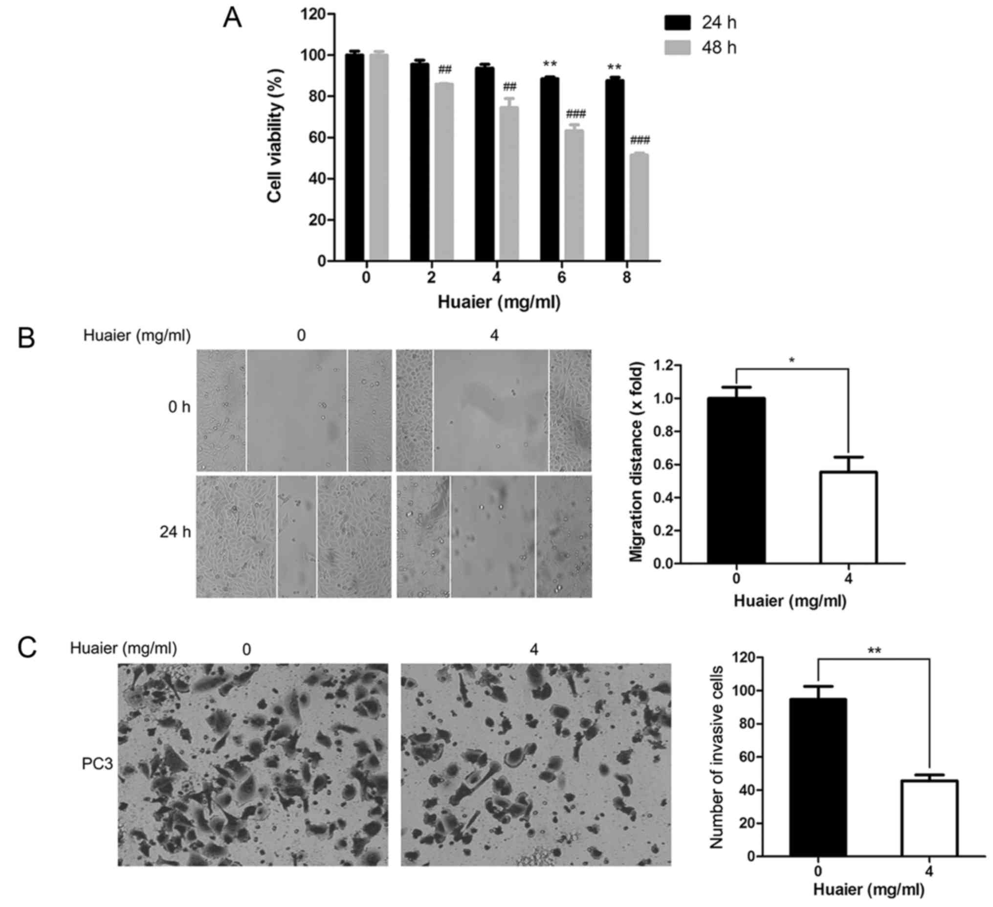
Huaier inhibits the proliferative and
metastatic potential of human prostate cancer PC3 cells
CCK-8 assay indicated that Huaier aqueous extract
significantly inhibited proliferation of the PC3 cells in a time-
and dose-dependent manner (Fig.
1A). The IC50 value of PC3 cells exposed to Huaier for 48 h was
8.18 mg/ml. The wound healing assay is widely used to evaluate the
migratory ability of cells in vitro. As shown in Fig. 1B, the migration capacity of PC3
cells was decreased after treatment with Huaier, and the migration
inhibition rate of PC3 cells treated with Huaier was 44.68±16.39%.
Moreover, the invasion capacity of cells in vitro is
frequently examined by using Transwell assay. As shown in Fig. 1C, after treatment of PC3 cells with
4 mg/ml Huaier aqueous extract for 36 h, the number of cells that
had successfully passed through the Matrigel-coated membrane was
markedly reduced compared with that of the control group cells.
Taken together, Huaier markedly suppressed the proliferative and
metastatic capability of the human prostate cancer cells.
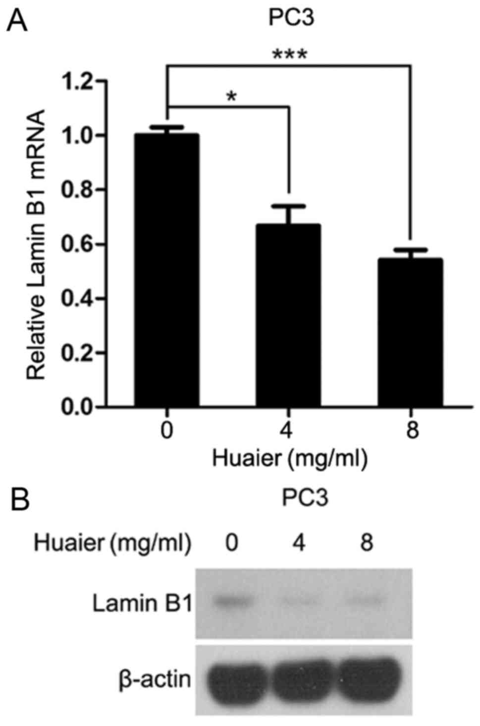
Huaier downregulates Lamin B1
expression in human prostate cancer PC3 cells
Lamin B1 is a member of the lamin family making up
the nuclear matrix and is abnormally overexpressed in multiple
types of cancers including prostate, hepatocellular carcinoma and
pancreatic cancer (19,23–25).
Moreover, Lamin B1 positively regulates the proliferation and
invasion of pancreatic cancer cells (25). Our previous study revealed that as a
pro-oncogenic gene, Lamin B1 was dramatically decreased in human
hepatoma SKHEP-1 cells exposed to Huaier (10). Intriguingly, qRT-PCR and western
blot analysis demonstrated that Huaier aqueous extract
significantly inhibited the mRNA and protein levels of Lamin B1 in
PC3 cells in a dose-dependent manner (Fig. 2A and B).
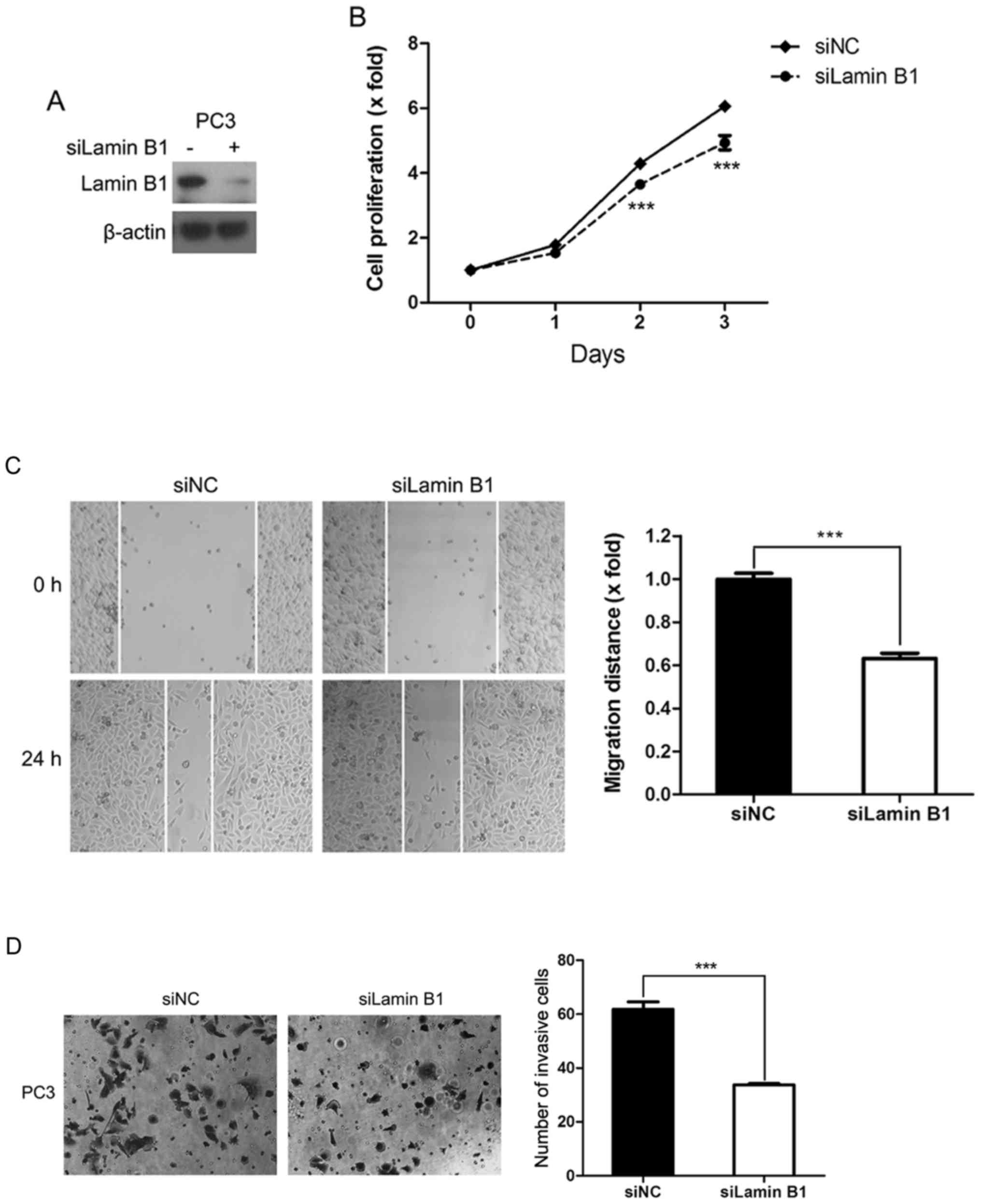
Lamin B1 is involved in the inhibition
of proliferation and metastatic potential of PC3 cells exposed to
Huaier
Next, we investigated whether downregulation of
Lamin B1 contributes to Huaier-mediated inhibition of proliferative
and metastatic capacity of PC3 cells by using RNA interference.
Transfection with siRNAs targeting Lamin B1 markedly reduced Lamin
B1 expression (Fig. 3A). Moreover,
depletion of Lamin B1 significantly blunted the proliferation of
PC3 cells (Fig. 3B). In addition,
reduction of Lamin B1 substantially suppressed the migration and
invasion of PC3 cells (Fig. 3C and
D). Thus, Lamin B1 positively regulates the proliferation and
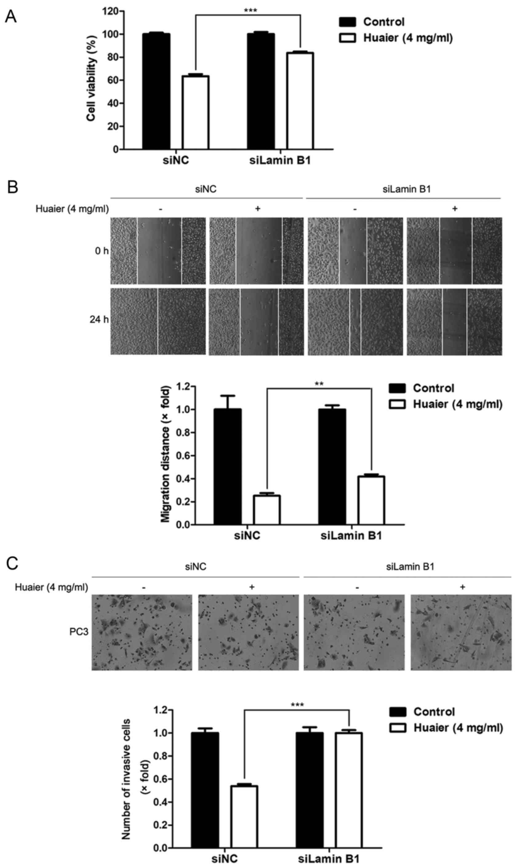
metastatic potential of human prostate cancer PC3 cells. To further
investigate the role of Lamin B1 in the inhibition of proliferative
and metastatic potential of PC3 cells in the presence of Huaier, we
treated PC3 cells transfected with Lamin B1 siRNAs or negative
control siRNAs with Huaier aqueous extract. As depicted in Fig. 4A, depletion of Lamin B1 attenuated
the inhibitory effect of Huaier on the proliferation of PC3 cells.
Additionally, the suppression of migration and invasion of PC3
cells caused by Huaier treatment were significantly impaired by
knockdown of Lamin B1 (Fig. 4B and
C). Collectively, decreased Lamin B1 is partially responsible
for the inhibition of proliferation and the metastatic potential of
PC3 cells in the presence of Huaier.
Huaier induces autophagy in PC3
cells
Autophagy is one of the underlying mechanisms for
drug-induced cell death (26,27).
In the present study we determined the effect of Huaier on
autophagy in PC3 cells. Observation of autophagosomes and other
related subcellular structures in the cytoplasm with transmission
electron microscope is widely used for autophagy assessment
(22,28,29).
Ultrastructural changes of PC3 cells treated with Huaier were
observed by transmission electron microscope. As shown in Fig. 5A, PC3 cells treated with Huaier
exhibited typical characteristics of autophagy: autophagosomes and
autolysosomes which contained cytoplasmic components. Moreover,
acridine orange staining was used to observe the accumulation of
autophagy vesicles. The number of acidic vesicles marked by orange
fluorescence increased in response to Huaier treatment in a
time-dependent manner (Fig. 5B). A
series of autophagy-related proteins participate in different
stages of autophagosome formation, such as Atg3, Atg5 and Beclin-1
(30–33). LC3 is an autophagosomal marker
protein (34). LC3-II, one form of
LC3, accumulates on the membranes of autophagosomes and is widely
used as a marker for autophagy evaluation (34,35).
Western blot analysis revealed that Huaier treatment led to
increase in the expression of Atg3, Atg5, Beclin-1 and LC3-II in a
dose- and time-dependent manner in PC3 cells (Fig. 5C and D). Moreover, Huaier treatment
markedly increased LC3 puncta in number and intensity in PC3 cells
under a fluorescence microscope (Fig.
5E), indicating that the number of autophagosomes was increased
in the presence of Huaier. Taken together, Huaier evidently
promoted autophagy in PC3 cells.
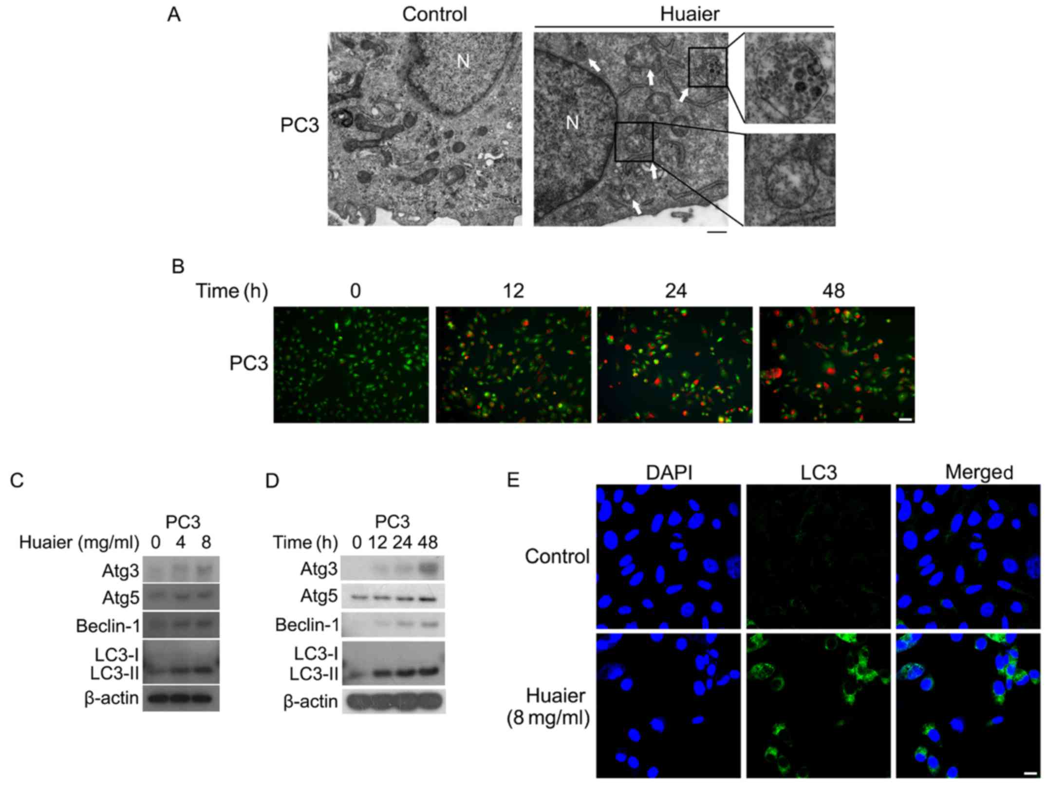 | Figure 5.Huaier induces autophagy in PC3
cells. (A) Transmission electron microscopy (TEM) analysis of PC3
cells treated with Huaier extract (8 mg/ml) for 48 h. White arrows
indicated autolysosomes/autophagosomes. Scale bar, 500 nm. N,
nucleus. (B) PC3 cells treated with Huaier extract (8 mg/ml) for 0,
12, 24 and 48 h were stained with acridine orange (10 µg/ml), and
observed under a fluorescence microscopy. Scale bar, 100 µm. (C and
D) Total cell lysates harvested from PC3 cells treated with Huaier
extract at the concentrations of 0, 4 and 8 mg/ml for 48 h (C) or
treated with 8 mg/ml Huaier extract for the indicated times (0, 12,
24 and 48 h) (D) were subjected to immunoblotting for detection of
Atg3, Atg5, Beclin-1 and LC3. (E) PC3 cells treated with 8 mg/ml
Huaier extract for 48 h were stained with anti-LC3 antibody. The
LC3 puncta (green) were observed through immunofluorescence. Nuclei
were stained with DAPI (blue). Representative images are presented.
Scale bar, 20 µm. |
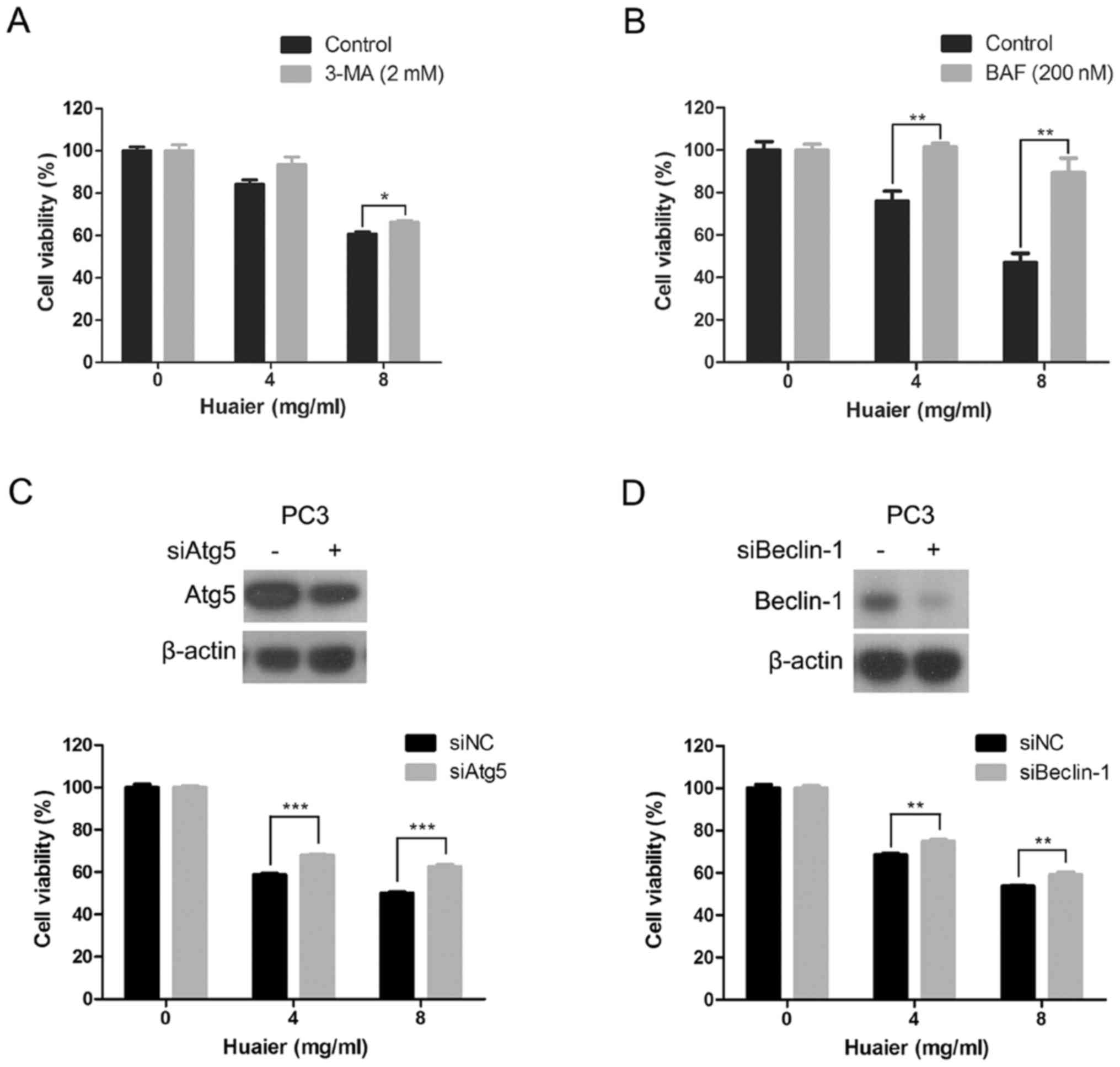
Autophagy inhibition attenuates
Huaier-induced inhibition of proliferation of PC3 cells
To investigate whether autophagy was involved in the
inhibition of proliferation of PC3 cells by Huaier, we suppressed
autophagy with two autophagy inhibitors and RNA interference. As
depicted in Fig. 6A and B,
inhibition of autophagy with 3-MA or bafilomycin A1 significantly
impaired the sensitivity of PC3 cells to Huaier treatment.
Moreover, Atg5 and Beclin-1, two essential components involved in
autophagosome formation (36), were
efficiently knocked down in the PC3 cells transfected with siRNAs
(Fig. 6C and D). Consistent with
the results of autophagy inhibitors, suppression of autophagy with
siRNAs against Atg5 or Beclin-1 remarkably blunted the inhibitory
effect of Huaier on the proliferation of PC3 cells (Fig. 6C and D). Collectively, the
inhibitory proliferation of PC3 cells exposed to Huaier is
partially mediated by activation of autophagy.
Discussion
In recent years, the antitumor effects of Huaier
have drawn the attention of cancer researchers. Many clinical
applications have demonstrated that Huaier can be used for the
treatment of multiple types of cancers including prostate cancer.
However, the anti-prostate cancer effect of Huaier and its
underlying mechanisms remain elusive. In the present study, we
demonstrated that Huaier inhibited the proliferation and metastatic
potential of human prostate cancer PC3 cells partially through
downregulation of Lamin B1. In addition, we revealed that Huaier
treatment induced autophagic cell death in PC3 cells.
Uncontrolled cell proliferation is one of the
hallmarks of cancer (37).
Inhibition of cancer cell proliferation can be used as a strategy
to treat cancer. Metastasis is a complex multi-step process which
plays a pivotal role in the progression of cancer (38). Cancer metastasis is responsible for
~90% of human cancer-related deaths (39). Metastases to distant organs such as
the bone, liver, lungs and brain frequently occur in the advanced
stage of prostate cancer (40,41).
Most prostate cancer-related deaths result from metastases. Herein
we revealed that Huaier had excellent anti-proliferative and
anti-metastatic effects in PC3 cells. Thus, Huaier can be used as a
candidate drug for targeting the proliferation and metastasis of
cancer cells in the treatment of human prostate cancer. Lamin B1 is
an important member of the lamin protein family (23) and was reported to be a carcinogenic
gene (25). The levels of Lamin B1
are elevated in tumors of patients with hepatocellular carcinoma
(24). Moreover, Lamin B1
expression was found to be markedly increased in malignant prostate
cancer (19). Knockdown of Lamin B1
expression by siRNAs induced apoptosis in HeLa cells (42). Our previous study demonstrated that
Lamin B1 was remarkably downregulated in human hepatoma SKHEP-1
cells exposed to Huaier (10). In
the present study, we revealed that Huaier treatment markedly
reduced Lamin B1 expression in human prostate cancer PC3 cells.
Furthermore, Lamin B1 was required for the inhibition of
proliferation and metastatic potential of PC3 cells by Huaier. As
an oncogenic protein, Lamin B1 is a novel target of Huaier for
prostate cancer treatment.
Autophagy is a normal physiological process in which
the cytoplasmic components including misfolded proteins and damaged
organelles are surrounded to form autophagosomes that are
eventually transported to lysosomes for degradation (20,43).
As one of the underlying mechanisms of cell death, autophagy
frequently occurs in cancer cells in response to antitumor
therapies (44–47). Huaier triggered autophagy in human
breast cancer MDA-MB-231, MDA-MB-468 and MCF7 cells, and autophagy
inhibition impaired Huaier-induced cell death in these cancer
cells. Huaier-induced cytotoxicity in human breast cancer cells was
partialy due to activation of autophagy (48). In addition, Huaier augmented
tamoxifen-induced autophagy in ER-positive breast cancer cells
(49). We demonstrated that Huaier
extract dramatically triggered autophagy in PC3 cells through
electron microscopy observation, acridine orange staining, western
blotting and immunofluorescence assay. Moreover, inhibition of
autophagy via drugs or siRNAs significantly abrogated
Huaier-induced cytotoxicity in PC3 cells. Therefore, autophagic
cell death is involved in Huaier-induced cytotoxicity in human
prostate cancer cells.
In this study, we demonstrated that Huaier-induced
cytotoxicity and decreased cell mobility were at least partially
mediated by downregulation of Lamin B1 and autophagic cell death in
prostate cancer PC3 cells. The multiple mechanisms reported in our
study contribute to the understanding of the complex anti-prostate
cancer effects of Huaier. The present study provides a new
theoretical basis for the clinical application of Huaier in
prostate cancer.
Acknowledgements
Not applicable.
Funding
The present study was financially supported by the
National Natural Science Foundation of China (nos. 81403147 and
81402219), the Excellent Young Scientist Foundation of Beijing
University of Chinese Medicine (no. 2015-JYB-XYQ-004), and the
Outstanding Young Talent Foundation of the Organization Department
of Beijing Municipal Party Committee (no. 2014000021469G221).
Availability of data and materials
All data generated or analyzed during this study are
included in this published article.
Authors' contributions
AY designed and performed the experiments, analyzed
data and wrote the manuscript. YaZ, YW, XZ and YuZ analyzed data
and revised the manuscript. PT and ZH supervised the study,
designed experiments and revised the manuscript. All authors read
and approved the manuscript and agree to be accountable for all
aspects of the research in ensuring that the accuracy or integrity
of any part of the work are appropriately investigated and
resolved.
Ethics approval and consent to
participate
Not applicable.
Consent for publication
Not applicable.
Competing interests
The authors declare that they have no competing
interests.
Glossary
Abbreviations
Abbreviations:
|
TCM
|
Traditional Chinese Medicine
|
|
FBS
|
fetal bovine serum
|
|
OD
|
optical density
|
|
CCK-8
|
Cell Counting Kit-8
|
|
TEM
|
transmission electron microscopy
|
References
|
1
|
Denmeade SR and Isaacs JT: A history of
prostate cancer treatment. Nat Rev Cancer. 2:389–396. 2002.
View Article : Google Scholar : PubMed/NCBI
|
|
2
|
Trewartha D and Carter K: Advances in
prostate cancer treatment. Nat Rev Drug Discov. 12:823–824. 2013.
View Article : Google Scholar : PubMed/NCBI
|
|
3
|
Kelloff GJ: Perspectives on cancer
chemoprevention research and drug development. Adv Cancer Res.
78:199–334. 2000. View Article : Google Scholar : PubMed/NCBI
|
|
4
|
Hwang C: Overcoming docetaxel resistance
in prostate cancer: A perspective review. Ther Adv Med Oncol.
4:329–340. 2012. View Article : Google Scholar : PubMed/NCBI
|
|
5
|
Wallace TJ, Torre T, Grob M, Yu J, Avital
I, Brücher B, Stojadinovic A and Man YG: Current approaches,
challenges and future directions for monitoring treatment response
in prostate cancer. J Cancer. 5:3–24. 2014. View Article : Google Scholar : PubMed/NCBI
|
|
6
|
Wang CY, Bai XY and Wang CH: Traditional
Chinese medicine: A treasured natural resource of anticancer drug
research and development. Am J Chin Med. 42:543–559. 2014.
View Article : Google Scholar : PubMed/NCBI
|
|
7
|
Xu H, Zhao X, Liu X, Xu P, Zhang K and Lin
X: Antitumor effects of traditional Chinese medicine targeting the
cellular apoptotic pathway. Drug Des Devel Ther. 9:2735–2744.
2015.PubMed/NCBI
|
|
8
|
Yan Z, Lai Z and Lin J: Anticancer
properties of traditional Chinese medicine. Comb Chem High
Throughput Screen. 20:423–429. 2017. View Article : Google Scholar : PubMed/NCBI
|
|
9
|
Song X, Li Y, Zhang H and Yang Q: The
anticancer effect of Huaier (Review). Oncol Rep. 34:12–21. 2015.
View Article : Google Scholar : PubMed/NCBI
|
|
10
|
Hu Z, Yang A, Su G, Zhao Y, Wang Y, Chai X
and Tu P: Huaier restrains proliferative and invasive potential of
human hepatoma SKHEP-1 cells partially through decreased Lamin B1
and elevated NOV. Sci Rep. 6:312982016. View Article : Google Scholar : PubMed/NCBI
|
|
11
|
Li Y, Qi W, Song X, Lv S, Zhang H and Yang
Q: Huaier extract suppresses breast cancer via regulating
tumor-associated macrophages. Sci Rep. 6:200492016. View Article : Google Scholar : PubMed/NCBI
|
|
12
|
Yan X, Lyu T, Jia N, Yu Y, Hua K and Feng
W: Huaier aqueous extract inhibits ovarian cancer cell motility via
the AKT/GSK3β/β-catenin pathway. PLoS One. 8:e637312013. View Article : Google Scholar : PubMed/NCBI
|
|
13
|
Wang X, Zhang N, Huo Q and Yang Q:
Anti-angiogenic and antitumor activities of Huaier aqueous extract.
Oncol Rep. 28:1167–1175. 2012. View Article : Google Scholar : PubMed/NCBI
|
|
14
|
Wu T, Chen W, Liu S, Lu H, Wang H, Kong D,
Huang X, Kong Q, Ning Y and Lu Z: Huaier suppresses proliferation
and induces apoptosis in human pulmonary cancer cells via
upregulation of miR-26b-5p. FEBS Lett. 588:2107–2114. 2014.
View Article : Google Scholar : PubMed/NCBI
|
|
15
|
Yang AL, Hu ZD and Tu PF: Research
progress on anti-tumor effect of Huaier. Zhongguo Zhong Yao Za Zhi.
40:4805–4810. 2015.PubMed/NCBI
|
|
16
|
Wang X, Zhang N, Huo Q, Sun M, Lv S and
Yang Q: Huaier aqueous extract suppresses human breast cancer cell
proliferation through inhibition of estrogen receptor α signaling.
Int J Oncol. 43:321–328. 2013. View Article : Google Scholar : PubMed/NCBI
|
|
17
|
Yan L, Liu X, Yin A, Wei Y, Yang Q and
Kong B: Huaier aqueous extract inhibits cervical cancer cell
proliferation via JNK/p38 pathway. Int J Oncol. 47:1054–1060. 2015.
View Article : Google Scholar : PubMed/NCBI
|
|
18
|
Yang A, Fan H, Zhao Y, Zha X, Zhang H, Hu
Z and Tu P: Huaier aqueous extract inhibits proliferation and
metastasis of tuberous sclerosis complex cell models through
downregulation of JAK2/STAT3 and MAPK signaling pathways. Oncol
Rep. 36:1491–1498. 2016. View Article : Google Scholar : PubMed/NCBI
|
|
19
|
Coradeghini R, Barboro P, Rubagotti A,
Boccardo F, Parodi S, Carmignani G, D'Arrigo C, Patrone E and Balbi
C: Differential expression of nuclear lamins in normal and
cancerous prostate tissues. Oncol Rep. 15:609–613. 2006.PubMed/NCBI
|
|
20
|
Klionsky DJ: Autophagy: From phenomenology
to molecular understanding in less than a decade. Nat Rev Mol Cell
Biol. 8:931–937. 2007. View
Article : Google Scholar : PubMed/NCBI
|
|
21
|
Hu Z, Wang Y, Huang F, Chen R, Li C, Wang
F, Goto J, Kwiatkowski DJ, Wdzieczak-Bakala J, Tu P, et al:
Brain-expressed X-linked 2 is pivotal for hyperactive mechanistic
target of rapamycin (mTOR)-mediated tumorigenesis. J Biol Chem.
290:25756–25765. 2015. View Article : Google Scholar : PubMed/NCBI
|
|
22
|
Ylä-Anttila P, Vihinen H, Jokitalo E and
Eskelinen EL: Monitoring autophagy by electron microscopy in
Mammalian cells. Methods Enzymol. 452:143–164. 2009. View Article : Google Scholar : PubMed/NCBI
|
|
23
|
Dittmer TA and Misteli T: The lamin
protein family. Genome Biol. 12:2222011. View Article : Google Scholar : PubMed/NCBI
|
|
24
|
Sun S, Xu MZ, Poon RT, Day PJ and Luk JM:
Circulating Lamin B1 (LMNB1) biomarker detects early stages of
liver cancer in patients. J Proteome Res. 9:70–78. 2010. View Article : Google Scholar : PubMed/NCBI
|
|
25
|
Li L, Du Y, Kong X, Li Z, Jia Z, Cui J,
Gao J, Wang G and Xie K: Lamin B1 is a novel therapeutic target of
betulinic acid in pancreatic cancer. Clin Cancer Res. 19:4651–4661.
2013. View Article : Google Scholar : PubMed/NCBI
|
|
26
|
Tsujimoto Y and Shimizu S: Another way to
die: Autophagic programmed cell death. Cell Death Differ. 12 Suppl
2:S1528–S1534. 2005. View Article : Google Scholar
|
|
27
|
Fulda S and Kögel D: Cell death by
autophagy: Emerging molecular mechanisms and implications for
cancer therapy. Oncogene. 34:5105–5113. 2015. View Article : Google Scholar : PubMed/NCBI
|
|
28
|
Petibone DM, Majeed W and Casciano DA:
Autophagy function and its relationship to pathology, clinical
applications, drug metabolism and toxicity. J Appl Toxicol.
37:23–37. 2017. View
Article : Google Scholar : PubMed/NCBI
|
|
29
|
Dunn WA Jr: Studies on the mechanisms of
autophagy: Formation of the autophagic vacuole. J Cell Biol.
110:1923–1933. 1990. View Article : Google Scholar : PubMed/NCBI
|
|
30
|
Kim AD, Kang KA, Kim HS, Kim DH, Choi YH,
Lee SJ, Kim HS and Hyun JW: A ginseng metabolite, compound K,
induces autophagy and apoptosis via generation of reactive oxygen
species and activation of JNK in human colon cancer cells. Cell
Death Dis. 4:e7502013. View Article : Google Scholar : PubMed/NCBI
|
|
31
|
Noda NN and Inagaki F: Mechanisms of
autophagy. Annu Rev Biophys. 44:101–122. 2015. View Article : Google Scholar : PubMed/NCBI
|
|
32
|
Li X, Li Y, Fang S, Su J, Jiang J, Liang
B, Huang J, Zhou B, Zang N, Ho W, et al: Downregulation of
autophagy-related gene ATG5 and GABARAP expression by IFN-λ1
contributes to its anti-HCV activity in human hepatoma cells.
Antiviral Res. 140:83–94. 2017. View Article : Google Scholar : PubMed/NCBI
|
|
33
|
Ma K, Fu W, Tang M, Zhang C, Hou T, Li R,
Lu X, Wang Y, Zhou J, Li X, et al: PTK2-mediated degradation of
ATG3 impedes cancer cells susceptible to DNA damage treatment.
Autophagy. 13:579–591. 2017. View Article : Google Scholar : PubMed/NCBI
|
|
34
|
Tanida I, Ueno T and Kominami E: LC3 and
autophagy. Methods Mol Biol. 445:77–88. 2008. View Article : Google Scholar : PubMed/NCBI
|
|
35
|
Mizushima N and Yoshimori T: How to
interpret LC3 immunoblotting. Autophagy. 3:542–545. 2007.
View Article : Google Scholar : PubMed/NCBI
|
|
36
|
Kang R, Zeh HJ, Lotze MT and Tang D: The
Beclin 1 network regulates autophagy and apoptosis. Cell Death
Differ. 18:571–580. 2011. View Article : Google Scholar : PubMed/NCBI
|
|
37
|
Hanahan D and Weinberg RA: Hallmarks of
cancer: The next generation. Cell. 144:646–674. 2011. View Article : Google Scholar : PubMed/NCBI
|
|
38
|
Valastyan S and Weinberg RA: Tumor
metastasis: Molecular insights and evolving paradigms. Cell.
147:275–292. 2011. View Article : Google Scholar : PubMed/NCBI
|
|
39
|
Mehlen P and Puisieux A: Metastasis: A
question of life or death. Nat Rev Cancer. 6:449–458. 2006.
View Article : Google Scholar : PubMed/NCBI
|
|
40
|
Bubendorf L, Schöpfer A, Wagner U, Sauter
G, Moch H, Willi N, Gasser TC and Mihatsch MJ: Metastatic patterns
of prostate cancer: An autopsy study of 1,589 patients. Hum Pathol.
31:578–583. 2000. View Article : Google Scholar : PubMed/NCBI
|
|
41
|
Park JC and Eisenberger MA: Advances in
the treatment of metastatic prostate cancer. Mayo Clin Proc.
90:1719–1733. 2015. View Article : Google Scholar : PubMed/NCBI
|
|
42
|
Harborth J, Elbashir SM, Bechert K, Tuschl
T and Weber K: Identification of essential genes in cultured
mammalian cells using small interfering RNAs. J Cell Sci.
114:4557–4565. 2001.PubMed/NCBI
|
|
43
|
Levine B and Klionsky DJ: Development by
self-digestion: Molecular mechanisms and biological functions of
autophagy. Dev Cell. 6:463–477. 2004. View Article : Google Scholar : PubMed/NCBI
|
|
44
|
Gozuacik D and Kimchi A: Autophagy as a
cell death and tumor suppressor mechanism. Oncogene. 23:2891–2906.
2004. View Article : Google Scholar : PubMed/NCBI
|
|
45
|
Yang WL, Perillo W, Liou D, Marambaud P
and Wang P: AMPK inhibitor compound C suppresses cell proliferation
by induction of apoptosis and autophagy in human colorectal cancer
cells. J Surg Oncol. 106:680–688. 2012. View Article : Google Scholar : PubMed/NCBI
|
|
46
|
Yao Z, Xie F, Li M, Liang Z, Xu W, Yang J,
Liu C, Li H, Zhou H and Qu LH: Oridonin induces autophagy via
inhibition of glucose metabolism in p53-mutated colorectal cancer
cells. Cell Death Dis. 8:e26332017. View Article : Google Scholar : PubMed/NCBI
|
|
47
|
Yang ZJ, Chee CE, Huang S and Sinicrope
FA: The role of autophagy in cancer: Therapeutic implications. Mol
Cancer Ther. 10:1533–1541. 2011. View Article : Google Scholar : PubMed/NCBI
|
|
48
|
Wang X, Qi W, Li Y, Zhang N, Dong L, Sun
M, Cun J, Zhang Y, Lv S and Yang Q: Huaier extract induces
autophagic cell death by inhibiting the mTOR/S6K pathway in breast
cancer cells. PLoS One. 10:e01317712015. View Article : Google Scholar : PubMed/NCBI
|
|
49
|
Qi W, Sun M, Kong X, Li Y, Wang X, Lv S,
Ding X, Gao S, Cun J, Cai C, et al: Huaier extract synergizes with
tamoxifen to induce autophagy and apoptosis in ER-positive breast
cancer cells. Oncotarget. 7:26003–26015. 2016. View Article : Google Scholar : PubMed/NCBI
|