Introduction
Cancer remains the second leading cause of mortality
in developed countries, exceeded only by heart disease (1). Liver and kidney cancer are among the
top ten most common malignancies worldwide. An increase in the
incidence rate has been observed and the two types of cancer have a
very high fatality rate (2–8). The incidence of kidney cancer varies
geographically: The rates are highest in more developed regions in
Europe, North America and Australia and they are lowest in Africa
and South-East Asia (7,8). Kidney cancer is more often diagnosed
in males globally (6,7). The development of this tumor is
dependent on intrinsic (genetic predisposition, sex and age) and
extrinsic risk factors (smoking, obesity and alcohol abuse)
(8). Hepatoblastoma is the most
common liver neoplasm in children that is diagnosed during the
early years of life; it is an embryonal malignancy of
hepatocellular origin (3,4,9).
Therapeutic strategies include surgical resection and chemotherapy
(3,4). Although 60% of tumors are unresectable
at presentation, adjuvant chemotherapy effectively reduces tumor
size, allowing surgical intervention in the majority of patients
(3). However, the prognosis for
children with advanced or chemotherapy-refractory cancer remains
poor (4). Renal cell carcinoma
(RCC) comprises 90% cases of all kidney cancer. RCC, as the most
frequent type of renal neoplasm, is also the most lethal among all
urological tumors (8). In 75–80%
cases, RCC can easily invade neighboring tissues and metastasize,
and is often resistant to currently available therapeutic
strategies. The patients respond poorly to conventional cytostatic
drugs and radiotherapy (10). The
5-year survival rates of RCC are estimated at only 50%; the fact
that at first diagnosis 20–30% of patients have metastatic disease
is one of the reasons for this prognosis (11,12).
In addition, >40% of patients develop metastases following
radical nephrectomy (13). However,
successful treatment, particularly at metastatic stages, remains a
challenge. Overall, metastatic RCC remains incurable, although
targeted therapeutic strategies, including mammalian target of
rapamycin inhibitors appeared to have improved efficacy (14). Despite recent advances, the outlook
for patients remains poor. The search and discovery of novel and
safer anticancer agents with higher cytotoxicity in cancer cells
may aid in improving the management of these types of cancer.
The increasing diversity of small molecule libraries
provides an important source for the discovery of novel drug
candidates. In this context, heterocycles and in particular
1,3-thiazolidin-4-ones are one of the most extensively investigated
classes of compounds. They are heterocyclic nuclei with an atom of
sulfur and nitrogen at position 1 and 3, respectively, and a
carbonyl group at position 4. Studies of the synthesis and
bioactivity of these derivatives have attracted significant
attention (15,16). These derivatives are known to
exhibit antimicrobial (17–21), anticonvulsant (22), antihistaminic (23) and cardioprotective (24) bioactivities. In our previous study,
the 2,3-disubstituted 1,3-thiazolidin-4-one derivatives were
revealed to exhibit significant antimicrobial activity (25). Among twenty compounds tested,
particularly
N-[2-(4-methylphenyl)-4-oxo-1,3-thiazolidin-3-yl]benzamide
possessed a spectrum of bioactivity against Gram-positive bacteria
(staphylococci, streptococci, micrococci and Bacillus spp.).
The aforementioned derivative also exhibited activity against
yeasts belonging to Candida spp., mainly Candida
albicans (25). The
1,3-thiazolidin-4-one derivatives are also known for their
anticancer activity (26,27). A review of current literature
revealed that 2,5-disubstituted 1,3-thiazolidin-4-ones had been
reported for their antiproliferative activity; for example, in
human colon adenocarcinoma cells (HT29), human gastric cancer cell
line, human colon cancer cell lines and sarcoma-derived cells
(28,29). It was reported that
5-benzylidene-4-thiazolidinone derivatives demonstrated significant
antitumor potential with various biotargets and mechanisms,
including sphingosine kinase (SK), JNK stimulating phosphatase-1
(JSP-1) or non-membrane protein tyrosine phosphatase (SHP-2)
(16,30). The 2,3,5-trisubstituted derivatives
were demonstrated to induce cell growth arrest in HT29 cells and
significant cytotoxicity towards human lung and human breast cancer
cultures (31). In turn,
2-phenylimino-3-alkyl-4-thiazolidinone derivatives hindered the
proliferation of HT29 cells, characterized by CDK1/cyclin B
inhibition. This effect was achieved by blocking cell progression
at the G2/M phase border and inducing apoptosis (16). The 2,3-disubstituted
4-thiazolidinone derivatives were also active against leukemia cell
lines (26). Due to the
aforementioned results, the antiproliferative potential of
1,3-thiazolidin-4-ones, as well as the general cellular mechanisms
associated with it, were investigated.
To the best of our knowledge, the present study was
the first scientific report of the anticancer potential of
1,3-thiazolidin-4-ones in RCC (769-P). In addition, the impact of
other derivatives from this chemical group on HepG2 cells was
studied only by George et al (32). The aim of the present study was to
synthesize and evaluate the cytotoxicity of the aforementioned
N-[2-(4-methylphenyl)-4-oxo-1,3-thiazolidin-3-yl]benzamide
and three other promising compounds (25) towards these human cancer cell lines.
The effects on cell viability were assessed by MTT assay, and the
effects on the cell cycle and apoptosis were also investigated. The
cell cycle assay has applicability to drug screening, as
dysregulation of the cell cycle is a common occurrence in
neoplasia, thereby providing the opportunity to discover novel
targets for anticancer agents. The Annexin V assay was also
selected as a useful tool in detecting apoptotic cells.
Materials and methods
Reagents
All required chemicals and solvents were purchased
from Sigma-Aldrich; Merck KGaA (Darmstadt, Germany) and Merck KGaA,
and were used without further purification. Melting points were
determined using Fisher-Johns blocks (Thermo Fisher Scientific,
Inc., Waltham, MA, USA) and were uncorrected. The 1H NMR
and 13C NMR spectra were recorded on the Bruker Avance
300 apparatus (Bruker Corporation, Ettlingen, Germany) in dimethyl
sulfoxide (DMSO)-d6 with tetramethylsilane as the
internal standard. The progress of the reaction and the purity of
the obtained compounds were monitored by thin-layer chromatography
using precoated aluminum sheet 60 F254 plates (Merck KGaA), in a
CHCl3/C2H5OH (10:1, v/v) solvent
system. The spots were detected by exposure to the UV lamp at 254
nm. The elemental analysis of obtained compounds was performed
using the AMZ 851 CHX analyzer (Gdańsk University of Technology,
Gdańsk, Poland). The results of elemental analysis (C, H, N) were
within ±0.4% of the calculated values.
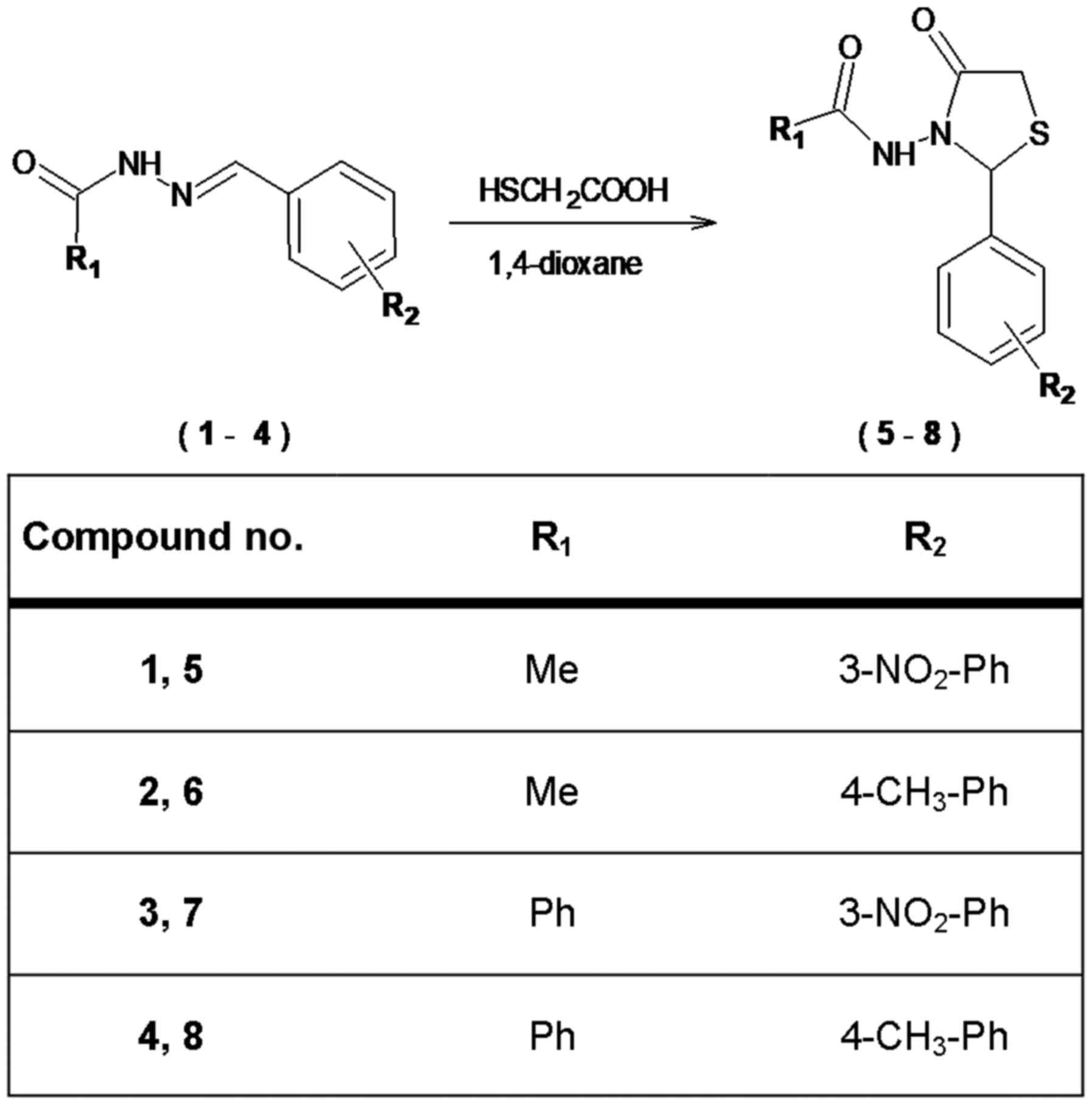
Preparation of 2,3-disubstituted
1,3-thiazolidin-4-one derivatives (5–8)
Synthesis was performed according to procedure
described previously by Popiołek et al (25). To the solution of corresponding
N-substituted hydrazide derivatives (1–4) (10
mmol) in 1,4-dioxane (15 ml), mercaptoacetic acid (0.92 g, 10 mmol)
was added dropwise. The mixture was stirred for 6 h at room
temperature. Next, the solvent was removed under reduced pressure
to obtain a crude product, which was purified by recrystallization
from ethanol. The following derivatives were prepared:
N-[2-(3-nitrophenyl)-4-oxo-1,3-thiazolidin-3-yl]acetamide
[compound 5; CAS Registry Number: 1644570-76-2; analytical and
spectral data are consistent with those reported by Popiołek et
al (25); yield, 69%; melting
point (m.p.), was 202-204°C];
N-[2-(4-methylphenyl)-4-oxo-1,3-thiazolidin-3-yl]acetamide
(compound 6; CAS Registry Number: 1644570-74-0; analytical and
spectral data are consistent with those reported by Popiołek et
al (25); yield, 81%; m.p.,
104–106°C);
N-[2-(3-nitrophenyl)-4-oxo-1,3-thiazolidin-3-yl]benzamide
(compound 7; CAS Registry Number: 1644570-79-5; analytical and
spectral data are consistent with those reported by Popiołek et
al (25); yield, 70%; m.p.,
176–178°C); and
N-[2-(4-methylphenyl)-4-oxo-1,3-thiazolidin-3-yl]benzamide
(compound 8; CAS Registry Number: 1644570-78-4; analytical and
spectral data are consistent with those reported by Popiołek et
al (25); yield, 87%; m.p.,
106–108°C).
Cell culture
The present study used normal green monkey kidney
cells (GMK) obtained from Biomed Serum and Vaccine Production Plant
Ltd. (Lublin, Poland) and tumor cell cultures supplied by American
Type Culture Collection (ATCC; Manassas, VA, USA), including HepG2
(human hepatoblastoma-derived cells; catalog no. HB-8065) and 769-P
(human renal cell adenocarcinoma; catalog no. CRL-1933) (9). The GMK cell line was cultured in basic
RPMI-1640 medium [with L-glutamine,
4-(2-hydroxethyl)-1-piperazineethanesulfonic acid (HEPES) and
sodium decarbonate] and the 769-P cell line was cultured in
rich-component RPMI-1640 medium (with L-glutamine, sodium pyruvate,
glucose, HEPES and sodium dicarbonate). The HepG2 cells were
cultured in Eagle's minimal essential medium (EMEM). All the media
were supplemented with 10% fetal bovine serum (FBS), 100 U/ml
penicillin, 100 µg/ml streptomycin and 2.5 µg/ml amphotericin B.
The cell cultures were grown in 75 cm2 tissue culture
flasks (EasYFlasks™ Nunclon™ Δ; Nalge Nunc International, Penfield,
NY, USA) as a monolayer in a humidified atmosphere of 5%
CO2 at 37°C in a cell incubator. The suspensions of GMK,
769-P and HepG2 cells were prepared at a density of
1×10⁶ cells/ml, prior to being transferred to 96-well
cell culture plates (SPL Life Sciences, Pocheon, Korea). The
prepared plates were incubated for 24 h in order for cells to
adhere to the plates.
Drugs and substances
Drugs and substances were as follows: MTT
(Sigma-Aldrich; Merck KGaA), dimethyl sulfoxide (DMSO; Avantor
Performance Materials, Inc., Gliwice, Poland), phosphate-buffered
saline (PBS; Biomed Serum and Vaccine Production Plant Ltd.), and
The Eagle's minimal essential medium (EMEM; ATCC). The basic and
rich-component RPMI-1640, FBS, and the penicillin, streptomycin,
amphotericin B and trypsin solution (0.25% trypsin and 0.02% EDTA
in PBS without Ca2+ or Mg2+, with phenol red)
were supplied by PAN-Biotech GmbH (Aidenbach, Germany).
Cell viability assay
Cell viability was assessed using an MTT assay based
on DB-ALM protocol no. 17 (European Centre for the Validation of
Alternative Methods, Database Service on Alternative Methods to
Animal Experimentation). Cell viability was determined in a
mitochondrial-dependent reaction (reduction in mitochondrial
dehydrogenase activity) by measurement of the formazan production
from MTT salt. Cell viability is expressed as the percentage (%) of
control cells. The examined compounds were first dissolved in DMSO
and subsequently diluted to the required concentration with the
respective cell culture medium. The solutions were prepared ex
tempore, were added to the cells in the same volume (100 µl/well)
and incubated for 24 h. Following incubation, 10 µl MTT solution (5
mg/ml) was added to each well of a microplate and was incubated for
3 h at 37°C. At the end of the incubation, the culture medium was
removed carefully from each well and 100 µl DMSO was added. The
absorbance of each well was measured at 550 nm using an automated
absorbance microplate reader ELx808IU (BioTek
Instruments Inc., Winooski, VT, USA). The experiments included the
determination of IC10, IC25 and
IC50 values for tested compounds. The cells were
analyzed under a phase-contrast microscope (magnification, ×150)
Nikon Eclipse Ti (Nikon Corporation, Tokyo, Japan). Next, the cell
cultures were incubated in the presence of tested compounds at
concentrations of IC10 and IC25 during 24, 48
and 72 h. All the parameters mentioned in the present study were
evaluated in the presence of the solvent of 1,3-thiazolidin-4-one
derivatives and there were no significant differences between the
control cells and the solvent-treated cells. The final
concentration of DMSO did not exceed 0.5% v/v. All the experiments
were performed at least five times.
The cell cycle and apoptosis of 769-P
cells
Two-step cell cycle analysis was performed using the
NucleoCounter NC-3000 system (ChemoMetec, Allerod, Denmark),
according to the manufacturer's protocol. This system enables the
rapid quantification of the DNA content of mammalian cells,
allowing determination of G0/G1, S and G2/M cell cycle phases. The
DNA content is measured using the fluorescent, DNA-selective stain,
DAPI, which exhibits emission signals proportional to DNA mass. The
Annexin V assay for the NucleoCounter NC-3000 system enables the
measurement of externalization of phosphatidylserine. The
translocation of the aforementioned phospholipid to the outer
membrane layer indicates early apoptosis and fluorescently labeled
Annexin V preferentially binds to negatively charged
phosphatidylserine. Annexin V is a cellular protein that also binds
to this phospholipid on late apoptotic and necrotic cells. However,
as the membrane integrity of these cells has been lost, these can
be distinguished from early apoptotic cells by the use of an
impermeant dye, such as propidium iodide (PI). Cells were stained
with an Annexin V-CF488A conjugate and with Hoechst 33342.
Immediately prior to analysis, the cells are mixed with PI to stain
non-viable cells. The quantification of early apoptotic cells was
based on Annexin V binding and PI exclusion.
Statistical analysis
Results are expressed as the mean ± standard error
of the mean. The statistical significance among the groups was
determined by analysis of variance, followed by Dunnett's post hoc
test using Statistica software (version 12; Statsoft, Inc., Tulsa,
OK, USA). P<0.05 was considered to indicate a statistically
significant difference.
Results
Chemistry
In the present study, 2,3-disubstituted
1,3-thiazolidin-4-one derivatives (5–8) were
synthesized by the cyclization reaction of N-substituted carboxylic
acid hydrazide derivatives (1–4) with
mercaptoacetic acid in the presence of 1,4-dioxane (Fig. 1), as previously described (25). The obtained compounds are stable
solids at room temperature and their spectral data (1H
NMR, 13C NMR) is in full agreement with the proposed
structures. Synthesized compounds were evaluated for in
vitro antiproliferative study.
In vitro cytotoxicity
The assessment of anticancer potential of
1,3-thiazolidin-4-one derivatives was performed in human renal cell
adenocarcinoma (769-P) and human hepatoblastoma-derived cells
(HepG2) (9), and green monkey
kidney cells (GMK) as a normal reference cell line, using the MTT
method. The IC10, IC25 and IC50
values for compounds 5–8 were determined based on dose-response
curves (Table I).
 | Table I.The determination of IC for
1,3-thiazolidin-4-one derivatives (compounds 5–8). |
Table I.
The determination of IC for
1,3-thiazolidin-4-one derivatives (compounds 5–8).
|
| Cell line |
|---|
|
|
|
|---|
| Compound no. | IC, mM | GMK | 769-P | HepG2 |
|---|
| 5 |
IC10 | 0.73 | nd | 0.95 |
|
|
IC25 | 2.34 | nd | 2.90 |
|
|
IC50 | 16.48 | nd | 18.62 |
| 6 |
IC10 | 0.64 | 0.69 | 0.31 |
|
|
IC25 | 1.09 | 1.15 | 0.7 |
|
|
IC50 | 2.66 | 2.67 | 2.70 |
| 7 |
IC10 | nd | nd | 0.64 |
|
|
IC25 | nd | nd | 2.03 |
|
|
IC50 | 33.85 | nd | 13.67 |
| 8 |
IC10 | 0.59 | 0.81 | 0.51 |
|
|
IC25 | 0.96 | 1.31 | 0.75 |
|
|
IC50 | 2.13 | 2.93 | 1.43 |
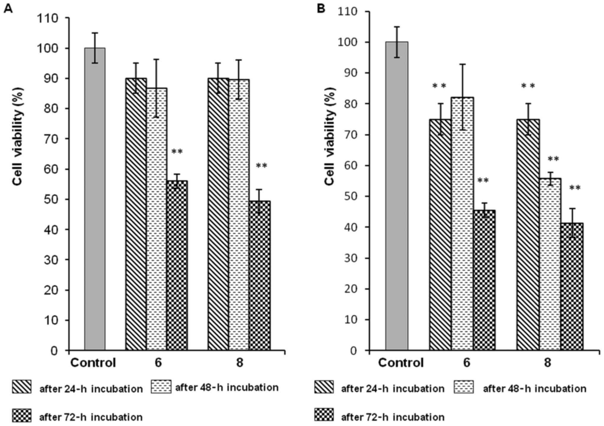
After 72 h of incubation of human renal
adenocarcinoma cells (769-P) with compound 6 at IC10 and
compound 8 at IC10 the significant reductions of cell
viability by ~50% were noted compared with the control (Fig. 2A). Additionally, following
incubation of 769-P cells with compound 6, the decreases in cell
viability were almost the same for IC10 and
IC25 (Fig. 2A and B).
Compound 8 at a concentration of IC25 evoked a
time-dependent decrease in the viability of 769-P cells (45% growth
inhibition after 48 h and 60% after 72 h, compared with the
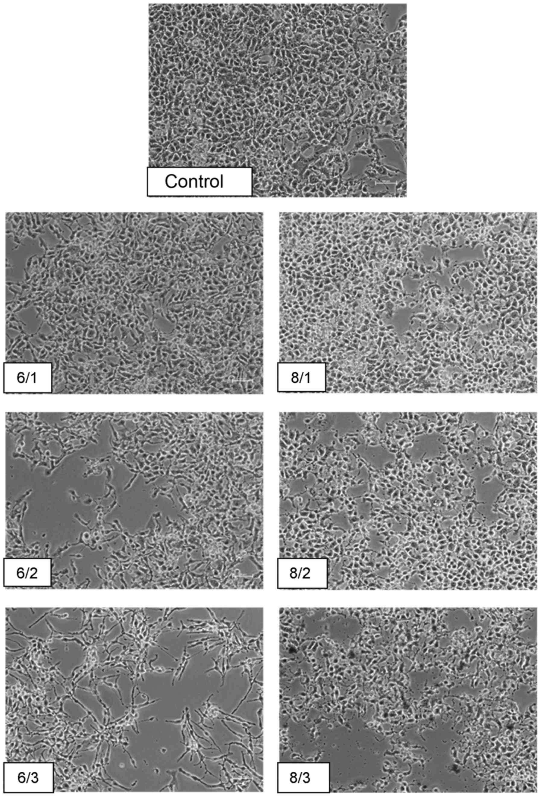
control; Fig. 2B). After a 24-h
incubation period of 769-P cells with the aforementioned compounds
at concentrations of IC10, IC25 and
IC50, they were evaluated using a phase-contrast
microscope (magnification, ×150). The adverse changes in their
general morphology and the reduction in the cell culture density
resulted from the inhibitory concentration used. The occurrence of
irregularly shaped cells resulting from shrinkage of the cytoplasm
and inhibition of contact growth were already observed at the
lowest IC10 concentration, particularly in the case of
compound 8. At the highest concentration corresponding to
IC50 in the field of vision beyond the aforementioned
changes, a small number of detached and dead cells were observed
(Fig. 3). This derivative used in
the all determined inhibitory concentrations caused clearly visible
cell damage after 24 h of incubation. The number of cell
deformations and the extent of growth inhibition corresponded with
the increasing concentration (Fig.
3).
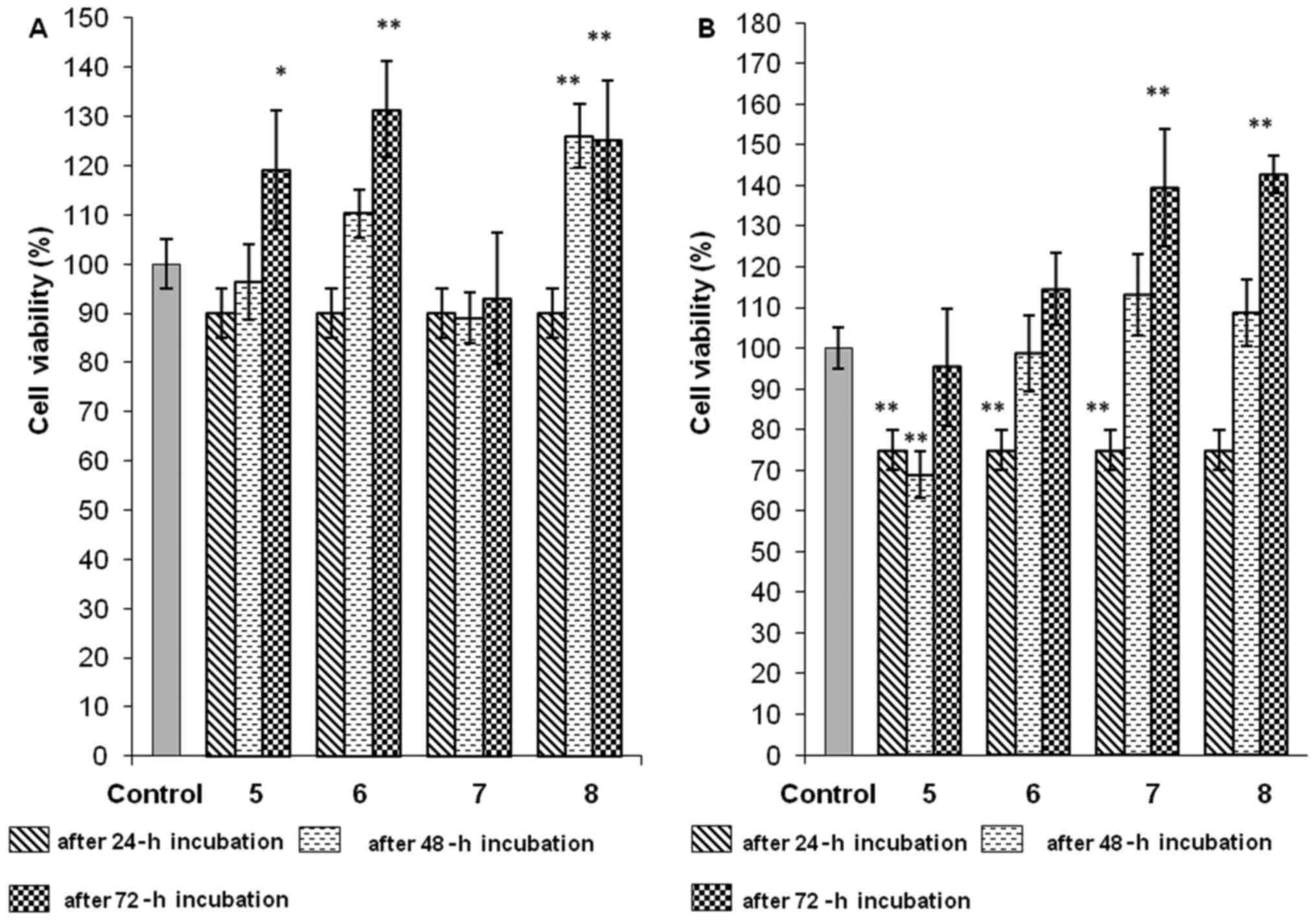
Compounds 5, 6 and 8, applied at concentrations of
IC10 to the cancer HepG2 cell line, significantly
increased cell viability by 20–30% in a time-dependent manner, when
compared with the control (Fig.
4A). Similar results were obtained for compound 8 at a
concentration of IC25 (Fig.
4B). Increasing the concentrations to IC25 for
compound 5 and 6 also caused the enhancement of HepG2 cell
viability after 48 h and 72 h of incubation, but not to above that
of the control cells (Fig. 4B). In
the case of compound 7 at IC10, the time of incubation
had no impact on the 10%-cytotoxic effect, but at IC25,
a very significant and time-dependent increase in cell viability
was observed (Fig. 4A and B).
The 48-h and 72-h incubation periods of GMK cells
with compound 6 at a concentration of IC10 caused an
increase in the cytotoxic effect by ~30 and 20%, respectively,
compared with the control (Table
II). A larger decrease in viability (~40% vs. control) was
noted after 72 h of incubation of the aforementioned cells with
compound 8 at IC10. The decrease in the viability of
cells incubated with derivative 8 was also time-dependent (Table II). Following 48-h and 72-h
incubation with compound 6 at IC25, the increase in
concentration resulted in slight increase in cytotoxic effect by
~35% (Table II). However, compound
8 at IC25 led to the most significant decrease (almost
60% vs. control) in cell viability after 72 h (Table II).
| Table II.Effect of 1,3-thiazolidin-4-one
derivatives (compounds 6 and 8) on the viability of GMK cells after
24, 48 or 72 h of incubation. |
Table II.
Effect of 1,3-thiazolidin-4-one
derivatives (compounds 6 and 8) on the viability of GMK cells after
24, 48 or 72 h of incubation.
|
| Viability of the
cells following incubation, % |
|---|
|
|
|
|---|
| Compound no. | Concentration | 24 h | 48 h | 72 h |
|---|
| 6 |
IC10 | 88.21±4.32 |
72.28±6.33a |
78.36±8.26a |
|
|
IC25 |
73.38±4.54a |
65.65±2.52a |
64.11±3.25a |
| 8 |
IC10 | 92.03±5.98 |
76.79±8.26a |
60.01±1.88a |
|
|
IC25 |
77.25±6.15a |
80.38±9.80a |
43.79±1.36a |
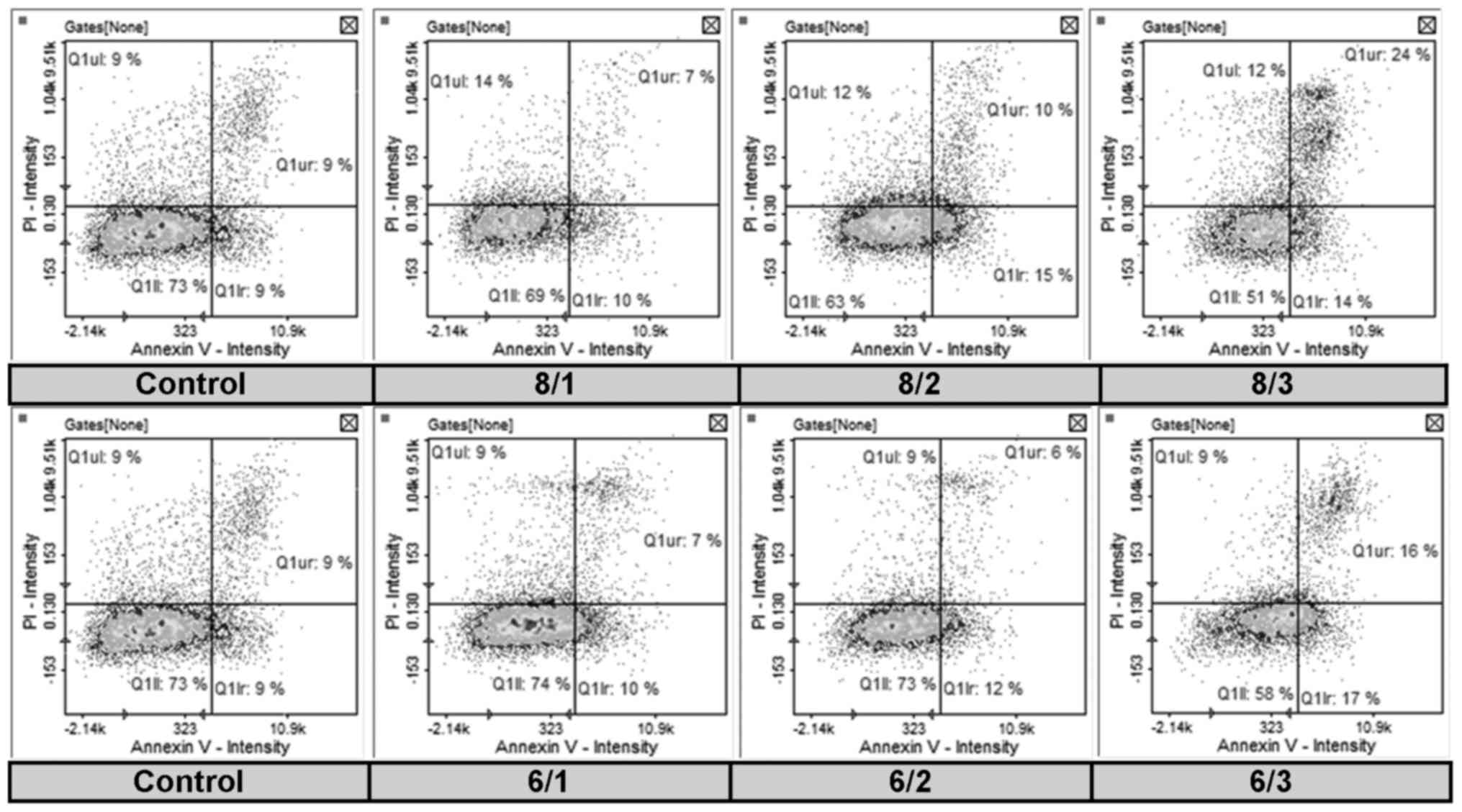
Annexin V assay
Since a significant dose- and time-dependent
inhibitory effect of compounds 6 and 8 on 769-P cells was observed,
it was investigated whether these novel derivatives caused
apoptosis in 769-P cells, which was determined by Annexin V and PI
double staining (Fig. 5). The
apoptosis rate of the cells treated with compounds 6 at
IC25 was almost two times higher than that in the
control group (Table III). When
compound 8 was applied at IC25 or IC50, the
number of early and late apoptotic cells was increased in
comparison to the untreated cells from 18.1 to 24.7 or 37.4%,
compared with the inhibitory concentration (Table III). The aforementioned increase
was also dose-dependent. In the cells treated with compound 8
applied at IC10, IC25 or IC50 when
compared with the control cells, a dose-dependent decrease in the
number of healthy cells (from 72.9 to a maximum of 50.9%) and an
increased in the number of necrotic cells (from 9 to a maximum of
15.7%) were observed (Table
III).
| Table III.Impact of different inhibitory
concentrations of 1,3-thiazolidin-4-one derivatives (compounds 6
and 8) on the apoptosis of 769-P cells after 24 h of
incubation. |
Table III.
Impact of different inhibitory
concentrations of 1,3-thiazolidin-4-one derivatives (compounds 6
and 8) on the apoptosis of 769-P cells after 24 h of
incubation.
| Compound
no./concentration | Healthy, cells,
% | Early apoptotic
cells, % | Late apoptotic
cells, % | Necrotic cells,
% |
|---|
| Control | 72.9 | 9.1 | 9.0 | 9.0 |
| 6/1 | 74.0 | 10.1 | 6.7 | 9.2 |
| 6/2 | 58.0 | 16.7 | 16.4 | 8.9 |
| 6/3 | 73.4 | 11.6 | 6.4 | 8.6 |
| 8/1 | 69.4 | 8.4 | 6.5 | 15.7 |
| 8/2 | 63.4 | 14.7 | 10.0 | 12.0 |
| 8/3 | 50.9 | 13.8 | 23.6 | 11.6 |
Cell cycle assay
The effect of compounds 6 and 8 on the cell cycle
progression of 769-P cells was determined. As demonstrated in
Table IV, compared with the
control, compound 6 increased the population of cells in the G1
phase from 69.4 to a maximum of 92.1% in a dose-dependent manner.
Additionally, a dose-dependent decrease in the S phase (from 3.9 to
a maximum of 1%) and G2 phase (from 21.1 to a maximum of 1.9%) cell
population was observed. 769-P cell accumulation in the G1 fraction
was also observed following cell exposure to compound 8 and the
number of cells increased from 69.4 to a maximum of 89.8% as the
inhibitory concentration increased (Table IV). Notably, cell treatment with
compound 8 increased cell distribution in the sub-G0 stage of the
cell cycle, which is consistent with the results of identification
of apoptotic cells (Tables III
and IV). Additionally, as
demonstrated in Table IV, the
number of 769-P cells at the G2 phase in the treatment groups was
lower than in the control group, and this decrease was
dose-dependent (from 21.1 to a maximum of 2%).
| Table IV.769-P cell cycle analysis following
24-h incubation with different inhibitory concentrations of
1,3-thiazolidin-4-one derivatives (compounds 6 and 8). |
Table IV.
769-P cell cycle analysis following
24-h incubation with different inhibitory concentrations of
1,3-thiazolidin-4-one derivatives (compounds 6 and 8).
|
| Stage |
|---|
|
|
|
|---|
| Compound
no./concentration | Sub-G0, % | G1, % | S, % | G2, % |
|---|
| Control | 4.5 | 69.4 | 3.9 | 21.1 |
| 6/1 | 4.7 | 89.3 | 1.3 | 4.3 |
| 6/2 | 4.4 | 89.7 | 1.2 | 4.4 |
| 6/3 | 4.7 | 92.1 | 1.0 | 1.9 |
| 8/1 | 7.1 | 77.9 | 3.4 | 11.0 |
| 8/2 | 7.9 | 79.9 | 4.3 | 8.2 |
| 8/3 | 6.0 | 89.8 | 1.9 | 2.0 |
Discussion
Taking into consideration the incidence and
mortality rates, there is an urgent requirement to investigate
novel molecules targeting tumors, including renal cell carcinoma or
hepatoblastoma (3–8). The 1,3-thiazolidin-4-ones are of great
importance on the design and synthesis of novel biologically active
agents (15,16). In the present study, when the
chemical structures of synthesized 1,3-thiazolidin-4-ones were
confirmed by spectral data, their anticancer potential was
evaluated in in vitro experiments. The thiazolidin-4-one
moiety is a biologically proven anticancer pharmacophore and
substitution in this scaffold may further enhance its activity
(15–16,30,33).
It is known that substituents may be varied but the greatest
significance in properties is exerted by the group attached to the
carbon atom at the 2-position of the thiazolidinone heterocycle
(16). In particular,
2-aryl-1,3-thiazolidin-4-ones demonstrated considerable cytotoxic
effect against human cancer cell cultures (16,34).
Therefore, the MTT cell viability assay of newly synthesized
2,3-disubstituted 1,3-thiazolidin-4-ones was performed against the
renal cell adenocarcinoma 769-P cell line, the
hepatoblastoma-derived HepG2 cell line and the normal cell line
(GMK) as reference (9). In the
present study, among novel 4-thiazolidinone derivatives, compound 6
was the most toxic against 769-P cells and compound 8 exhibited
similar properties, which may be associated with 4-methylphenyl at
the 2-position of the thiazolidin-4-one moiety. They inhibited
cancer cell growth with IC50 values estimated at 2.67 mM
(compound 6) and 2.93 mM (compound 8). Compound 8 caused a
significant, dose- and time-dependent decrease in cell viability up
to 60% (P<0.01). Whereas derivative 6 possessed more promising
activity by being more selective with respect to the cancer line.
With regards to cancer cell culture (769-P), the incubation time
appeared to also be crucial in the case of compound 6, and after 72
h a significant (P<0.01) ~50% growth inhibition was noted for
the two concentrations used. However, in the normal cell line,
minor decreases in viability were observed with regard to
increasing dose or time of incubation. It was hypothesized that
1,3-thiazolidin-4-one structure, which is 2-substituted with
p-methylphenyl fragment, may be essential for the
antiproliferative properties against the cancer 769-P cell line.
The compounds with electron donating groups at the C-terminal of
the phenyl ring were revealed to exhibit increased activity in
inducing cancer cell death (15).
It was demonstrated that, among 2,3-disubstituted thiazolidinone
derivatives with phenyl ring at position 2, the groups attached to
phenyl moiety are essential for anticancer activity (30,34).
The nitro group is a structural moiety that is frequently observed
in biologically active molecules (15,16).
Joseph et al (30) reported
that the o-nitro group at the phenyl ring of
thiazolidin-4-one scaffold conferred maximum activity in the human
breast adenocarcinoma MCF-7 cell line. However, a previous study on
human breast cancer BT-549 cells and HeLa cells revealed that
2,3-diaryl thiazolidinones substituted with nitro group at ortho
and para positions of the phenyl ring exhibited moderate
antiproliferative properties (34).
It may be concluded that our newly-synthesized substances,
compounds 5 and 7 with meta-nitro phenyl,
demonstrated slight anticancer activity in HepG2. The electronic
influences of the substituents in the phenyl ring appear to serve
an important role in anticancer activity, but this behavior also
depends on the cancer cell lines being used (29). With regards to IC50
values estimated in 769-P and HepG2 cell lines, it may be stated
that the substitution at the 2 position of thiazolidinone
heterocycle with 4-methylphenyl is more favorable than with
3-nitrophenyl and that this significantly enhances the
antiproliferative properties of the derivative tested. In turn,
Kunzler et al (35) revealed
that when 2,3-dubstituted 1,3-thiazolidin-4-ones with a phenyl ring
at position 2 was tested on the normal monkey kidney Vero cell
line, compared with GMK, the 3-nitro group in the phenyl ring was
more cytotoxic than 4-methylphenyl (35). However, the results of the present
study demonstrated the opposite. Therefore, it may be hypothesized
that the scaffold at position 3 of thiazolidin-4-one moiety is of a
great importance for normal cell line viability. The results of the
present study demonstrated that N-substitution of newly
developed compounds with acetamide moiety is less harmful than with
benzamide one.
The more detailed analysis of the antiproliferative
potential of compounds 6 and 8 in 769-P cells included a cell cycle
assay and apoptosis analysis. It demonstrated that the cancer cells
accumulated particularly in the G1 phase, while the number of 769-P
cells in the G2 stage was lower than the number of untreated cells
in the G2 stage. This decrease was dose-dependent, and may be
associated with the inhibition of tubulin synthesis, which prevents
mitosis. Another recent study demonstrated that the combination of
mTOR inhibitor approved for advanced RCC and MEK1 inhibitor, which
is currently used in clinical trials, also causes G1 cell cycle
arrest (36). Therefore, it is
known that PI3K/AKT/mTOR and RAS/MEK/ERK are the most critical
pathways in carcinogenesis and tumor progression, but further
investigation into the possible mechanisms of this are required for
the aforementioned novel molecules (36). In addition, the application of
compound 8 to 769-P cells also caused a decrease in the number of
cells in the S phase in a dose-dependent manner. Taken together,
these results suggested that the two molecule treatments were
associated with cytostatic cell growth arrest. It is known that
during the G1 phase, various enzymes required for S-phase
replication are synthesized. G1 cell cycle arrest and a decrease in
the number of cells in the S phase indicated serious disturbances
in cellular enzymatic activity. This is supported by the results of
MTT assay, which revealed the impairment of the enzymes involved in
tetrazolic salt metabolism in mitochondria. This suggested
metabolic dysfunction and decreased mitochondrial reserve capacity
in 769-P cells following exposure to 1,3-thiazolidin-4-one
derivatives. Mitochondria serve a pivotal role in the initiation
and amplification of the majority of apoptotic pathways. The
mitochondrial membrane permeability increases when mitochondria are
stimulated by apoptotic signals (28,37).
The results of the apoptosis assay indicated that the two tested
derivatives increased the number of apoptotic cells in 769-P cell
culture when they were added at higher inhibitory concentrations.
Compound 8 had a clear ability to induce apoptotic cell death and
it was demonstrated in a more consistent way, compared with
compound 6. It may be associated with benzamide scaffold at
position 3 of the thiazolidin-4-one moiety and the presence of a
phenyl ring may be crucial for the aforementioned changes. In
brief, all the aforementioned assays confirmed the
antiproliferative and pro-apoptotic effects of the two compounds on
renal cell adenocarcinoma cells (769-P). Considering the molecular
basis of RCC, the development of targeted agents should also
include the inhibitory potential associated with intracellular
signal transduction pathways that drive angiogenesis (36). Furthermore, a recent study
demonstrated that newly identified steroidal thiazolidin-4-ones
exhibited significant anti-angiogenic effects (33). Our future studies may include these
novel perspectives.
The research performed on the cancer HepG2 cell line
indicated compound 8 as the most promising with IC50
estimated at 1.43 mM. Unfortunately, prolonged incubation revealed
undesirable increases in cell viability. Compound 7 exhibited an
IC50 value that was three times higher than that for
normal cells, compared with analogous values for cancer cells.
Additionally, compound 7 applied at IC10 preserved
antiproliferative activity during 48- and 72-h incubation periods.
The more detailed studies demonstrated that compound 7 incubated
with HepG2 cells at a higher concentration caused, like derivative
8, undesirable cell growth enhancement.
In conclusion, 2,3-disubstituted
1,3-thiazolidin-4-one derivatives were successfully prepared in the
present study using the cyclization reaction of appropriate
N-substituted carboxylic acid hydrazides with mercaptoacetic
acid, as previously described (25). All prepared compounds were subjected
to in vitro study of cytotoxicity towards human cancer cell
lines. It was observed that generally 2,3-substituted
thiazolidinones were characterized by diverse cytotoxicity, whereas
N-[2-(4-methylphenyl)-4-oxo-1,3-thiazolidin-3-yl]acetamide
(6) and
N-[2-(4-methylphenyl)-4-oxo-1,3-thiazolidin-3-yl]benzamide
(8) appeared to be the most active
against human renal adenocarcinoma 769-P cells and significantly
decreased their viability in a dose- and time-dependent manner. The
results obtained suggested that the most prominent cytotoxicity
could be partially attributed to the electron-donating methyl group
at the 4-position of phenyl ring. These compounds were responsible
for G1 cell cycle arrest in 769-P cells. The two derivatives also
decreased cell distribution in the G2 phase in a dose-dependent
manner. In turn, cell treatment with compound 8 caused an increase
in the number of cells in the sub-G0 stage, which is consistent
with the results of identification of apoptotic cells. For the
aforementioned newly developed molecule, the ability to induce
apoptotic cell death may be associated with the benzamide moiety.
The presented results clearly indicated that the aforementioned
novel 2,3-disubstituted 1,3-thiazolidin-4-ones induced cell cycle
arrest and apoptosis in human renal adenocarcinoma cells (769-P) in
a dose-dependent manner.
Acknowledgements
Not applicable.
Funding
The present study was supported by Funds for
Statutory Activity of Medical University of Lublin, Poland (grant
no. DS38/2014-2015).
Availability of data and materials
All data generated and/or analyzed during this study
are included in this published article.
Authors' contributions
MGG, ŁP and DNC designed and directed the
experiment. MGG wrote the manuscript (excluding part describing the
synthesis and chemical structure identification of tested
compounds). ŁP designed, synthesized, purified and identified by
spectral methods 1,3-thiazolidin-4-one derivatives and wrote the
part of manuscript regarding these processes. MGG, DNC and IPC
performed the in vitro cytotoxicity study. DNC and MIz
analyzed/collected the data. DNC performed the statistical
analysis. DNC and MH interpreted the results of statistical
analysis. MH was involved in drafting the manuscript and revised
critically the final version of the manuscript. MIw performed the
cell cycle assay and Annexin V assay and microscopic analysis and
collected the data. AK, JD and MW were involved in the conception
of the study. AK supervised cell cycle and Annexin V assays, JD
supervised the in vitro cytotoxicity study, MW supervised
the synthesis and spectral identification of novel derivatives. AK,
JD and MW revised critically the final version of the manuscript.
All authors read and approved the final manuscript.
Ethics approval and consent to
participate
Not applicable.
Patient consent for publication
Not applicable.
Competing interests
The authors declare that they have no competing
interests.
References
|
1
|
Gregorić T, Sedić M, Grbčić P, Tomljenović
Paravić A, Kraljević Pavelić S, Cetina M, Vianello R and Raić-Malić
S: Novel pyrimidine-2,4-dione-1,2,3-triazole and
furo[2,3-d]pyrimidine-2-one-1,2,3-triazole hybrids as potential
anti-cancer agents: Synthesis, computational and X-ray analysis and
biological evaluation. Eur J Med Chem. 125:pp. 1247–1267. 2017,
View Article : Google Scholar : PubMed/NCBI
|
|
2
|
Escudier B, Porta C, Schmidinger M, Algaba
F, Patard JJ, Khoo V, Eisen T and Horwich A: ESMO Guidelines
Working Group: Renal cell carcinoma: ESMO Clinical Practice
Guidelines for diagnosis, treatment and follow-up. Ann Oncol.
25((Suppl 3)): iii49–iii56. 2014. View Article : Google Scholar : PubMed/NCBI
|
|
3
|
Meyers RL, Tiao G, de Ville de Goyet J,
Superina R and Aronson DC: Hepatoblastoma state of the art:
Pre-treatment extent of disease, surgical resection guidelines and
the role of liver transplantation. Curr Opin Pediatr. 26:29–36.
2014. View Article : Google Scholar : PubMed/NCBI
|
|
4
|
Sumazin P, Chen Y, Treviño LR, Sarabia SF,
Hampton OA, Patel K, Mistretta TA, Zorman B, Thompson P, Heczey A,
et al: Genomic analysis of hepatoblastoma identifies distinct
molecular and prognostic subgroups. Hepatology. 65:104–121. 2017.
View Article : Google Scholar : PubMed/NCBI
|
|
5
|
Ridge CA, Pua BB and Madoff DC:
Epidemiology and staging of renal cell carcinoma. Semin Intervent
Radiol. 31:3–8. 2014. View Article : Google Scholar : PubMed/NCBI
|
|
6
|
Stewart BW and Wild CP: World Cancer
Report. 2014, International Agency for Research on Cancer Lyon.
2014
|
|
7
|
Znaor A, Lortet-Tieulent J, Laversanne M,
Jemal A and Bray F: International variations and trends in renal
cell carcinoma incidence and mortality. Eur Urol. 67:519–530. 2015.
View Article : Google Scholar : PubMed/NCBI
|
|
8
|
Kabaria R, Klaassen Z and Terris MK: Renal
cell carcinoma: Links and risks. Int J Nephrol Renovasc Dis.
9:45–52. 2016.PubMed/NCBI
|
|
9
|
López-Terrada D, Cheung SW, Finegold MJ
and Knowles BB: Hep G2 is a hepatoblastoma-derived cell line. Hum
Pathol. 40:1512–1515. 2009. View Article : Google Scholar
|
|
10
|
Chen Z, Zhang J, Zhang Z, Feng Z, Wei J,
Lu J, Fang Y, Liang Y, Cen J, Pan Y, et al: The putative tumor
suppressor microRNA-30a-5p modulates clear cell renal cell
carcinoma aggressiveness through repression of ZEB2. Cell Death
Dis. 8:e28592017. View Article : Google Scholar : PubMed/NCBI
|
|
11
|
Chen T, Ji B and Chen Y: Tetrandrine
triggers apoptosis and cell cycle arrest in human renal cell
carcinoma cells. J Nat Med. 68:46–52. 2014. View Article : Google Scholar : PubMed/NCBI
|
|
12
|
Roseweir AK, Qayyum T, Lim Z, Hammond R,
MacDonald AI, Fraser S, Oades GM, Aitchison M, Jones RJ and Edwards
J: Nuclear expression of Lyn, a Src family kinase member, is
associated with poor prognosis in renal cancer patients. BMC
Cancer. 16:2292016. View Article : Google Scholar : PubMed/NCBI
|
|
13
|
Fang Y, Wei J, Cao J, Zhao H, Liao B, Qiu
S, Wang D, Luo J and Chen W: Protein expression of ZEB2 in renal
cell carcinoma and its prognostic significance in patient survival.
PLoS One. 8:e625582013. View Article : Google Scholar : PubMed/NCBI
|
|
14
|
Yuan ZX, Mo J, Zhao G, Shu G, Fu HL and
Zhao W: Targeting strategies for renal cell carcinoma: From renal
cancer cells to renal cancer stem cells. Front Pharmacol.
7:4232016. View Article : Google Scholar : PubMed/NCBI
|
|
15
|
Jain AK, Vaidya A, Ravichandran V, Kashaw
SK and Agrawal RK: Recent developments and biological activities of
thiazolidinone derivatives: A review. Bioorg Med Chem.
20:3378–3395. 2012. View Article : Google Scholar : PubMed/NCBI
|
|
16
|
Tripathi AC, Gupta SJ, Fatima GN, Sonar
PK, Verma A and Saraf SK: 4-Thiazolidinones: The advances
continue…. Eur J Med Chem. 72:52–77. 2014. View Article : Google Scholar : PubMed/NCBI
|
|
17
|
Murugesan V, Tiwari VS, Saxena R, Tripathi
R, Paranjape R, Kulkarni S, Makwana N, Suryawanshi R and Katti SB:
Lead optimization at C-2 and N-3 positions of thiazolidin-4-ones as
HIV-1 non-nucleoside reverse transcriptase inhibitors. Bioorg Med
Chem. 19:6919–6926. 2011. View Article : Google Scholar : PubMed/NCBI
|
|
18
|
Omar K, Geronikaki A, Zoumpoulakis P,
Camoutsis C, Soković M, Cirić A and Glamoclija J: Novel
4-thiazolidinone derivatives as potential antifungal and
antibacterial drugs. Bioorg Med Chem. 18:426–432. 2010. View Article : Google Scholar : PubMed/NCBI
|
|
19
|
Popiołek Ł, Stefańska J, Kiełczykowska M,
Musik I, Biernasiuk A, Malm A and Wujec M: Synthesis, dissociation
constants, and antimicrobial activity of novel
2,3-disubstituted-1,3-thiazolidin-4-one derivatives. J Chem. 53:pp.
393–402. 2016
|
|
20
|
Rawal RK, Tripathi R, Katti SB,
Pannecouque C and De Clercq E: Synthesis and evaluation of
2-(2,6-dihalophenyl)-3-pyrimidinyl-1,3-thiazolidin-4-one analogues
as anti-HIV-1 agents. Bioorg Med Chem. 15:pp. 3134–3142. 2007,
View Article : Google Scholar : PubMed/NCBI
|
|
21
|
Bielenica A, Szulczyk D, Olejarz W,
Madeddu S, Giliberti G, Materek IB, Koziol AE and Struga M:
1H-Tetrazol-5-amine and 1,3-thiazolidin-4-one derivatives
containing 3-(trifluoromethyl)phenyl scaffold: Synthesis, cytotoxic
and anti-HIV studies. Biomed Pharmacother. 94:pp. 804–812. 2017,
View Article : Google Scholar : PubMed/NCBI
|
|
22
|
Archana, Srivastava VK and Kumar A:
Synthesis of newer thiadiazolyl and thiazolidinonyl quinazolin-4
3H-ones as potential anticonvulsant agents. Eur J Med Chem. 37:pp.
873–882. 2002, View Article : Google Scholar : PubMed/NCBI
|
|
23
|
Diurno MV, Mazzoni O, Correale G, Gomez
Monterrey I, Calignano A, La Rana G and Bolognese A: Synthesis and
structure-activity relationships of 2-(substituted
phenyl)-3-[3-(N,N-dimethylamino)propyl]-1,3-thiazolidin-4-ones
acting as H1-histamine antagonists. Farmaco. 54:pp. 579–583. 1999,
View Article : Google Scholar : PubMed/NCBI
|
|
24
|
Firke SD, Firake BM, Chaudhari RY and
Patil R: Synthetic and pharmacological evaluation of some pyridine
containing thiazolidinones. Asian J Res Chem. 2:157–161. 2009.
|
|
25
|
Popiołek Ł, Biernasiuk A and Malm A:
Synthesis and antimicrobial activity of new 1,3-thiazolidin-4-one
derivatives obtained from carboxylic acid hydrazides. Phosphorus
Sulfur. 190:pp. 251–260. 2015, View Article : Google Scholar
|
|
26
|
Wang S, Zhao Y, Zhang G, Lv Y, Zhang N and
Gong P: Design, synthesis and biological evaluation of novel
4-thiazolidinones containing indolin-2-one moiety as potential
antitumor agent. Eur J Med Chem. 46:pp. 3509–3518. 2011, View Article : Google Scholar : PubMed/NCBI
|
|
27
|
Zhou H, Wu S, Zhai S, Liu A, Sun Y, Li R,
Zhang Y, Ekins S, Swaan PW, Fang B, et al: Design, synthesis,
cytoselective toxicity, structure-activity relationships, and
pharmacophore of thiazolidinone derivatives targeting
drug-resistant lung cancer cells. J Med Chem. 51:pp. 1242–1251.
2008, View Article : Google Scholar : PubMed/NCBI
|
|
28
|
Sharath Kumar KS, Hanumappa A, Vetrivel M,
Hegde M, Girish YR, Byregowda TR, Rao S, Raghavan SC and Rangappa
KS: Antiproliferative and tumor inhibitory studies of 2,3
disubstituted 4-thiazolidinone derivatives. Bioorg Med Chem Lett.
25:3616–3620. 2015. View Article : Google Scholar : PubMed/NCBI
|
|
29
|
Wang S, Zhao Y, Zhu W, Liu Y, Guo K and
Gong P: Synthesis and anticancer activity of Indolin-2-one
derivatives bearing the 4-thiazolidinone moiety. Arch Pharm
(Weinheim). 345:pp. 73–80. 2012, View Article : Google Scholar : PubMed/NCBI
|
|
30
|
Joseph A, Shah CS, Kumar SS, Alex AT,
Maliyakkal N, Moorkoth S and Mathew JE: Synthesis, in vitro
anticancer and antioxidant activity of thiadiazole substituted
thiazolidin-4-ones. Acta Pharm. 63:pp. 397–408. 2013, View Article : Google Scholar : PubMed/NCBI
|
|
31
|
Ottanà R, Carotti S, Maccari R, Landini I,
Chiricosta G, Caciagli B, Vigorita MG and Mini E: In vitro
antiproliferative activity against human colon cancer cell lines of
representative 4-thiazolidinones. Part I. Bioorg Med Chem Lett.
15:3930–3933. 2005. View Article : Google Scholar : PubMed/NCBI
|
|
32
|
George RF: Stereoselective synthesis and
QSAR study of cytotoxic
2-(4-oxo-thiazolidin-2-ylidene)-2-cyano-N-arylacetamides. Eur J Med
Chem. 47:pp. 377–386. 2012, View Article : Google Scholar : PubMed/NCBI
|
|
33
|
Živković MB, Matić IZ, Rodić MV, Novaković
IT, Krivokuća AM, Sladić DM and Krstić NM: Anticancer potential of
new steroidal thiazolidin-4-one derivatives. Mechanisms of
cytotoxic action and effects on angiogenesis in vitro. J Steroid
Biochem Mol Biol. 174:72–85. 2017. View Article : Google Scholar : PubMed/NCBI
|
|
34
|
Suthar SK, Jaiswal V, Lohan S, Bansal S,
Chaudhary A, Tiwari A, Alex AT and Joesph A: Novel quinolone
substituted thiazolidin-4-ones as anti-inflammatory, anticancer
agents: Design, synthesis and biological screening. Eur J Med Chem.
63:pp. 589–602. 2013, View Article : Google Scholar : PubMed/NCBI
|
|
35
|
Kunzler A, Neuenfeldt PD, das Neves AM,
Pereira CM, Marques GH, Nascente PS, Fernandes MH, Hübner SO and
Cunico W: Synthesis, antifungal and cytotoxic activities of
2-aryl-3-((piperidin-1-yl)ethyl)thiazolidinones. Eur J Med Chem.
64:pp. 74–80. 2013, View Article : Google Scholar : PubMed/NCBI
|
|
36
|
Zou Y, Wang J, Leng X, Huang J, Xue W,
Zhang J and Huang Y: The selective MEK1 inhibitor Selumetinib
enhances the antitumor activity of everolimus against renal cell
carcinoma in vitro and in vivo. Oncotarget. 8:20825–20833. 2017.
View Article : Google Scholar : PubMed/NCBI
|
|
37
|
Wang Y, Xia C, Lun Z, Lv Y, Chen W and Li
T: Crosstalk between p38 MAPK and caspase-9 regulates
mitochondria-mediated apoptosis induced by
tetra-α-(4-carboxyphenoxy) phthalocyanine zinc photodynamic therapy
in LoVo cells. Oncol Rep. 39:61–70. 2018.PubMed/NCBI
|