Introduction
Hepatocellular carcinoma (HCC) is one of the most
prevalent and lethal malignancies in the world (1), with a 5-year survival rate of only
30–40% (2). HCC has a poor prognosis
due to chemotherapy resistance and a high recurrence rate (3). Given the unsatisfactory state of current
liver cancer treatment effectiveness, there is a great need for the
development of new strategies for targeting HCC (4).
Drug repurposing is an important approach in cancer
drug discovery and development (5,6). Notably,
we previously found that pimozide, an FDA-approved compound used to
treat chronic psychosis, exhibits anticancer effects on various
types of carcinomas, including HCC (7), prostate cancer (8) and osteosarcoma (9). Pimozide has been reported to inhibit
cell proliferation, colony formation, and sphere formation of these
three cancer types (7–9). Pimozide also reduced migration of
prostate and liver cancer cells (7).
Additionally, pimozide increased the sensitivity of breast cancer
cells to γ-irradiation treatment (10) and enhanced the anticancer effect of
L-methyl-tryptophan (an indoleamine 2, 3-dioxygenase inhibitor) on
melanoma cells (11). Furthermore,
previously, we found that pimozide inhibited cancer cell growth
through suppression of signal transducer and activator of
transcription 3 (STAT3) activation (7–9) or
Wnt/β-catenin signaling (12,13). In addition, pimozide has been shown to
inhibit maintenance and tumorigenicity of HCC stem-like cells
(14) and to induce reactive oxygen
species (ROS) generation, which can impede cell proliferation
(9).
In light of the aforementioned findings, the
potential clinical anticancer efficacy of pimozide has piqued our
interest. The toxicology profile of pimozide in human patients is
well established owing to its long-term use in psychiatry. In
addition, molecular targets involved in the suppression of cancer
cell growth by pimozide have been identified. However, various
factors affecting the drug's pharmacodynamic effects remain to be
clarified, including whether the suppression of HCC cell
proliferation by pimozide is reversible. Irreversible mechanisms
include cell death, cell senescence and terminal differentiation
(15–17), whereas reversible mechanisms include
principally cell cycle arrest and cellular quiescence (18). The potential anticancer effect
reversibility of pimozide would have direct effects on the dosage
and administration cycle of the drug. Elucidation of these dynamics
can accelerate the clinical translation of pimozide for oncological
applications.
The aim of the present study was to investigate the
type of proliferative inhibition produced by pimozide on liver
cancer cells. We used cell proliferation assays with pan-caspase
and RIP (receptor-interacting protein) kinase inhibitors to
identify whether the effects of pimozide are associated with
apoptosis or necrosis. In addition, we analyzed the effects of
pimozide on the cell cycle. Importantly, we assessed the
reversibility of the anti-proliferative effects of pimozide on
liver cancer cells, the potential associations of the efficacy of
pimozide with quiescence and ROS production, and the interaction of
pimozide with sorafenib which is a tyrosine kinase inhibitor used
to treat cancers affecting various organs.
Materials and methods
Cell lines and culture
The human liver cancer cell lines Sk-Hep1, Huh7 and
HepG2 (provided by the Guangdong Provincial Key Laboratory of Liver
Disease Research, Guangzhou, China) were cultured in Dulbecco's
modified Eagle's medium (DMEM; Gibco; Thermo Fisher Scientific,
Inc.) containing 10% fetal bovine serum (FBS; Sigma-Aldrich; Merck
KGaA), penicillin (100 U/ml) and streptomycin (100 µg/ml). The cell
lines were maintained in 5% CO2 at 37°C. Huh7 and
Sk-Hep1 cell lines were authenticated by short tandem repeat
profiling analysis conducted by Procell Life Science &
Technology Co., Ltd. (Wuhan, China).
Cell viability assay
Cell proliferation was assessed using the Cell
Counting Kit-8 (CCK-8) assays (MedChem Express, Monmouth Junction,
NJ, USA). Liver cancer cells were plated in 96-well plates (10,000
cells/well) and exposed to different concentrations of pimozide (5
and 10 µM) (MedChem Express) for 48 h. Kit-provided solution was
added to each well and incubated at 37°C for 4 h. Absorbance at 450
nm was detected by a multi-well plate reader (Bio-Rad Laboratories,
Inc., Hercules, CA, USA). In addition, Sk-Hep1 and Huh7 cells were
treated with 10 µM pimozide alone or with combination plus
pan-caspase inhibitor Z-VAD-FMK (10 µM) or RIP kinase inhibitor
necrostain 1 (10 µM) or 2 mM NAC (MedChem Express) for 48 h before
being subjected to cell viability assay. In addition, Sk-Hep1 and
Huh7 cells were treated with the 5 µM pimozide alone, sorafenib
(MedChem Express) alone or combination for 48 h and then were
subjected to cell viability assay.
Colony formation assay
The colony formation assay was performed as
described previously (19). Liver
cancer cells (500 cells/well) were plated in 10% FBS medium and
treated with different concentrations of pimozide (5 and 10 µM).
After incubation for 2 weeks, the cells were fixed with methanol
and stained with crystal violet. Colony morphology was imaged under
a stereomicroscope (magnification ×100). The colonies, defined as a
cluster of at least 50 cells, were counted.
Cell cycle distribution assay
Cell cycle distribution was determined by propidium
iodide (PI, Sigma-Aldrich; Merck KGaA) staining. Equal cell
aliquots were seeded in 6-well plates and treated with pimozide for
48 h. The cells were harvested, washed with phosphate-buffered
saline containing 0.1% bovine serum albumin, and vortexed with cold
absolute ethanol. After addition of PI buffer (40 µg/ml, containing
100 µg/ml RNase), cells were analyzed by flow cytometry (CytoFLEX,
Beckman Coulter, Inc.).
For quiescent cell detection, cells were washed,
fixed with 0.5 ml of 4% paraformaldehyde in phosphate-buffered
saline, and then incubated with an equal volume of 0.2% Triton-X.
The fixed and permeabilized cells were washed, resuspended in
staining buffer, and stained with Ki-67-FITC (BD Pharmigen). The
stained cells were washed and resuspended in propidium iodide (PI)
staining buffer. Cells in G0 phase were analyzed by flow cytometry
(CytoFLEX, Beckman Coulter, Inc.) based on Ki-67 negativity and PI
incorporation.
Annexin V/PI staining apoptosis
assay
Apoptotic cells were detected with an Annexin
V-FITC/PI Apoptosis Detection kit (Absin Bioscience, Shanghai,
China) according to the manufacturer's instructions. Briefly, cells
were treated with 10 µM pimozide for 48 h, submitted to Annexin
V-FITC/PI staining, and then analyzed by flow cytometry (CytoFLEX,
Beckman Coulter, Inc.).
ROS assay and ROS inhibition
ROS products were detected with
2,7-dichlorofluorescein diacetate (DCFH-DA), a fluorescent dye
(Beyotime, Jiangsu, China). Briefly, after 10 µM pimozide treatment
for 48 h, cells were washed and incubated with DCFH-DA (10 µM) for
30 min at 37°C in the dark. Then, cells were washed twice with DMEM
and analyzed by flow cytometry at an excitation/emission wavelength
of 488/525 nm. ROS production was inhibited with
N-acetyl-l-cysteine (NAC).
Western blotting
Equal amounts of protein (20 µg/well) collected from
cells with RIPA lysis buffer were subjected to sodium dodecyl
sulfate-polyacrylamide gel electrophoresis (SDS-PAGE) and
transferred to polyvinylidene difluoride membranes (Merck
Millipore, Billerica MA, USA). Blots were detected using primary
antibodies against cell division protein kinase 6 (CDK6) (cat. no.
ab124821; dilution 1:1,000), p-Rb (cat. no. ab173289; dilution
1:1,000) (Abcam, Cambridge, MA, USA), p27 (#3686; dilution
1:1,000), cyclin D1 (#2978; dilution 1:1,000), STAT3 (#12640;
dilution 1:1,000), phosphorylated (p)-STAT3 (at tyrosine 705)
(#9145; dilution 1:1,000), ERK1/2 (extracellular signal-regulated
kinase 1 and 2) (#4695; dilution 1:1,000), phosphorylated
(p)-ERK1/2 (#4376; dilution 1:1,000) (Cell Signaling Technology,
Beverly, MA, USA) and β-actin (cat. no. sc-47778; dilution 1:1,000)
(Santa Cruz Biotechnology, Santa Cruz, CA, USA). Antibody binding
was detected with an enhanced chemiluminescence kit (Immobilon
Western Chemilum HRP Substrate, WBKLS0500, Merck Millipore).
Statistical analysis
The data were analyzed in GraphPad Prism 6.0
(GraphPad Software, Inc., San Diego, CA, USA) and are presented as
means with standard deviations (SDs). Student's t tests or analysis
of one-way variance (ANOVA) followed by Dunnett's post hoc test for
multiple comparisons were applied to assess the statistical
differences between groups. P-values <0.05 were considered
significant.
Results
The anti-proliferative effect of
pimozide on liver cancer cells is independent of drug-induced cell
death
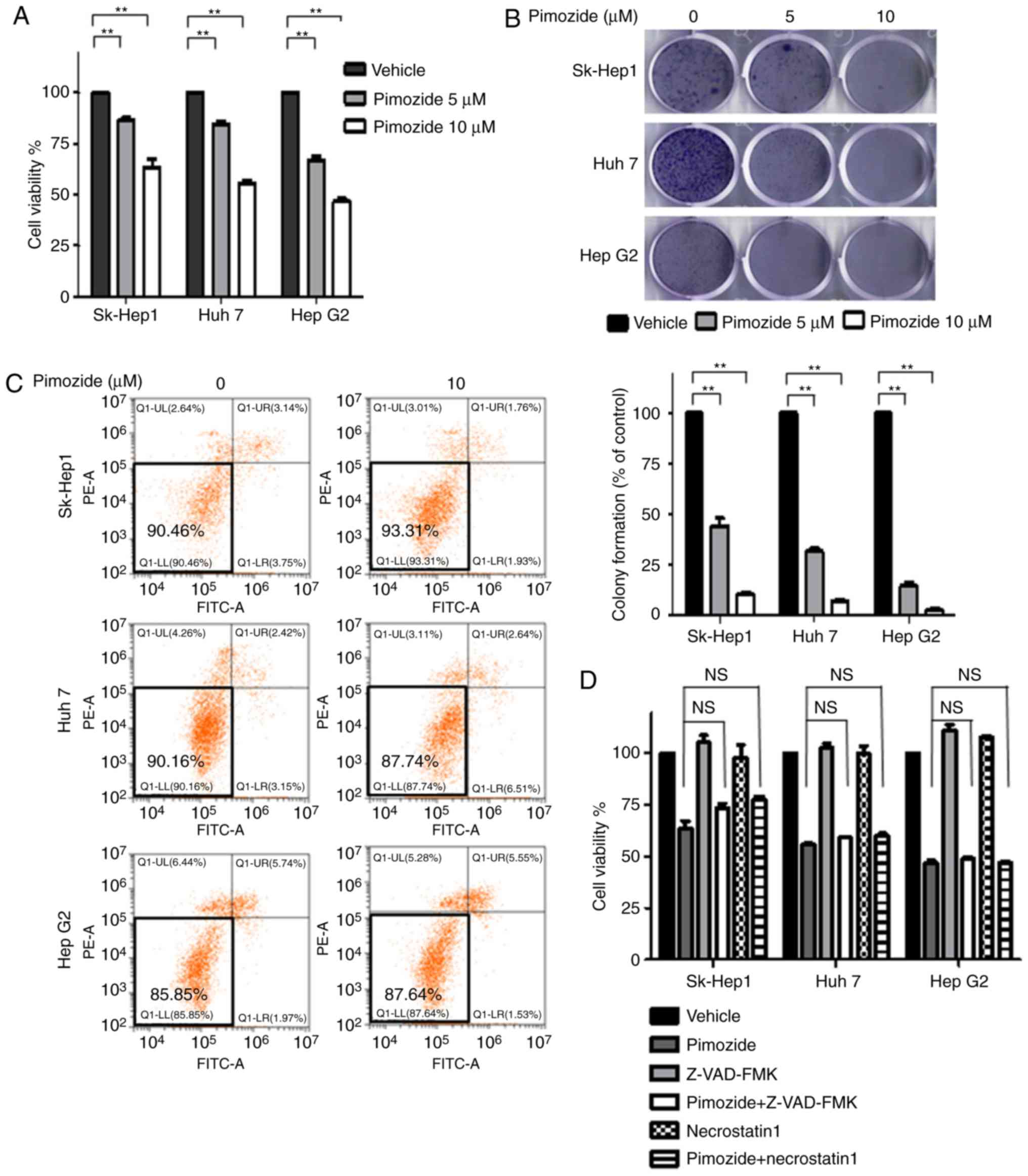
Proliferative assays showed that pimozide inhibited
the growth of Sk-Hep1, Huh7 and HepG2 liver cancer cells in a
dose-dependent manner (Fig. 1A).
Colony formation assays showed similar inhibition of proliferation
across the three cell lines (Fig.
1B). Annexin-V-FITC/PI double staining cell apoptosis assays
showed that 10 µM pimozide did not induce obvious apoptosis in the
liver cancer cells (Fig. 1C). Cell
proliferation assays further showed that pan-caspase and RIP kinase
inhibitors did not attenuate pimozide inhibition of cell growth in
liver cancer cells (Fig. 1D),
indicating that the suppression of proliferation was not associated
with apoptosis or necrosis.
Pimozide increases cell cycle arrest
in HCC cells
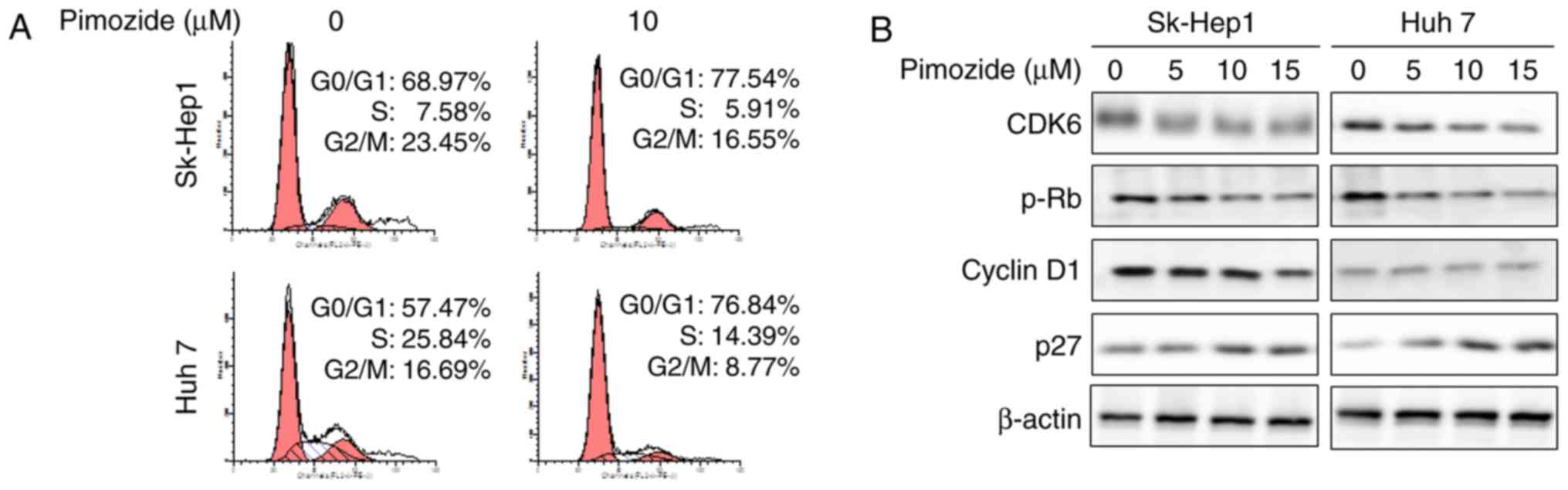
As shown in Fig. 2A,
PI staining revealed that 14 h of 10 µΜ pimozide treatment
significantly increased the number of cells in the G0/G1 phase in
the Sk-Hep1 (68.97 to 77.54%; P<0.01) and Huh7 cells (57.47 to
76.84%). Western blots showed reduced expression of the cell cycle
markers CDK6, p-Rb and cyclin D1 together with markedly increased
p27 levels (Fig. 2B). These results
indicated that the anti-proliferative effect of pimozide is
associated with cell cycle arrest.
Reversibility of proliferative
suppression by pimozide due to quiescence
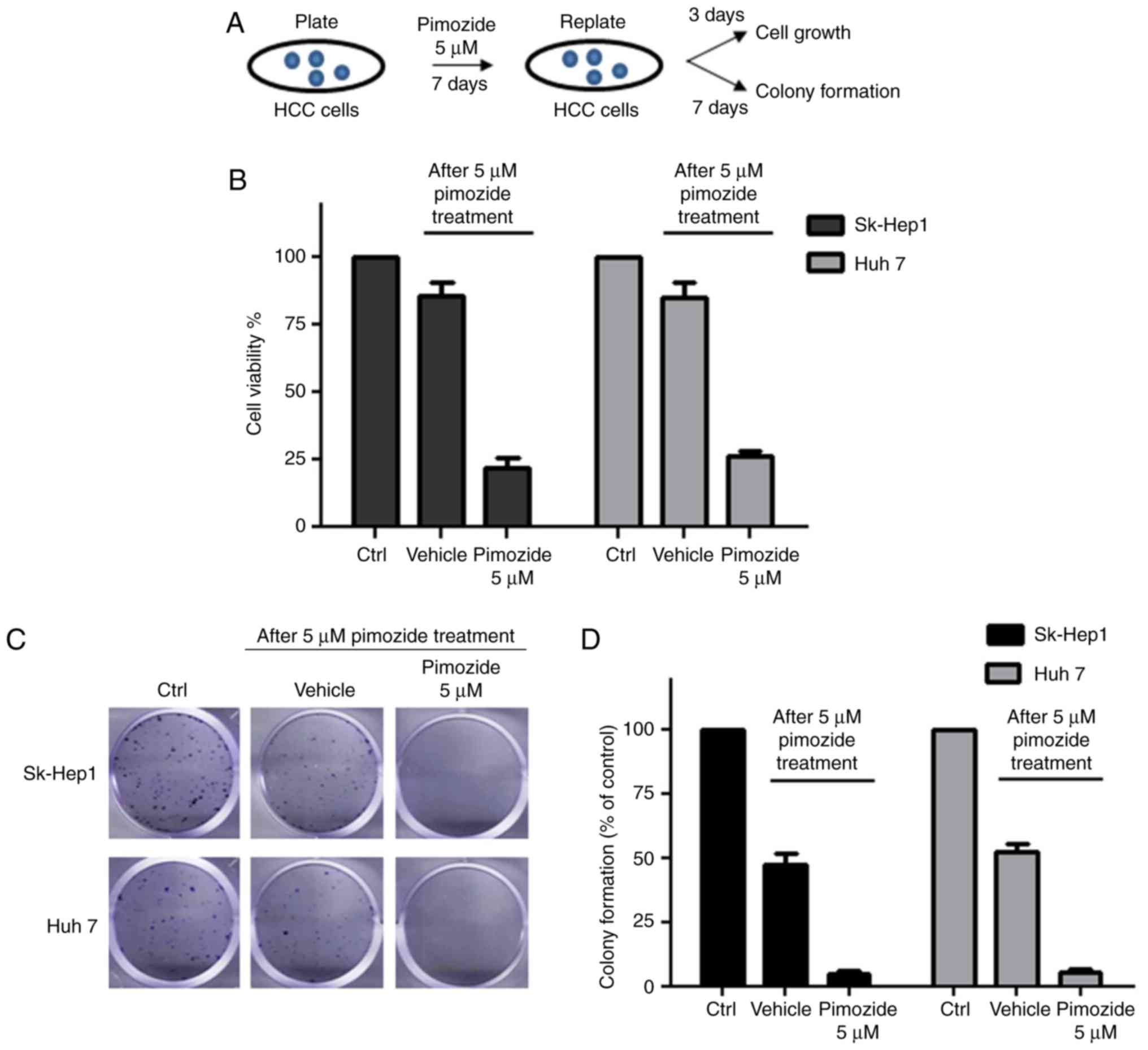
After treatment with 5 µM pimozide, HCC cells were
re-plated in preparation for cell proliferative and colony
formation assays, as illustrated in Fig.
3A. Increased cell counts and colony formation were observed in
the pimozide-treated Sk-Hep1 and Huh7 cells following pimozide
withdrawal, demonstrating restoration of proliferation and colony
forming abilities (Fig. 3B and C).
Following treatment with 5 µΜ pimozide for 1 week, Sk-Hep1 cells
showed a decrease of 96.6±1.1% in colony number (Fig. 3D), while Sk-Hep1 cells without
pimozide treatment displayed significant ability of colony
formation (48.6±2.3%). Similar results were achieved in the Huh7
cells.
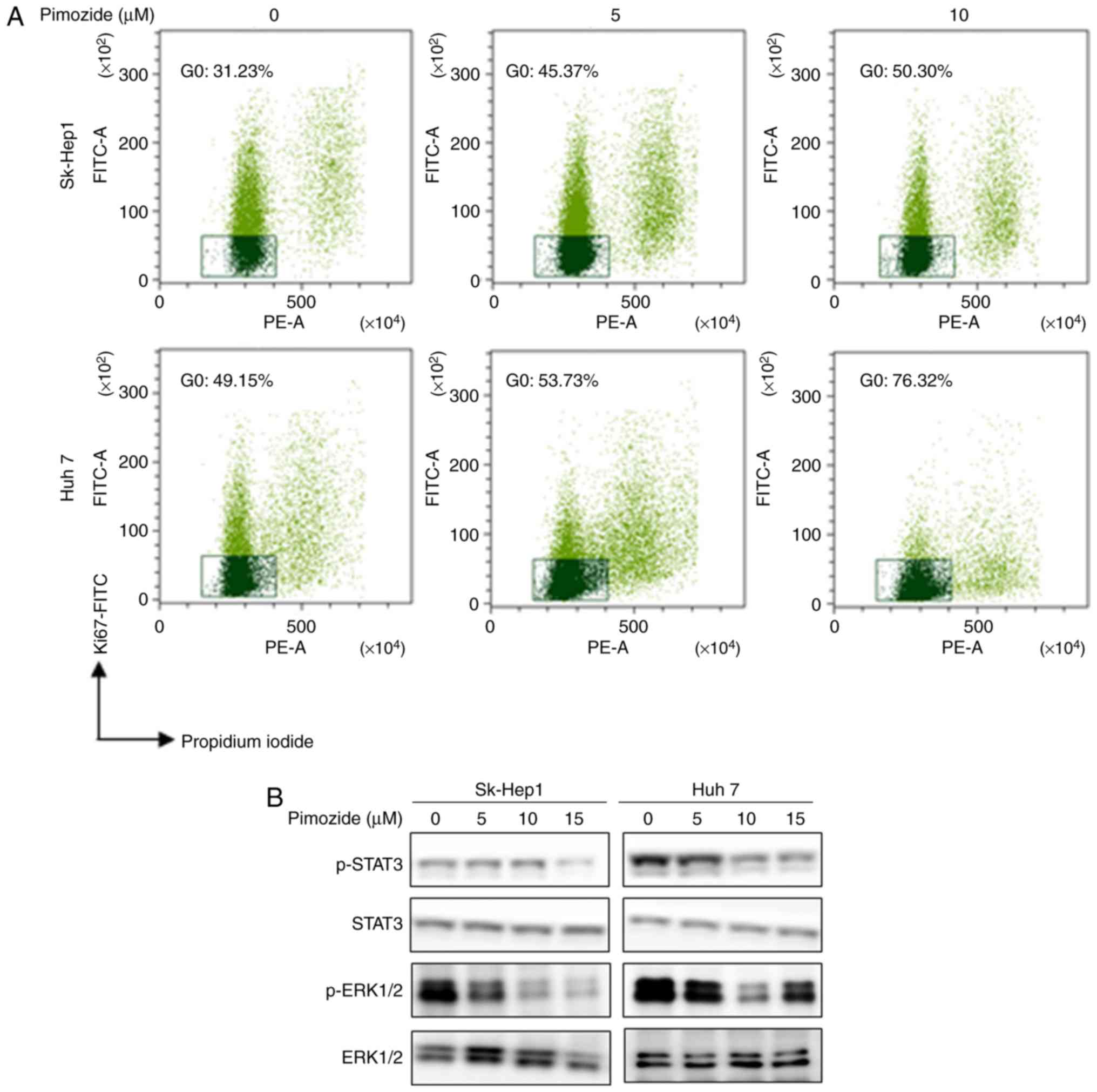
Ki-67 labelling, PI staining, and flow cytometry
showed that pimozide increased the portion of cells in the
quiescent G0 phase in a dose-dependent manner (Fig. 4A). After 10 µM pimozide treatment,
Sk-Hep1 (31.23 to 50.30%) and Huh7 (49.15 to 76.32%) cells had
significantly increased percentages of cells in the G0 phase.
Western blot analysis showed that pimozide reduced p-STAT3 and
p-ERK1/2 levels (Fig. 4B), indicating
that pimozide induced HCC cell quiescence.
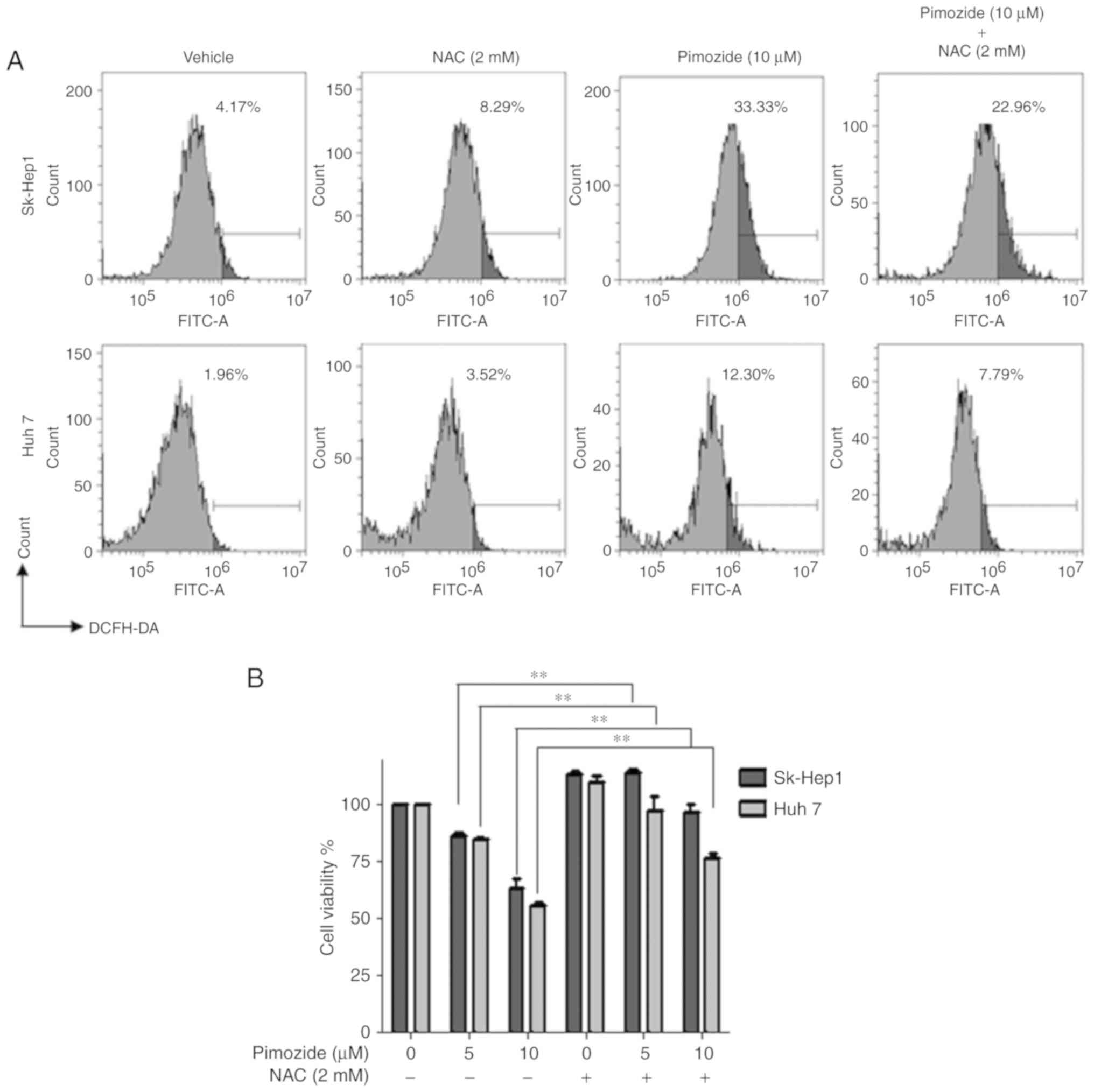
Pimozide induces ROS generation in HCC
cells
Flow cytometry after pimozide exposure (10 µΜ for 48
h) demonstrated increased ROS levels in the Sk-Hep1 and Huh7 cells
and this effect was attenuated by the ROS scavenger NAC (Fig. 5A). NAC also reversed the
pimozide-mediated inhibition of Sk-Hep1 and Huh7 cell proliferation
(Fig. 5B).
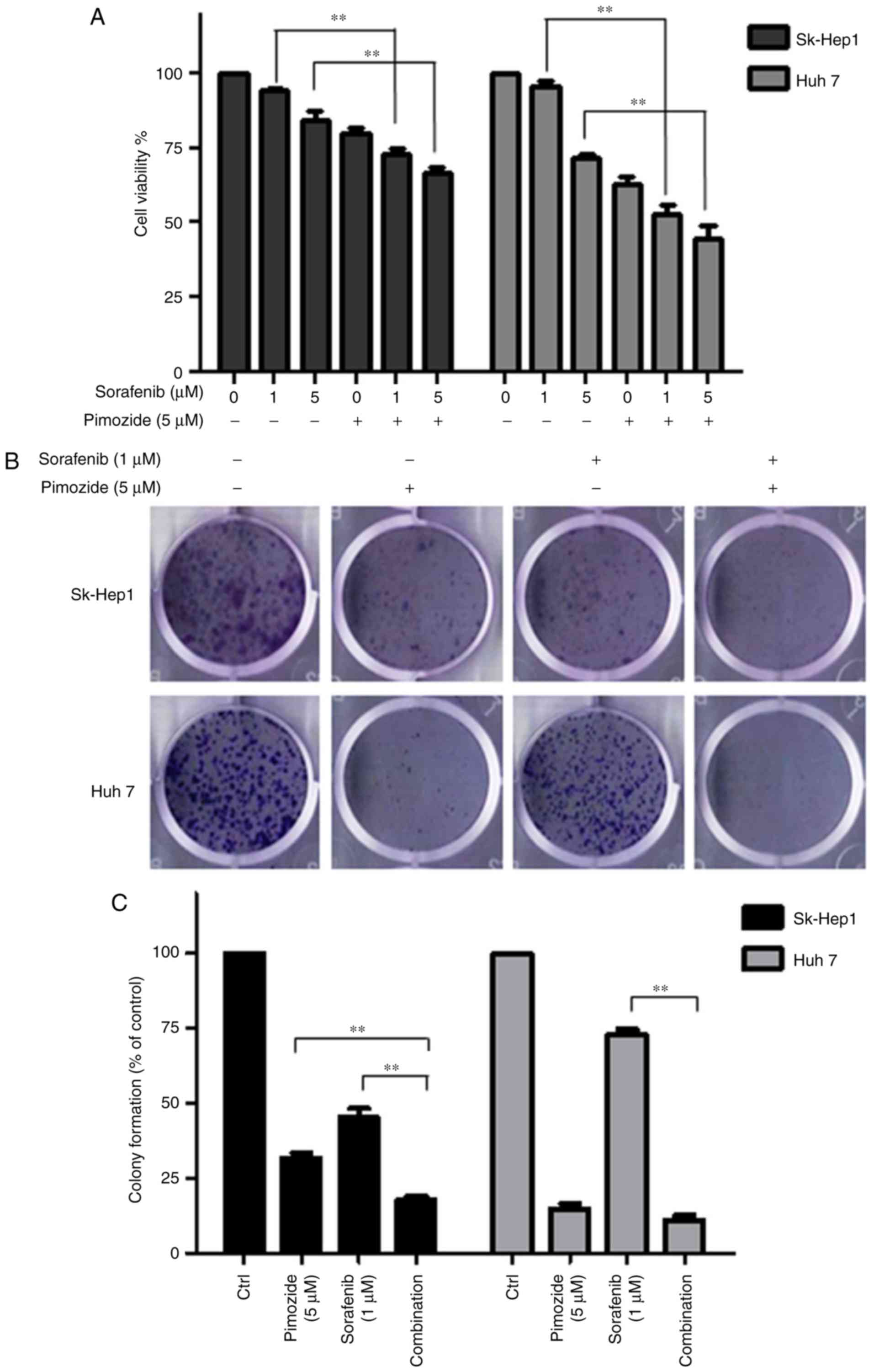
Pimozide enhances the proliferative
inhibition induced by tyrosine kinase inhibitor in HCC cells
Cell viability (Fig.
6A) and colony formation assays (Fig.
6B and C) showed that 5 µΜ pimozide significantly enhanced the
anticancer effect of 1 µΜ sorafenib in the Sk-Hep1 and Huh7 cell
lines.
Discussion
Drug effect reversibility is an important factor in
determining drug administration mode for optimizing efficacy and
persistence while avoiding adverse secondary effects (20). The same drug may produce different
effects on different types of tumor cells, mainly due to the
particular cell type's genetic background (14). Although pimozide was found to promote
the apoptosis of breast cancer (10,21,22),
prostate cancer and melanoma cells (23), our data showed that pimozide did not
promote apoptosis of liver cancer cells. Moreover, the inhibition
of liver cancer cell proliferation by primozide was not affected by
pan-caspase or RIP kinase inhibition. These results indicate that
the mechanism of the anti-proliferative effects of pimozide on
liver cancer cells may not involve drug-induced cell death.
The present observation of the reversible
suppression of liver cancer cell proliferation was associated with
pimozide-induced cellular quiescence (the resting,
non-proliferative G0 phase of the cell cycle) (24,25).
Quiescence has been posited to be a drug effect-escape mechanism
(26). However, while in this dormant
state, cancer cells are not causing harm (27). Because proliferative suppression due
to cellular quiescence is transient and reversible, HCC cells will
return to a proliferating state if they re-enter the cell cycle
(28).
With respect to the mechanism, it was found that HCC
cellular quiescence was associated with reduced phosphorylation of
STAT3 and ERK1/2. The time-sensitive antitumor activity of pimozide
observed suggests that long-term maintenance of pimozide would be
needed to maintain its anticancer efficacy. Because pimozide is a
well-known antagonist of serotonin 5-hydroxytryptamine receptor 7
(5HT7), we examined 5HT7 expression in liver cancer cell lines and
found that they indeed had elevated 5HT7 expression (data not
shown). Further research is needed to probe the association of 5HT7
expression with cellular quiescence in HCC cells.
Pimozide is an FDA approved drug shown to have no
adverse effects on hepatic or hematopoietic cells. Pimozide is well
tolerated in mice without significant effects on body weight
(7,29). To date, a lethal dose for pimozide in
humans has not been established. The median lethal doses for
pimozide in mice and rats are 228 and 5,120 mg/kg, respectively
[DrugBank no. DB01100 (30)]. The
dose of pimozide used in this study is substantially lower than
commonly used clinical doses. Therefore, there is good reason to be
optimistic that pimozide may be a safe drug for treating
cancer.
Drug repurposing requires a new drug application
clinical entry point (31). For
example, metformin, a biguanide-class antidiabetic medication, was
repurposed as an adjuvant therapy for breast (32) and bladder cancer (33). Similarly, we have been exploring the
potential of pimozide to be an adjuvant anticancer therapy. Given
the mediocre clinical efficacy of sorafenib alone in patients with
HCC, it is hoped that its efficacy may be enhanced by combining it
with another intervention (34).
Previously, carfilzomib has been shown to work synergistically with
sorafenib in inhibiting the proliferation, survival and metastasis
of HCC cells in vitro (35).
Here, we showed that pimozide can enhance the proliferative
inhibition of sorafenib in HCC cells, suggesting that it has the
potential to improve HCC outcomes as an adjuvant therapy.
In conclusion, suppression of liver cancer cell
proliferation by pimozide was found to involve promotion of
cellular quiescence and to be reversible. Furthermore, pimozide was
shown to act synergistically with sorafenib, suggesting that it
should be further explored as an adjuvant therapy for use with
anticancer drugs such as sorafenib in liver cancer treatment with
its dosage respecting the reversibility of its effects.
Acknowledgements
We thank the other members of the laboratory of Ji
Kunmei for their critical comments.
Funding
This work was supported by the National Natural
Science Foundation of China (81602595 to JJC), the Natural Science
Foundation of Shenzhen City (JCY20170818142053544 to JJC), the
Medical Scientific Research Foundation of Guangdong Province
(A2019522 to JJC) and 2016 Discipline Construction of Shenzhen
City.
Availability of data and materials
The datasets used during the present study are
available from the corresponding author upon reasonable
request.
Authors' contributions
JJC and KJ conceived and designed the study. JJC and
LNZ collected and analyzed the data. JJC, LNZ NC and ZZ performed
the experiments. JJC and KJ were responsible for the
reagents/materials/analysis tools. JJC and KJ wrote the manuscript.
All authors read and approved the manuscript and agree to be
accountable for all aspects of the research in ensuring that the
accuracy or integrity of any part of the work are appropriately
investigated and resolved.
Ethics approval and consent to
participate
Not applicable.
Patient consent for publication
Not applicable.
Competing interests
The authors declare that they have no competing
interests.
References
|
1
|
Crissien AM and Frenette C: Current
management of hepatocellular carcinoma. Gastroenterol Hepatol (N
Y). 10:153–161. 2014.PubMed/NCBI
|
|
2
|
Altekruse SF, McGlynn KA and Reichman ME:
Hepatocellular carcinoma incidence, mortality, and survival trends
in the united states from 1975 to 2005. J Clin Oncol. 27:1485–1491.
2009. View Article : Google Scholar : PubMed/NCBI
|
|
3
|
Hoshida Y, Fuchs BC and Tanabe KK:
Prevention of hepatocellular carcinoma: Potential targets,
experimental models, and clinical challenges. Curr Can Drug
Targets. 12:1129–1159. 2012. View Article : Google Scholar
|
|
4
|
Cervello M, McCubrey JA, Cusimano A,
Lampiasi N, Azzolina A and Montalto G: Targeted therapy for
hepatocellular carcinoma: Novel agents on the horizon. Oncotarget.
3:236–260. 2012. View Article : Google Scholar : PubMed/NCBI
|
|
5
|
Boguski MS, Mandl KD and Sukhatme VP: Drug
discovery. Repurposing with a difference. Science. 324:1394–1395.
2009. View Article : Google Scholar : PubMed/NCBI
|
|
6
|
Knapp S: New opportunities for kinase drug
repurposing and target discovery. Br J Cancer. 118:936–937. 2018.
View Article : Google Scholar : PubMed/NCBI
|
|
7
|
Chen JJ, Cai N, Chen GZ, Jia CC, Qiu DB,
Du C, Liu W, Yang Y, Long ZJ and Zhang Q: The neuroleptic drug
pimozide inhibits stem-like cell maintenance and tumorigenicity in
hepatocellular carcinoma. Oncotarget. 8:17593–17609.
2017.PubMed/NCBI
|
|
8
|
Zhou W, Chen MK, Yu HT, Zhong ZH, Cai N,
Chen GZ, Zhang P and Chen JJ: The antipsychotic drug pimozide
inhibits cell growth in prostate cancer through suppression of
STAT3 activation. Int J Oncol. 48:322–328. 2016. View Article : Google Scholar : PubMed/NCBI
|
|
9
|
Cai N, Zhou W, Ye LL, Chen J, Liang QN,
Chang G and Chen JJ: The STAT3 inhibitor pimozide impedes cell
proliferation and induces ROS generation in human osteosarcoma by
suppressing catalase expression. Am J Transl Res. 9:3853–3866.
2017.PubMed/NCBI
|
|
10
|
Strobl JS, Melkoumian Z, Peterson VA and
Hylton H: The cell death response to gamma-radiation in MCF-7 cells
is enhanced by a neuroleptic drug, pimozide. Breast Cancer Res
Treat. 51:83–95. 1998. View Article : Google Scholar : PubMed/NCBI
|
|
11
|
Jia H, Ren W, Feng Y, Wei T, Guo M, Guo J,
Zhao J, Song X, Wang M, Zhao T, et al: The enhanced antitumour
response of pimozide combined with the IDO inhibitor L-MT in
melanoma. Int J Oncol. 53:949–960. 2018.PubMed/NCBI
|
|
12
|
Fako V, Yu Z, Henrich CJ, Ransom T, Budhu
AS and Wang XW: Inhibition of wnt/β-catenin signaling in
hepatocellular carcinoma by an antipsychotic drug pimozide. Int J
Biol Sci. 12:768–775. 2016. View Article : Google Scholar : PubMed/NCBI
|
|
13
|
Ren Y, Tao J, Jiang Z, Guo D and Tang J:
Pimozide suppresses colorectal cancer via inhibition of
Wnt/β-catenin signaling pathway. Life Sci. 209:267–273. 2018.
View Article : Google Scholar : PubMed/NCBI
|
|
14
|
Gonçalves JM, Silva CAB, Rivero ERC and
Cordeiro MMR: Inhibition of cancer stem cells promoted by Pimozide.
Clin Exp Pharmacol Physiol. 46:116–125. 2019. View Article : Google Scholar : PubMed/NCBI
|
|
15
|
Yan D, Parker RE, Wang X, Frye SV, Earp HS
III, DeRyckere D and Graham DK: MERTK promotes resistance to
irreversible EGFR tyrosine kinase inhibitors in non-small cell lung
cancers expressing wild-type EGFR family members. Clin Can Res.
24:6523–6535. 2018. View Article : Google Scholar
|
|
16
|
Abdallah SM and Hirsh V: Irreversible
tyrosine kinase inhibition of epidermal growth factor receptor with
afatinib in EGFR activating mutation-positive advanced
non-small-cell lung cancer. Curr Oncol. 25 (Suppl 1):S9–S17. 2018.
View Article : Google Scholar : PubMed/NCBI
|
|
17
|
Sagot I and Laporte D: Quiescence, an
individual journey. Curr Genet. 65:695–699. 2019. View Article : Google Scholar : PubMed/NCBI
|
|
18
|
Heldt FS, Barr AR, Cooper S, Bakal C and
Novák B: A comprehensive model for the proliferation-quiescence
decision in response to endogenous DNA damage in human cells. Proc
Natl Acad Sci USA. 115:2532–2537. 2018. View Article : Google Scholar : PubMed/NCBI
|
|
19
|
Cai N, Xie SJ, Qiu DB, Jia CC, Du C, Liu
W, Chen JJ and Zhang Q: Potential effects of α-mangostin in the
prevention and treatment of hepatocellular carcinoma. J Funct
Foods. 26:309–318. 2016. View Article : Google Scholar
|
|
20
|
Agarwal SM, Pal D, Gupta M and Saini R:
Insight into discovery of next generation reversible TMLR
inhibitors targeting EGFR activating and drug resistant T790M
mutants. Curr Can Drug Targets. 17:617–636. 2017.
|
|
21
|
Rybczynska M, Spitaler M, Knebel NG, Boeck
G, Grunicke H and Hofmann J: Effects of miltefosine on various
biochemical parameters in a panel of tumor cell lines with
different sensitivities. Biochem Pharmacol. 62:765–772. 2001.
View Article : Google Scholar : PubMed/NCBI
|
|
22
|
Dakir EH, Pickard A, Srivastava K,
McCrudden CM, Gross SR, Lloyd S, Zhang SD, Margariti A, Morgan R,
Rudl PS and El-Tanani M: The anti-psychotic drug pimozide is a
novel chemotherapeutic for breast cancer. Oncotarget.
9:34889–34910. 2018. View Article : Google Scholar : PubMed/NCBI
|
|
23
|
Strobl JS, Kirkwood KL, Lantz TK, Lewine
MA, Peterson VA and Worley JF III: Inhibition of human breast
cancer cell proliferation in tissue culture by the neuroleptic
agents pimozide and thioridazine. Cancer Res. 50:5399–5405.
1990.PubMed/NCBI
|
|
24
|
Coller HA, Sang L and Roberts JM: A new
description of cellular quiescence. PLoS Biol. 4:e832006.
View Article : Google Scholar : PubMed/NCBI
|
|
25
|
Aguirre-Ghiso JA: Models, mechanisms and
clinical evidence for cancer dormancy. Nat Rev Cancer. 7:834–846.
2007. View
Article : Google Scholar : PubMed/NCBI
|
|
26
|
Alarcón T and Jensen HJ: Quiescence: A
mechanism for escaping the effects of drug on cell populations. J R
Soc Interface. 8:99–106. 2011. View Article : Google Scholar : PubMed/NCBI
|
|
27
|
Fiore APZP, Ribeiro PF and Bruni-Cardoso
A: Sleeping beauty and the microenvironment enchantment:
Microenvironmental regulation of the proliferation-quiescence
decision in normal tissues and in cancer development. Front Cell
Dev Biol. 6:592018. View Article : Google Scholar : PubMed/NCBI
|
|
28
|
Recasens A and Munoz L: Targeting cancer
cell dormancy. Trends Pharmacol Sci. 40:30229–30233. 2019.
View Article : Google Scholar
|
|
29
|
Nelson EA, Walker SR, Xiang M, Weisberg E,
Bar-Natan M, Barrett R, Liu S, Kharbanda S, Christie AL, Nicolais
M, et al: The STAT5 inhibitor pimozide displays efficacy in models
of acute myelogenous leukemia driven by FLT3 mutations. Genes
Cancer. 3:503–511. 2012. View Article : Google Scholar : PubMed/NCBI
|
|
30
|
Wishart DS, Feunang YD, Guo AC, Lo EJ,
Marcu A, Grant JR, Sajed T, Johnson D, Li C, Sayeeda Z, et al:
DrugBank 5.0: A major update to the DrugBank database for 2018.
Nucleic Acids Res. 46(D1): D1074–D1082. 2018. View Article : Google Scholar : PubMed/NCBI
|
|
31
|
Bhattarai D, Singh S, Jang Y, Hyeon Han S,
Lee K and Choi Y: An insight into drug repositioning for the
development of novel anti-cancer drugs. Curr Top Med Chem.
16:2156–2168. 2016. View Article : Google Scholar : PubMed/NCBI
|
|
32
|
Samuel SM, Varghese E, Varghese S and
Büsselberg D: Challenges and perspectives in the treatment of
diabetes associated breast cancer. Cancer Treat Rev. 70:98–111.
2018. View Article : Google Scholar : PubMed/NCBI
|
|
33
|
El-Arabey AA: Correction to: New insight
for metformin against bladder cancer. Genes Environ. 40:162018.
View Article : Google Scholar : PubMed/NCBI
|
|
34
|
Forner A, Reig M and Bruix J:
Hepatocellular carcinoma. Lancet. 391:1301–1314. 2018. View Article : Google Scholar : PubMed/NCBI
|
|
35
|
Jiang C, Xu R, Li XX, Zhou YF, Xu XY, Yang
Y, Wang HY and Zheng XFS: Sorafenib and carfilzomib synergistically
inhibit the proliferation, survival, and metastasis of
hepatocellular carcinoma. Mol Cancer Ther. 17:2610–2621. 2018.
View Article : Google Scholar : PubMed/NCBI
|