Introduction
Patients with similar clinicopathological risk
classification can display striking clinical heterogeneity in the
progression of their prostate cancer (1). This limits clinical decision making for
personalized management strategies. The molecular mechanisms
underlying prostate cancer progression involve alterations in a
variety of signaling pathways. Elucidating underlying mechanisms
influencing prostate cancer progression can contribute to our
understanding of how they drive different clinical phenotypes in
prostate cancer (2), while providing
potential candidates as novel biomarkers.
MicroRNAs (miRNAs) are a class of short non-protein
coding RNA molecules which act within the RNA interference pathway.
Each miRNA is a single-stranded RNA molecule approximately 22
nucleotides in length, and guides messenger RNA (mRNA) to
post-transcriptional regulation machinery via binding to a sequence
in the mRNA 3′ untranslated region (3′UTR). Through inhibition of
targets which are tumor suppressors or oncogenes, miRNAs have been
revealed to influence each of the classic hallmarks of cancer, and
individual miRNAs often alter multiple hallmarks (3).
Evidence for the role of microRNA-198 (miR-198) as a
tumor suppressor stems from its downregulation in numerous cancer
types including multiple myeloma, gastric cancer, colorectal
cancer, glioblastoma, osteosarcoma, and tumors of the breast,
pancreas, liver and lungs (4–12). Low miR-198 levels have been revealed
to predict poor survival in pancreatic and gastric cancer cohorts,
and overexpression of miR-198 in vitro regulates several
hallmarks of cancer (5,8). However, there is limited research on the
role of miR-198 in prostate cancer.
The present findings demonstrated that miR-198 is
reduced in high Gleason score tumors, and functions as a tumor
suppressor in prostate cancer by inhibiting proliferation,
anchorage-independent growth, and tumor growth, using in
vitro and in vivo models. Mindbomb E3 ubiquitin protein
ligase 1 (MIB1) was identified as a novel effector gene
downstream of miR-198, which appears to function in a Notch
pathway-independent manner. This research enhances our
understanding of prostate cancer biology by identifying miR-198 as
a novel tumor suppressor miRNA.
Materials and methods
Patient analyses
The Canadian Prostate Cancer Genome Network
(CPC-GENE) dataset is comprised of 162 samples, of which 137 are
intermediate risk Gleason 7 (3+4=99 and 4+3=38), 12 are low risk
(Gleason 6), and 13 are high risk (Gleason >7). Differential
expression between Gleason ≤7 and >7 was evaluated using
two-sided Wilcoxon test, and all statistical analyses were
completed using R statistical environment (v3.4.0). CPC-GENE is
part of the international genome consortium (ICGC) and data access
control is regulated via the ICGC-DACO (www.icgc.org). Informed consent, consistent with local
Research Ethics Board (REB) and International Cancer Genome
Consortium (ICGC) guidelines, was obtained at the time of clinical
follow-up. Previously collected tumor tissues were used, following
University Health Network REB-approved study protocols (UHN
06-0822-CE, UHN 11-0024-CE, CHUQ 2012-913:H12-03-192).
Cell culture
Human prostate carcinoma cell lines LNCaP
(castrate-sensitive) and DU145 (castrate-resistant) were purchased
from the American Type Culture Collection (ATCC). LNCaP cells were
maintained in RPMI-1640 medium (Gibco; Thermo Fisher Scientific,
Inc.) supplemented with 10% fetal bovine serum (FBS; Invitrogen;
Thermo Fisher Scientific, Inc.) and 1% penicillin (100
U/ml)-streptomycin (100 µg/ml) (PS; Invitrogen; Thermo Fisher
Scientific, Inc.). DU145 cells were cultured in Dulbecco's modified
Eagle's medium containing 4.5 g/l D-glucose and GlutaMAX (DMEM;
Gibco; Thermo Fisher Scientific, Inc.) supplemented with 10% FBS
and 1% PS. All cell lines were maintained in tissue-culture flasks
within a humidified 37°C incubator with 5% CO2, and
passaged when they reached 80% confluency. LS174T Notch-Luciferase
cells were previously generated by our laboratory (13), and maintained in DMEM with 10% FBS and
1% PS. Cell lines were regularly confirmed to be free from
mycoplasma contamination using MycoAlert Detection Kit (Lonza
Group, Ltd.).
Transfection
Negative control and human miR-198 mimics (denoted
as ‘Control’ and ‘miR-198’ respectively in figures; sequences in
Table SI) were purchased from
Shanghai GenePharma Co., Ltd. Three-sequence pooled siRNA for human
MIB1 (sc-75781; sequence in Table
SI) was obtained from Santa Cruz Biotechnology, Inc., along
with a control siRNA (sc-37007; sequence in Table SI). MIB1 cDNA ORF in
pcDNA3.1+ vector was purchased from GenScript (ID:
OHu21837) and used in parallel with pcDNA3.1+ backbone
plasmid as a control. All RNA products were stored at −80°C and
exposed to a maximum of two freeze-thaw cycles. Sequences are
listed in Table SI. Passaged cells
were counted using a Countess automated cell counter (Invitrogen;
Thermo Fisher Scientific, Inc.), and a total of 2.5×10⁵
cells (DU145) or 4×10⁵ cells (LNCaP) were seeded in
6-well plates, and transfected the following day using
Lipofectamine 2000 as per the manufacturer's instructions (Thermo
Fisher Scientific, Inc.). Cells were transfected for 24 h prior to
being trypsinized, collected, and used in subsequent assays.
Quantitative real-time PCR
(RT-qPCR)
For miRNA abundance, cells were lysed and total
miRNA was extracted using mirVana miRNA isolation kit according to
the manufacturer's instructions (Life Technologies; Thermo Fisher
Scientific, Inc.). For gene expression, RNA was extracted using the
RNeasy Mini Kit (Qiagen). RNA quality measured by absorbance at 260
nm was evaluated using a NanoDrop 2000 (Thermo Fisher Scientific).
cDNA was synthesized using the miScript II RT kit for miRNA
(Qiagen, Inc.) or Superscript VILO cDNA kit (Thermo Fisher
Scientific, Inc.) for mRNA. Real-time quantitative PCR to assess
gene expression was performed on a StepOnePlus Real-Time PCR system
(Applied Biosystems; Thermo Fisher Scientific, Inc.) using the
miScript SYBR Green PCR kit (Qiagen, Inc.) or SYBR Select Master
Mix (Applied Biosystems; Thermo Fisher Scientific, Inc.) for mature
miRNA or mRNA respectively. Primers for miRNA profiling were
obtained from miScript Primer Assays for RNU6-2 and miR-198
(Qiagen, Inc.), whereas primers for mRNA abundance were designed
using Primer-BLAST software (NCBI) and synthesized by Invitrogen;
Thermo Fisher Scientific, Inc. (Table
SII). Expression levels were calculated with the ΔΔCq method
(14) using StepOne Software (Applied
Biosystems; Thermo Fisher Scientific, Inc.), and relative abundance
was normalized to RNU6-2 for miRNA or GADPH for mRNA.
Proliferation assay
Transfected cells were seeded in technical
triplicates at 5×10⁴ cells/well in a 6-well plate. After
incubation for 4 days (LNCaP) or 5 days (DU145), cells were
trypsinized and the total number of viable cells in each well was
determined by mixing the cell suspension 1:1 (v/v) with 0.4% trypan
blue solution, incubating for 1 min at room temperature, then
measuring with a Countess automated cell counter. Results were
graphed as the change in cell number from original number plated,
relative to the amount of growth in the control condition.
Cell cycle analysis
LNCaP cells were seeded at 4×10⁵
cells/well in a 6-well plate, and 24 h later transfected with miRNA
mimics. The cells were incubated undisturbed for 48 h after
transfection, at which time they were trypsinized, washed with
phosphate-buffered saline (PBS), and fixed on ice for 30 min in
cold 80% ethanol and Hank's balanced salt solution. Cells were
pelleted by 5-min centrifugation at 200 × g, resuspended in RNAse A
in HBSS (2 mg/ml), and stained with propidium iodide (0.1 mg/ml;
Sigma-Aldrich; Merck KGaA) with 0.6% NP-40 (Thermo Fisher
Scientific, Inc.). After 30 min of incubation in the dark, 50,000
events per sample were captured using a FACSCalibur flow cytometer
(BD Biosciences), and the cell cycle profile was generated using
FlowJo software (Version 10.0.4; FlowJo LLC).
Soft agar assay
Bottom soft agar layers were created in 24-well
plates by adding a mixture containing 2X DMEM-F12 (Gibco; Thermo
Fisher Scientific, Inc.), 10% FBS, 1% PS, and 0.8% w/v liquefied
Agar A (Bio Basic, Inc.). Transfected cells were added to the upper
agar layer mixture, which contained the same components as the base
with the exception of the use of 0.5% liquefied Agar A, and plated
in technical triplicates on top of the base layers at 700
cells/well. Approximately 30 days later, the plates were imaged
using a Leica MZ FLIII stereomicroscope (Leica Microsystems GmbH),
and the number of colonies were manually counted.
Generation of stable cell lines
Pre-miR-198 was cloned into a pBabe-puro vector, and
transfected into Phoenix-AMPHO cells (a retroviral packaging cell
line, ATCC), parallel to empty-pBabe vector as a matching control.
After 24 h, the culture media containing pBabe-miR198 virus or
pBabe-empty virus was collected. Viral media and polybrene
(Sigma-Aldrich; Merck KGaA) were added onto adhered LNCaP cells in
a 6-well plate, which were then spin transduced at 1,000 × g for 90
min in a 37°C centrifuge. Viral media was removed the following
day, and puromycin (0.75 µg/ml) was added 3 days post
spin-transduction for selection. After selection, the cells were
maintained in puromycin (0.75 µg/ml) and miR-198 expression was
determined using RT-qPCR.
In vivo tumor xenograft formation
All experiments involving animals were performed in
accordance with the University of Toronto and Sunnybrook Research
Institute Animal Care Committee guidelines using a peer-reviewed
protocol (AUP #17-509). As per this AUP, humane endpoints included
tumors >1.5 cm diameter, weight loss >20%, tumor ulceration,
or abnormal posture. After reaching any of these endpoint criteria,
mice were anaesthetized with isofluorane (4%) and sacrificed by
cervical dislocation. Mice were housed and fed according to
standard animal care policies, and welfare-related assessments were
performed on a regular basis. Six to seven-week-old male athymic
nude mice purchased from Charles River Laboratories were injected
subcutaneously into the right flank with 1.5×10⁶
LNCaP-ctrl or LNCaP-198 stable cell lines mixed in a 1:1 (v:v)
ratio with Matrigel (Corning Incorporated). Prior to injection, the
mice were weighed and distributed evenly into two experimental
groups (LNCaP-ctrl or LNCaP-198) each containing four mice. Mice
were monitored every 2–3 days and the tumor volume was measured
using calipers and calculated using the modified ellipsoid formula:
Volume=1/2 (length × width2). Tumor formation was
graphed as the percent of mice tumor-free, defined as >60
mm3, and analyzed using log-rank test to compare
survival curves.
Western blotting
Cells were rinsed with PBS and lysed in ice-cold
radioimmunoassay precipitation lysis buffer containing Complete
Mini protease inhibitor cocktail and PhosSTOP phosphatase inhibitor
cocktail (Roche Diagnostics). Collected lysate was sonicated, and
then centrifuged to pellet cell debris. Protein quantification was
performed using the Bradford protein assay (Bio-Rad Laboratories,
Inc.) with a Nanodrop spectrophotometer. Protein samples were
combined with β-mercaptoethanol, and denatured by boiling. Protein
lysate was run on a 4–20% polyacrimide gradient gel (Bio-Rad
Laboratories, Inc.), wet-transferred to polyvinylidene difluoride
membranes (Thermo Fisher Scientific, Inc.), and then blocked
against non-specific binding for 1 h with gentle agitation in TBST
containing either 5% non-fat dry milk or 5% bovine serum albumin
(BSA). A primary antibody in appropriate solution (5% milk or 5%
BSA) was added and incubated overnight at 4°C. The membranes were
then washed three times with TBST, and then incubated with a
horseradish peroxidase-conjugated anti-rabbit IgG secondary
antibody (1:5,000; cat. no. 7074; Cell Signaling Technologies,
Inc.) for 1 h at room temperature, and afterwards washed again
three times in TBST. Protein detection was performed using
incubation with an enhanced chemiluminescence solution (1.25 mM
Luminol, 2 mM 4IPBA, 100 mM Tris-HCL pH 8.8), and imaged using a
ChemiDoc Imaging System (Bio-Rad Laboratories, Inc.). The following
antibodies from Cell Signaling Technologies, Inc. were used:
β-actin (1:2,000; cat. no. 4967; anti-rabbit), MIB1 (1:1,000; cat.
no. 4400; anti-rabbit).
Transcriptomic analysis
Total RNA from transfected cell lines was extracted
using RNeasy Mini Kit (Qiagen, Inc.) as per the manufacturer's
instructions. RNA quality was assessed using a spectrophotometer,
and all samples were ethanol-precipitated if any sample was found
to have an absorbance at 260 nm ≤1.8. Gene expression profiling was
performed by the Centre for Applied Genomics (The Hospital for Sick
Children, Toronto, Canada) using a GeneChip Human Gene 2.0 ST array
(Affymetrix; Thermo Fisher Scientific, Inc.). Transcriptomic data
was normalized using default parameters in Expression Console
software (V.1.2; Affymetrix).
In silico analysis
miRNA target prediction was performed using miRWalk
2.0 atlas of predicted and published miRNA-gene interactions
(15).
Notch-luciferase reporter assay
A Notch reporter cell line was previously generated
and validated (13) by transducing a
human colon adenocarcinoma line (LS174T) with lentivirus containing
a minimal promoter with multiple recombination signal-binding
protein 1 for J-Kappa (RBP-jκ) Notch response elements
(5′-CGTGGGAA-3′) driving expression of the firefly luciferase gene
(Qiagen, Inc.). The same lentivirus was used to transduce LNCaP
cells to create a prostate cancer Notch reporter cell line. To
determine Notch-luciferase activity, cells were seeded in a 24-well
plate, transfected or treated with inhibitor for 24 h, then lysed
and luciferase activity was assessed using a Luciferase Assay
System (Promega Corporation) and a luminometer.
3′UTR luciferase assay
LNCaP cells were seeded in triplicate in a 96-well
plate and co-transfected the following day with a luciferase
reporter plasmid containing either wild-type MIB1 3′UTR or MIB1
3′UTR containing mutations to disrupt binding in the miR-198
predicted binding site (SwitchGear Genomics; Active Motif), and
control or miR-198 mimic. After 24 h, luciferase activity was
assayed according to the manufacturer's protocol using LightSwitch
Luciferase Reagent (SwitchGear Genomics; Active Motif).
Statistical analyses
Statistical analyses were performed using GraphPad
Prism 5.0 (GraphPad Software), except for analyses conducted on
patient samples which were performed in R. All in vitro
experiments consisted of at least three experimental replicates,
unless otherwise stated. Differences in means were compared using
two-sided Student's t-tests, and graphed as the mean ± SEM.
Statistical significance was defined as P<0.05, P<0.01 and
P<0.001.
Results
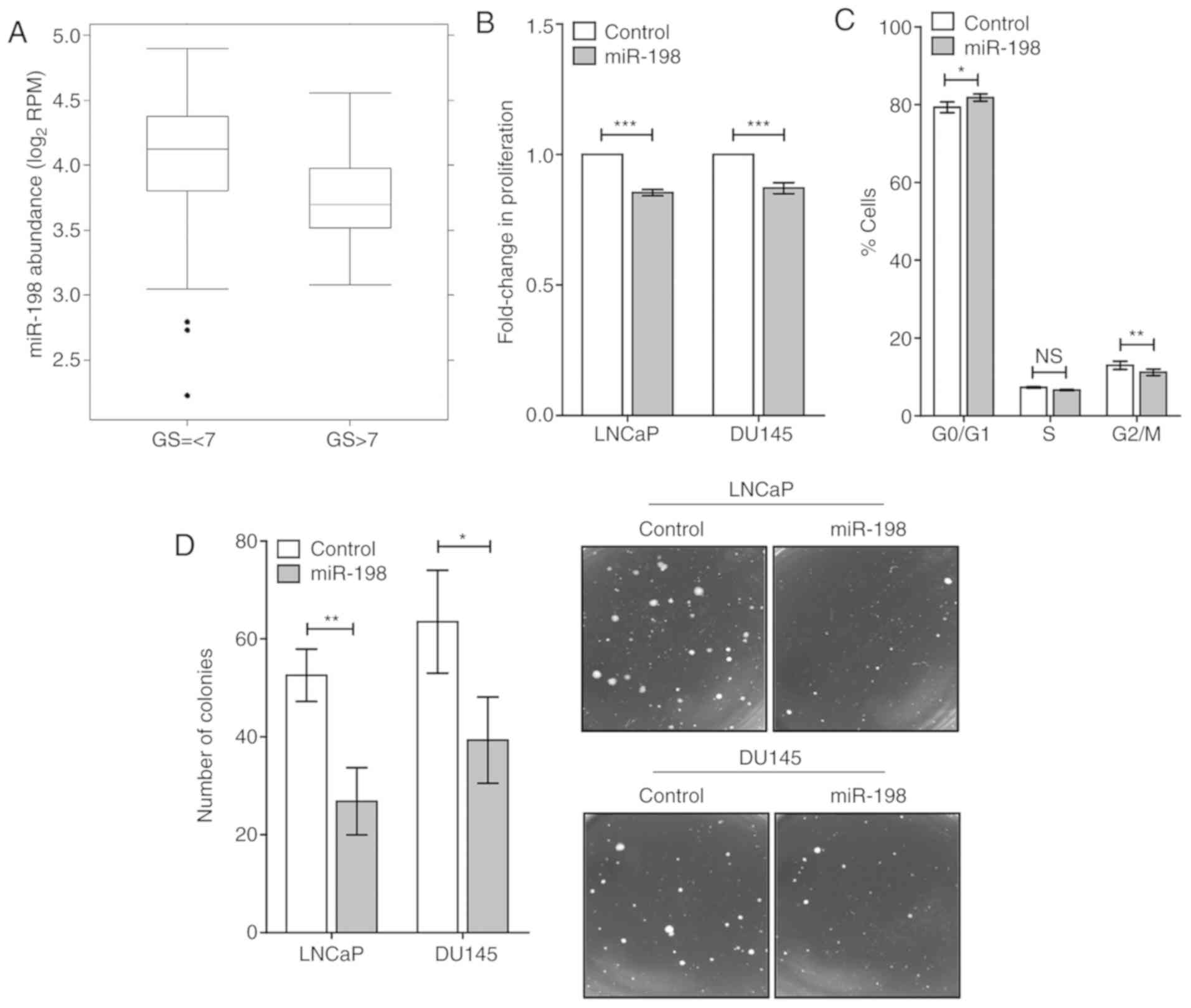
miR-198 is downregulated in aggressive
disease
Given the role of miR-198 as a potential biomarker
in other cancers, it was of interest in determining whether miR-198
abundance was altered in patient samples. We examined the Canadian
Prostate Cancer Genome Network (CPC-GENE) sequencing project data,
which contains miRNA abundance, and clinical information from 162
localized prostate cancer patients. An association with Gleason
score was observed in the CPC-GENE dataset (Fig. 1A). In high Gleason score patients
(Gleason >7), miR-198 abundance was significantly lower than in
intermediate and low Gleason score patients (Gleason score ≤7;
1.2-fold decrease compared with GS ≤7, P<0.05). This analysis
indicated a potential role for miR-198 in prostate cancer
aggression, and warranted investigation into its function in
prostate cancer.
Increased miR-198 suppresses
aggressive phenotype
We assayed for important hallmarks of tumor
aggression, including cellular proliferation, anchorage-independent
growth, and invasive capacity in two human prostate cancer cell
lines LNCaP and DU145. Transient transfection of miR-198 mimic in
DU145 and LNCaP cells was performed, and overexpression was
confirmed by RT-qPCR (Fig. S1A).
This resulted in significantly reduced proliferation compared with
the control mimic using viable cell counting (Fig. 1B). Analysis of cell cycle profiles was
performed in LNCaP cells using flow cytometry and significant
differences were revealed in the cell cycle distributions between
miR-198 and control conditions (Fig.
1C). A greater percentage of miR-198 cells were observed in the
G0/G1 phase (P<0.05), with less in the S
phase (P=0.055) and G2/M phase (P<0.01) relative to
the control, which was consistent with reduced cellular
proliferation. To assay tumorigenic potential in vitro,
anchorage-independent growth assays were performed and it was
determined that miR-198 mimic-transfected DU145 and LNCaP cells had
a significantly reduced ability to form colonies in soft agar
(Fig. 1D).
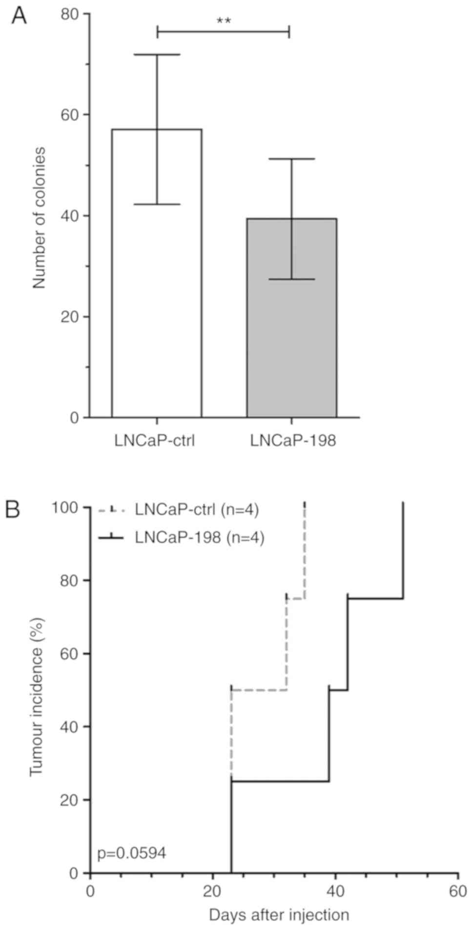
miR-198 reduces tumorigenicity in
vivo
In order to assay the influence of miR-198 on
tumorigenicity in vivo, stably overexpressing cell lines
were generated by transducing LNCaP cells with a lentivirus
containing a miR-198 sequence (LNCaP-198) or a control sequence
(LNCaP-ctrl). After antibiotic selection, surviving cells were
pooled and assayed to confirm overexpression of miR-198 (Fig. S1B), and the ability to suppress
colony formation in soft agar (Fig.
2A). Subsequently, male athymic nude mice were injected
subcutaneously with either LNCaP-ctrl or LNCaP-198 cells, and tumor
growth was monitored. At 35 days post-injection, only one LNCaP-198
mouse had an observable tumor, whereas all four LNCaP-ctrl mice
developed tumors. Tumors ultimately formed in the entire LNCaP-198
group; however, the mean number of days required was 38.75 days vs.
28.25 in LNCaP-ctrl. Tumor-free survival analysis demonstrated
miR-198 overexpression trended towards slower tumor formation in
mice (Fig. 2B, log-rank
P=0.0594).
MIB1 is directly targeted by
miR-198
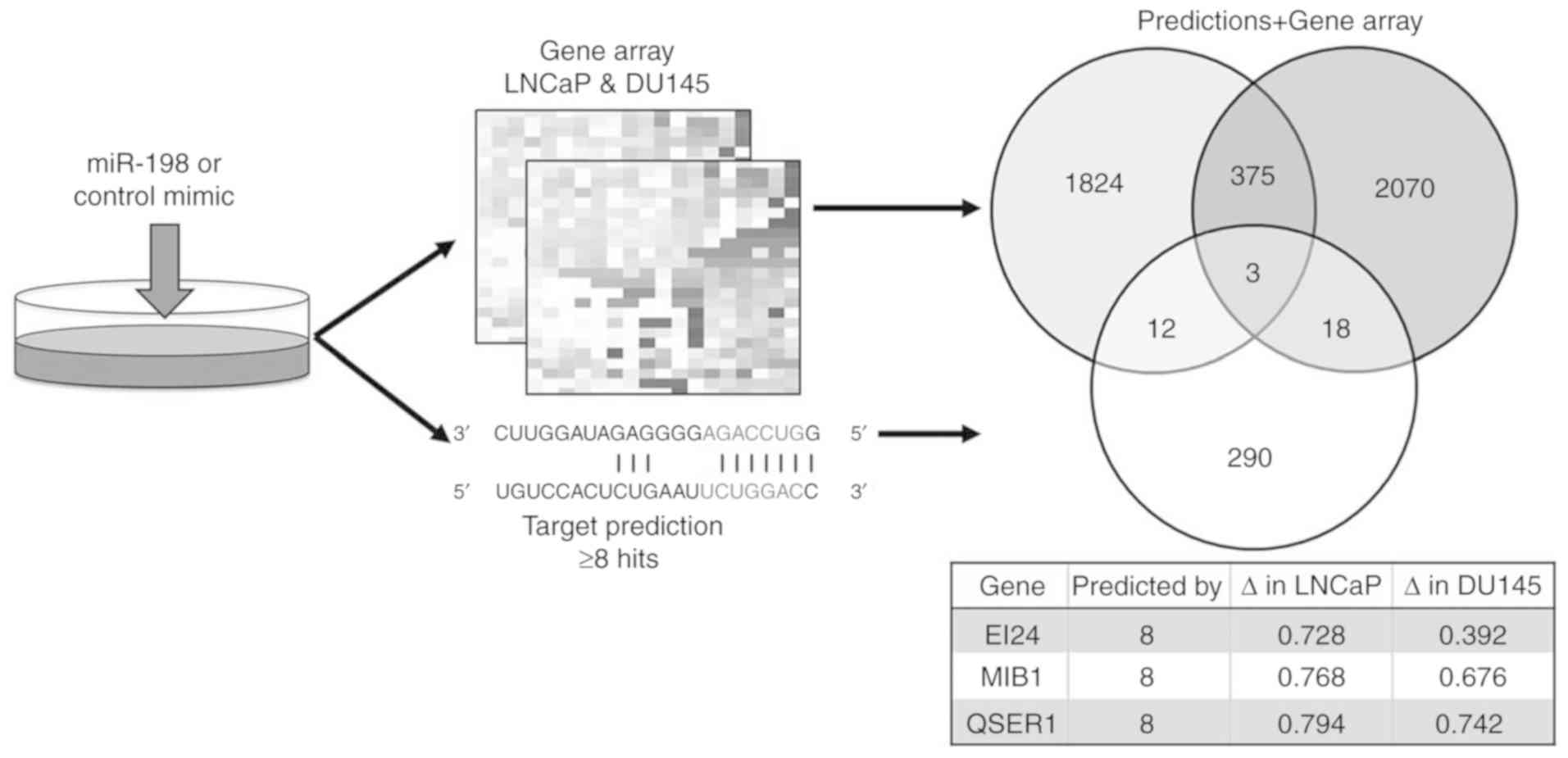
To identify targets of miR-198 which regulate
tumorigenicity, transcriptomic analyses from DU145 and LNCaP cells
transiently transfected with miR-198 or control mimics were
performed, and these data were combined with in silico
predicted targets from miRWalk 2.0 (Fig.
3). The three highest predicted targets were EI24 autophagy
associated transmembrane protein (EI24), mindbomb E3 ubiquitin
protein ligase 1 (MIB1), and glutamine and serine rich 1 (QSER1).
QSER1 is a protein of unknown function, EI24 encodes a putative
tumor suppressor (16), whereas MIB1
has a possible oncogenic function through promotion of Notch
signaling and thus is a potential target of interest. In the
transcriptomic analyses, miR-198 overexpression decreased MIB1
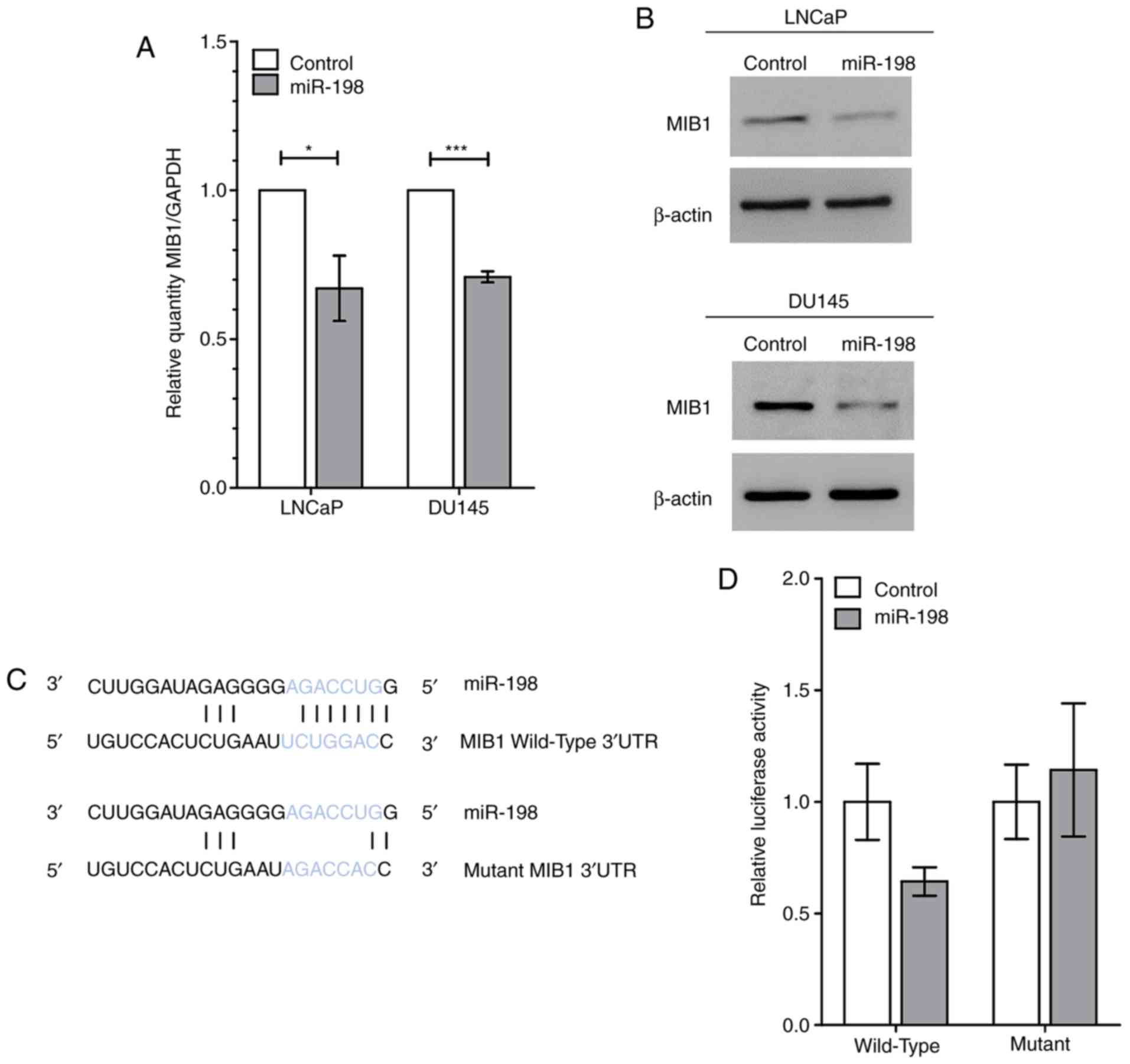
abundance by 32% in DU145 cells and 23% in LNCaP cells (Fig. 3). RT-qPCR performed after transient
miR-198 transfection confirmed that MIB1 abundance was reduced by
29 and 33% in DU145 and LNCaP cells, respectively (Fig. 4A). In addition, western blot analysis
of MIB1 confirmed a decrease at the protein level (Fig. 4B). Next, a luciferase reporter was
designed and created, containing the predicted wild-type or a
mutated miR-198 binding site from the MIB1 3′UTR in order to
confirm direct binding of miR-198 to MIB1 (Fig. 4C). Luciferase activity was
significantly decreased in LNCaP cells co-transfected with miR-198
mimic and wild-type MIB1 3′UTR compared to the control mimic, and
exhibited no differences with miR-198 or control mimics with the
mutant 3′UTR (Fig. 4D). These
experiments established MIB1 as an authentic target of miR-198 in
prostate cancer.
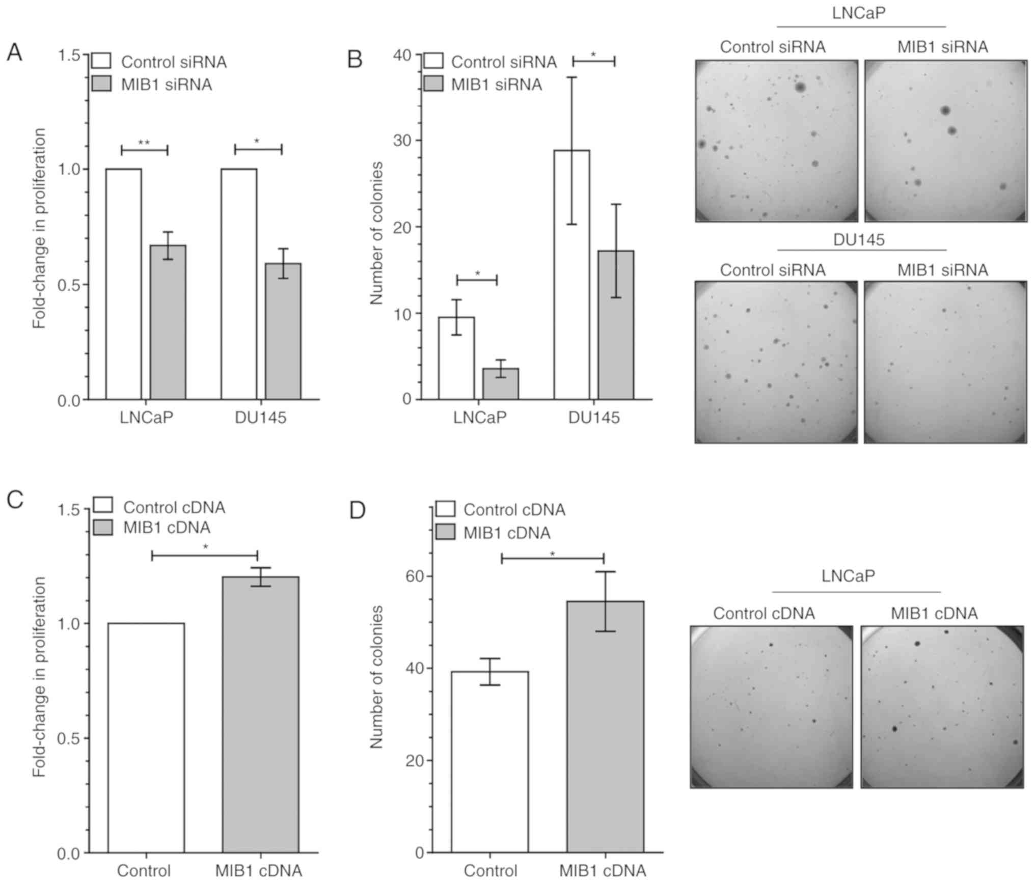
MIB1 alteration recapitulates
phenotype
It was next determined whether MIB1 knockdown could
result in a similar phenotype as miR-198. Knockdown of MIB1 was
first confirmed at the mRNA and protein level, after siRNA
transfection in DU145 and LNCaP cells using RT-qPCR and western
blotting, respectively (Fig. S1C and
D). Knockdown of MIB1 significantly reduced proliferation in
both DU145 and LNCaP cells (Fig. 5A).
Tumorigenic potential in soft agar was also decreased with MIB1
knockdown (Fig. 5B). In addition,
MIB1 abundance was increased in LNCaP cells by transfecting with a
vector containing MIB1 ORF cDNA, and subsequently assayed for
phenotype. Cells with elevated MIB1 displayed significantly more
proliferation (Fig. 5C) and colony
formation ability (Fig. 5D) compared
with the control vector. Thus, MIB1 knockdown effectively
recapitulated the suppressive effects of miR-198 on cellular
proliferation and tumorigenic potential, which was conversely
promoted by elevation of MIB1.
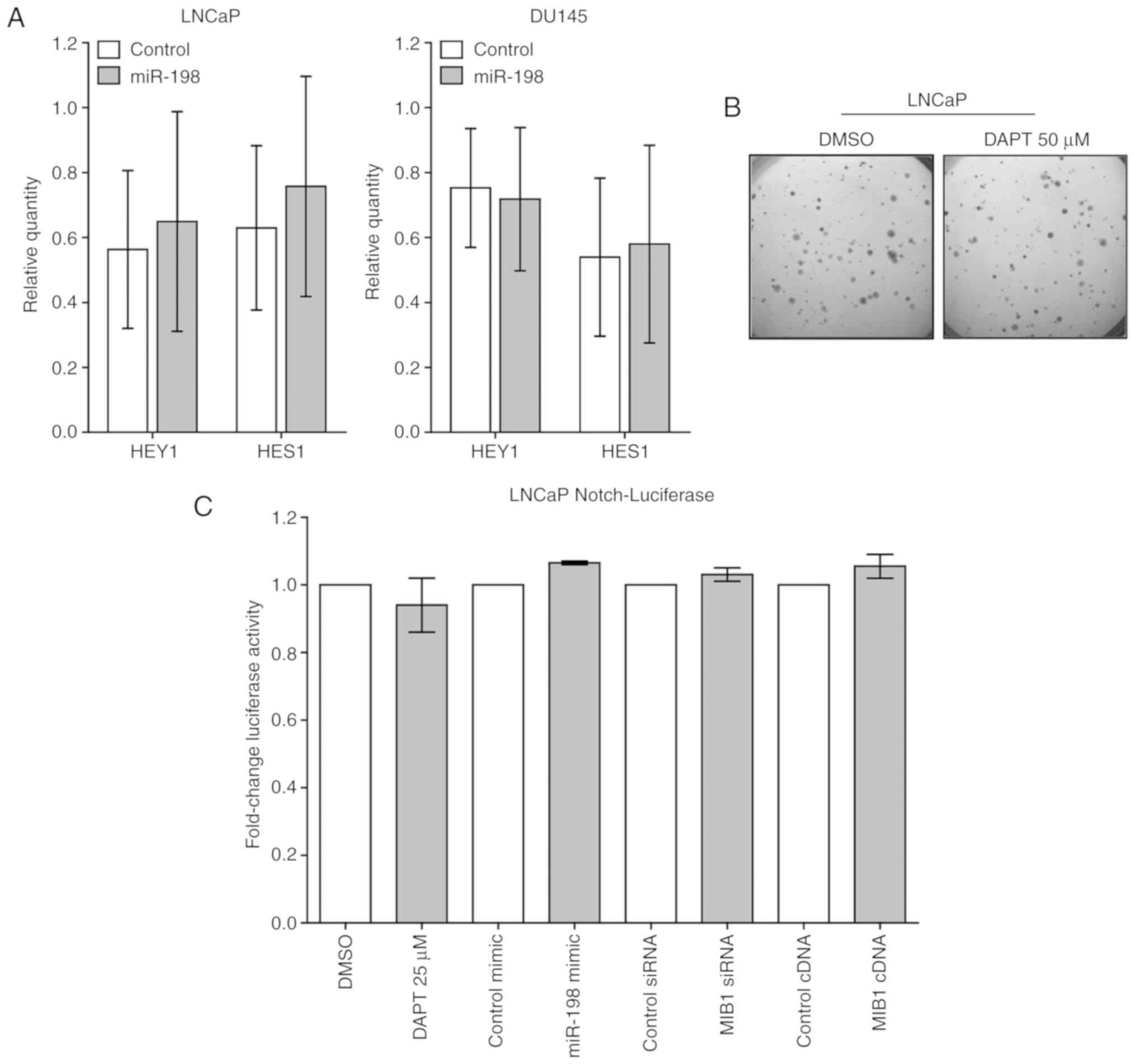
miR198/MIB1 effects on prostate cancer
are likely Notch-independent
The most well-characterized role of MIB1 is as an E3
ubiquitin ligase which interacts with Delta to increase Notch
signaling (17). To examine the
effect of miR-198 on Notch signaling, the expression of the Notch
transcription factors HEY1 and HES1 were examined, which are used
as surrogate markers for Notch activation. RT-qPCR revealed no
alterations in the expression of either mRNA with miR-198
transfection in either LNCaP or DU145 cells (Fig. 6A). Additionally, HEY1 and HES1 were
unchanged by miR-198 transfection in both LNCaP and DU145
transcriptomic arrays (data not shown). To examine if Notch
suppression could contribute to the miR-198 phenotype, soft agar
colony formation assays were performed with DAPT, a Notch
inhibitor, which revealed no difference in tumorigenic potential
(Fig. 6B). Lastly, a LNCaP
Notch-luciferase reporter cell line was generated to examine Notch
activity, which exhibited no appreciable differences in Notch
activity in LNCaP cells either after DAPT treatment, MIB1 siRNA, or
miR-198 mimic transfection (Fig. 6C).
To validate the Notch-luciferase assay, the effect of these
manipulations in the previously validated LS174T cell line were
examined, which confirmed reduced luciferase activity after
treatment with the Notch inhibitor, but no effect from miR-198 or
MIB1 transfections (Fig. S2).
Collectively, our data strongly indicated that miR-198 and MIB1
exert their tumor suppressive effects independent of the Notch
pathway.
Discussion
The present study revealed lower abundance of
miR-198 in high Gleason grade tumors, highlighting its potential
role as a tumor suppressor miRNA. In vitro experiments
demonstrated that miR-198 overexpression reduced proliferation and
anchorage-independent growth, and induced a
G0/G1 cell cycle block. Tumor formation in
vivo was reduced with elevated miR-198 abundance, confirming
miR-198 functions as a tumor suppressive miRNA in prostate cancer.
In prostate cancer, miR-198 is a low-abundance miRNA which may
limit is utility as a biomarker; this contrasts to its higher
abundance in other cancer types. For example, studies have reported
miR-198 as a biomarker in other TCGA cancer cohorts, such as
elevated abundance associated with increased overall survival in
glioblastoma patients (9). However,
miR-198 was undetectable (abundance level=zero, below detection
threshold) in >90% of TCGA normal prostate and prostate cancer
samples, which limited our ability to perform analyses in this
dataset. CPC-GENE utilized a different platform to assess miRNA,
which appears more sensitive to this low abundance miRNA, and thus
was able to provide non-zero values for miR-198 abundance.
The previously identified targets of miR-198 vary
widely in structure and function depending on the cancer type
investigated. miR-198 has one previously described target in
prostate cancer, Livin (or BIRC7) (18). However, no consistent decrease was
observed in this mRNA after miR-198 transfection in both LNCaP and
DU145 cells. In addition to Livin, previously identified targets
from other cancer types were assayed, however, decreased abundance
of these genes in LNCaP and DU145 cells (Table SIII) was not consistently observed.
Combination of transcriptomic and in silico analyses
identified MIB1 as a putative target of miR-198, as it contained a
miR-198 binding site in its 3′UTR, and was consistently reduced by
miR-198 overexpression in both cell line arrays. This effect was
also observed using RT-qPCR and western blotting to analyze protein
abundance. Subsequent MIB1 knockdown studies phenocopied miR-198
effects on reducing proliferation and tumorigenic potential, while
a converse effect was observed with MIB1 elevation using an
overexpressing vector.
MIB1 is described primarily as an integral positive
regulator of Notch signaling, however, significant responsiveness
in LNCaP cells was not observed. The role of Notch in prostate
cancer is complex with clinical studies yielding conflicting
results, demonstrating both up- and downregulation of components in
prostate cancer samples and varying effects on deregulation in
vitro (19,20). Several studies have concluded that
aberrant activation of Notch signaling is consistently observed in
metastatic samples, especially increases in NOTCH1 receptor and
JAG1 ligand (19). In the present
study, treatment of LNCaP cells with the Notch inhibitor DAPT did
not reduce anchorage-independent growth. In addition, RT-qPCR
analysis of downstream Notch reporter genes HEY1 and HES1 exhibited
no change in their abundance after miR-198 transfection.
Transfection with miR-198 mimic or treatment with a Notch inhibitor
both failed to alter luciferase reporter activity in LNCaP cells,
indicating that the key function of miR-198/MIB1 in prostate cancer
may occur independently of the effects on Notch signaling. The
present study is the first to report a miRNA targeting MIB1 in
cancer, and therefore requires future exploration of pathways
external to Notch signaling in which miR-198/MIB1 may exert a
non-canonical function.
In summary, this research establishes a tumor
suppressive role of miR-198 in prostate cancer. miR-198 displayed
lower abundance in high Gleason grade tumors, and overexpression
impaired tumor formation in mice. MIB1 was identified as a novel
miR-198 target, through which miR-198 reduced proliferation and
tumourigenicity of prostate cancer. This mechanism likely occurs in
a Notch pathway-independent manner, which can be further explored
to develop new therapeutic strategies. These study findings
reinforce the importance and complexity of miRNA in regulating
prostate cancer aggression.
Supplementary Material
Supporting Data
Acknowledgements
Not applicable.
Funding
SKL is a Movember Rising Star award recipient
proudly funded by the Movember Foundation (grant. nos. RS2014-03,
D2015-12 and D2017-1811), the Telus Motorcycle Ride For Dad
(Huronia Branch), and a Ministry of Research and Innovation Early
Researcher Award.
Availability of data and materials
All data generated or analyzed during the preset
study are included in this published article.
Authors' contributions
JR designed and executed the majority of
experiments, interpreted results, performed statistical analyses,
and drafted the manuscript. SKL conceived the project, supervised
direction, and revised the manuscript. CH, XH, MRD and ST assisted
with the experiments and data interpretation. JJ, supervised by
PCB, performed bioinformatic analyses of patient datasets. All
authors read and approved the final manuscript and agree to be
accountable for all aspects of the research in ensuring that the
accuracy or integrity of any part of the work are appropriately
investigated and resolved.
Ethics approval and consent to
participate
CPC-GENE is part of the international genome
consortium (ICGC) and data access control is regulated via the
ICGC-DACO (www.icgc.org). Informed consent,
consistent with local Research Ethics Board (REB) and International
Cancer Genome Consortium (ICGC) guidelines, was obtained at the
time of clinical follow-up. Previously collected tumor tissues were
used, following University Health Network REB-approved study
protocols (UHN 06-0822-CE, UHN 11-0024-CE, CHUQ
2012-913:H12-03-192). All experiments involving animals were
performed in accordance with the University of Toronto and
Sunnybrook Research Institute Animal Care Committee guidelines
using a peer-reviewed protocol (AUP #17-509).
Patient consent for publication
Not applicable.
Competing interests
The authors declare that they have no competing
interests.
Glossary
Abbreviations
Abbreviations:
|
3′UTR
|
3′ untranslated region
|
|
CPC-GENE
|
Canadian Prostate Cancer Genome
Network
|
|
HES1
|
hairy and enhancer of split 1
|
|
HEY1
|
hes-related family bHLH transcription
factor with YRPW motif 1
|
|
MIB1
|
mindbomb E3 ubiquitin protein ligase
1
|
|
miRNA
|
microRNA
|
|
miR-198
|
microRNA-198
|
|
siRNA
|
small interfering RNA
|
References
|
1
|
Boutros PC, Fraser M, Harding NJ, de Borja
R, Trudel D, Lalonde E, Meng A, Hennings-Yeomans PH, McPherson A,
Sabelnykova VY, et al: Spatial genomic heterogeneity within
localized, multifocal prostate cancer. Nat Genet. 47:736–745. 2015.
View Article : Google Scholar : PubMed/NCBI
|
|
2
|
Fraser M, Sabelnykova VY, Yamaguchi TN,
Heisler LE, Livingstone J, Huang V, Shiah YJ, Yousif F, Lin X,
Masella AP, et al: Genomic hallmarks of localized, non-indolent
prostate cancer. Nature. 541:359–364. 2017. View Article : Google Scholar : PubMed/NCBI
|
|
3
|
Iorio MV and Croce CM: MicroRNA
dysregulation in cancer: Diagnostics, monitoring and therapeutics.
A comprehensive review. EMBO Mol Med. 4:143–159. 2012. View Article : Google Scholar : PubMed/NCBI
|
|
4
|
Bi C, Chung TH, Huang G, Zhou J1, Yan J,
Ahmann GJ, Fonseca R and Chng WJ: Genome-wide pharmacologic
unmasking identifies tumor suppressive microRNAs in multiple
myeloma. Oncotarget. 6:26508–26518. 2015. View Article : Google Scholar : PubMed/NCBI
|
|
5
|
Cui Z, Zheng X and Kong D: Decreased
miR-198 expression and its prognostic significance in human gastric
cancer. World J Surg Oncol. 14:332016. View Article : Google Scholar : PubMed/NCBI
|
|
6
|
Hu Y, Tang Z, Jiang B, Chen J and Fu Z:
miR-198 functions as a tumor suppressor in breast cancer by
targeting CUB domain-containing protein 1. Oncol Lett.
13:1753–1760. 2017. View Article : Google Scholar : PubMed/NCBI
|
|
7
|
Huang WT, Wang HL, Yang H, Ren FH, Luo YH,
Huang CQ, Liang YY, Liang HW, Chen G and Dang YW: Lower expressed
miR-198 and its potential targets in hepatocellular carcinoma: A
clinicopathological and in silico study. Onco Targets Ther.
9:5163–5180. 2016. View Article : Google Scholar : PubMed/NCBI
|
|
8
|
Marin-Muller C, Li D, Bharadwaj U, Li M,
Chen C, Hodges SE, Fisher WE, Mo Q, Hung MC and Yao Q: A
tumorigenic factor interactome connected through tumor suppressor
microRNA-198 in human pancreatic cancer. Clin Cancer Res.
19:5901–5913. 2013. View Article : Google Scholar : PubMed/NCBI
|
|
9
|
Nie E, Jin X, Wu W, Yu T, Zhou X, Shi Z,
Zhang J, Liu N and You Y: MiR-198 enhances temozolomide sensitivity
in glioblastoma by targeting MGMT. J Neurooncol. 133:59–68. 2017.
View Article : Google Scholar : PubMed/NCBI
|
|
10
|
Wang M, Wang J, Kong X, Chen H, Wang Y,
Qin M, Lin Y, Chen H, Xu J, Hong J, et al: MiR-198 represses tumor
growth and metastasis in colorectal cancer by targeting fucosyl
transferase 8. Sci Rep. 4:61452014. View Article : Google Scholar : PubMed/NCBI
|
|
11
|
Wu S, Zhang G, Li P, Chen S, Zhang F, Li
J, Jiang C, Chen X, Wang Y, Du Y, et al: miR-198 targets SHMT1 to
inhibit cell proliferation and enhance cell apoptosis in lung
adenocarcinoma. Tumour Biol. 37:5193–5202. 2016. View Article : Google Scholar : PubMed/NCBI
|
|
12
|
Zhang S, Zhao Y and Wang L: MicroRNA-198
inhibited tumorous behaviors of human osteosarcoma through directly
targeting ROCK1. Biochem Biophys Res Commun. 472:557–565. 2016.
View Article : Google Scholar : PubMed/NCBI
|
|
13
|
Liu SK, Bham SA, Fokas E, Beech J, Im J,
Cho S, Harris AL and Muschel RJ: Delta-like ligand 4-notch blockade
and tumor radiation response. J Natl Cancer Inst. 103:1778–1798.
2011. View Article : Google Scholar : PubMed/NCBI
|
|
14
|
Livak KJ and Schmittgen TD: Analysis of
relative gene expression data using real-time quantitative PCR and
the 2(-Delta Delta C(T)) method. Methods. 25:402–408. 2001.
View Article : Google Scholar : PubMed/NCBI
|
|
15
|
Dweep H, Sticht C, Pandey P and Gretz N:
miRWalk-database: Prediction of possible miRNA binding sites by
‘walking’ the genes of three genomes. J Biomed Inform. 44:839–847.
2011. View Article : Google Scholar : PubMed/NCBI
|
|
16
|
Mork CN, Faller DV and Spanjaard RA: Loss
of putative tumor suppressor EI24/PIG8 confers resistance to
etoposide. FEBS Lett. 581:5440–5444. 2007. View Article : Google Scholar : PubMed/NCBI
|
|
17
|
Itoh M, Kim CH, Palardy G, Oda T, Jiang
YJ, Maust D, Yeo SY, Lorick K, Wright GJ, Ariza-McNaughton L, et
al: Mind bomb is a ubiquitin ligase that is essential for efficient
activation of Notch signaling by Delta. Dev Cell. 4:67–82. 2003.
View Article : Google Scholar : PubMed/NCBI
|
|
18
|
Ye L, Li S, Ye D, Yang D, Yue F, Guo Y,
Chen X, Chen F, Zhang J and Song X: Livin expression may be
regulated by miR-198 in human prostate cancer cell lines. Eur J
Cancer. 49:734–740. 2013. View Article : Google Scholar : PubMed/NCBI
|
|
19
|
Deng G, Ma L, Meng Q, Ju X, Jiang K, Jiang
P and Yu Z: Notch signaling in the prostate: Critical roles during
development and in the hallmarks of prostate cancer biology. J
Cancer Res Clin Oncol. 142:531–547. 2016. View Article : Google Scholar : PubMed/NCBI
|
|
20
|
Su Q and Xin L: Notch signaling in
prostate cancer: Refining a therapeutic opportunity. Histol
Histopathol. 31:149–157. 2016.PubMed/NCBI
|