Introduction
Bone morphogenetic proteins (BMPs) are members of
the TGF-β superfamily and are known for their diverse function in
cancer progression (1). BMPs were
originally believed to play a role solely in the development of
bone and cartilage as they were isolated from bone extracts
(2); however, the various functions
of BMPs have since been discovered. BMPs display aberrant
expression in a variety of malignancies (3). In breast cancer, BMP4 and BMP7 are
reported to be the most frequently overexpressed BMPs (4). However, their biological functions in
breast cancer remain controversial. Some studies have reported that
BMPs suppress breast cancer growth, whereas others have reported
that BMPs promote it. BMP4 was reported to suppress breast cancer
metastasis by upregulating immune responses and inhibition of
myeloid-derived suppressor cells (5),
whereas it was also reported that high expression of BMP4 promoted
cancer cell migration, invasion and metastasis (6). BMP7 was also shown to both facilitate
and inhibit cancer growth, migration, invasiveness and metastasis
in different breast cancer cell lines (7). BMP receptors (BMPRs) have also reported
to play a role in the carcinogenesis and progression of breast
cancer (8). As such, there are
multiple conflicting reports concerning the role of BMPs in breast
cancer.
TGF-β regulates immune cell infiltration in the
tumor microenvironment. TGF-β suppresses anticancer immune cell
infiltration such as cytotoxic T cells and natural killer cells,
whereas it promotes the function of pro-cancer immune cells, such
as regulatory T cells (Tregs) and M2 macrophages (9). BMP6 was also found to induce hormone
therapy resistance through macrophage-derived cytokines in prostate
cancer (10). Thus, we hypothesized
that BMPs regulate immune cell infiltration, resulting in
differential survival of breast cancer patients based on the
expression levels of BMPs.
Here, we investigated the association of BMP
expression and prognosis in breast cancer utilizing The Cancer
Genome Atlas (TCGA) (11,12). Furthermore, we explored the difference
in the association between BMP expression and survival by estrogen
receptor (ER) status and sought possible underlying mechanisms
using a bioinformatical approach.
Materials and methods
Data acquisition
Clinical data and gene expression from RNA sequences
in TCGA breast cancer cohort were downloaded through cBioPortal
(13,14) and UCSC Genome Browser (http://genome.ucsc.edu/) and processed, as previously
described (15–20). As a validation cohort, we utilized
GSE1456, in which there are 62 Luminal A/B patients with gene
expression and survival information from Gene Enrichment Omnibus
(21). TCGA and GSE1456 are
de-identified and publicly available cohorts, thus institutional
review board approval was waived.
Gene set enrichment analysis
(GSEA)
GSEA was conducted comparing BMP7 high and low as
well as BMP6 high and low in both ER-positive (ER+) and
ER-negative (ER−) cohorts with the same cutoffs of
survival analyses among 50 hallmark sets (23) using software provided by the Broad
Institute (http://software.broadinstitute.org/gsea/index.jsp) as
described previously (24).
CIBERSORT
Tumor infiltrating immune cell composition was
estimated from the gene expression profiles by CIBERSORT algorism
(25). Immune cell fraction data were
downloaded through TCIA (https://tcia.at/home) (26).
Cytolytic activity
Cytolytic activity was estimated utilizing GZMA and
PRF1 expression (27) as described
previously (28).
Statistical analysis
Overall survival (OS) was estimated by Kaplan-Meier
curve with log-rank test, comparing high and low expression of each
gene of interest. The cutoffs were determined by analyzing multiple
cutoff points within the range of values and the optimal cutoff
points were chosen based on the statistical significance of the
varying points as previously reported (22).
Statistical comparisons of gene expression were
performed using the Student t-test, and the clinical demographics
were compared using the Fisher exact test. Pearson correlations
were calculated based on the expression levels of the genes of
interest for correlation matrix (Fig.
2C). All statistical analyses were performed using R software
(http://www.r-project.org/) and
Bioconductor (http://bioconductor.org/).
Results
Various BMPs and BMPRs are associated
with better OS, whereas others are associated with worse OS in
breast cancer
There have been conflicting reports on the
association between the expression levels of BMPs and patient
survival (29). Thus, we analyzed the
association between the expression of each BMP and OS of the breast
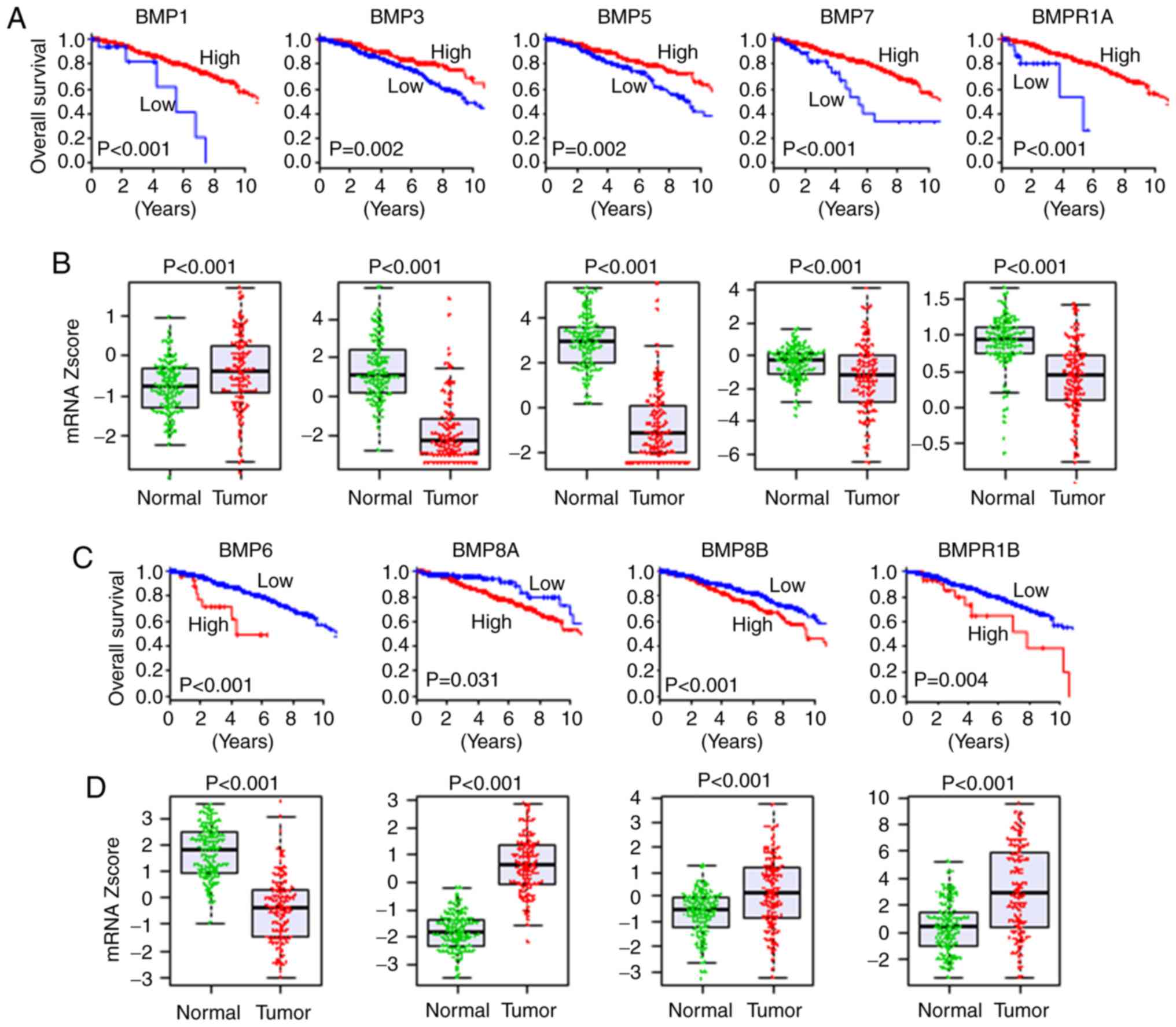
cancer patients. High expression of BMP1 (P<0.001), BMP3
(P=0.002), BMP5 (P=0.002), BMP7 (P<0.001) and BMPR1A
(P<0.001) was indicative of a significantly better OS of the
breast cancer patients (Fig. 1A).
Conversely, high expression of BMP6 (P<0.001), BMP8A (P=0.031),
BMP8B (P<0.001) and BMPR1B (P=0.004) was indicative of a worse
OS (Fig. 1C). These findings indicate
that the former may act as tumor suppressors, and the latter as
oncogenic factors. On the other hand, the expression levels of BMP2
(P=0.083), BMP4 (P=0.071), BMP10 (P=0.441), BMP13 (P=0.112) and
BMP15 (P=0.050) were not associated with OS of breast cancer
patients (Fig. S1). Of interest,
cutoff points of BMP expression with the highest impact on OS were
extremely low in some tumor-suppressive BMPs, and hence a poorer
prognosis was shown in <10% of patients for BMP1, BMP7 and
BMPR1A (Fig. 1A). Similar trends were
shown in some oncogenic BMPs, BMP6 and BMPR1B, in which cutoff
points with the highest impact of OS were extremely high and
<10% of patients were classified as high expression with worse
prognosis (Fig. 1C). In summary, we
found that extremely low expression of tumor-suppressive BMPs and
extremely high expression of oncogenic BMPs were associated with
worse OS in breast cancer.
Majority of oncogenic BMPs are
expressed at a high level, and tumor-suppressive BMPs are expressed
at a low in the tumors compared to the normal tissues
Given the associations between the expression levels
of the BMPs and worse or better survival, we hypothesized that
oncogenic BMPs are expressed at a higher level and
tumor-suppressive BMPs are expressed at a lower level in tumors
compared to adjacent normal tissues. A total of 114 breast cancers
with matched tumor and adjacent normal tissues were present in
TCGA, where the expression of BMPs and BMPRs was compared. All of
the BMPs and BMPRs, which were associated with survival, were
expressed at significantly different levels between the normal and
tumor tissues. All of the tumor-suppressive BMPs except BMP1; i.e.
BMP3, BMP5, BMP7 and BMPR1A, were expressed at a significantly
lower level in the tumors, whereas all of the oncogenic BMPs except
BMP6; i.e. BMP8A, BMP8B and BMPR1B, were expressed at a higher
level in the tumors compared to the normal tissues (Fig. 1B and D). The expression levels of
BMP10 and BMP15, which showed no association with OS, were not
detectable in the vast majority of samples (106 normal and 108
tumor tissues, and 104 normal and 100 tumor tissues, respectively).
BMP2 and BMP4, that did not show an association with OS, were also
expressed at a lower level in the tumors (Fig. S1). These results support the notion
that oncogenic BMPs are expressed higher, and tumor-suppressive
BMPs are expressed lower in the tumors, except BMP1 and BMP6.
Expression of BMPs differs according
to ER status
It is well known that certain signaling, such as
HER2, crosstalks with ER signaling and the targeted drug
sensitivity is enhanced by ER blockade (30). Signaling of BMPs is also known to
crosstalk with that of ER (31,32).
Therefore, it was of interest to investigate the association of ER
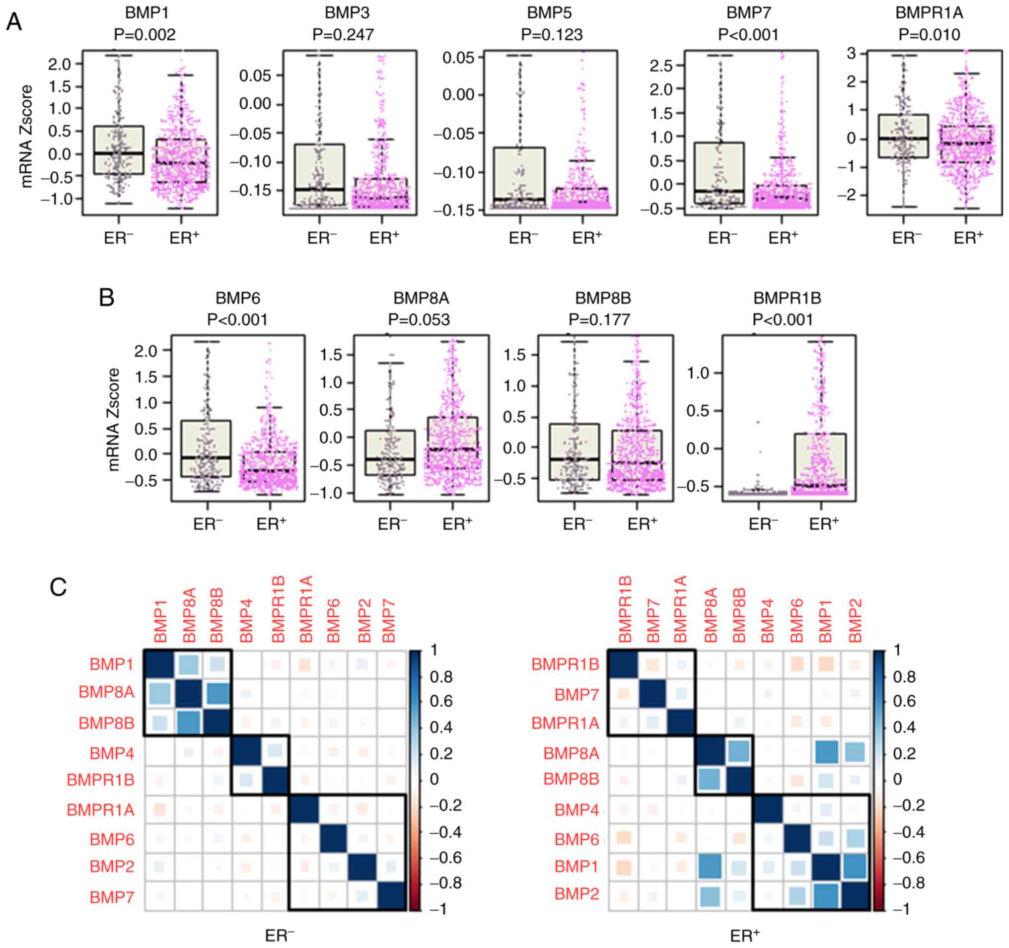
positivity and BMP expression. We found that among the
tumor-suppressive BMPs, BMP1 (P=0.002), BMP7 (P<0.001) and
BMPR1A (P=0.010) were expressed significantly higher in the
ER− tumors compared to the ER+ tumors,
whereas there was no significant difference in the expression
levels of BMP3 (P=0.247) and BMP5 (P=0.123) (Fig. 2A). On the other hand, among the
oncogenic BMPs, BMP6 expression was significantly higher in the
ER− tumors (P<0.001), whereas BMPR1B expression was
significantly higher in the ER+ tumors (P<0.001)
(Fig. 2B). Interactions of BMPs and
BMPRs were also different between ER+ and ER−
tumors (Fig. 2C). In the
ER+ tumors, BMP6 was in the same cluster as BMP1, BMP2
and BMP4, where BMP6 exhibited a higher correlation with BMP1 and
BMP2, and BMP7 was in the same cluster of BMPRs (BMPR1A and BMPR1B)
(Fig. 2C). Whereas in the
ER− tumors, BMP6 and BMP7 were in the same cluster with
BMP2 and BMPR1A, showing a very weak correlation with each other
(Fig. 2C).
Higher BMP7 expression is associated
with better OS in both ER+ and ER−
tumors
Since BMP7 was reported to have roles in both
promotion and inhibition of cancer growth (4), and showed significant differential
expression as well as differential interaction with other BMPs and
BMPRs between ER+ and ER− tumors, we focused
on BMP7 among the tumor-suppressive BMPs. Higher expression of BMP7
demonstrated better survival in both ER+ (P<0.001)
and ER− (P<0.001) cohorts (Fig. 3A and B). Although higher expression of
BMP7 was found to be associated with better prognosis in the both
ER+ and ER− tumors, GSEA demonstrated that
different gene sets were enriched between the ER+ and
ER− tumors (Tables
SI–SIII). In the ER+
tumors, immune response-related gene sets, such as inflammatory
response (P<0.001), IL2/STAT5 signaling (P<0.001), TNFα
signaling via NF-κB (P<0.001), IL6/JAK/STAT3 signaling
(P<0.001) and allograft rejection (P=0.003), were enriched in
the BMP7 high tumors (Fig. 3C,
Table SI). As tumor aggressiveness
feature-related gene sets, the epithelial-mesenchymal transition
(EMT) gene set was also enriched (P<0.001), while cell
proliferation, cell cycle-related gene sets were not enriched in
the BMP7 high ER+ tumors (Table SI). On the other hand, no gene set
was significantly enriched in the BMP7 low tumors. When we compared
demographics of the patients with BMP7 high and low ER+
tumors, the patients with BMP7 high ER+ tumors were
younger than those with BMP7 low ER+ tumors (Table I). In the ER− tumors, no
immune reaction-related gene set was enriched neither in BMP7 high
nor low tumors (Tables SII and
SIII). These findings suggest that
BMP7 plays a tumor-suppressive role in the ER+ tumor by
evoking anticancer immunity, which has a more predominant effect
than promotion of EMT, whereas it has different mechanisms in the
ER− tumors.
 | Table I.Patient demographics comparing BMP7
high and low as well as BMP6 high and low expression in the
ER+ breast cancer patients. |
Table I.
Patient demographics comparing BMP7
high and low as well as BMP6 high and low expression in the
ER+ breast cancer patients.
|
| BMP7
expression | BMP6
expression |
|---|
|
|
|
|
|---|
|
Characteristics | High (n=770) n
(%) | Low (n=34) n
(%) | P-value | High (n=608) n
(%) | Low (n=196) n
(%) | P-value |
|---|
| Age, years |
|
| 0.001a |
|
| 0.033a |
|
<60 | 395 (51.3) | 8 (23.5) |
| 318 (52.7) | 85 (43.4) |
|
|
≥60 | 375 (48.7) | 26 (76.5) |
| 290 (47.7) | 111 (56.6) |
|
| Race |
|
| 0.658 |
|
| 0.167 |
|
Asian | 36 (5.2) | 2 (6.7) |
| 26 (4.6) | 12 (7.5) |
|
|
African-American | 106 (15.9) | 3 (10.0) |
| 81 (14.4) | 28 (17.6) |
|
|
Caucasian | 548 (79.4) | 25 (83.3) |
| 454 (80.9) | 119 (74.8) |
|
| Menopause
status |
|
| 0.056 |
|
|
<0.001a |
|
Pre | 160 (24.4) | 3 (9.1) |
| 142 (27.1) | 21 (12.7) |
|
|
Post | 497 (75.6) | 30 (90.9) |
| 382 (72.9) | 145 (87.3) |
|
| pT |
|
| 0.343 |
|
| 0.663 |
|
T1/2 | 640 (83.1) | 25 (75.8) |
| 501 (82.4) | 164 (84.1) |
|
|
T2/3 | 130 (16.9) | 8 (24.2) |
| 107 (17.6) | 31 (15.9) |
|
| pN |
|
| 0.286 |
|
| 0.559 |
| N0 | 337 (44.6) | 18 (54.5) |
| 266 (44.4) | 89 (46.8) |
|
|
N1/2/3 | 419 (55.4) | 15 (45.5) |
| 333 (55.6) | 101 (53.2) |
|
| pM |
|
| 0.474 |
|
| 0.084 |
| M0 | 628 (97.7) | 25 (96.2) |
| 494 (98.2) | 159 (95.8) |
|
| M1 | 15 (2.3) | 1 (3.8) |
| 9 (1.8) | 7 (4.2) |
|
| Stage |
|
| 0.842 |
|
| 0.572 |
| Stage
I/II | 557 (73.9) | 24 (81.8) |
| 437 (73.2) | 144 (75.4) |
|
| Stage
III/IV | 198 (26.1) | 9 (18.2) |
| 160 (26.8) | 47 (24.6) |
|
| Histology |
|
| 0.475 |
|
|
<0.001a |
|
Infiltrating ductal
carcinoma | 523 (73.9) | 18 (81.8) |
| 381 (68.7) | 160 (93.0) |
|
|
Infiltrating lobular
carcinoma | 185 (26.1) | 4 (18.2) |
| 177 (31.7) | 12 (7.0) |
|
Higher BMP6 expression is associated
with better OS in ER+ tumors, whereas it is associated
with worse OS in ER− tumors
Since high BMP6 expression was associated with
significantly worse survival with lower expression in the tumor
tissues, and different expression of BMP6 as well as different
correlation with other BMPs and BMPRs between ER+ and
ER− tumors were observed, we focused on BMP6 among the
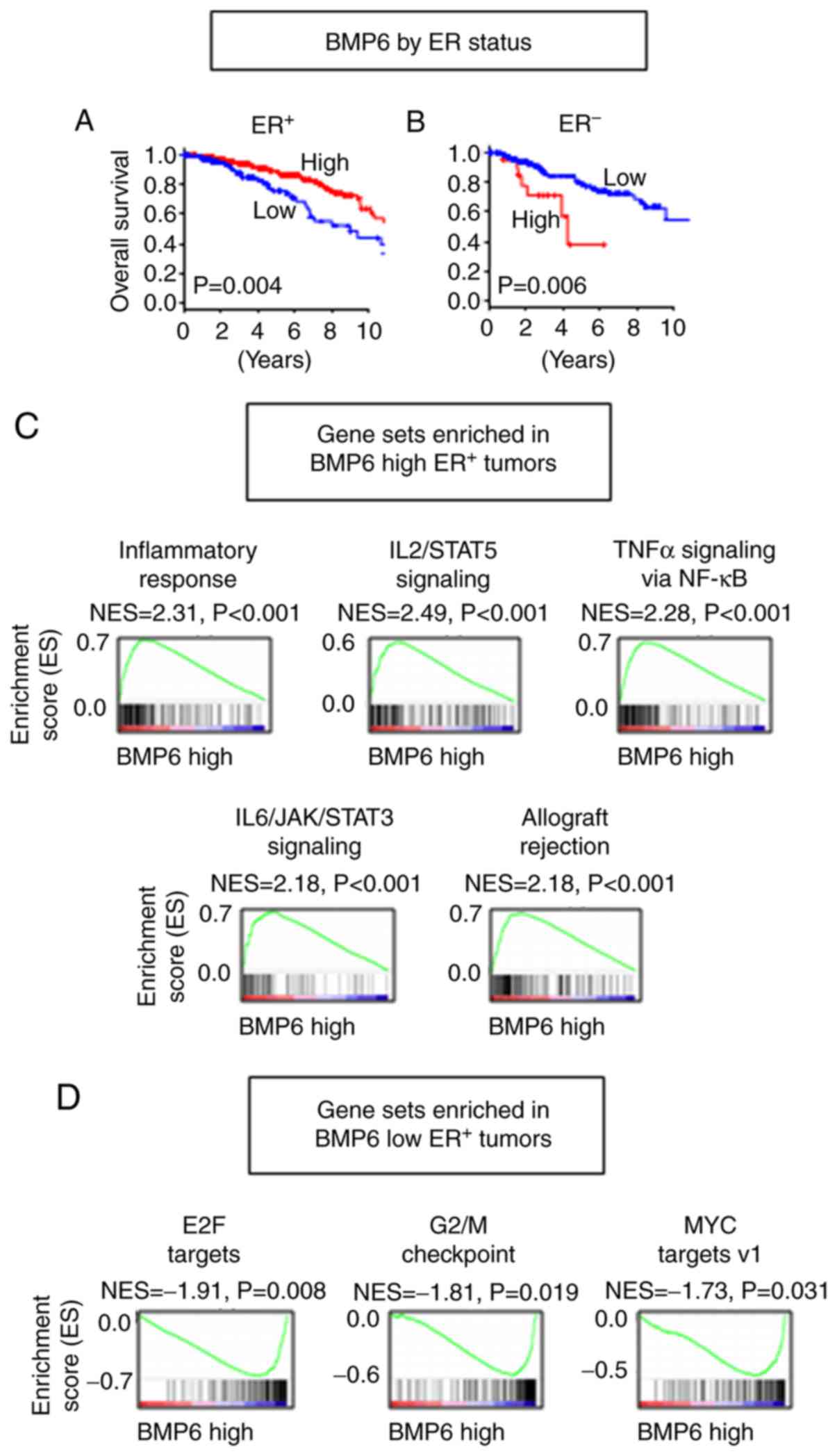
oncogenic BMPs. Despite the fact that BMP6 high tumors demonstrated
worse OS in the whole cohort, that trend was only apparent in the
ER− tumors (P=0.006) (Fig.
4B). Conversely, high expression of BMP6 was found to be
associated with better survival in the ER+ tumors
(P=0.004) (Fig. 4A). GSEA revealed
that immune reaction-related gene sets, including inflammatory
response (P<0.001), IL2/STAT5 signaling (P<0.001), TNFα
signaling via NF-κB (P<0.001), IL6/JAK/STAT3 signaling
(P<0.001) and allograft rejection (P<0.001) were enriched in
the BMP6 high ER+ tumors (Fig.
4C and Table SIV). Similar to
the BMP7 high ER+ tumors, the EMT gene set was also
enriched (P<0.001) in the BMP6 high ER+ tumors
(Table SIV). Interestingly, cell
proliferation and cell cycle-related gene sets, such as E2F targets
(P=0.008), G2/M checkpoint (P=0.019) and MYC targets v1 (33) (P=0.031), were enriched in the BMP6 low
ER+ tumors, which suggest that a different mechanism may
be in place than BMP7 (Fig. 4D and
Table SV). The patients with BMP6
high ER+ tumors showed a higher proportion of younger
age, pre-menopausal status, and infiltrating lobular carcinoma in
histology compared to those with BMP6 low ER+ tumors
(Table I). In the ER−
tumors, MYC targets v2 (34) gene set
was enriched in the BMP6 high tumors (P=0.041), whereas no immune
reaction or cell proliferation gene set was enriched (Tables SVI and SVII). These findings suggest that the BMP6
high tumors have more anticancer immunity and the BMP6 low tumors
have accelerated cell cycle; thus, high expression of BMP6 is
associated with better survival in the ER+ tumors,
whereas higher expression of MYC target genes may play a role in
the BMP6 high ER− tumors, resulting in worse
survival.
BMP7 high as well as BMP6 high
ER+ tumors have higher anticancer immune cell
infiltration and cytolytic activity
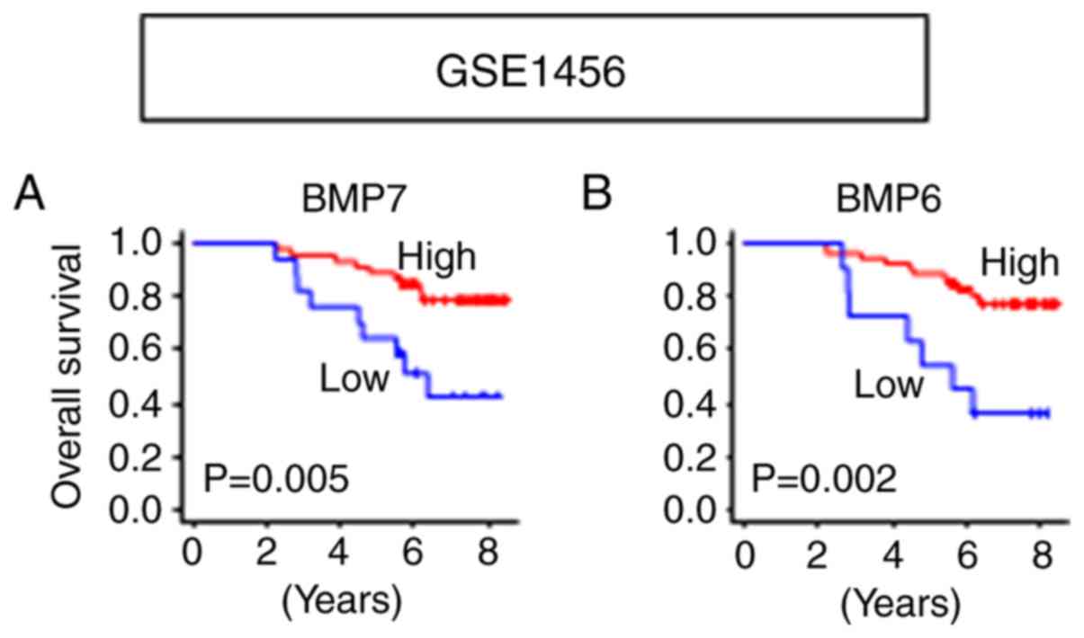
The results that both BMP7 and BMP6 function as
tumor suppressors in ER+ tumors were validated in
another cohort, GSE1456 (21), in
which the patients with high expression of BMP7 (P=0.005) as well
as BMP6 (P=0.002) showed significantly better OS in Luminal A/B
tumors (Fig. 5). Given these results
and the BMP7 and BMP6 high expressing tumor enriched immune-related
gene sets, we further hypothesized that the ER+ tumors
with high BMP7 as well as high BMP6 expressions have higher
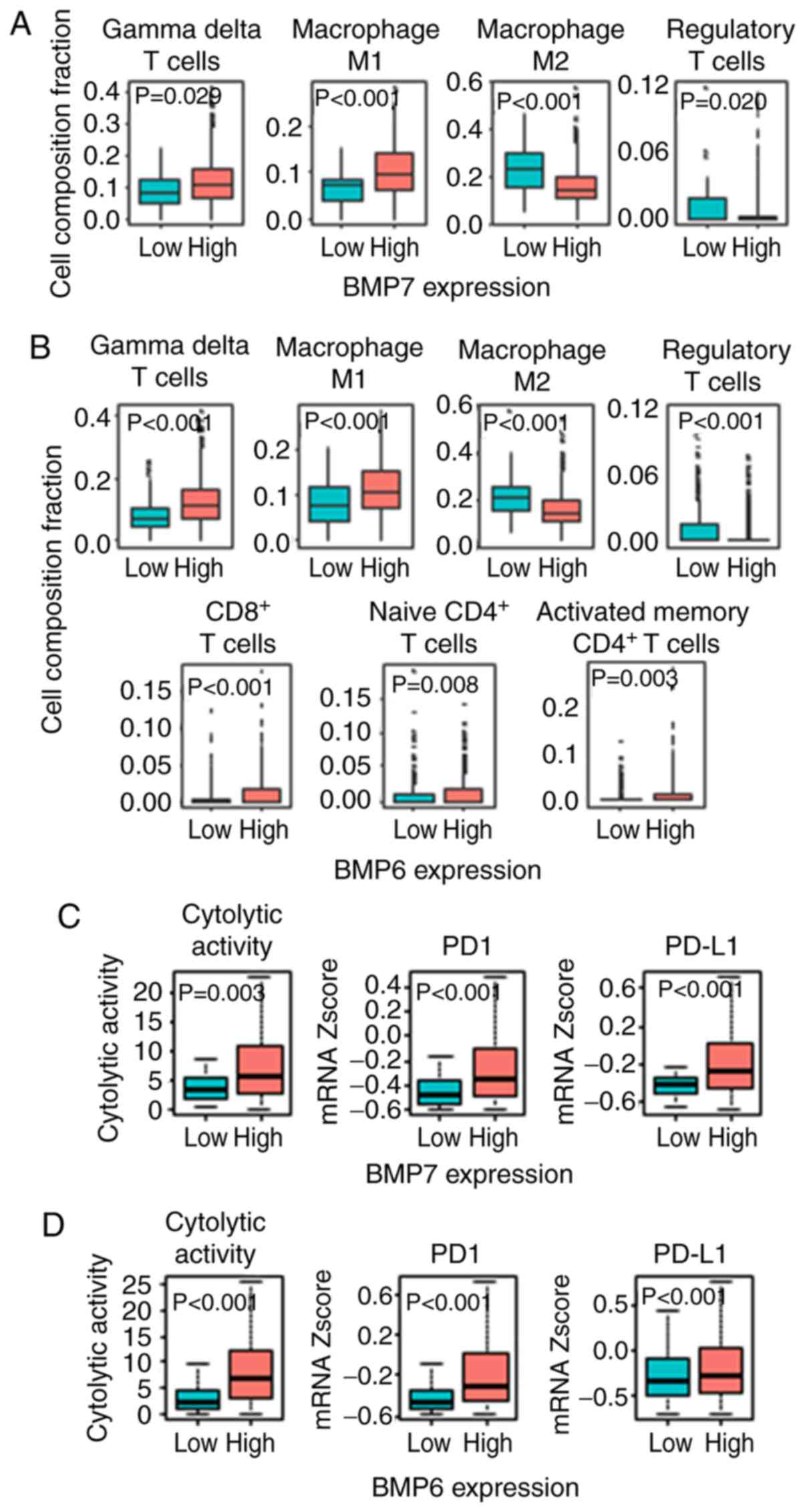
anticancer immune cell infiltration. Utilizing CIBERSORT algorithm
(25), we found higher anticancer
immune cell infiltration, such as gamma-delta (γδ) T cells
(P=0.029), M1 macrophages (P<0.001), and less pro-cancer immune
cell infiltrations, such as M2 macrophages (P<0.001) and Tregs
(P=0.020) in the BMP7 high ER+ tumors (Figs. 6A and S2). A similar trend was found in the BMP6
high ER+ tumors, where higher infiltration of anticancer
immune cells, not only γδ T cells (P<0.001) and M1 macrophages
(P<0.001), but also CD8+ T cells (P<0.001),
CD4+ naïve T cells (P=0.008) and CD4+
activated memory T cells (P=0.003), and less infiltration of
pro-cancer immune cells, M2 macrophages (P<0.001) and Tregs
(P<0.001) in the BMP6 high ER+ tumors were observed
(Figs. 6B and S3). Since higher anticancer immune cell
infiltration was observed in both BMP7 and BMP6 high expressing
ER+ tumors, we further analyzed the cytolytic activity
of those tumors. As shown in Fig. 6C and
D, high BMP7 as well as high BMP6 in the ER+ tumors
demonstrated higher cytolytic activity compared to those with low
expression tumors. These findings imply that ER+ tumors
with high BMP7 as well as high BMP6 expression have not only higher
immune cell infiltration, but also higher cytolytic activity,
resulting in better prognosis.
To ascertain whether these tumors are good
candidates for immune check point inhibition, PD1 and PD-L1
expressions were compared between BMP7 high and low as well as BMP6
high and low expression in the ER+ tumors. PD1 and PD-L1
expression levels were significantly higher in the BMP7 high
expressing as well as BMP6 high expressing ER+ tumors
(Fig. 6C and D). These findings
suggest that high expression of BMP7 and BMP6 in the ER+
tumors may be good candidates for immune check point
inhibition.
Discussion
In the present study, we found that each BMP was
associated differentially with OS of breast cancer patients. High
expression levels of BMP1, BMP3, BMP5, BMP7 and BMPR1A were
associated with better prognosis in the whole cohort of breast
cancer patients, whereas those of BMP6, BMP8A, BMP8B and BMPR1B
were associated with worse prognosis. BMP7 was associated with
better prognosis in both the ER+ and ER−
tumors. In contrast, higher BMP6 expression was found to be
associated with better prognosis in the ER+ tumors, but
was associated with worse prognosis in the ER− tumors.
High expression of BMP7 as well as BMP6 was associated with higher
immune response in the ER+ tumors, but not in the
ER− tumors.
BMPs play complex roles in cancer progression,
specifically in breast cancer (34).
Our findings, that high expression levels of BMP7 and BMP6 are
associated with better prognosis in ER+ breast cancer,
is consistent with previous reports. Utilizing breast cancer cell
lines, Takahashi et al demonstrated that BMP6 and BMP7
inhibit estrogen-induced proliferation (35). It was reported that higher BMP7
expression is associated with higher rates of bone metastasis,
however, lower BMP7 expression is associated with local relapse in
breast cancer (36). In our analysis
BMP7 expression was found to be associated with a better overall
survival, irrespective of ER status. In the same token, BMP6
expression has been demonstrated to suppress breast cancer
metastasis in both ER+ and ER− tumors
(37,38). It was also found that BMP6 silencing
increased chemo-resistance to doxorubicin by upregulating multiple
drug resistance glycoproteins in breast cancer (39). In our analysis, BMP6 expression was
found to be associated with worse survival in the whole patient
cohort. Notably, the reverse finding was observed in the
ER+ tumors, where BMP6 plays a tumor-suppressive role.
This suggests a complex interplay with the hormonal pathway.
There have been few reports that link BMP7 or BMP6
to immune response. During the wound healing process, BMP7 plays an
anti-fibrotic role by causing inflammation (40). BMP6 expression was found to be
elevated in the ovary of polycystic ovarian syndrome which is known
to be associated with inflammation, when compared to normal ovarian
tissues (41). Overall, the role of
BMPs in breast cancer remains poorly understood and is associated
with complex interactions with a multitude of pathways and
effectors that may act to completely alter the effector function of
BMP signaling.
Although novel findings were discovered, there are
limitations to the present study. First, our results were confirmed
in a few cohorts due to availability. Second, all of our findings
were based on gene expression of the primary tumor, thus
experimental approaches are needed to elucidate further molecular
mechanisms.
In the context of ER expression, individualized
patient BMP expression profiles may act as a prognostic tool to
guide therapy or may even be used as a therapeutic target in
itself. This revelation in findings will continue to evolve, and
the correlation between the BMP activation in regards to different
pathways and the effect on prognosis in the clinical realm remain
to be clearly elucidated.
Supplementary Material
Supporting Data
Acknowledgements
Not applicable.
Funding
This work was supported by NIH grant R01CA160688 to
KT and the National Cancer Institute (NCI) grant P30CA016056 and
U24CA232979 involving the use of Roswell Park Comprehensive Cancer
Center Shared Resources.
Availability of data and materials
Clinical data and gene expression from RNA sequence
in TCGA breast cancer cohort were downloaded through cBioPortal
(http://www.cbioportal.org/) and UCSC
Genome Browser (http://genome.ucsc.edu/). Clinical data and gene
expression of GSE1456 were downloaded from Gene Expression
Omnibus.
Author's contributions
EK conceptualized the research design and performed
the analysis and prepared the manuscript. AAM conceptualized the
research design and prepared the manuscript. LY carried out
supervision of the analyses. KT carried out supervision of the
analysis and prepared the manuscript. All authors read and approved
the manuscript and agree to be accountable for all aspects of the
research in ensuring that the accuracy or integrity of any part of
the work are appropriately investigated and resolved.
Ethics approval and consent to
participate
TCGA and GSE1456 include data which have been
de-identified and are publicly available cohorts, thus
institutional review board approval and patient consent were
waived.
Patient consent for publication
Not applicable.
Competing interests
The authors declare that they have no competing
interests.
Glossary
Abbreviations
Abbreviations:
|
BMP
|
bone morphogenetic protein
|
|
BMPR
|
BMP receptor
|
|
TCGA
|
The Cancer Genome Atlas
|
|
OS
|
overall survival
|
|
ER
|
estrogen receptor
|
|
EMT
|
epithelial-mesenchymal transition
|
|
Treg
|
regulatory T cell
|
|
GSEA
|
Gene set enrichment analysis
|
References
|
1
|
Guo X and Wang XF: Signaling cross-talk
between TGF-beta/BMP and other pathways. Cell Res. 19:71–88. 2009.
View Article : Google Scholar : PubMed/NCBI
|
|
2
|
Wozney JM, Rosen V, Celeste AJ, Mitsock
LM, Whitters MJ, Kriz RW, Hewick RM and Wang EA: Novel regulators
of bone formation: Molecular clones and activities. Science.
242:1528–1534. 1988. View Article : Google Scholar : PubMed/NCBI
|
|
3
|
Langenfeld E, Kong Y and Langenfeld J:
Bone morphogenetic protein 2 stimulation of tumor growth involves
the activation of Smad-1/5. Oncogene. 25:685–692. 2006. View Article : Google Scholar : PubMed/NCBI
|
|
4
|
Alarmo EL, Kuukasjärvi T, Karhu R and
Kallioniemi A: A comprehensive expression survey of bone
morphogenetic proteins in breast cancer highlights the importance
of BMP4 and BMP7. Breast Cancer Res Treat. 103:239–246. 2007.
View Article : Google Scholar : PubMed/NCBI
|
|
5
|
Cao Y, Slaney CY, Bidwell BN, Parker BS,
Johnstone CN, Rautela J, Eckhardt BL and Anderson RL: BMP4 inhibits
breast cancer metastasis by blocking myeloid-derived suppressor
cell activity. Cancer Res. 74:5091–5102. 2014. View Article : Google Scholar : PubMed/NCBI
|
|
6
|
Guo D, Huang J and Gong J: Bone
morphogenetic protein 4 (BMP4) is required for migration and
invasion of breast cancer. Mol Cell Biochem. 363:179–190. 2012.
View Article : Google Scholar : PubMed/NCBI
|
|
7
|
Alarmo EL, Pärssinen J, Ketolainen JM,
Savinainen K, Karhu R and Kallioniemi A: BMP7 influences
proliferation, migration, and invasion of breast cancer cells.
Cancer Lett. 275:35–43. 2009. View Article : Google Scholar : PubMed/NCBI
|
|
8
|
Pickup MW, Hover LD, Guo Y, Gorska AE,
Chytil A, Novitskiy SV, Moses HL and Owens P: Deletion of the BMP
receptor BMPR1a impairs mammary tumor formation and metastasis.
Oncotarget. 6:22890–22904. 2015. View Article : Google Scholar : PubMed/NCBI
|
|
9
|
Yang L, Pang Y and Moses HL: TGF-beta and
immune cells: An important regulatory axis in the tumor
microenvironment and progression. Trends Immunol. 31:220–227. 2010.
View Article : Google Scholar : PubMed/NCBI
|
|
10
|
Lee GT, Jung YS, Ha YS, Kim JH, Kim WJ and
Kim IY: Bone morphogenetic protein-6 induces castration resistance
in prostate cancer cells through tumor infiltrating macrophages.
Cancer Sci. 104:1027–1032. 2013. View Article : Google Scholar : PubMed/NCBI
|
|
11
|
Green TP, Fennell M, Whittaker R, Curwen
J, Jacobs V, Allen J, Logie A, Hargreaves J, Hickinson DM,
Wilkinson RW, et al: Preclinical anticancer activity of the potent,
oral Src inhibitor AZD0530. Mol Oncol. 3:248–261. 2009. View Article : Google Scholar : PubMed/NCBI
|
|
12
|
Chin L, Andersen JN and Futreal PA: Cancer
genomics: From discovery science to personalized medicine. Nat Med.
17:297–303. 2011. View Article : Google Scholar : PubMed/NCBI
|
|
13
|
Cerami E, Gao J, Dogrusoz U, Gross BE,
Sumer SO, Aksoy BA, Jacobsen A, Byrne CJ, Heuer ML, Larsson E, et
al: The cBio cancer genomics portal: An open platform for exploring
multidimensional cancer genomics data. Cancer Discov. 2:401–404.
2012. View Article : Google Scholar : PubMed/NCBI
|
|
14
|
Gao J, Aksoy BA, Dogrusoz U, Dresdner G,
Gross B, Sumer SO, Sun Y, Jacobsen A, Sinha R, Larsson E, et al:
Integrative analysis of complex cancer genomics and clinical
profiles using the cBioPortal. Sci Signal. 6:pl12013. View Article : Google Scholar : PubMed/NCBI
|
|
15
|
Terakawa T, Katsuta E, Yan L, Turaga N,
McDonald KA, Fujisawa M, Guru KA and Takabe K: High expression of
SLCO2B1 is associated with prostate cancer recurrence after radical
prostatectomy. Oncotarget. 9:14207–14218. 2018. View Article : Google Scholar : PubMed/NCBI
|
|
16
|
Hirose Y, Nagahashi M, Katsuta E, Yuza K,
Miura K, Sakata J, Kobayashi T, Ichikawa H, Shimada Y, Kameyama H,
et al: Generation of sphingosine-1-phosphate is enhanced in biliary
tract cancer patients and is associated with lymphatic metastasis.
Sci Rep. 8:108142018. View Article : Google Scholar : PubMed/NCBI
|
|
17
|
Young J, Kawaguchi T, Yan L, Qi Q, Liu S
and Takabe K: Tamoxifen sensitivity-related microRNA-342 is a
useful biomarker for breast cancer survival. Oncotarget.
8:99978–99989. 2017. View Article : Google Scholar : PubMed/NCBI
|
|
18
|
Hoki T, Katsuta E, Yan L, Takabe K and Ito
F: Low DMT1 expression associates with increased oxidative
phosphorylation and early recurrence in hepatocellular carcinoma. J
Surg Res. 234:343–352. 2019. View Article : Google Scholar : PubMed/NCBI
|
|
19
|
Sporn JC, Katsuta E, Yan L and Takabe K:
Expression of microRNA-9 is associated with overall survival in
breast cancer patients. J Surg Res. 233:426–435. 2019. View Article : Google Scholar : PubMed/NCBI
|
|
20
|
Katsuta E, Qi Q, Peng X, Hochwald SN, Yan
L and Takabe K: Pancreatic adenocarcinomas with mature blood
vessels have better overall survival. Sci Rep. 9:13102019.
View Article : Google Scholar : PubMed/NCBI
|
|
21
|
Pawitan Y, Bjöhle J, Amler L, Borg AL,
Egyhazi S, Hall P, Han X, Holmberg L, Huang F, Klaar S, et al: Gene
expression profiling spares early breast cancer patients from
adjuvant therapy: Derived and validated in two population-based
cohorts. Breast Cancer Res. 7:R953–R964. 2005. View Article : Google Scholar : PubMed/NCBI
|
|
22
|
Goldhirsch A, Wood WC, Coates AS, Gelber
RD, Thürlimann B, Senn HJ and Panel members: Strategies for
subtypes-dealing with the diversity of breast cancer: Highlights of
the St Gallen international expert consensus on the primary therapy
of early breast cancer 2011. Ann Oncol. 22:1736–1747. 2011.
View Article : Google Scholar : PubMed/NCBI
|
|
23
|
Subramanian A, Tamayo P, Mootha VK,
Mukherjee S, Ebert BL, Gillette MA, Paulovich A, Pomeroy SL, Golub
TR, Lander ES and Mesirov JP: Gene set enrichment analysis: A
knowledge-based approach for interpreting genome-wide expression
profiles. Proc Natl Acad Sci USA. 102:15545–15550. 2005. View Article : Google Scholar : PubMed/NCBI
|
|
24
|
Katsuta E, Yan L, Nagahashi M, Raza A,
Sturgill JL, Lyon DE, Rashid OM, Hait NC and Takabe K: Doxorubicin
effect is enhanced by sphingosine-1-phosphate signaling antagonist
in breast cancer. J Surg Res. 219:202–213. 2017. View Article : Google Scholar : PubMed/NCBI
|
|
25
|
Newman AM, Liu CL, Green MR, Gentles AJ,
Feng W, Xu Y, Hoang CD, Diehn M and Alizadeh AA: Robust enumeration
of cell subsets from tissue expression profiles. Nat Methods.
12:453–457. 2015. View Article : Google Scholar : PubMed/NCBI
|
|
26
|
Charoentong P, Finotello F, Angelova M,
Mayer C, Efremova M, Rieder D, Hackl H and Trajanoski Z: Pan-cancer
immunogenomic analyses reveal genotype-immunophenotype
relationships and predictors of response to checkpoint blockade.
Cell Rep. 18:248–262. 2017. View Article : Google Scholar : PubMed/NCBI
|
|
27
|
Rooney MS, Shukla SA, Wu CJ, Getz G and
Hacohen N: Molecular and genetic properties of tumors associated
with local immune cytolytic activity. Cell. 160:48–61. 2015.
View Article : Google Scholar : PubMed/NCBI
|
|
28
|
Narayanan S, Kawaguchi T, Yan L, Peng X,
Qi Q and Takabe K: Cytolytic activity score to assess anticancer
immunity in colorectal cancer. Ann Surg Oncol. 25:2323–2331. 2018.
View Article : Google Scholar : PubMed/NCBI
|
|
29
|
Ye L, Bokobza S, Li J, Moazzam M, Chen J,
Mansel RE and Jiang WG: Bone morphogenetic protein-10 (BMP-10)
inhibits aggressiveness of breast cancer cells and correlates with
poor prognosis in breast cancer. Cancer Sci. 101:2137–2144. 2010.
View Article : Google Scholar : PubMed/NCBI
|
|
30
|
Sudhan DR, Schwarz LJ, Guerrero-Zotano A,
Formisano L, Nixon MJ, Croessmann S, González Ericsson PI, Sanders
M, Balko JM, Avogadri-Connors F, et al: Extended adjuvant therapy
with neratinib plus fulvestrant blocks ER/HER2 crosstalk and
maintains complete responses of ER+/HER2+
breast cancers: Implications to the ExteNET trial. Clin Cancer Res.
25:771–783. 2019. View Article : Google Scholar : PubMed/NCBI
|
|
31
|
Athanasios F, Afrodite N, Effstratios P
and Demetrios K: Co-expression of bone morphogenetic protein 6 with
estrogen receptor a in endometriosis. Arch Gynecol Obstet.
285:1001–1007. 2012. View Article : Google Scholar : PubMed/NCBI
|
|
32
|
Shee K, Jiang A, Varn FS, Liu S, Traphagen
NA, Owens P, Ma CX, Hoog J, Cheng C, Golub TR, et al: Cytokine
sensitivity screening highlights BMP4 pathway signaling as a
therapeutic opportunity in ER(+) breast cancer. FASEB J.
33:1644–1657. 2019. View Article : Google Scholar : PubMed/NCBI
|
|
33
|
Liberzon A, Birger C, Thorvaldsdóttir H,
Ghandi M, Mesirov JP and Tamayo P: The molecular signatures
database (MSigDB) hallmark gene set collection. Cell Syst.
1:417–425. 2015. View Article : Google Scholar : PubMed/NCBI
|
|
34
|
Davies SR, Watkins G, Douglas-Jones A,
Mansel RE and Jiang WG: Bone morphogenetic proteins 1 to 7 in human
breast cancer, expression pattern and clinical/prognostic
relevance. J Exp Ther Oncol. 7:327–338. 2008.PubMed/NCBI
|
|
35
|
Takahashi M, Otsuka F, Miyoshi T, Otani H,
Goto J, Yamashita M, Ogura T, Makino H and Doihara H: Bone
morphogenetic protein 6 (BMP6) and BMP7 inhibit estrogen-induced
proliferation of breast cancer cells by suppressing p38
mitogen-activated protein kinase activation. J Endocrinol.
199:445–455. 2008. View Article : Google Scholar : PubMed/NCBI
|
|
36
|
Alarmo EL, Korhonen T, Kuukasjärvi T,
Huhtala H, Holli K and Kallioniemi A: Bone morphogenetic protein 7
expression associates with bone metastasis in breast carcinomas.
Ann Oncol. 19:308–314. 2008. View Article : Google Scholar : PubMed/NCBI
|
|
37
|
Yang S, Du J, Wang Z, Yuan W, Qiao Y,
Zhang M, Zhang J, Gao S, Yin J, Sun B and Zhu T: BMP-6 promotes
E-cadherin expression through repressing deltaEF1 in breast cancer
cells. BMC Cancer. 7:2112007. View Article : Google Scholar : PubMed/NCBI
|
|
38
|
Du J, Yang S, An D, Hu F, Yuan W, Zhai C
and Zhu T: BMP-6 inhibits microRNA-21 expression in breast cancer
through repressing deltaEF1 and AP-1. Cell Res. 19:487–496. 2009.
View Article : Google Scholar : PubMed/NCBI
|
|
39
|
Lian WJ, Liu G, Liu YJ, Zhao ZW, Yi T and
Zhou HY: Downregulation of BMP6 enhances cell proliferation and
chemoresistance via activation of the ERK signaling pathway in
breast cancer. Oncol Rep. 30:193–200. 2013. View Article : Google Scholar : PubMed/NCBI
|
|
40
|
Tandon A, Sharma A, Rodier JT, Klibanov
AM, Rieger FG and Mohan RR: BMP7 gene transfer via gold
nanoparticles into stroma inhibits corneal fibrosis in vivo. PLoS
One. 8:e664342013. View Article : Google Scholar : PubMed/NCBI
|
|
41
|
Schmidt J, Weijdegård B, Mikkelsen AL,
Lindenberg S, Nilsson L and Brännström M: Differential expression
of inflammation-related genes in the ovarian stroma and granulosa
cells of PCOS women. Mol Hum Reprod. 20:49–58. 2014. View Article : Google Scholar : PubMed/NCBI
|