Introduction
Primary liver cancer is responsible for a large
proportion of cancer-related deaths globally. More than half of all
hepatocellular carcinoma (HCC) patients are in China, and Chinese
HCC mortality rates are the highest in the world (1,2).
Despite the advances in clinical and experimental research on HCC,
the survival rate of HCC patients remains low (3). Emerging studies have reported that
carcinogenesis is a complex process that involves multiple
molecules (4). To this end, HCC
carcinogenesis must be more extensively elucidated, and it is
crucial to develop new approaches to improve HCC treatment and
increase the long-term survival rate of HCC patients.
mRNAs that encode proteins account for <2% of the
human transcriptome (5). Increasing
evidence indicates that even though many transcripts are not
translated into proteins, they function at the transcriptional
level. Long non-coding RNAs (lncRNAs) are a new class of
transcripts that consist of >200 nucleotides but are not
translated into proteins (6). Some
lncRNAs have been reported to be abnormally expressed in cancer,
while others have been found to be related to cancer occurrence and
development (7). LINC01554
has been reported as a significant lncRNA involved in the
pathogenesis of non-alcoholic fatty liver disease and oesophageal
cancer (8,9). In the present study, we found that
LINC01554 was significantly downregulated in HCC tissues,
and its expression levels were closely associated with the
prognosis of HCC patients. Moreover, LINC01554 inhibited HCC
proliferation and metastasis in vitro. Further experiments
were performed to identify the mechanism of LINC01554 in HCC
and determine its potential suitability as a diagnostic biomarker
for early HCC detection and a potential target for HCC therapy.
Materials and methods
Gene expression profiles
We downloaded gene expression profiling data from
the Gene Expression Omnibus database (GEO, http://www.ncbi.nlm.nih.gov/gds/). The GSE113850
dataset, which consists of 39 tissue samples including 32 HCC and 7
normal liver tissues, was used (10). We also downloaded gene expression
profiling data from The Cancer Genome Atlas database (TCGA,
http://portal.gdc.cancer.gov/). This
dataset consisted of 424 tissues, including 371 primary HCC, 3
recurrent HCC, and 50 normal liver tissues.
Screening of HCC patients for
differentially expressed genes (DEGs)
We used the GEO2R statistical analysis tool
(https://www.ncbi.nlm.nih.gov/geo/geo2r/) and Student's
t-test to identify DEGs in the GSE113850 dataset. The adjusted
P-value was set at <0.05, and the logFC value was set at >2
(upregulated genes) or <-2 (downregulated genes). Data from the
TCGA database were analysed using the R packages, DESeq and edgeR
(https://www.bioconductor.org/), and
Student's t-test to identify DEGs. Meanwhile, heat maps and volcano
plots were created by a hierarchical clustering method.
Comparison of the expression levels of
three lncRNAs and the clinicopathological characteristics of the
HCC patients
Gene Expression Profiling Interactive Analysis
(GEPIA, http://gepia.cancer-pku.cn/index.html) is an online
tool used for analysing RNA sequencing (RNA-Seq) data from TCGA and
GTEx databases. It includes RNA sequencing expression data of 9,736
tumors and 8,587 normal samples and related clinical datasets of
TCGA and the GTEx projects. It provides key interactive and
customizable functions including differential expression analysis,
profiling plotting, correlation analysis, patient survival
analysis, similar gene detection and dimensionality reduction
analysis (11). Using GEPIA, we
compared the gene expression levels of HCC and normal liver
tissues. We used log-rank test to analyse the correlation between
the levels of gene expression and the duration of patient survival.
Moreover, we analysed the gene expression levels according to the
tumour stage of the HCC patients. The adjusted P-value was set at
<0.05, and the datasets were set at LIHC (liver hepatocellular
carcinoma).
Clinical specimens and cell
culture
Forty patients (age range, 20–76 years; mean age,
54.78±10.64 years; males, 33; females, 7), who attended the
Zhongnan Hospital of Wuhan University from April 2018 to April
2019, were enrolled in this study. All patients were diagnosed with
HCC by histological examination, and they provided written informed
consent for their tissues to be used for medical research. Ethical
approval for this study was obtained from the Zhongnan Hospital of
Wuhan University. Liver cancer cell lines (SK-Hep1, HCCLM9 and
HepG2) were obtained from the Cell Bank of the Type Culture
Collection (Chinese Academy of Sciences, Shanghai, China). Cells
were cultured in Dulbecco's modified Eagles medium (DMEM;
Invitrogen; Thermo Fisher Scientific, Inc.), supplemented with 10%
foetal bovine serum (FBS) and 1% penicillin-streptomycin, at 37°C
in a 5% CO2 humidified atmosphere.
Lentiviral construction and cell
transfections
The sequence of lncRNA LINC01554 was sub-cloned into
the lentiviral expression vector Lv105 (GeneCopoeia, Guangzhou,
China). 293T cells were plated into 6-well plates. After 293T cell
confluence reached 80%, cells were co-transfected with
LINC01554-Lv105 (or control vector EX-EGFP-Lv105) and Lenti-Pac CMV
Expression Packaging vectors (gag-pol-RRE vector, REV vector and
VSV-G vector) (GeneCopoeia). The mass of each vector in each well
is 1.0 µg lentiviral plasmid, 1.2 µg gag-pol-RRE vector, 1.0 µg REV
vector, and 1.0 µg VSV-G vector. The transfection was performed by
using LipoFectMax™ Transfection Reagent (ABP Biosciences, USA).
After incubation for 48 h, the lentiviral particles were harvested
from the cell medium supernatant by centrifuging at 500 × g for 10
min. The SK-Hep1 and HCCLM9 cells were then transduced with
Lv-LINC01554 lentivirus controlled with the Lv-NC lentivirus.
Quantitative reverse transcription
polymerase chain reaction (RT-qPCR) analysis
Total RNA was isolated from the clinical specimens
and cells using TRIzol reagent (Yeasen Biotech). cDNA was generated
using a Hieff First Strand cDNA Synthesis Super Mix for RT-qPCR kit
(Yeasen Biotech). RT-qPCR analysis was performed using the Hieff
qPCR SYBR Green Master Mix kit (Yeasen Biotech) and a StepOnePlus
instrument (Applied Biosystems; Thermo Fisher Scientific, Inc.).
Relative lncRNA expression was analysed using the 2−ΔΔCq
method (12), with normalisation to
β-actin expression. Primer sequences used for RT-qPCR were as
follows: LINC01554 forward, 5′-TGTGGCAAACGCAAACGA-3′ and
reverse, 5′-GCCCAGAGTAAAGGGAAATGTA-3′; β-actin forward,
5′-TGGCACCCAGCACAATGAA-3′ and reverse,
5′-CTAAGTCATAGTCCGCCTAGAAGCA-3′.
Western blot analysis
Total protein was isolated from the HCCLM9 and
SK-Hep1 cells using RIPA lysis buffer (Wuhan Servicebio
Biotechnology). Protein concentrations were determined using the
bicinchoninic acid assay (Wuhan Servicebio Biotechnology). The mass
of protein loaded per lane was 25 µg. Proteins were separated using
10% SDS-PAGE gels and electro-transferred to PVDF membranes. The
membranes were then blocked by TBST (with 5% skim milk powder) for
2 h at room temperature and incubated overnight, respectively, with
primary antibodies at 4°C. After washing with TBST three times, the
membranes were incubated, respectively, with goat anti-rabbit
secondary antibody at 37°C for 2 h. The dilutions and suppliers of
the primary antibodies and secondary antibodies are documented in
Table I. Protein blots were
detected using a chemiluminescence kit (NCM Biotech) and Tanon 4500
Immunodetection System (Tanon). The protein bands were visualised
with chemiluminescence ECL reagent and recorded with X-ray film.
The signal intensity was analysed by ImageJ v1.42q software
(National Institutes of Health). GAPDH content was used to
standardise sample loading.
 | Table I.Diluted concentrations and supplier
information for the antibodies used for western blotting. |
Table I.
Diluted concentrations and supplier
information for the antibodies used for western blotting.
| Antibodies | Catalog no. |
Company/Supplier | Dilution |
|---|
| ZO-1 | 21773-1-AP | Wuhan Proteintech
Group | 1:1,000 |
| E-cadherin | AF0131 | Affinity
Biosciences | 1:1,000 |
| N-cadherin | GB11135 | Wuhan Servicebio
Biotechnology | 1:1,000 |
| Vimentin | GB11192 | Wuhan Servicebio
Biotechnology | 1:1,000 |
| AKT | 10176-2-AP | Wuhan Proteintech
Group | 1:1,000 |
| p-AKT | AF0908 | Affinity
Biosciences | 1:1,000 |
| GSK3β | GB11099 | Wuhan Servicebio
Biotechnology | 1:1,000 |
| p-GSK3β | 5558 | Cell Signaling
Technology | 1:1,000 |
| β-catenin | GB11015 | Wuhan Servicebio
Biotechnology | 1:1,000 |
| GAPDH | GB12002 | Wuhan Servicebio
Biotechnology | 1:1,000 |
| Goat anti-rabbit
IgG | GB23303 | Wuhan Servicebio
Biotechnology | 1:3,000 |
| Goat anti-mouse
IgG | GB23301 | Wuhan Servicebio
Biotechnology | 1:3,000 |
Flow cytometric analysis
After transfection, the cells were collected and
washed twice with phosphate-buffered saline (PBS). For cell cycle
analysis, the cells were stained with a cell cycle staining kit
(KeyGEN) for 15 min. Apoptosis was detected using an Annexin V-FITC
and Propidium Iodide Apoptosis Detection Kit (KeyGEN). Finally, the
proportion of cells in different phases of the cell cycle and the
apoptosis rate were measured using a FACSCalibur flow cytometer (BD
Biosciences).
Fluorescence in situ hybridisation
(FISH)
Briefly, the SK-Hep1 and HCCLM9 cells were first
fixed with 4% formaldehyde. They were then placed on slides, and
hybridisation reactions were performed using LINC01554-,
U6-, and 18S rRNA-specific probes. Cells were then
counterstained with DAPI. Fluorescence images were captured using a
confocal microscope. The FAM-labelled LINC01554,
TAMRA-labelled 18S rRNA, and TAMRA-labelled U6 probe
sequences were 5′-ACTTCCCGACTGCTCATCAACCGACCTCCCTGGGGCCGAGAGGCA-3′,
5′-CGCTGAGCCAGTCAGTGTAGCGCGCGTGCAGCCCCGGACATCTAAGGGCATCACAGACCTGTTATTGCTCAATCTCGGGTGGCTGAACGCCACTTGTCCCTCTAAGAAGTTGGGGGACGCCGACCGCTCGGGGGTCGCGTAACTAGTTAGCATGCCAGAGTCTCGTTCGTTATCGGAATTAACCAGACAAATCGCTCCACCAACTAAGAACGGCCAT-3′
and
5′-AAAAATATGGAACGCTTCACGAATTTGCGTGTCATCCTTGCGCAGGGGCCATGCTAATCTTCTCTGTATCGTTCCAATTTTAGTATATGTGCTGCCGAAGCGAGCAC-3′,
respectively. The probes were purchased from AxlBioCo., Ltd.
Cell Counting Kit-8 (CCK-8) assay
Cells in the logarithmic growth phase were digested
with trypsin and seeded (2×10³) in a 96-well plate.
CCK-8 reagent (10 µl) (Dojindo Molecular Technologies) was added to
each well at 0, 24, 48, and 72 h. After incubating the cells with
the CCK-8 reagent for 4 h, we measured the optical density (OD) at
450 nm.
Scratch test assay
Cells were seeded in 6-well plates at 50% density of
the cells. When they were approximately 90% confluent, the tip of a
sterile 1,000-µl pipette was used to scratch a line wound. Cells
that migrated to the wound region were observed under a microscope
at 0 and 24 h.
Transwell assays
A 24-well Transwell chamber with Matrigel was used
to evaluate the cell invasion rates. Cells (5×10⁴) from
each group were added to the upper Transwell chamber, and DMEM (600
µl), containing 10% FBS, was added to the lower Transwell chamber.
Cells were then incubated at 37°C in 5% CO2 for 48 h.
Finally, after fixation with 4% paraformaldehyde for 20 min and
crystal violet staining for 15 min at room temperature, the number
of cells passing through the membrane was observed under a light
microscope. Cells were counted in five random fields, and the
average number of cells per field was calculated.
Gene set enrichment analysis
(GSEA)
Thirty-two HCC samples and 7 normal liver samples
from GSE113850 were divided into two groups (HCC vs. NC). In order
to investigate the further function of LINC01554, GSEA (http://software.broadinstitute.org/gsea/index.jsp) was
carried out between the two groups. Annotated gene were selected as
the reference gene sets. False discovery rate (FDR) <0.25, and
gene size ≥100 were regarded as the cut-off criteria.
Bioinformatics analysis of the
co-expressed genes (CEGs)
Co-LncRNA (http://bio-bigdata.hrbmu.edu.cn/Co-LncRNA/) is a
web-based computational tool that predicts the CEGs of single or
multiple lncRNAs. We analysed the same RNA-Seq datasets to identify
the CEGs. The regression/correlation method was set as Linear
Regression. The corresponding coefficient threshold values were set
at >2, and the adjusted P-value was set at <0.01.
Gene Ontology (GO) and Kyoto
Encyclopaedia of Genes and Genomes (KEGG) pathway analyses of
CEGs
The GO resource (http://www.geneontology.org/) describes gene functions
and groups them into three categories: Molecular function, cellular
component, and biological process. KEGG (http://www.genome.jp/kegg/) contains a set of genomes
and biological pathways that provide information on genome
functions and the relationships among genes. We visualised all
known GO functional annotations and KEGG pathways of the CEGs using
DAVID (http://david.ncifcrf.gov). GO and KEGG
enrichment plots were also created.
Statistical analysis
Statistical analysis was performed using the SPSS
22.0 software (IBM Corp.). All experiments were performed in
triplicate, and all data are presented as means ± standard error of
the mean. All data are presented as a comparison between the groups
and analysed by Student's t-test or analysis of variance (ANOVA).
Receiver operating characteristic (ROC) curves were used to
determine the correlation between the true- and false-positive
rates, and this was important for assessing the diagnostic value of
the genes. Results were regarded as statistically significant at
P<0.05.
Results
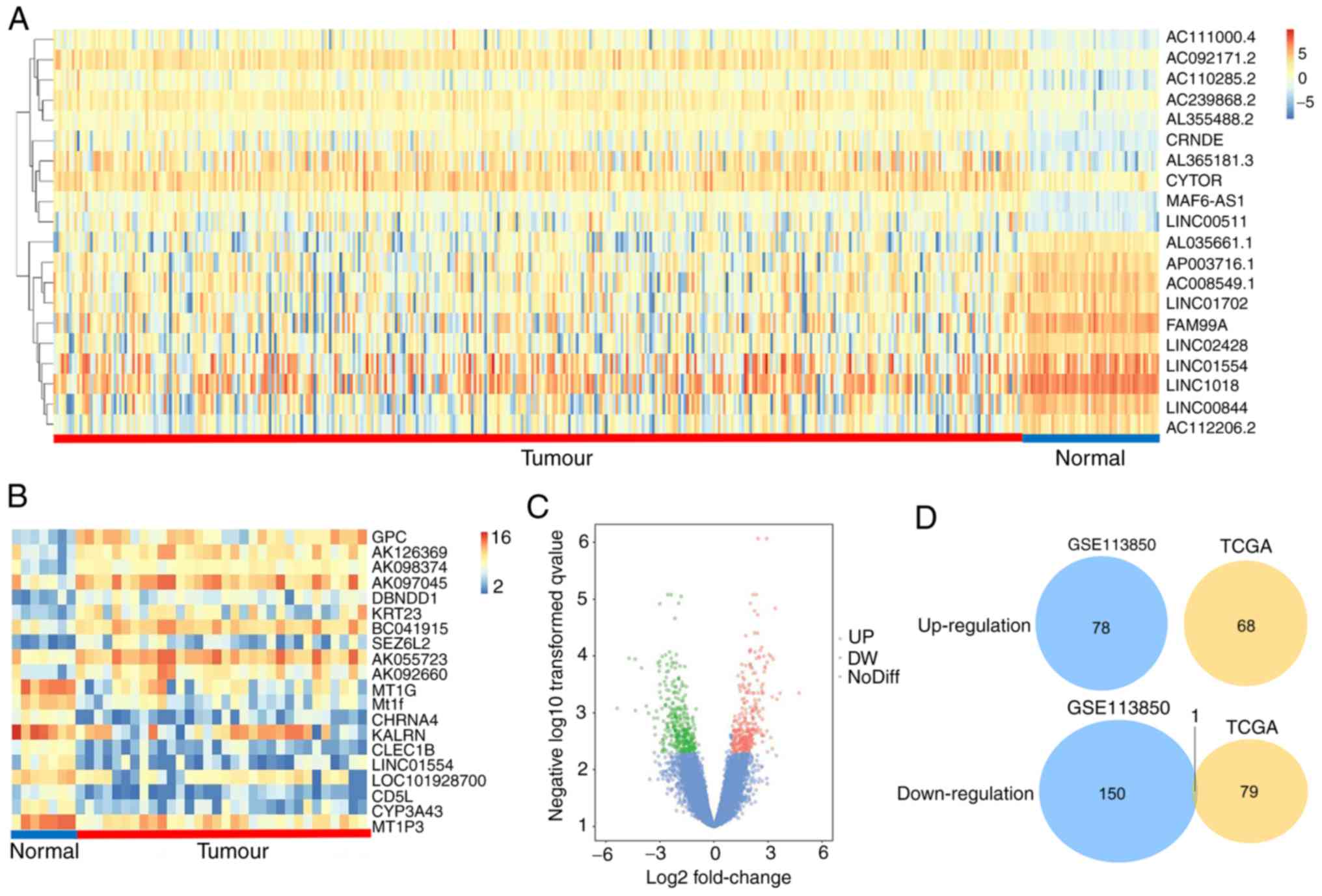
Identification of the DEGs
After analysing the GSE113850 dataset, we identified
229 DEGs, of which 151 were downregulated and 78 were upregulated,
as shown in a volcano plot (Fig.
1C). Hierarchical clustering analysis of the top 10 DEGs with a
fold change >2 is presented as a heat map (Fig. 1B). Three lncRNA genes (DIO3OS,
LINC01554, and LINC01093) were among these DEGs. After
analysing TCGA data, we identified 148 differentially expressed
lncRNAs, and hierarchical clustering analysis of the top 10 lncRNAs
with a fold change >1 is presented as a heat map (Fig. 1A). The Venn plot shows that
LINC01554 was the only differentially expressed lncRNA in
both the GSE113850 and TCGA datasets (Fig. 1D).
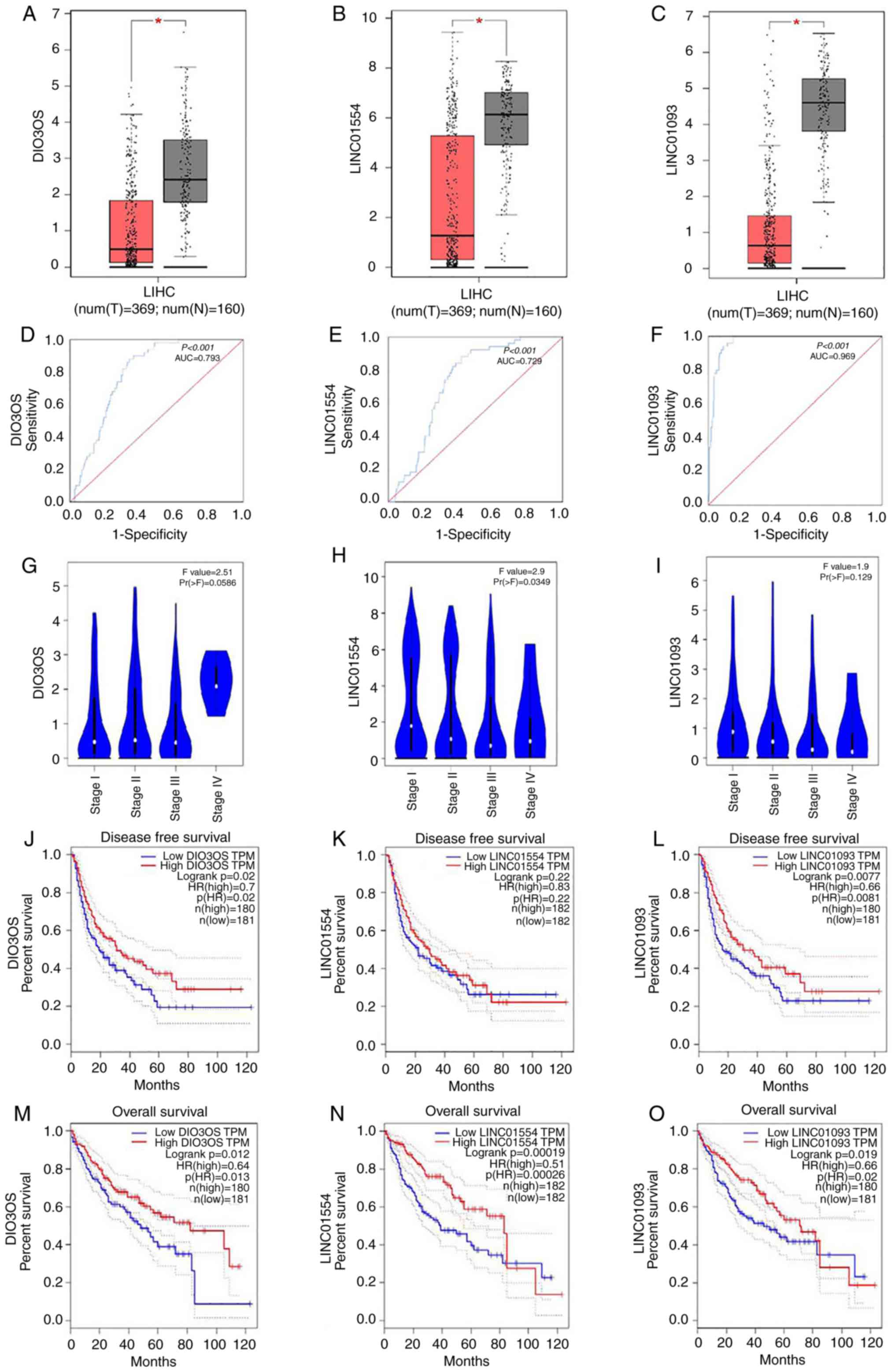
Relationship between the expression
levels of three lncRNAs and the prognosis of HCC patients
Using GEPIA, we compared the expression levels of
the three lncRNAs, DIO3OS, LINC01554 and LINC01093,
in the HCC and normal liver tissues. The results showed that the
lncRNA expression levels were lower in HCC tissues than these in
the normal liver tissues (Fig.
2A-C). The area under the curve (AUC) values for DIO3OS,
LINC01554 and LINC01093 were 0.793, 0.729 and 0.969,
respectively (Fig. 2D-F),
indicating a moderate diagnostic value of their expression levels
in HCC. These results were based on the original data from TCGA. We
also analysed the lncRNA expression levels according to the tumour
stage of HCC patients. Among the three lncRNAs, LINC01554
showed expression levels that significantly differed according to
the HCC stage (Fig. 2G-I). We
further explored the effects of these lncRNAs on the survival of
HCC patients. These results showed that decreased DIO3OS
LINC01554 and LINC01093 were significantly correlated
with disease-free survival (DFS) of all HCC patients (Fig. 2J-L), and decreased levels of all
three lncRNAs were significantly correlated with overall survival
(OS) of all HCC patients (Fig.
2M-O). Meanwhile, we also explored the correlation between
LINC01554 expression and the clinicopathological features in
40 HCC patients to evaluate the clinical role of LINC01554
in HCC (Table II). Lower
LINC01554 expression levels were strongly linked to certain
clinical characteristics, including HBV infection (P=0.0044),
pathological stage (P=0.0079), tumour size (P=0.0252), portal vein
tumour thrombus (P=0.0471), and TNM stage (P=0.0022).
| Table II.Associations of LINC1554
expression with clinicopathological parameters in HCC. |
Table II.
Associations of LINC1554
expression with clinicopathological parameters in HCC.
|
|
| LINC01554
expression |
|
|---|
|
|
|
|
|
|---|
| Variable | No. of cases | Low (n=20) | High (n=20) | P-value |
|---|
| Sex |
|
|
| 0.4075 |
|
Male | 33 | 15 | 18 |
|
|
Female | 7 | 5 | 2 |
|
| Age (years) |
|
|
| 0.2351 |
|
≤50 | 8 | 6 | 2 |
|
|
>50 | 32 | 14 | 18 |
|
| HBV infection |
|
|
|
0.0044a |
|
Negative | 19 | 5 | 14 |
|
|
Positive | 21 | 15 | 6 |
|
| Serum AFP
(ng/ml) |
|
|
| 0.1274 |
|
≤8.78 | 9 | 2 | 7 |
|
|
>8.78 | 31 | 18 | 13 |
|
| Pathological
stage |
|
|
|
0.0079a |
|
I+II | 25 | 8 | 17 |
|
|
III+IV | 15 | 12 | 3 |
|
| Cirrhosis |
|
|
| 0.7233 |
| No | 11 | 6 | 5 |
|
|
Yes | 29 | 14 | 15 |
|
| Tumour number |
|
|
| 0.1274 |
|
Singular | 31 | 18 | 13 |
|
|
Multifocal | 9 | 2 | 7 |
|
| Tumour size
(cm) |
|
|
|
0.0252a |
| ≤5 | 23 | 8 | 15 |
|
|
>5 | 17 | 12 | 5 |
|
| PVTT |
|
|
|
0.0471a |
| No | 35 | 15 | 20 |
|
|
Yes | 5 | 5 | 0 |
|
| TNM (AJCC) |
|
|
|
0.0022a |
|
I+II | 26 | 8 | 18 |
|
|
III+IV | 14 | 12 | 2 |
|
LINC01554 expression is downregulated in
human HCC samples. We analysed LINC01554 expression in 40
paired HCC and normal tumour-adjacent tissue samples by RT-qPCR.
The results showed that LINC01554 expression was
significantly lower in the HCC samples (P<0.01, Fig. 3B), and downregulated
LINC01554 expression was observed in 70.0% (28/40) of the
HCC samples (Fig. 3A). We also
investigated LINC01554 expression in the liver cancer cell
lines (SK-Hep1, HepG2, and HCCLM9). RT-qPCR results showed that
LINC01554 expression was relatively lower in the SK-Hep1 and
HCCLM9 cells than in the HepG2 cells (Fig. 3C). Therefore, SK-Hep1 and HCCLM9
cells were used for subsequent experiments. To study
LINC01554 functions in HCC cell lines, a recombinant
lentivirus that carried the LINC01554 DNA sequence
(Lv-LINC01554) and a recombinant lentivirus without the
LINC01554 DNA sequence (Lv-NC), as a control, were
transfected into HCC cells. Transfection efficiencies were
confirmed by RT-qPCR (P<0.01; Fig.
3D).
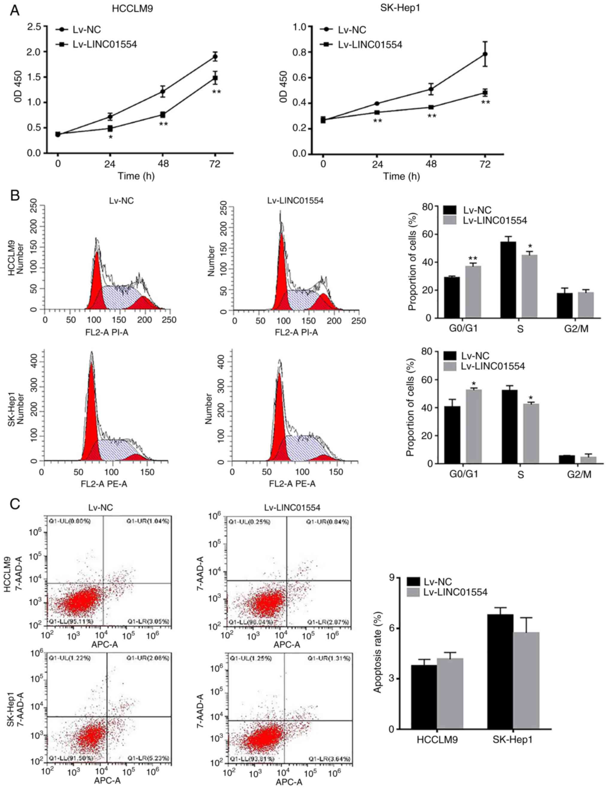
LINC01554 overexpression inhibits
SK-Hep1 and HCCLM9 cell proliferation and promotes G0/G1 arrest,
but it does not significantly affect apoptosis
According to the CCK-8 assay results, the OD450
values of the SK-Hep1 and HCCLM9 cell lines in the
Lv-LINC01554 group at 0, 24, 48 and 72 h were significantly
lower than those in the Lv-NC group (P<0.01; Fig. 4A). We examined the distribution of
cells at different stages of the cell cycle using flow cytometry.
The results revealed that LINC01554 overexpression promoted
G0/G1 arrest (P<0.05; Fig. 4B),
but it did not significantly affect apoptosis (Fig. 4C).
LINC01554 inhibits
epithelial-mesenchymal transition (EMT) and tumour invasion in the
SK-Hep1 and HCCLM9 cells
Wound-healing and Transwell invasion assays were
used to investigate the role of LINC01554 in HCC cell
migration and invasion. Scratch assays showed that the migration
rate of the Lv-LINC01554 transfection group was
significantly lower than that of the Lv-NC transfection group
(P<0.01; Fig. 5A). Transwell
experimental results showed that the number of migrated cells in
the Lv-LINC01554 transfection group was significantly lower
than that in the Lv-NC transfection group (P<0.01; Fig. 5B). Next, we assessed EMT-related
gene expression in the HCCLM9 and SK-Hep1 cells. The results of the
western blot analysis showed that LINC01554 overexpression
led to increased ZO-1 and E-cadherin expression, but decreased
N-cadherin and vimentin expression (Figs. 5C and S1). Moreover, FISH assays showed that
LINC01554 was mainly located in the nuclei of the SK-Hep1
and HCCLM9 cells (Fig. 5D).
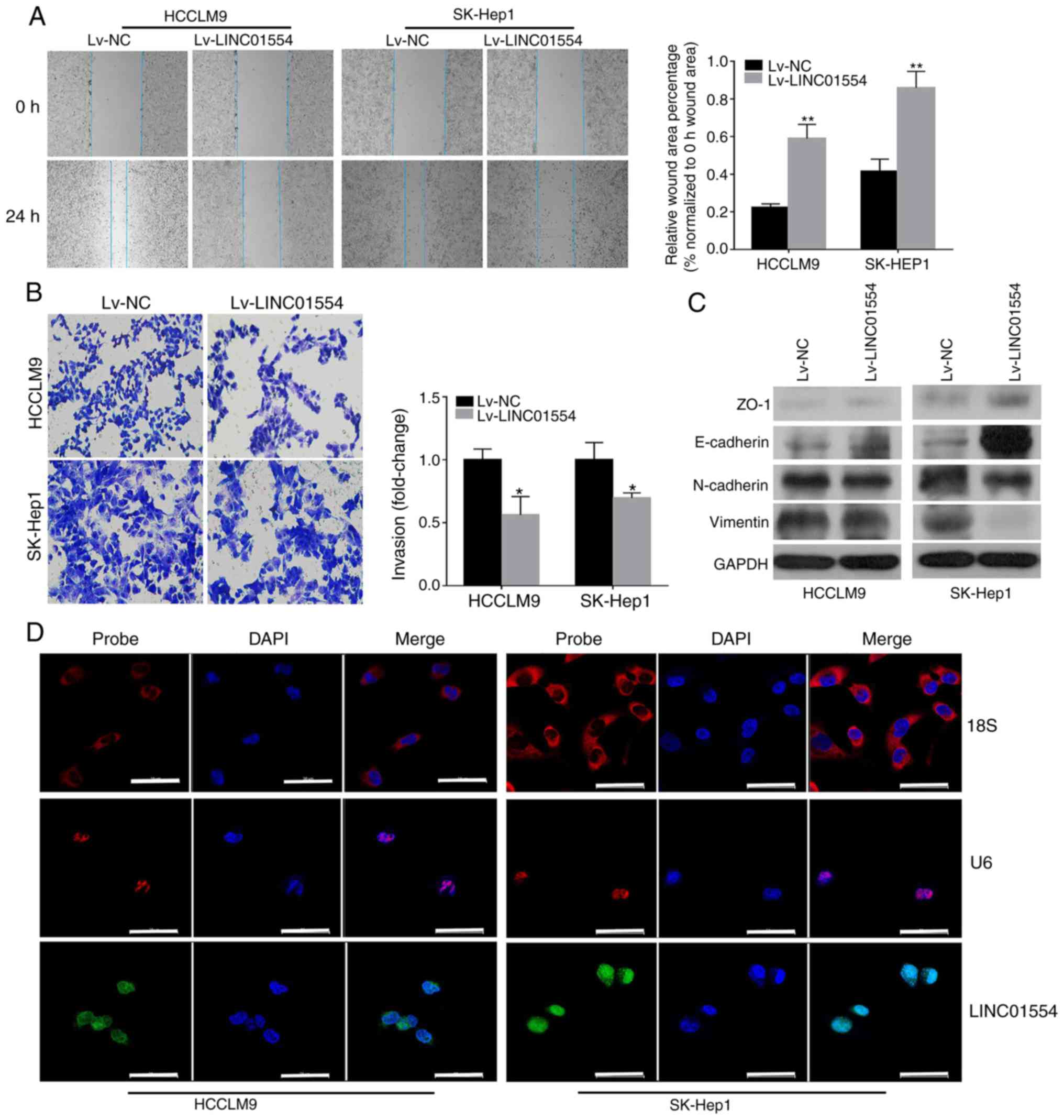 | Figure 5.LINC01554 overexpression
suppresses migration, invasion, and EMT-related gene expression in
HCC cells. (A) Wound-healing assay in
LINC01554-overexpressing SK-Hep1 and HCCLM9 cells. (B)
Invasion ability of LINC01554-overexpressing SK-Hep1 and
HCCLM9 cells. (C) Expression levels of EMT markers (ZO-1,
E-cadherin, N-cadherin and vimentin) in
LINC01554-overexpressing HCCLM9 and SK-Hep1 cells are
analysed by western blotting. (D) FISH analysis of LINC01554
in the SK-Hep1 and HCCLM9 cells. The U6 and 18S rRNA
probes are labelled red, the LINC01554 RNA probe is labelled
green, and nuclear DNA with DAPI staining is labelled blue. Scale
bar, 50 µm. All results are from at least three independent
experiments. *P<0.05, **P<0.01, comparison with the Lv-NC
group. HCC, hepatocellular carcinoma; EMT, epithelial-mesenchymal
transition. |
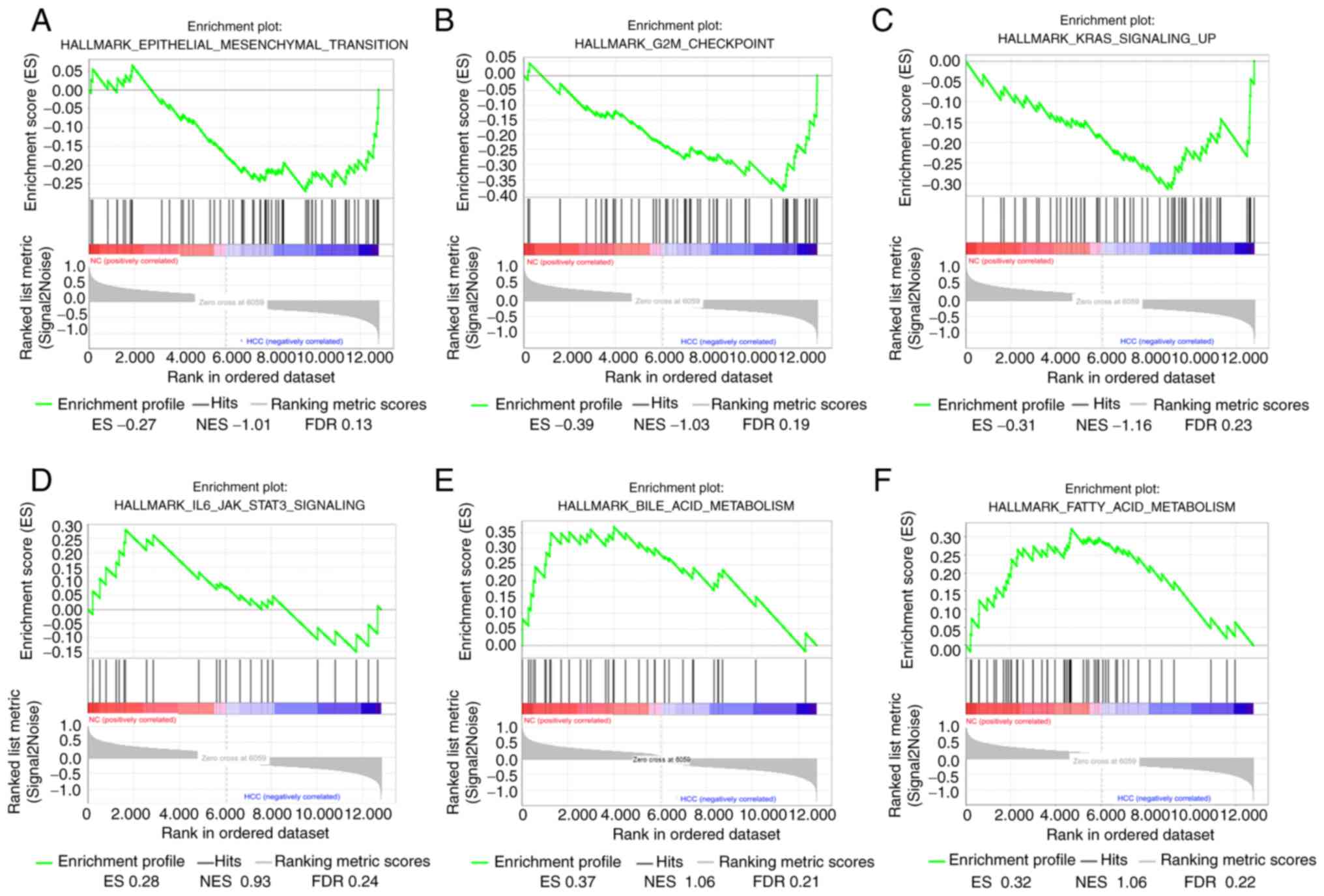
Gene set enriched analysis
GSEA was used to map into 6 functional gene sets. A
total of 6 functional gene sets were enriched, which were mainly
‘Epithelial mesenchymal transition’, ‘G2M checkpoint’, ‘Kras
signaling up’, ‘IL6/JAK/STAT3 signaling’, ‘Bile acid metabolism’
and ‘Fatty acid metabolism’ (Fig.
6).
GO and KEGG pathway enrichment
analysis
We analysed the GO functions and KEGG pathways of
the genes co-expressed with LINC01554 using the DAVID online
database. Only the top 10 pathways for each category are listed
(Fig. 7A-D). GO analysis showed
that the CEGs were mainly enriched in ‘intracellular signal
transduction’, ‘transcription, DNA-templated’, ‘negative regulation
of transcription by the RNA polymerase II promoter’, ‘negative
regulation of cell proliferation’, ‘response to hypoxia’,
‘canonical Wnt signalling pathway’, ‘cell migration’, and ‘cell
cycle arrest’. KEGG pathway analysis showed that these genes were
related to PI3K-Akt, Ras, Wnt, chemokine, and tumour necrosis
factor (TNF) signalling pathways; viral carcinogenesis; and
toxoplasmosis, hepatitis B, and non-alcoholic fatty liver disease
pathways.
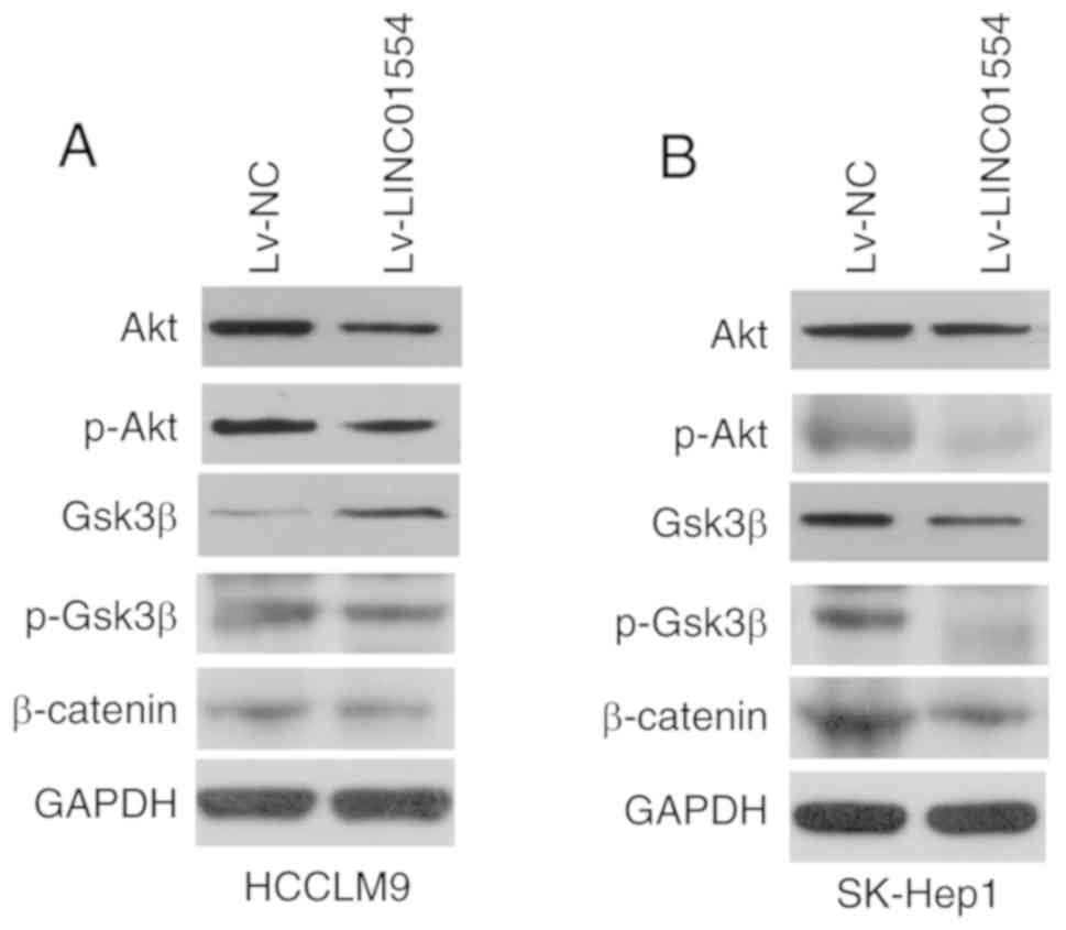
LINC01554 overexpression affected the
expression of proteins related to Wnt and PI3K-Akt signalling in
HCC cells
To examine whether LINC01554 exerts its
function through Wnt and PI3K-Akt signalling in HCC cells, the
expression levels of proteins related to these pathways (Akt,
p-Akt, Gsk3β, p-Gsk3β and β-catenin) were measured in the
LINC01554-overexpressing HCCLM9 and SK-Hep1 cells (Figs. 8A and B and S2). The results showed that
LINC01554 overexpression inhibited Akt, p-Akt, β-catenin,
and p-Gsk3β expression.
Discussion
As key regulators of tumour pathogenesis, long
non-coding RNAs (lncRNAs) are involved in the multilevel regulation
of gene expression in cancer (13).
However, the lncRNAs with aberrant expression in HCC remain
unknown. In the present study, we screened hepatocellular carcinoma
(HCC) patients for differentially expressed genes (DEGs) from the
Gene Expression Omnibus (GEO) dataset, GSE113850, and found that
DIO3OS, LINC01554, and LINC01093 were in the top 30
DEGs. Based on a literature review, we chose DIO3OS,
LINC01554, and LINC01093 for further investigation as
they showed relevant biological functions. For example, previous
studies have shown that DIO3OS is highly expressed in
pancreatic cancer and DIO3OS was found to regulate
pancreatic cancer cell proliferation and metastasis by affecting
the miR-122/ALDOA axis (14). LINC01093 is a liver-specific
lncRNA that was found to be significantly downregulated in HCC
samples and could inhibit HCC cell growth and metastasis (15). LINC01554 downregulation was
found to promote high aerobic glycolysis in cancer cells and cell
growth by regulating the PKM2 and Akt/mTOR signalling pathways
(16). We used GEPIA datasets to
verify the differences in DIO3OS, LINC01554, and
LINC01093 expression levels between the HCC and normal liver
tissues. We further explored the effects of DIO3OS,
LINC01554, and LINC01093 expression levels on overall
survival (OS) and disease-free survival (DFS). These results showed
that decreased DIO3OS, LINC01554, and LINC01093
levels were significantly correlated with OS and decreased
DIO3OS levels were significantly associated with DFS. The
AUC values of DIO3OS, LINC01554, and LINC01093
indicated that their expression levels were of moderate diagnostic
value in HCC. Therefore, we suggested that these three lncRNAs
could be used as tumour biomarkers of HCC. Meanwhile, we also
screened the patients for differentially expressed lncRNAs using
TCGA datasets. Venn plots showed that LINC01554 was a common
differentially expressed lncRNA in the GSE113850 and TCGA datasets.
Moreover, we found that lower LINC01554 expression levels were
positively correlated with pathological stage, hepatitis B
infection, tumour size, portal vein tumour thrombus, and TNM stage
in 40 HCC patients. Therefore, we chose LINC01554 as the
focus of our subsequent experiments.
A search of the NCBI databases (https://www.ncbi.nlm.nih.gov/) revealed that
LINC01554 is a 1,931-bp lncRNA, with three exons, encoded by
a gene on chromosome 5q15. Using RT-qPCR, we confirmed that
LINC01554 expression levels were significantly lower in the
HCC tissues than these levels in the paired normal tumour-adjacent
tissues obtained from 40 HCC patients. This finding was
consistently observed in the GSE113850 and TCGA datasets. A
gain-of-function assay further showed that LINC01554
inhibited HCCLM9 and SK-Hep1 cell proliferation, migration, and
invasion and promoted G0/G1 arrest, but it did not significantly
affect apoptosis. Cancer cell proliferation and metastasis are
known to play significant roles in cancer development, and the
adhesion of cancer cells in the microcirculation is related to
cancer metastasis through the vessels (17,18).
Epithelial-mesenchymal transition (EMT) is an important step in
liver cancer metastasis, and extracellular matrix (ECM)-receptor
interactions lead to the direct or indirect control of cellular
adhesion and migration (19,20).
Moreover, EMT is an early event that is critical for cancer
invasion and metastasis (21). In
our study, LINC01554 affected the expression levels of EMT
markers in the HCCLM9 and SK-Hep1 cells. LINC01554
overexpression promoted the expression of epithelial markers
(E-cadherin and ZO-1) but inhibited the expression of mesenchymal
markers (N-cadherin and vimentin) in the HCCLM9 and SK-Hep1 cells.
These findings strongly implicate the downregulation of
LINC01554 in the metastatic growth of HCC.
The mechanism of action of lncRNAs is mainly
associated with their subcellular localisation. Nuclear lncRNAs
mostly show effects at the transcriptional levels, while
cytoplasmic lncRNAs mostly function as endogenous sponges for
miRNAs or show effects at post-transcriptional levels (22,23).
Furthermore, FISH assays showed that LINC01554 is mainly
located in the nuclei of HCC cells, indicating its function in
transcriptional regulation. To gain a better understanding of the
potential molecular mechanism of LINC01554 in HCC, we
performed LINC01554 co-expression analysis using the
Co-LncRNA website. lncRNA-mRNA co-expression analyses are commonly
used to identify potential target genes of lncRNAs and research the
molecular mechanisms of lncRNAs further (24). We identified 139 genes as potential
LINC01554 targets using this analysis. Meanwhile, we
performed GO and KEGG pathway analyses of these 139 genes using the
DAVID online database. The analyses identified multiple signalling
pathways that involved these genes. The pathways included PI3K-Akt,
Ras, Wnt, chemokine, and TNF signalling pathways; viral
carcinogenesis, and hepatitis B disease pathway. Importantly, the
abnormal activation of the Wnt and PI3K/Akt pathways has been
widely reported in tumour development. The Wnt signalling pathway
is commonly affected in HCC of different aetiologies, which most
likely involve common pathogenetic mechanisms. Furthermore, the Wnt
signalling pathway is reported to have a close association with EMT
(25,26). Emerging evidence indicates that some
lncRNAs participate in tumour invasion and metastasis by regulating
the Wnt/β-catenin signalling pathway and inhibiting E-cadherin
expression. The mitogen-activated protein kinase (MAPK) and
RAS/RAF/MEK/ERK pathways, which regulate cell growth and involve a
number of receptors such as epidermal growth factor receptor (EGFR)
and Kirsten rat sarcoma viral oncogene homologue (KRAS), also play
key roles in HCC development (27–30).
OTUD6B was found to inhibit clear cell renal cell carcinoma by
regulating the Wnt/β-catenin pathway and suppressing EMT-related
protein expression (31). Moreover,
it has been reported that the PI3K/Akt signalling pathway mediates
EMT to increase tumour aggressiveness (32,33).
PI3K/Akt promotes non-small-cell lung carcinoma metastasis by
regulating EMT (34). Additionally,
emerging studies have reported that some lncRNAs promote cancer
progression by regulating the PI3K/Akt pathway (35). One study demonstrated that knockdown
of the lncRNA NR027113 inhibited the activity of the
PI3K/Akt signalling pathway and restrained EMT. Furthermore, this
study reported that PTEN silencing promoted Akt protein
phosphorylation and HCC cell function (36). These data suggest that the Wnt and
PI3K/Akt pathways are tightly linked to each other and both promote
EMT. In the present study, the Wnt and PI3K/Akt pathways were
repressed by LINC01554 overexpression in in vitro
investigations. However, the mechanism by which LINC01554
regulates EMT through the Wnt and PI3K/Akt pathways warrants
further exploration. KEGG pathway analysis also showed that genes
co-expressed with LINC01554 were associated with
precancerous diseases, such as hepatitis B virus infection and
viral carcinogenesis, which are factors critical for triggering
liver carcinogenesis (37). These
studies consistently suggest that low LINC01554 levels may
play a role in tumour pathogenesis.
The present study has some limitations. Firstly, the
functions and potential mechanisms of LINC01554 in HCC were
not confirmed in vivo. We did perform inhibition experiments
of LINC01554. Regrettably, the efficiencies of interference were
extremely low. The possible reason may be the location of LINC01554
in the nucleus of HCC cells. Secondly, the experimental validation
of LINC01554 expression was only performed in 40 pairs of
HCC and matched normal tissues. Therefore, investigations and
corresponding experiments with a larger clinical sample size will
be the focus of our future research.
In conclusion, we report the novel finding that
LINC01554 inhibited HCC cell proliferation, migration, and
invasion and promoted G0/G1 arrest. Taken together, our findings
provide new insights into the molecular mechanisms underlying HCC
tumourigenesis and implicated LINC01554 as a potential
target for HCC therapy.
Supplementary Material
Supporting Data
Acknowledgements
Not applicable.
Funding
This project was supported by the Health Commission
of the Hubei Province Scientific Research Project (grant no.
WJ2019H074), The Medical Science Advancement Program (Clinical
Medicine) of Wuhan University (grant no. TFLC2018003) and National
Key Laboratory of Virology (grant no. 2018KF005).
Availability of data and materials
The datasets used during the present study are
available from the corresponding author upon reasonable
request.
Authors' contributions
QY and GP designed the research and revised the
manuscript. LL, KH and ZL performed the experiments and wrote the
draft manuscript. LL, KH, ZL, HZ and HL analyzed the experimental
results. All authors read and approved the manuscript and agree to
be accountable for all aspects of the research in ensuring that the
accuracy or integrity of any part of the work are appropriately
investigated and resolved.
Ethics approval and consent to
participate
All patients provided written informed consent for
their tissues to be used for medical research. Ethical approval
(2019073) for this study was obtained from the Zhongnan Hospital of
Wuhan University (Wuhan, Hubei, China).
Patient consent for publication
Not applicable.
Competing interests
The authors declare that they have no competing
interests.
References
|
1
|
De Martel C, Maucort-Boulch D, Plummer M
and Franceschi S: World-wide relative contribution of hepatitis B
and C viruses in hepatocellular carcinoma. Hepatology.
62:1190–1200. 2015. View Article : Google Scholar : PubMed/NCBI
|
|
2
|
Chen W, Zheng R, Baade PD, Zhang S, Zeng
H, Bray F, Jemal A, Yu XQ and He J: Cancer statistics in China,
2015. CA Cancer J Clin. 66:115–132. 2016. View Article : Google Scholar : PubMed/NCBI
|
|
3
|
Aravalli RN, Steer CJ and Cressman EN:
Molecular mechanism of hepatocellular carcinoma. Hepatology.
48:2047–2063. 2008. View Article : Google Scholar : PubMed/NCBI
|
|
4
|
Furuta M, Kozaki KI, Tanaka S, Arii S,
Imoto I and Inazawa J: miR-124 and miR-203 are epigenetically
silenced tumour-suppressive microRNAs in hepatocellular carcinoma.
Carcinogenesis. 31:766–776. 2010. View Article : Google Scholar : PubMed/NCBI
|
|
5
|
Quinn JJ and Chang HY: Unique features of
long non-coding RNA biogenesis and function. Nat Rev Genet.
17:47–62. 2016. View Article : Google Scholar : PubMed/NCBI
|
|
6
|
St Laurent G, Wahlestedt C and Kapranov P:
The landscape of long noncoding RNA classification. Trends Genet.
31:239–251. 2015. View Article : Google Scholar : PubMed/NCBI
|
|
7
|
Maruyama R and Suzuki H: Long noncoding
RNA involvement in cancer. BMB Rep. 45:604–611. 2012. View Article : Google Scholar : PubMed/NCBI
|
|
8
|
Ryaboshapkina M and Hammar M: Human
hepatic gene expression signature of non-alcoholic fatty liver
disease progression, a meta-analysis. Sci Rep. 7:123612017.
View Article : Google Scholar : PubMed/NCBI
|
|
9
|
Fan Q and Liu B: Identification of a
RNA-Seq based 8-long non-coding RNA signature predicting survival
in esophageal cancer. Med Sci Monit. 22:5163–5172. 2016. View Article : Google Scholar : PubMed/NCBI
|
|
10
|
Klingenberg M, Groß M, Goyal A,
Polycarpou-Schwarz M, Miersch T, Ernst A, Leupold J, Patil N,
Warnken U, Allgayer H, et al: The long noncoding RNA cancer
susceptibility 9 and RNA binding protein heterogeneous nuclear
ribonucleoprotein L form a complex and coregulate genes linked to
AKT signaling. Hepatology. 68:1817–1832. 2018. View Article : Google Scholar : PubMed/NCBI
|
|
11
|
Tang Z, Li C, Kang B, Gao G, Li C and
Zhang Z: GEPIA: A web server for cancer and normal gene expression
profiling and interactive analyses. Nucleic Acids Res. 45:W98–W102.
2017. View Article : Google Scholar : PubMed/NCBI
|
|
12
|
Livak KJ and Schmittgen TD: Analysis of
relative gene expression data using real-time quantitative PCR and
the 2(-Delta Delta C(T)) method. Methods. 25:402–408. 2001.
View Article : Google Scholar : PubMed/NCBI
|
|
13
|
Lim LJ, Wong SYS, Huang F, Lim S, Chong
SS, Ooi LL, Kon OL and Lee CG: Roles and regulation of long
noncoding RNAs in hepatocellular carcinoma. Cancer Res.
79:5131–5139. 2019. View Article : Google Scholar : PubMed/NCBI
|
|
14
|
Cui K, Jin S, Du Y, Yu J, Feng H, Fan Q
and Ma W: Long noncoding RNA DIO3OS interacts with miR-122 to
promote proliferation and invasion of pancreatic cancer cells
through upregulating ALDOA. Cancer Cell Int. 19:2022019. View Article : Google Scholar : PubMed/NCBI
|
|
15
|
He J, Zuo Q, Hu B, Jin H, Wang C, Cheng Z,
Deng X, Yang C, Ruan H, Yu C, et al: A noval, liver-specific long
noncoding RNA LINC01093 suppresses HCC progression by interaction
with IGF2BP1 to facilitate decay of GLI1 mRNA. Cancer Lett.
450:98–109. 2019. View Article : Google Scholar : PubMed/NCBI
|
|
16
|
Zhang YL, Li L, Jia YX, Zhang BZ, Li JC,
Zhu YH, Li MQ, He JZ, Zeng TT, Ban XJ, et al: LIN01554-mediated
glucose metabolism reprogramming suppresses tumourigenicity in
hepatocellular carcinoma via downregulating PKM2 expression and
inhibiting Akt/mTOR signaling pathway. Theranostics. 9:796–810.
2019. View Article : Google Scholar : PubMed/NCBI
|
|
17
|
Labernadie A, Kato T, Brugues A,
Serra-Picamal X, Derzsi S, Arwert E, Weston A, Gonzalez-Tarrago V,
Elosegui-Artola A, Albertazzi L, et al: A mechanically active
heterotypic E-cadherin/N-cadherin adhesion enables fibroblasts to
drive cancer cell invasion. Nat Cell Biol. 19:224–237. 2017.
View Article : Google Scholar : PubMed/NCBI
|
|
18
|
Saitoh M: Involvement of partial EMT in
cancer progression. J Biochem. 164:257–264. 2018. View Article : Google Scholar : PubMed/NCBI
|
|
19
|
Cheng N, Li Y and Han ZG: Argonaute2
promotes tumour metastasis by way of up-regulating focal adhesion
kinase expression in hepatocellular carcinoma. Hepatology.
57:1906–1918. 2013. View Article : Google Scholar : PubMed/NCBI
|
|
20
|
Liu YA, Liang BY, Guan Y, You J, Zhu L,
Chen XP and Huang ZY: Loss of N-cadherin is associated with loss of
E-cadherin expression and poor outcomes of liver resection in
hepatocellular carcinoma. J Surg Res. 194:167–176. 2015. View Article : Google Scholar : PubMed/NCBI
|
|
21
|
Cho ES, Kang HE, Kim NH and Yook JI:
Therapeutic implications of cancer epithelial-mesenchymal
transition (EMT). Arch Pharm Res. 42:14–24. 2019. View Article : Google Scholar : PubMed/NCBI
|
|
22
|
Yu S, Yang D, Ye Y, Liu P, Chen Z, Lei T,
Pu J, Liu L and Wang Z: Long noncoding RNA actin
filament-associated protein 1 antisense RNA 1 promotes malignant
phenotype through binging with lysine-specific demethylase 1 and
repressing HMG box-containing protein 1 in non-small-cell lung
cancer. Cancer Sci. 110:2211–2225. 2019. View Article : Google Scholar : PubMed/NCBI
|
|
23
|
Tang YH, He GL, Huang SZ, Zhong KB, Liao
H, Cai L, Gao Y, Peng ZW and Fu SJ: The long noncoding RNA AK002107
negatively modulates miR-140-5p and targets TGFBR1 to induce
epithelial-mesenchymal transition in hepatocellular carcinoma. Mol
Oncol. 13:1296–1310. 2019. View Article : Google Scholar : PubMed/NCBI
|
|
24
|
Zhang J, Le TD, Liu L and Li J: Inferring
and analysing module-specific lncRNA-mRNA causal regulatory
networks in human cancer. Brief Bioinform. 20:1403–1419. 2019.
View Article : Google Scholar : PubMed/NCBI
|
|
25
|
Chen J, Rajasekaran M, Xia H, Zhang X,
Kong SN, Sekar K, Seshachalam VP, Deivasigamani A, Goh BKP, Ooi LL,
et al: The microtubule-associated protein PRC1 promotes early
recurrence of hepatocellular carcinomain association with the
Wnt/β-catenin signalling pathway. Gut. 65:1522–1534. 2016.
View Article : Google Scholar : PubMed/NCBI
|
|
26
|
Zhang J, Cai H, Sun L, Zhan P, Chen M,
Zhang F, Ran Y and Wan J: LGR5, a novel functional glioma stem cell
marker, promotes EMT by activating the Wnt/β-catenin pathway and
predicts poor survival of glioma patients. J Exp Clin Cancer Res.
37:2252018. View Article : Google Scholar : PubMed/NCBI
|
|
27
|
Heuberger J and Birchmeier W: Interplay of
cadherin-mediated cell adhesion and canonical Wnt signaling. Cold
Spring Harb Perspect Biol. 2:a0029152010. View Article : Google Scholar : PubMed/NCBI
|
|
28
|
Zhang Z, Zhou C, Chang Y, Zhang Z, Hu Y,
Zhang F, Lu Y, Zheng L, Zhang W and Li X and Li X: Long non-coding
RNA CASC11 interacts with hnRNA-K and activates the WNT/β-catenin
pathway to promote growth and metastasis in colorectal cancer.
Cancer Lett. 376:62–73. 2016. View Article : Google Scholar : PubMed/NCBI
|
|
29
|
Chen L, Guo P, He Y, Chen Z, Chen L, Luo
Y, Qi L, Liu Y, Wu Q, Cui Y, et al: HCC-derived exosomes elicit HCC
progression and recurrence by epithelial-mesenchymal transition
through MAPK/ERK signaling pathway. Cell Death Dis. 9:5132018.
View Article : Google Scholar : PubMed/NCBI
|
|
30
|
Ding D, Huang H, Jiang W, Yu W, Zhu H, Liu
J, Saiyin H, Wu J, Huang H, Jiang S and Yu L: Reticulocalbin-2
enhances hepatocellular carcinoma proliferation via modulating the
EGFR-ERK pathway. Oncogene. 36:6691–6700. 2017. View Article : Google Scholar : PubMed/NCBI
|
|
31
|
Wang G, Zhang ZJ, Jian WG, Liu PH, Xue W,
Wang TD, Meng YY, Yuan C, Li HM, Yu YP, et al: Novel long noncoding
RNA OTUD6B-AS1 indicates poor prognosis and inhibits clear cell
renal cell carcinoma proliferation via the Wnt/β-catenin signaling
pathway. Mol Cancer. 18:152019. View Article : Google Scholar : PubMed/NCBI
|
|
32
|
Meng J, Zhang XT, Liu XL, Fan L, Li C, Sun
Y, Liang XH, Wang JB, Mei QB, Zhang F and Zhang T: WSTF promotes
proliferation and invasion of lung cancer cells by inducing EMT via
PI3K/Akt and IL-6/STAT3 signaling pathways. Cell Signal.
28:1673–1682. 2016. View Article : Google Scholar : PubMed/NCBI
|
|
33
|
Wang W, Pan Q, Fuhler GM, Smits R and
Peppelenbosch MP: Action and function of Wnt/β-catenin signaling in
the progression from chronic hepatitis C to hepatocellular
carcinoma. J Gastroenterol. 52:419–431. 2017. View Article : Google Scholar : PubMed/NCBI
|
|
34
|
Wang R, Yu Z, Chen F, Xu H, Shen S, Chen
W, Chen L, Su Q, Zhang L, Bi J, et al: miR-300 regulates the
epithelial-mesenchymal transition and invasion of hepatocellular
carcinoma by targeting the FAK/PI3K/AKT signaling pathway. Biomed
Pharmacother. 103:1632–1642. 2018. View Article : Google Scholar : PubMed/NCBI
|
|
35
|
Wang H, Wu Q, Liu Z, Luo X, Fan Y, Liu Y,
Zhang Y, Hua S, Fu Q, Zhao M, et al: Downregulation of FAP
suppresses cell proliferation and metastasis through PTEN/PI3K/AKT
and Ras-ERK signaling in oral squamous cell carcinoma. Cell Death
Dis. 5:e11552014. View Article : Google Scholar : PubMed/NCBI
|
|
36
|
Chen Z, Zhou ZY, He CC, Zhang JL, Wang J
and Xiao ZY: Down-regulation of LncRNA NR027113 inhibits cells
proliferration and metastasis via PTEN/PI3K/Akt signaling pathway
in hepatocellular carcinoma. Eur Rev Med Pharmacol Sci.
22:7222–7232. 2018.PubMed/NCBI
|
|
37
|
Chaturvedi VK, Singh A, Dudey SK, Hetta
HF, John J and Singh MP: Molecular mechanistic insight of hepatitis
B virus mediated hepatocellular carcinoma. Microb Pathog.
128:184–194. 2019. View Article : Google Scholar : PubMed/NCBI
|