Introduction
Pancreatic cancer is one of the deadliest types of
human cancer. Globally, there were an estimated 458,918 new cases
of pancreatic cancer and 432,242 pancreatic cancer deaths in 2018
(1,2). In the USA, there will be an estimated
57,600 new cases of pancreatic cancer and 47,050 pancreatic
cancer-related deaths in 2020 (3).
Several modifiable risk factors i.e., smoking, alcohol, obesity,
Helicobacter pylori infection and non-modifiable factors
such as age, sex (male predominance), race/ethnicity (higher
incidence in black population), blood group (A, B and AB), family
history and genetic susceptibility (e.g., BRCA2 mutation) have been
identified that increase the likelihood of developing pancreatic
cancer (2–6). Despite preventive measures (e.g.,
avoidance of above risk factors) and numerous advances in the
imaging technologies and the development of cancer therapeutics in
the past few decades, around 80–85% of patients with pancreatic
cancer have an unresectable tumor at the time of presentation with
the 5-year survival rate of only 9% (2–6).
Therefore, there is an urgent need not only to identify biomarkers
of diagnostic, prognostic and predictive value but also to develop
more effective and less toxic therapeutic interventions.
Growth factor receptor tyrosine kinases (RTKs) with
tyrosine kinase activity and their downstream cell signaling
molecules are important not only in normal development but also
when aberrantly expressed or activated in the pathogenesis of human
cancers and therapeutic targets. One such example is the human
epidermal growth factor receptor subfamily (also called EGFR or
ErbB family) which consists of four members; ErbB1/HER1/(also
called EGFR), ErbB2/HER2, ErbB3/HER3 and ErbB4/HER4 (7). Upon ligand binding (EGF, AR and TGFα
for EGFR, HB-EGF, BTC, Epiregulin for EGFR/HER4 and NGR1-4 for
HER3/4) conformational changes in the extracellular domain of ErbB
receptor are induced resulting in the formation of homodimers or
heterodimers between ErbB family members. This leads to an auto
and/or trans-phosphorylation of C-terminal region of the
intracellular tyrosine kinase domain leading to the activation of
various downstream signaling pathways such as PI3K-AKT,
RAS/RAF/MAPK, JAK-STAT and PLC-γ1 that regulates
proliferation, metabolism, angiogenesis, cell progression and
survival (7–9). In some studies, aberrant expression
and activation of HER-mediated signaling has been associated with
tumor aggressiveness and a poorer prognosis in patients with
pancreatic cancer (7,10–13).
However, of the HER inhibitors, only the reversible EGFR-specific
erlotinib has been approved by the US FDA for the treatment of
locally advanced, unresectable or metastatic pancreatic cancer when
used in combination with gemcitabine (14). While erlotinib treatment improved
patient survival, the duration of response is usually short with
both primary and secondary resistance being the major causes of the
treatment failure (14). Therefore,
it is imperative to discover more effective and less toxic
therapeutic agents for the treatment of patients with pancreatic
cancer (7).
In addition to the HER family members, deregulation
of other subfamilies of RTKs including insulin-like growth factor 1
receptor (IGF-IR), c-MET and anaplastic lymphoma kinase (ALK) as
well as non-RTKs including SRC have been reported in pancreatic
cancer (7,15–19),
For example, overexpression and activation of IGF-IR has been
associated with the aggressiveness, poor prognosis and resistance
to anti-HER targeted therapy in some patients with pancreatic
cancer (15). Increased SRC
activity has also been reported in more than 60% of pancreatic
cancers resulting in increased crosstalk between tumor cells and
the surrounding stroma ultimately leading to increased
tumorigenesis, metastasis and a poorer response to therapy
(20–22). Preclinical studies have shown the
effectiveness of dasatinib in treating patients with pancreatic
cancer; however, it has failed to demonstrate significant results
in clinical trials (23) suggesting
that dasatinib monotherapy may not be of therapeutic value in
patients with pancreatic cancer. Therefore, further studies of such
agents in combination with other agents are warranted.
In recent years, both the heterogeneous nature of
pancreatic cancer and its complex biology and tumor
microenvironment have been associated with resistance to therapy
(24–26). In our previous study, we
demonstrated that the second generation pan-HER family blocker,
afatinib, was more effective than the first generation EGFR TKIs
erlotinib and gefitinib in inhibiting the growth of human
pancreatic cancer cells (27). We
also reported the superiority of afatinib when used in combination
with the IGF-IR inhibitor NVP-AEW541 (15). The aim of this study was to
investigate the sensitivity of a panel of human pancreatic cancer
cell lines established from patients at different stages of the
disease to treatment with agents targeting various growth factor
receptors such as HER family members (afatinib and erlotinib),
c-MET (crizotinib and capmatinib), ALK (ceritinib and brigatinib),
IGF-IR (NVP-AEW742 and linsitinib), SRC family (dasatinib,
bosutinib and ponatinib), CDK inhibitors (palbociclib and
dinaciclib), STAT3 inhibitor (stattic) and cytotoxic agent
(gemcitabine) both as single agents and/or in combination. We also
investigated the association between the cell surface expression of
various growth factor receptors and their response to treatment
with the above agents. Furthermore, the effect of selected agents
on the phosphorylation of HER receptors and subsequent downstream
signaling molecules, cell cycle distribution and migration of human
pancreatic cancer cell lines were determined.
Materials and methods
Tumor cell lines
A panel of seven human pancreatic cancer cell lines
(HPCCLs) was used in this study including BxPC-3, Capan-1, FA-6,
Panc-1, Mia-Paca-2, Hs766T and CF-PAC-1. All cell lines were
cultured routinely at 37°C in a humidified atmosphere (5%
CO2) as described previously (15,28).
BxPC-3, Capan-1 and FA-6 were cultured in RPMI-1640 medium
(Sigma-Aldrich; Merck KGaA), Panc-1, Mia-Paca-2 and Hs766T were
cultured in Dulbecco's modified Eagle's medium (DMEM) (Merck) and
CF-PAC-1 was cultured in Iscove's modified medium each supplemented
with 10% fetal bovine serum (FBS) (heat inactivated)
(Sigma-Aldrich; Merck KGaA), antibiotics penicillin (50 µg/ml),
streptomycin (50 µg/ml) and neomycin (50 µg/ml) (Sigma-Aldrich;
Merck KGaA). RPMI-1640 and Iscove's modified Dulbecco's medium were
supplemented with 2 and 8 mM L-glutamine (Sigma-Aldrich; Merck
KGaA) respectively.
Tyrosine kinase inhibitors and
antibodies
Gemcitabine and crizotinib were purchased from
Healthcare at Home (Burton on Trent, UK) and Tocris (Avonmouth,
UK), respectively. Afatinib, NVP-AEW742, stattic, brigatinib,
linsitinib, ceritinib, crenolanib, ponatinib, dasatinib, bosutinib,
AZD4547, palbociclib, erlotinib and dinaciclib were all purchased
from Selleckchem (Europe Ltd. UK). The antibodies for flow
cytometry including mouse anti-EGFR (HM43.16B) and anti-HER2
(HM50.67A) were raised in-house against the external domain of
these receptors (29) whereas mouse
anti-HER3 (MAB3481), anti-HER4 (MAB11311), ALK7 (MAB77491), HGF
R/c-MET (MAB3582), PDGFRα (MAB1264), PDGFRβ (MAB1263) and IGF-IR
(MAB391) were purchased from R&D Systems (Europe Ltd. UK) and
Insight Biotechnology (Middlesex, UK), respectively. The anti-mouse
IgG FITC-conjugated STAR9B was purchased from Serotec Ltd. (Oxford,
UK). The antibodies for western blot analysis including mouse
anti-EGFR monoclonal antibody (mAb) (clone F4) and phosphor-Tyr-100
were obtained from Merck (Dorset UK) and Cell Signaling Technology,
Inc. (Hitchin, UK) respectively. The rabbit anti-phospho-EGFR (Tyr
1068), MAPK, phospho-MAPK (Tyr204), Akt, phospho-AKt (Ser473),
STAT3, phospho-STAT3 (Tyr705), SRC, phospho-SRC (Tyr416),
phospho-IGF-IR and β-actin were all obtained from Cell Signaling
Technology, Inc. Both the goat anti-mouse IgG IRDye 800CW and
donkey anti-rabbit IgG IRDye 680RD was purchased from LI-COR Ltd.
(Cambridge, UK).
Flow cytometry
The surface expression of various growth factor
receptors on HPCCLs was accessed by flow cytometry as described
previously (27,30). Approximately 1×10⁶ cells
suspended in 2% FBS medium were added to 1.5 ml Eppendorf,
centrifuged (254 × g for 3 min), washed once with cold PBS and
incubated with or without 10 µg/ml of the primary antibody by
rotation at 4°C for 1 h. Following that, the cells were washed
thrice with 1 ml of cold PBS by centrifugation (254 × g for 3 min)
and incubated with secondary antibody STAR9B (1:200 dilution) by
rotation at 4°C for 1 h. Finally, the cells were washed thrice with
cold PBS by centrifugation and re-suspended in 1 ml of FACS flow
buffer (Becton Dickinson UK, Ltd., Oxford). FACS analysis was
carried out using Cell Quest Pro software (Becton Dickinson,
version 6.0). A minimum of 10,000 events were measured through
excitation of argon laser at 488 nm using an FITC detector (525 nm)
as part of the BD FACS Calibur Flow cytometer (BD Biosciences).
Growth inhibition studies
In order to determine the effect of various agents
on the proliferation of HPCCLs, sulforhodamine B (SRB;
Sigma-Aldrich; Merck KGaA) colorimetric assay was used as described
previously (27). Briefly,
5×10³ cells/well were seeded in 100 µl of growth medium
supplemented with 2% FBS in a 96-well plate and incubated at 37°C
(in a humidified atmosphere in 5% CO2). Following a 4-h
incubation, ‘time zero’ plate (representing the number of cells
prior to treatment) was fixed with 10% trichloroacetic acid (Fisher
Scientific, Loughborough, UK) for 1 h at room temperature, washed
thrice with tap water and left to air dry overnight. For other
plates, 100 µl of doubling dilutions of agents were added to each
well in triplicate and incubated at 37°C until the controls (medium
only) became confluent. These plates were then fixed as mentioned
above, stained with 0.04% (w/v) SRB in 1% acetic acid for 1 h,
washed thoroughly with 1% acetic acid and left to air dry
overnight. The stained cells were solubilized with 100 µl/well of
10 mM Tris-Base and the absorbance of each well was measured at 565
nm using an Epoch plate reader (Thermo Fisher Scientific, Inc.).
Growth as a percentage of control was determined using the
following formula:
% Cell Growth=X-YZ-Y×100;
where X is the absorbance of the drug-treated well
at 565 nm, Y is the absorbance prior to treatment at 565 nm and Z
is the absorbance of the untreated cells at 565 nm.
The 50% inhibitory concentration of each agent
(IC50) was calculated using the non-linear least squares
curve fitting (four parameter analysis, log (inhibitor) vs
response, variable slope) using Gen5 software (BioTeck, UK).
Cell cycle distribution analysis
The effect of selected agents including inhibitors
of HER family members, CDK, SRC, STAT3 and cytotoxic agent on the
cell cycle distribution of HPCCLs was investigated using flow
cytometry. Approximately, 0.5×10⁶ cells/well were seeded
in 5 ml of 2% FBS medium with or without drugs at IC70
and incubated at 37°C until the control wells (no drugs) became
almost confluent. Following that, the cells were harvested by
trypsinization and pooled with their respective supernatants,
washed once with cold PBS by centrifugation (264 × g for 4 min) and
fixed with 70% ice-cold ethanol for minimum of 3 h at −20°C. The
cells were collected by centrifugation (450 × g for 5 min) washed
thrice with cold PBS and stained with Guava cell cycle reagent
(Luminex). Cells were then run through a Guava EasyCyte™ flow
cytometer (Luminex Corp.) where 5,000 events were recorded by
excitation with a argon laser (488 nm) using Yellow-B fluorescence
(583/26 nm) and analyzed using Incyte™ soft 3.3 (Luminex
Corp.).
Determination of the combination
index
The effect of selected agents on the growth of
HPCCLs when used in combination was assessed using SRB assay as
described previously (27). For
each combination, two agents (TKIs or cytotoxic agent) were mixed
at their respective 4× IC50 value (determined previously
as a single agent) followed by eight doubling dilutions. Data
analysis was performed using Calcusyn software (Biosoft, UK) and
interpreted as follow: <0.9=synergistic effect, 0.9–1.1=additive
effect, >1.1=antagonistic effect.
Western blot analysis
The effect of various agents on downstream signaling
molecules of BxPC-3 and Capan-1 cells was investigated using
western blot analysis. Briefly, 0.5×10⁶ cells/well were
grown in 5 ml of 10% FBS RPMI-1640 medium in 6-well plate to near
confluency. Cells were washed once with 5 ml of 0.5% FBS RPMI-1640
medium and incubated at 37°C with the desired drug at a final
concentration of 400 nM (or no inhibitor/medium only as a negative
control) in 5 ml of fresh 0.5% FBS RPMI-1640 medium for 1 h. After
that, the cancer cells were incubated for a further 15 min with
EGF, HB-EGF or IGF-II or no ligand. The cells were then washed once
with PBS and lysed with 400 µl of preheated lysis buffer
(Invitrogen; Thermo Fisher Scientific, Inc.) containing protease
inhibitor cocktail (Sigma-Aldrich; Merck KGaA) and homogenized
using 25×5/8′ gauge needles in order to reduce its viscosity.
Protein samples (30 µg) were separated on 4–12% Bis-Tris gel
(Invitrogen; Thermo Fisher Scientific, Inc.) using the XCell II
Surelock Mini-Cell system (Invitrogen; Thermo Fisher Scientific,
Inc.) and transferred onto Immobilon-FL PVDF membranes (Merck)
using XCell II Mini-Cell Blot Module kit (Invitrogen; Thermo Fisher
Scientific, Inc.). The PVDF membranes were probed with various
antibodies at the manufacturer's recommended dilutions and
visualized using the LI-COR Image Studio software (version
1.x-2.x).
Scratch wound healing assay
The effect of selected agents on the migration of
HPCCLs was investigated using scratch wound healing experiment.
Briefly, 1×10⁵ cells/100 µl of 10% FBS medium per well
were seeded in a corning CELLBIND™ 96-well clear flat bottom
sterile plate (Sigma-Aldrich; Merck KGaA). Following 24 h of
incubation at 37°C, wounds were created using a wound maker,
carefully aspirating medium from each well. After washing with PBS,
cells were treated with 200 µl of 10% FBS medium containing various
drugs or medium only as positive control. The plate was then placed
onto the IncuCyte Zoom® instrument at 37°C for 72 h
where cells were analyzed every 3 h using Incucyte Zoom®
software (Essen Bioscience, version 2018A).
Statistical analysis
The statistical analysis was carried out using SPSS
software (IBM®, SPSS statistics version 26). Linear
regression analysis was used to assess the relationship between the
expression of HER family members and response to treatment with
various TKIs, CDK inhibitor, STAT3 inhibitor and cytotoxic agent.
The effect of selected agents on the migration of pancreatic cancer
cell lines were tested by paired t-test analysis. A P-value of
≤0.05 was considered to be statistically significant and an
R2 value closer to 1 showed the reliability of the
association between the IC50 value of each drug and
expression level of each marker. All statistical analyses were
carried out using SPSS statistics 26 (SPSS Inc.).
Results
Cell surface expression of various
growth factor receptors in pancreatic cancer cell lines
We determined the expression level of various
membrane bound growth factor receptors including HER family
members, IGF-IR, c-MET and ALK7 in seven HPCCLs using flow
cytometry and the results are represented as mean fluorescence
intensity (MFI) and histograms (Table
I, Fig. 1). Most cell lines
were found to have high expression for EGFR and moderate expression
for HER2. In comparison to control EGFR-overexpressing breast cell
line MDA-MB-468 (MFI=806), the MFI values for EGFR expression in
our panel ranged from moderate (Mia-Paca-2, MFI=21.1) to high
(Hs766T, MFI=236.9). Similarly, in comparison to control
HER2-overexpressing ovarian cancer cell line SKOV3 (MFI=385), the
MFI values for HER2 expression in our panel of HPCCLs ranged from
low (Hs766T, MFI=11.2) to moderate (Capan-1, MFI=33.1). The
expression level of HER3 and HER4 was undetectable to very low in
most of the cell lines. Finally, in comparsion to the IGF-IR and
ALK7-positive breast cancer cell lines MDA-MB231 (MFI of 23 and 20,
respectively) and c-MET positive MDA-MB468 (MFI=35), our panel of
human pancreatic cancer cells had low to moderate level of
expression with BxPC-3 cells having the highest expression of both
IGFIR (MFI=28.4) and ALK (MFI=50.2) and Hs766T having the highest
level of expression of C-MET with an MFI value of 50.8 respectively
(Table I).
 | Table I.Surface expression of various growth
factor receptors in human pancreatic cancer cell lines. |
Table I.
Surface expression of various growth
factor receptors in human pancreatic cancer cell lines.
|
| Mean fluorescence
intensity |
|---|
|
|
|
|---|
| Pancreatic cancer
cell lines | Control | EGFR | HER2 | HER3 | HER4 | IGF-IR | C-MET | ALK7 |
|---|
| BxPC-3 (primary
tumor) | 4.1±0.3 | 195.6±36.1 | 22.5±4.5 | 12.7±1.3 | 11.1±0.2 | 28.4±2.2 | 40.5±5.3 | 50.2±9.7 |
| Capan-1 (liver
metastasis) | 5.3±0.4 | 63.6±6.8 | 33.12±3.2 | 8.4±0.5 | 7.5±0.4 | 22.3±0.2 | 23.5±0.9 | 24.3±0.6 |
| FA-6 (unknown) | 4.1±0.5 | 133.6±11.5 | 13.9±0.3 | 8.67±0.1 | 6.48±0.5 | 9.1±0.4 | 17.2±0.5 | 24.0±0.3 |
| Panc-1 (primary
tumor) | 5.0±0.5 | 232.6±4.4 | 32.3±4.6 | 7.37±0.4 | 11.41±0.1 | 20.3±1.5 | 5.0±0.0 | 23.5±1.3 |
| Mia-Paca2 (primary
tumor) | 4.9±0.1 | 26.0±9.1 | 22.6±0.5 | 5.26±0.2 | 7.93±0.3 | 16.8±0.5 | 6.0±0.2 | 6.8±0.0.4 |
| Hs766T (lymph node
metastasis) | 4.1±0.6 | 241±4.5 | 11.2±0.4 | 10.4±0.1 | 15.5±0.4 | 16.1±0.8 | 50.8±7.2 | 37.5±3.0 |
| CF-PAC1 (liver
metastasis) | 4.5±0.5 | 96.7±7.8 | 48.3±1.4 | 18.1±0.8 | 9.52±0.2 | 26.2±3.2 | 38.8±4.2 | 26.3±0.6 |
|
| Controls |
|
|
|
|
|
|
|
|
|
| MDA-MB-468 | 4.4±0.3 | 806.1±0.5 | N/A | N/A | N/A | N/A | 35.3±1.7 | N/A |
| SKOV3
(ovarian) | 4.1±0.2 | N/A | 385.2±0.4 | N/A | N/A | N/A | N/A | N/A |
| MDA-MB-231 | 4.0±0.3 | N/A | N/A | N/A | N/A | 23.2±0.6 | N/A | 20.3±1.9 |
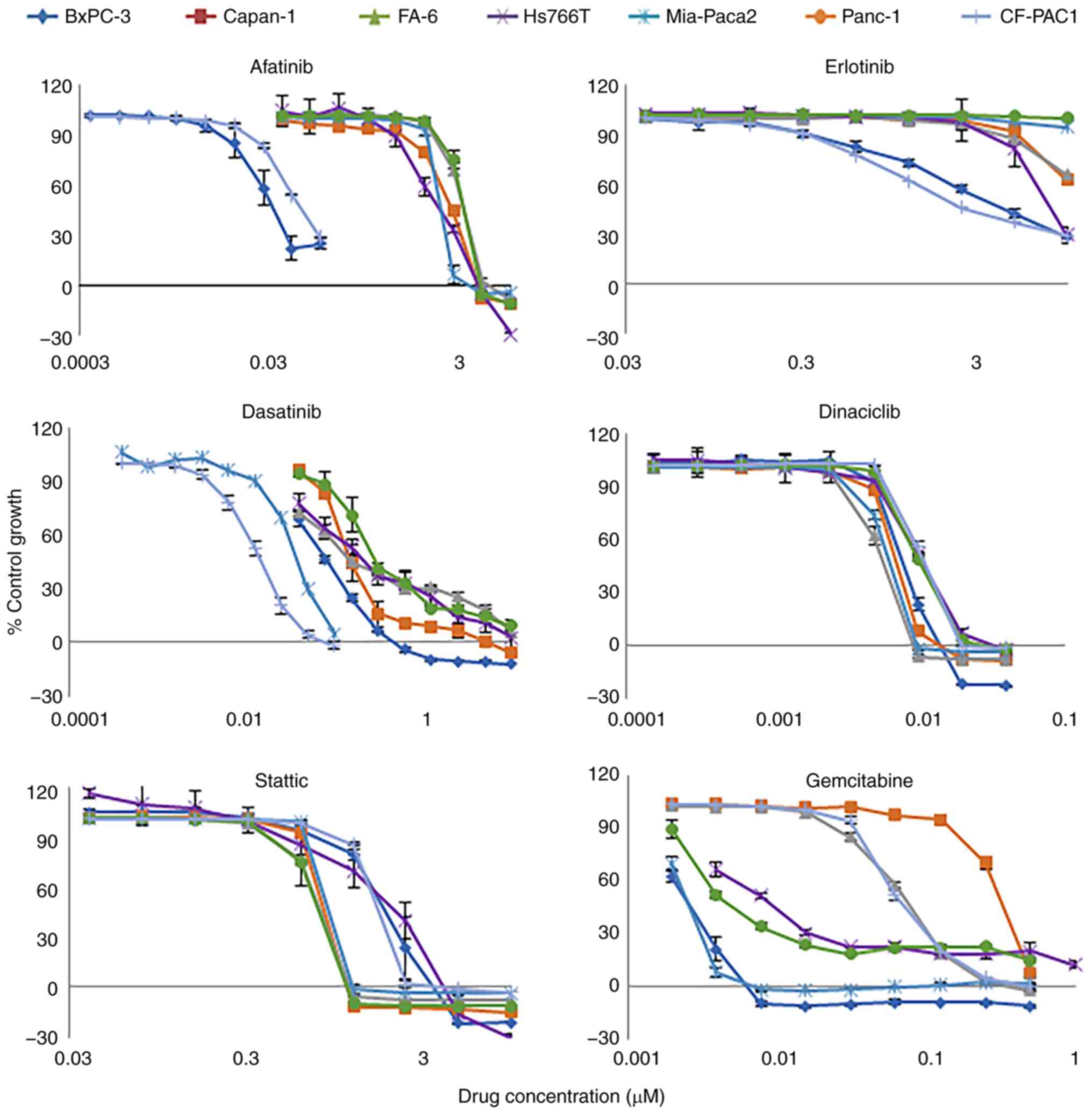
Growth response of human pancreatic
cancer cell lines to treatment with various TKIs, CDK inhibitors,
STAT3 inhibitor and cytotoxic agent
The effect of various agents on the growth of HPCCLs
was determined using SRB assay and the results are expressed as
IC50 (Table II,
Fig. 2). Of all the agents tested,
the CDK1/2/5/9 inhibitor dinaciclib was the most potent agent
inhibiting the growth of all seven cell lines with the
IC50 values of ≤10 nM. This was followed by the
Abl/Src/c-kit TKI dasatinib with IC50 values of 13 nM
(CF-PAC1) to 258 nM (Panc-1); gemcitabine with IC50 of
≤330 nM; the STAT3 inhibitor, static, with IC50 values
of 0.74 µM (Panc-1) to 2 µM (BxPC-3) and the irreversible pan-HER
TKI afatinib with IC50 of 27 nM (BxPC-3) to 2.95 µM
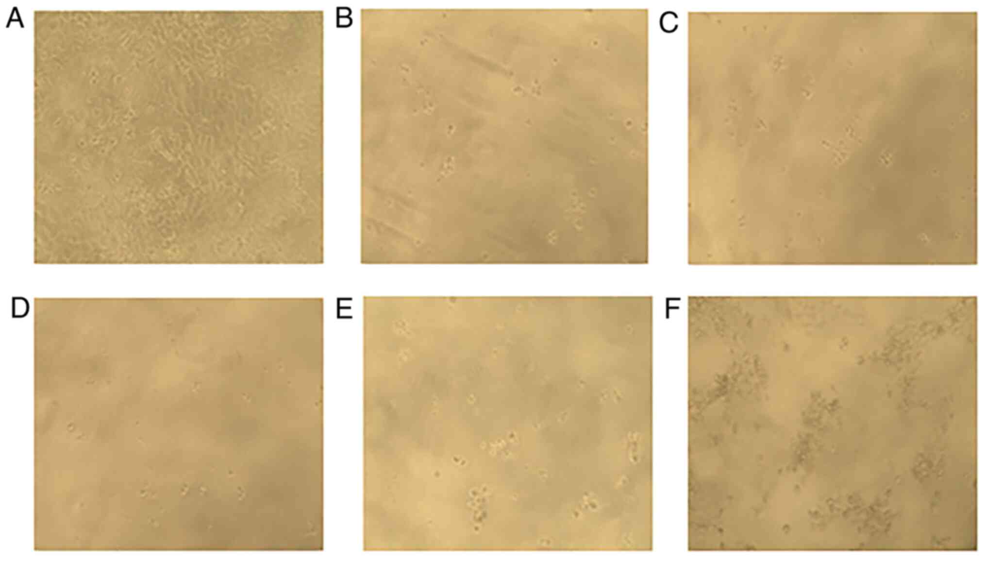
(FA-6). The morphology of BxPC-3 cells following treatment with the
above agents compared to the control are presented in Fig. 3. The effect of c-MET/ALK inhibitor,
crizotinib, ALK/IGF-IR/InsR inhibitor ceritinib,
Abl/PDGFRα/VEGFR2/FGFR1/Src inhibitor ponatinib and Src/Abl
inhibitor, bosutinib, on the growth of all HPCCLs was found to be
moderate (Table II). However,
treatment with the PDGFRα/β inhibitor crenolanib, FGFR1/2/3
inhibitor AZD4547, c-MET inhibitor capmatinib, IGF-IR inhibitor
NVP-AEW742, and IGF-IR/InsR inhibitor brigatinib had minimum to no
inhibitory effect on HPCCLs (Table
II).
| Table II.IC50 values of various
agents on HPCCLs as assessed by SRB colorimetric assay. |
Table II.
IC50 values of various
agents on HPCCLs as assessed by SRB colorimetric assay.
|
| IC50
value (µM) |
|---|
|
|
|
|---|
| Cell lines | Palbociclib (CDK4/6
inhibitor) | Dinacicilib
(CDK1/2/5/9 inhibitor) | Crizotinib
(c-MET/ALK TKI) | Capmatinib (c-MET
TKI) | Ceritinib
(ALK/IGF-IR/InsR TKI) | Brigatinib
(ALK/ROS1 TKI) | Erlotinib
(reversible EGFR TKI) | Afatinib
(irreversible pan-HER TKI) | Gemcitabine
(cytotoxic agent) |
|---|
| BxPC-3 | 3.87±1.58 | 0.010±0.21 | 1.83±0.30 | >10.00 | 1.43±0.17 | >10.00 | 1.70±0.05 | 0.027±0.01 | 0.003±0.02 |
| Capan-1 | 2.24±0.24 | 0.007±0.03 | 1.74±0.06 | >10.00 | 1.39±0.01 | 3.33±0.01 | >10.00 | 2.21±0.03 | 0.33±0.00 |
| FA-6 | 4.28±0.18 | 0.005±0.15 | 1.21±0.00 | >10.00 | 1.30±0.00 | 5.30±0.02 | >10.00 | 2.95±0.04 | 0.06±0.01 |
| Panc-1 | >10.00 | 0.010±0.02 | 2.93±0.14 | >10.00 | 2.65±0.29 | >10.00 | >10.00 | 2.63±0.50 | 0.02±0.02 |
| Mia-Paca2 | 6.90±0.14 | 0.006±0.02 | 1.73±0.10 | >10.00 | 1.33±0.02 | >10.00 | >10.00 | 1.76±0.05 | 0.003±0.00 |
| Hs766T | 4.31±0.41 | 0.010±0.34 | 3.13±0.22 | >10.00 | 1.55±0.03 | >10.00 | 7.7±0.04 | 1.72±0.01 | 0.008±0.01 |
| CF-PAC1 | >10.00 | 0.010±0.06 | 3.29± 0.20 | >10.00 | 5.02±0.20 | >10.00 | 0.96±0.12 | 0.054±0.01 | 0.06± 0.00 |
|
| Cell
lines | Dasatinib
(Abl/Src/c-kit TKI) | NVP-AEW742
(IGF-IR TKI) | Linsitinib
(IGF-IR/InsR TKI) | Stattic (STAT3
inhibitor) | Crenolanib
(PDGFRα/β KI) | Bosutinib
(Src/Abl TKI) | AZD4547
(FGFR1/2/3 TKI) | Ponatinib
(Abl/PDGFRα/ VEGFR2/FGFR1/Src TKI) |
|
| BxPC-3 | 0.07±0.00 | 6.74±0.31 | 5.43±0.22 | 2.00±0.22 | 4.59±0.48 | 0.73±0.02 | 4.39±0.05 | 0.40±0.04 |
| Capan-1 | 0.16±0.02 | >10.00 | 9.00±0.01 | 0.75±0.06 | 3.54±0.03 | 0.98±0.03 | 5.86±0.19 | 0.35±0.02 |
| FA-6 | 0.15±0.01 | >10.00 | 7.52±0.48 | 0.77±0.06 | 2.51±0.09 | 1.51± 0.10 | 2.51± 0.01 | 0.33±0.02 |
| Panc-1 | 0.258±0.03 | >10.00 | >10.00 | 0.74±0.03 | 6.32±0.06 | 5.01±0.40 | >10.00 | 4.85±0.13 |
| Mia-Paca2 | 0.08±0.01 | >10.00 | 5.65±1.21 | 0.80±0.12 | 3.08±1.03 | 3.81±0.11 | 4.72±0.02 | 2.52±0.06 |
| Hs766T | 0.21±0.05 | >10.00 | >10.00 | 1.85±0.07 | 1.29±0.32 | 0.32±0.12 | 3.37±0.12 | 3.69±0.04 |
| CF-PAC1 | 0.013±0.03 | >10.00 | >10.00 | 1.51±0.12 | 3.49±0.11 | 1.13±0.16 | 9.59±0.30 | 0.51±0.02 |
Cell cycle distribution analysis
The effect of various agents on the cell cycle
distribution of four HPCCLs was determined using flow cytometry and
the results are summarized in Table
III. Treatment with afatinib, dinaciclib, dasatinib, stattic
and gemcitabine increased the percentage of cells in the sub-G1
phase (apoptotic/dead cells) with subsequent reduction in the G0/G1
phase. Furthermore, treatment with afatinib and dasatinib increased
cells in the S and G2/M phase whereas treatment with dinaciclib and
gemcitabine increased cells in the S-phase in most of the cell
lines examined. The representative flow cytometry plots of cell
cycle distribution of BxPC-3 cells following treatment with these
agents is shown in Fig. S1.
| Table III.Effect of various agents on the cell
cycle distribution of human pancreatic cancer cell lines. |
Table III.
Effect of various agents on the cell
cycle distribution of human pancreatic cancer cell lines.
| Cell
line/treatment | Sub G1 | G0/G1 | S | G2/M |
|---|
| BxPC3/ |
|
|
|
|
|
Control | 5.28±0.36 | 65.65±3.54 | 16.70±1.65 | 11.63±2.60 |
|
Afatinib | 8.88±1.84 | 52.25±4.60 | 18.60±2.40 | 19.36±5.45 |
|
Dinaciclib | 24.30±9.93 | 41.36±13.85 | 22.75±3.26 | 10.76±3.23 |
|
Dasatinib | 21.51±8.26 | 38.57±15.23 | 19.52±0.37 | 16.57±4.55 |
|
Stattic | 39.05±11.89 | 38.89±8.63 | 15.00±0.16 | 6.86±0.56 |
|
Gemcitabine | 20.20±7.98 | 46.04±20.80 | 22.55±12.52 | 10.37±2.31 |
| Capan-1/ |
|
|
|
|
|
Control | 18.37±5.98 | 60.75±0.41 | 13.63±3.98 | 7.53±3.20 |
|
Afatinib | 44.77±15.22 | 44.92±11.96 | 8.26±3.37 | 2.32±1.42 |
|
Dinaciclib | 58.17±15.68 | 35.03±15.63 | 6.79±2.50 | 1.00±0.30 |
|
Dasatinib | 42.79±19.48 | 39.45±13.99 | 14.20±5.96 | 4.00±1.92 |
|
Stattic | 67.61±37.94 | 25.45±29.59 | 6.70±8.70 | 2.03±2.66 |
|
Gemcitabine | 62.72±18.94 | 32.91±17.64 | 4.35±3.16 | 0.59±0.50 |
| FA-6/ |
|
|
|
|
|
Control | 3.73±2.26 | 57.63±1.85 | 24.00±0.81 | 13.88±4.01 |
|
Afatinib | 16.60±9.44 | 30.99±4.25 | 31.47±0.33 | 18.79±3.44 |
|
Dinaciclib | 17.45±9.18 | 29.61±5.42 | 33.25±3.01 | 18.48±4.99 |
|
Dasatinib | 5.70±5.87 | 28.86±9.48 | 43.17±10.81 | 19.19±4.88 |
|
Stattic | 31.94±2.81 | 20.61±1.88 | 31.49±2.26 | 14.02±0.30 |
|
Gemcitabine | 11.86±6.78 | 42.07±18.45 | 35.83±8.05 | 9.47±3.11 |
| Mia-Paca2/ |
|
|
|
|
|
Control | 1.82±1.85 | 68.67±2.14 | 16.46±1.59 | 12.63±3.25 |
|
Afatinib | 17.70±23.89 | 22.57±16.79 | 39.67±7.94 | 20.95±17.16 |
|
Dinaciclib | 17.58±20.83 | 40.94±15.47 | 29.90±0.51 | 11.69±8.92 |
|
Dasatinib | 2.10±0.36 | 52.91±9.14 | 24.93±5.55 | 18.68±0.49 |
|
Stattic | 24.19±33.06 | 32.28±25.82 | 22.16±10.65 | 20.61±1.56 |
|
Gemcitabine | 3.14±1.23 | 38.97±12.76 | 43.88±25.77 | 14.50±10.83 |
Afatinib and dasatinib blocks the
phosphorylation of EGFR and SRC respectively
Next we investigated the effects of treatment with
afatinib, dinaciclib, dasatinib and stattic on the phosphorylation
of growth factor receptors and downstream cell signaling molecules
in BxPC-3 and Capan-1 cells. As expected, afatinib blocked the
ligand-induced phosphorylation of tyrosine and phosphorylation of
EGFR at position 1,068 which in turn resulted in reduction in the
phosphorylation of downstream signaling molecules such as MAPK and
AKT in both cell lines examined (BxPC-3 and Capan-1) (Fig. 4A and B). Treatment with dasatinib
alone was accompanied by the EGF, HB-EGF and IGF-II induced
phosphorylation of SRC. However, no phosphorylation of the IGF-IR
was evident following treatment with EGF, HB-EGF and IGF-II in
these two cancer cell lines (data not shown).
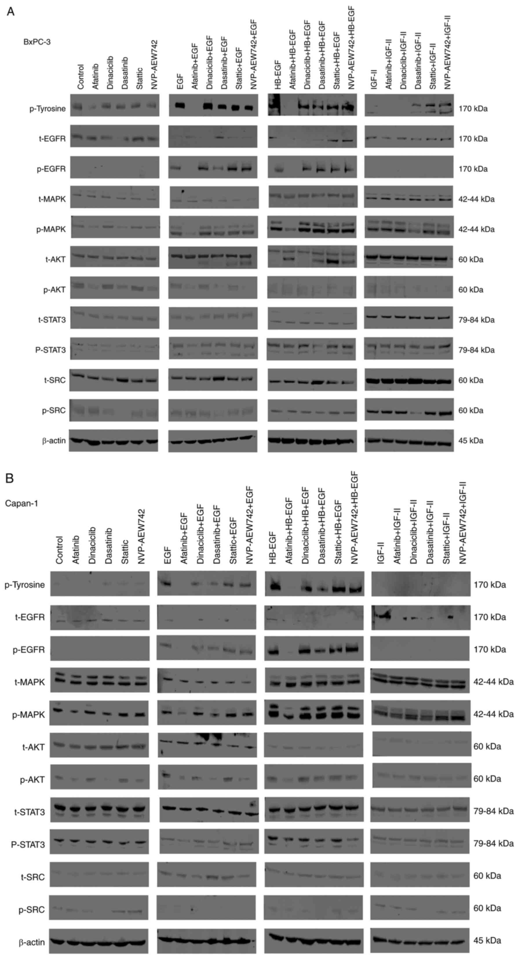 | Figure 4.Effect of afatinib, dinaciclib,
dasatinib, stattic and NVP-AEW742 with or without ligands (EGF,
HB-EGF, IGF-II) on the phosphorylation of EGFR and downstream cell
signaling molecules including MAPK, AKT, STAT3, SRC and IGF-IR in
BxPC-3 (A) and Capan-1 (B) cells. The cells were cultured in 10%
FBS RPMI-1640 medium to near confluency. Cells were washed once
with 0.5% FBS RPMI-1640 medium and incubated with selected agents
(400 nM) for 1 h and then stimulated with 40 nM ligands (EGF,
HB-EGF and IGF-II) for 15 min. Cells were then lysed, separated
using SDS-PAGE, transferred onto PDVF membranes, probed with the
antibodies of interest and visualized using LI-COR software. EGF,
epidermal growth factor; HB-EGF, heparin-binding EGF-like growth
factor; IGF-II, insulin-like growth factor II; EGFR, epidermal
growth factor receptor; MAPK, mitogen-activated protein kinase;
AKT, protein kinase B or PKB; STAT3, signal transducer and
activator of transcription 3; SRC, proto-oncogene tyrosine kinase
SRC; IGF-IR, insulin-like growth fact) or 1 receptor. |
Synergistic and antagonistic effect of
various drug combinations in pancreatic cancer cell lines
The combined effect of various agents including
afatinib, dinaciclib, dasatinib, stattic and gemcitabine on the
growth of HPCCLs was investigated. Only treatment with a
combination of dasatinib with afatinib or dasatinib in combination
with gemcitabine led to synergistic growth inhibition of four
HPCCLs (Table IV). In contrast,
treatment with a combination of afatinib with dinaciclib was found
to be antagonistic in all of four HPCCLs examined (Table IV). Finally, treatment with other
drug combinations resulted in mixed effects in all cell lines
examined (data not shown).
| Table IV.Combination index (CI) values of
dinaciclib plus afatinib, dasatinib plus afatinib and dasatinib
plus gemcitabine in human pancreatic cancer cell lines. |
Table IV.
Combination index (CI) values of
dinaciclib plus afatinib, dasatinib plus afatinib and dasatinib
plus gemcitabine in human pancreatic cancer cell lines.
| Cell lines | Dinaciclib +
afatinib combination index (effect) | Afatinib +
dasatinib combination index (effect) | Dasatinib +
gemcitabine combination index (effect) |
|---|
| BxPC-3 | 1.32 (slight
antagonism) | 0.97 (nearly
additive) | 0.85 (moderate
synergism) |
| Capan-1 | 1.85
(antagonism) | 0.39
(synergism) | 0.35
(synergism) |
| FA-6 | 1.60
(antagonism) | 0.53
(synergism) | 0.66
(synergism) |
| Mia-Paca2 | 1.66
(antagonism) | 0.58
(synergism) | 0.85 (moderate
synergism) |
Linear regression analysis
The association between the expression level of
various growth factor receptors and their response to treatment
with various agents was assessed using SPSS software (Table V). There was no correlation between
expression level of EGFR and the response to treatment with various
agents. However, there were some statistically significant
associations between HER2 expression and the response to treatment
with ALK/IGF-IR/InsR inhibitor ceritinib (R2=0.698,
P=0.019) and the FGFR1/2/3 inhibitor AZD4547 (R2=0.751,
P=0.012); HER3 expression and the response to treatment with HER
family targeting TKI erlotinib (R2=0.830, P=0.004) and
afatinib (R2=0.599, P=0.041); IGF-IR expression and the
response to treatment with erlotinib (R2=0.608, P=0.039)
and afatinib (R2=0.672, P=0.024). In addition, a
statistically significant association was found between c-MET
expression and the response to treatment with STAT3 inhibitor
stattic (R2=0.809, P=0.006) and SRC/Abl inhibitor
bosutinib (R2=0.747, P=0.012) and finally between ALK7
expression and the response to treatment with STAT3 inhibitor
stattic (R2=0.682, P=0.022). HER4 was not tested due to
its negative expression in all cell lines.
| Table V.Linear regression analysis of the
expression of various receptors against the sensitivity of human
pancreatic cancer cell lines to treatment with various TKIs, CDK
inhibitors, STAT3 inhibitor and a cytotoxic agent. |
Table V.
Linear regression analysis of the
expression of various receptors against the sensitivity of human
pancreatic cancer cell lines to treatment with various TKIs, CDK
inhibitors, STAT3 inhibitor and a cytotoxic agent.
| Drugs/cell surface
markers | EGFR R2
(P-value) | HER2 R2
(P-value) | HER3 R2
(P-value) | IGF-IR
R2 (P-value) | C-MET R2
(P-value) | ALK7 R2
(P-value) |
|---|
| Palbociclib | 0.004 (0.895) | 0.345 (0.165) | 0.046 (0.646) | 0.001 (0.940) | 0.091 (0.511) | 0.125 (0.437) |
| Crizotinib | 0.203 (0.310) | 0.185 (0.336) | 0.230 (0.276) | 0.017 (0.779) | 0.158 (0.377) | 0.029 (0.715) |
| Ceritinib | 0.001 (0.948) | 0.698
(0.019) | 0.496 (0.077) | 0.038 (0.674) | 0.031 (0.704) | 0.002 (0.916) |
| Brigatinib | 0.184 (0.692) | 0.180 (0.343) | 0.132 (0.424) | 0.069 (0.569) | 0.031 (0.705) | 0.021 (0.756) |
| Erlotinib | 0.018 (0.772) | 0.204 (0.309) | 0.830
(0.004) | 0.608
(0.039) | 0.449 (0.100) | 0.371 (0.147) |
| Afatinib | 0.000 (0.965) | 0.188 (0.331) | 0.599
(0.041) | 0.672
(0.024) | 0.386 (0.137) | 0.248 (0.256) |
| Gemcitabine | 0.197 (0.318) | 0.087 (0.522) | 0.021 (0.785) | 0.005 (0.882) | 0.006 (0.865) | 0.026 (0.732) |
| Dasatinib | 0.231 (0.275) | 0.034 (0.691) | 0.105 (0.478) | 0.007 (0.854) | 0.268 (0.234) | 0.020 (0.763) |
| Linsitinib | 0.114 (0.459) | 0.133 (0.421) | 0.062 (0.589) | 0.088 (0.519) | 0.038 (0.674) | 0.000 (0.972) |
| Stattic | 0.232 (0.274) | 0.008 (0.845) | 0.483 (0.083) | 0.412 (0.120) | 0.809
(0.006) | 0.682
(0.022) |
| Crenolanib | 0.034 (0.694) | 0.236 (0.269) | 0.010 (0.828) | 0.189 (0.329) | 0.232 (0.274) | 0.000 (0.983) |
| Bosutinib | 0.003 (0.903) | 0.020 (0.763) | 0.355 (0.158) | 0.074 (0.555) | 0.747
(0.012) | 0.397 (0.129) |
| AZD4547 | 0.001 (0.959) | 0.751
(0.012) | 0.063 (0.587) | 0.053 (0.620) | 0.054 (0.616) | 0.040 (0.667) |
| Ponatinib | 0.261 (0.241) | 0.041 (0.663) | 0.186 (0.334) | 0.091 (0.512) | 0.066 (0.577) | 0.037 (0.681) |
Effect of selected agents on the
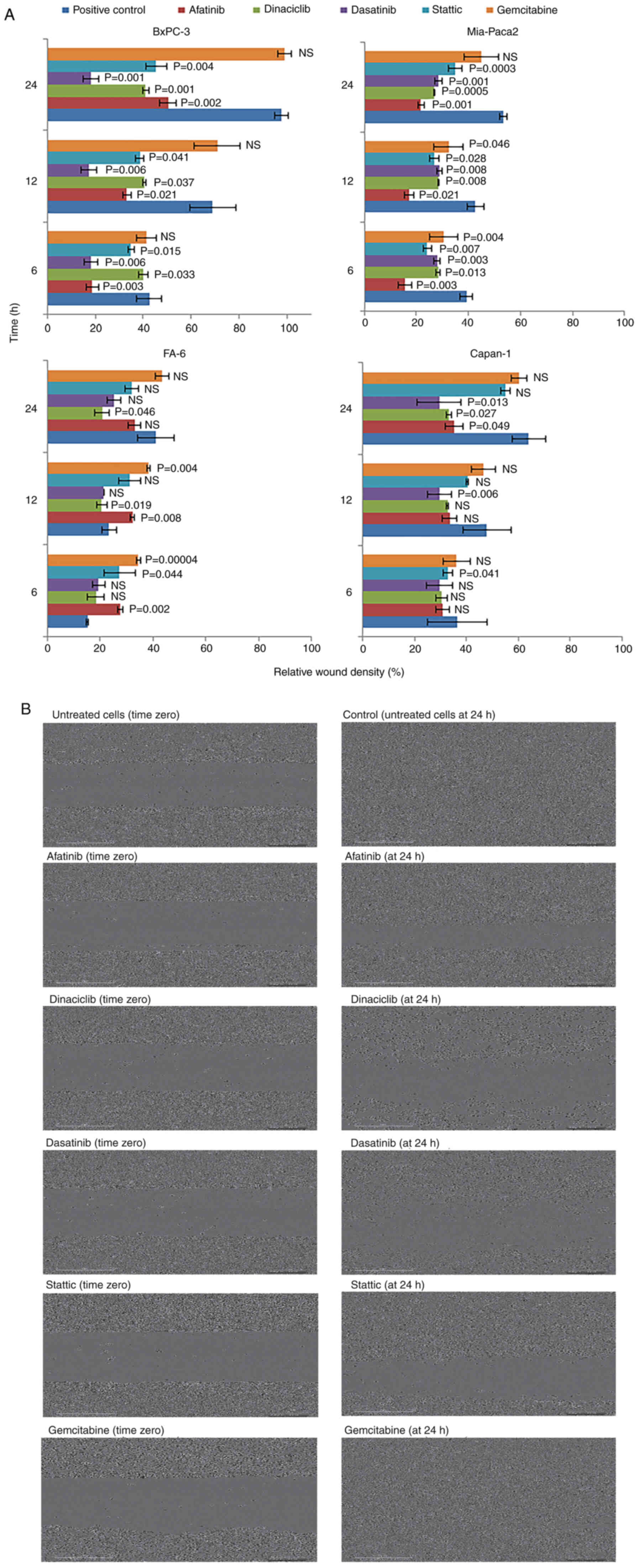
migration of pancreatic cancer cell lines
The effect of selected agents on the migration of
four HPCCLs was determined using scratch wound healing assay and
the results at time points 6, 12 and 24 h are summarized in
Fig. 5A. As an example, the effect
of these agents on the migration of BxPC-3 cells at 24 h is shown
in Fig. 5B. In comparison to
positive control (i.e., no treatment, 10% FBS medium only) most
drugs inhibited the migration of HPCCLs with SRC targeting TKI
dasatinib, and the CDK inhibitor dinaciclib and the irreversible
pan-HER TKI afatinib being most effective (Fig. 5A and B).
Discussion
Despite major advances in the early diagnosis and
treatment of solid tumors in the past three decades, pancreatic
cancer remains as one of the most aggressive and deadliest forms of
cancer. By 2030, it is predicted to become the second leading cause
of cancer-related deaths after lung cancer (31). Due to its heterogenous nature, its
retroperitoneal location, non-specific symptoms and lack of
screening methods, the overwhelming majority of pancreatic cancer
patients are diagnosed at an advanced stage of the disease at the
time of presentation (32). To
date, of the HER inhibitors, only the EGFR-specific tyrosine kinase
inhibitor (TKI), erlotinib has gained the FDA approval for the
targeted therapy of locally advanced, unresectable or metastatic
pancreatic cancer in combination with the cytotoxic agent
gemcitabine (14). However, the
majority of pancreatic cancer patients have either primary
resistance, or develop seondary resistance following a short course
of erlotinib. As a result, the duration of response can be short in
many patients (7,30). Therefore, there is an urgent need to
discover more effective therapeutic interventions for patients
diagnosed with different stages of pancreatic cancer (7).
We previously demonstrated that of the pan HER
family blocker, afatinib was more effective than erlotinib in
inhibiting the growth of human pancreatic cancer cells (27). We also reported that treatment with
afatinib in combination the insulin-like growth factor 1 receptor
(IGF-IR) inhibitor NVP-AEW541 resulted in synergistic growth
inhibition of pancreatic cancer cells (15). Due to the heterogeneous nature of
pancreatic cancer, in this study we investigated the growth
response of human pancreatic cancer cell lines (HPCCLs),
established from patients at different stages of the disease, to
the treatment with agents targeting different cyclin-dependent
kinases (CDKs), various growth factor receptors and cell signaling
molecules.
Of all the agents examined, the CDK1/2/5/9
inhibitor, dinaciclib was the most potent agent and inhibited the
proliferation of all seven primary and metastatic HPCCLs with
IC50 values of ≤10 nM. Pre-clinical testing of
dinaciclib exhibited an acceptable toxicity profile and effective
inhibition in mouse models and it was found to be safe and well
tolerated in phase I trials (33,34).
The second most effective agent with anti-proliferative activity
was the SRC/c-kit/Abl inhibitor dasatinib. It inhibited the growth
of all seven primary and metastatic HPCCLs with IC50 of
≤258 nM). However, the most sensitive cell line to growth inbition
by dasatinib were Bx-PC3 and Mia-PaCa-2, which were established
from two primary tumors and with IC50 values of 70 and
80 nM, respectively. The third most effective agent was the STAT3
inhibitor stattic which inhibited the growth of all HPCCLs with
IC50 of ≤2 µM. The pan-HER family blocker also inhibited
the growth of all seven HPCCLs with IC50 values ≤2.95
µM). However, the most sensitivite cell lines to treatment with
afatinib were the EGFR, HER2 and HER3-positive BxPC-3 and CF-PAC1
cells whereas the reversible EGFR TKI erlotinib was only effective
in BxPC-3, CF-PAC1 and Hs766T cells. We found a statistically
significant association between HER3 expression and their response
to treatment with TKIs targeting HER family members. In another
study, Frolov and colleagues suggested that the higher sensitivity
of HER3-positive cell lines to treatment with pan-HER TKIs could be
due to blockade of HER-3 transactivation via EGFR therefore
inhibiting PI3K/AKT signaling pathways (35). Other studies have also supported the
association between the expression level of HER3 and the
sensitivity of pancreatic cancer cell lines to treatment with
erlotinib (35,36). Although our panel of cell lines was
moderately positive for IGF-IR and showed moderate sensitivity to
agents targeting IGF-IR and fibroblast growth factor receptor
(FGFR), some statistically significant associations were found
between IGF-IR expression and their response to treatment with HER
family TKIs as well as HER2 expression and treatment with
ALK/IGF-IR/InsR inhibitor ceritinib and FGFR inhibitor AZD4547.
Both IGF-IR and FGFR-mediated signaling pathways have been found to
participate in the resistance to anti-HER targeted therapy
(15,37,38).
Moreover, we and other researchers have shown that the co-targeting
of IGF-IR and HER family members results in synergistic growth
inhibition of human pancreatic cancer cells (15,39).
The synchronous activation of bypass pathways via
receptor tyrosine kinases (RTKs) including c-MET and ALK7 has been
shown to induce resistance to current treatments and have emerged
as important therapeutic targets for pancreatic cancer treatments.
The expression level of both these receptors in our panel of cell
lines was moderate. While the growth of all seven HPCCLs were
inhibited by the dual c-MET/ALK TKI crizotinib (i.e.,
IC50 values ≤3.3 µM), none of the cell lines were
sensitive to treatment with the c-MET-specific TKI, capmatinib
(i.e., IC50 values >10 µM). However, a significant
association was found between c-MET and ALK-7 expression and their
response to treatment with the STAT3 inhibitor stattic. STAT3 lies
at the convergence of multiple oncogenic signaling pathways
stimulated by upstream activated receptors such as c-MET and ALK
(19,40). A study reported that hyperactivated
STAT3 signaling in non-small cell lung cancer (NSCLC) was induced
by the activation of MET (41). In
addition, a significant association was found between c-MET
expression and Src/Abl inhibitor bosutinib. The SRC and Abl are
some of the intracellular effector molecules recruited following
auto-phosphorylation of docking site of activated c-MET (40). Overexpression and activation of the
c-MET receptor have been shown to be involved in SRC kinase
activation (42).
Cell cycle distribution analysis revealed that the
treatment of HPCCLs with afatinib, dasatinib, dinaciclib, stattic
and gemcitabine increased the population of cells in sub-G1 with
concomitant decrease in the G1 phase. Afatinib treatment also
increased cells in the S and G2/M phase in most of the cell lines
and similar results were reported following the treatment of
nasopharyngeal carcinomas with afatinib (43). Dinaciclib treatment caused S-phase
arrest in most of the cell lines which is consistent with the
blockage of CDK2, one of the targets of dinaciclib (44). Treatment with dasatinib arrested
cells in the S and G2/M phase which is consistent with a study
where increasing the dasatinib concentration from 0.5–1.0 µM
increased the percentage of BxPC-3 cells in the S-phase (21). Treatment with the cytotoxic agent
gemcitabine increased cells in S-phase consistent with the
inhibition of DNA replication (45). Treatment with afatinib, dastainib
and stattic inhibited the phophorylation of EGFR, SRC and STAT3,
respectively and the migration of four human pancreatic cancer cell
lines, established from patients with either a primary tumor or a
meastatic tumor. Our results suggest that treatment with these
agents result in inhibition of two hallmarks of cancer which are
tumour cell proliferation and migration.
Next, we investigated the combinational potential of
most effective drugs on four HPCCLs. We found that treatment with a
combination of SRC targeting TKI dasatinib with the pan-HER TKI
afatinib resulted in synergistic growth inhibition of all four
HPCCLs examined. Treatment with the combination of dasatinib with
gemcitabine also resulted in synergistic growth inhibition of four
HPCCLs, including those established from patients with a primary
pancreatic tumor (e.g., Bx-PC-3) or liver metastasis (e.g.,
Capan-1). Consistent with the results of a present study, the
latter combination has been reported to have synergistic effect in
two other HPCCLs (46,47). Although, dasatinib and gemcitabine
combination has been shown to promote stable disease and to induce
a partial response in patients with pancreatic cancer (48), it failed to improve patient survival
in a phase II trial setting due to increased toxicity of such a
combination (49). This combination
is currently being used in a phase II trial in pancreatic cancer
patients (ClinicalTrials.gov Identifier:
NCT01234935). The triple combination of SRC inhibitor (dasatinib),
EGFR inhibitor (erlotinib) and gemcitabine demonstrated a
synergistic antitumor effect in pancreatic ductal adenocarcinoma
(PDAC) as well as encouraging preliminary activity in patients with
advanced pancreatic cancer (50,51).
However, when afatinib was used in combination with the CDK1/2/5/9
inhibitor, dinaciclib, such a combination had agonistic effect in
all cell lines examined. This highlights the importance of
selecting the appropriate partner when such drugs are used in
combination.
In conclusion, we demonstrated that of various
targeting agents employed in our study, the CDK inhibitor
dinaciclib, the irreversible pan-HER TKI afatinib, and the SRC
targeting TKI dasatinib were most effective at inhibiting the
proliferation and migration of HPCCLs, established from both a
primary pancreatic cancer and from a metastatic pancreatic cancer.
The combination of dasatinib with afatinib and dasatinib with
gemcitabine led to synergistic growth inhibition in pancreatic
cancer cell lines. Our results support the need for further
investigation of the therapeutic potential of these combinations in
the treatment of pancreatic cancer.
Supplementary Material
Supporting Data
Acknowledgements
We would like to thank Dr Soozana Puvanenthiran,
School of Life Science, Pharmacy and Chemistry, Kingston University
London, Kingston, UK for her help with preparation with various
figures.
Funding
This study was supported by the Kingston University
as part of a self-funded PhD project at the Kingston University
London, UK.
Availability of data and material
The datasets used and/or analyzed during the current
study are available from the corresponding author on reasonable
request.
Authors' contributions
TK performed the experiments and conducted data
analysis as part of her PhD project. HM conceived the original
research idea, read, and revised the manuscript. TK and HM wrote
the manuscript. SK and NI carried out data analysis. AMS, AGD and
SM were the other PhD supervisors on this study and thus were also
involved in all aspects of the experiments, data analysis and
writing. All authors have read and approved the study.
Ethics approval and consent to
participate
Not applicable.
Patient consent for publication
Not applicable.
Competing interests
The authors declare that they have no competing
interests.
Glossary
Abbreviations
Abbreviations:
|
EGFR
|
epidermal growth factor receptor
|
|
CDK
|
cyclin-dependent kinase
|
|
HPCCLs
|
human pancreatic cancer cell lines
|
|
TKIs
|
tyrosine kinase inhibitors
|
|
CDKI
|
cyclin-dependent kinase inhibitor
|
References
|
1
|
Bray F, Ferlay J, Soerjomataram I, Siegel
RL, Torre LA and Jemal A: Global cancer statistics 2018: GLOBOCAN
estimates of incidence and mortality worldwide for 36 cancers in
185 countries. CA Cancer J Clin. 68:394–424. 2018. View Article : Google Scholar : PubMed/NCBI
|
|
2
|
Rawla P, Sunkara T and Gaduputi V:
Epidemiology of pancreatic cancer: Global trends, etiology and risk
factors. World J Oncol. 10:10–27. 2019. View Article : Google Scholar : PubMed/NCBI
|
|
3
|
Siegel RL, Miller KD and Jemal A: Cancer
statistics, 2020. CA Cancer J Clin. 70:7–30. 2020. View Article : Google Scholar : PubMed/NCBI
|
|
4
|
Carreras-Torres R, Johansson M, Gaborieau
V, Haycock PC, Wade KH, Relton CL, Martin RM, Davey Smith G and
Brennan P: The role of obesity, type 2 diabetes, and metabolic
factors in pancreatic cancer: A mendelian randomization study. J
Natl Cancer Inst. 109:djx0122017. View Article : Google Scholar
|
|
5
|
Lauby-Secretan B, Scoccianti C, Loomis D,
Grosse Y, Bianchini F and Straif K; International Agency for
Research on Cancer Handbook Working Group, : Body Fatness and
Cancer-Viewpoint of the IARC Working Group. N Engl J Med.
375:794–798. 2016. View Article : Google Scholar : PubMed/NCBI
|
|
6
|
Wolfgang CL, Herman JM, Laheru DA, Klein
AP, Erdek MA, Fishman EK and Hruban RH: Recent progress in
pancreatic cancer. CA Cancer J Clin. 63:318–348. 2013. View Article : Google Scholar : PubMed/NCBI
|
|
7
|
Ioannou N, Seddon AM, Dalgleish A,
Mackintosh D and Modjtahedi H: Expression pattern and targeting of
HER family members and IGF-IR in pancreatic cancer. Front Biosci
(Landmark Ed). 2012(17): 2698–2724. 2012. View Article : Google Scholar
|
|
8
|
Seshacharyulu P, Ponnusamy MP, Haridas D,
Jain M, Ganti AK and Batra SK: Targeting the EGFR signaling pathway
in cancer therapy. Expert Opin Ther Targets. 16:15–31. 2012.
View Article : Google Scholar : PubMed/NCBI
|
|
9
|
Tebbutt N, Pedersen MW and Johns TG:
Targeting the ERBB family in cancer: Couples therapy. Nat Rev
Cancer. 13:663–673. 2013. View
Article : Google Scholar : PubMed/NCBI
|
|
10
|
Li Q, Zhang L, Li X, Yan H, Yang L, Li Y,
Li T, Wang J and Cao B: The prognostic significance of human
epidermal growth factor receptor family protein expression in
operable pancreatic cancer: HER1-4 protein expression and prognosis
in pancreatic cancer. BMC Cancer. 16:9102016. View Article : Google Scholar : PubMed/NCBI
|
|
11
|
Perini MV, Montagnini AL, Coudry R,
Patzina R, Penteado S, Abdo EE, Diniz A, Jukemura J and da Cunha
JE: Prognostic significance of epidermal growth factor receptor
overexpression in pancreas cancer and nodal metastasis. ANZ J Surg.
85:174–178. 2015. View Article : Google Scholar : PubMed/NCBI
|
|
12
|
Mahipal A, Mcdonald MJ, Witkiewicz A and
Carr BI: Cell membrane and cytoplasmic epidermal growth factor
receptor expression in pancreatic ductal adenocarcinoma. Med Oncol.
29:134–139. 2012. View Article : Google Scholar : PubMed/NCBI
|
|
13
|
Einama T, Ueda S, Tsuda H, Ogasawara K,
Hatsuse K, Matsubara O, Todo S and Yamamoto J: Membranous and
cytoplasmic expression of epidermal growth factor receptor in
metastatic pancreatic ductal adenocarcinoma. Exp Ther Med.
3:931–936. 2012. View Article : Google Scholar : PubMed/NCBI
|
|
14
|
Moore MJ, Goldstein D, Hamm J, Figer A,
Hecht JR, Gallinger S, Au HJ, Murawa P, Walde D, Wolff RA, et al:
Erlotinib plus gemcitabine compared with gemcitabine alone in
patients with advanced pancreatic cancer: A phase III trial of the
National Cancer Institute of Canada clinical trials group. J Clin
Oncol. 25:1960–1966. 2007. View Article : Google Scholar : PubMed/NCBI
|
|
15
|
Ioannou N, Seddon AM, Dalgleish A,
Mackintosh D and Modjtahedi H: Treatment with a combination of the
ErbB (HER) family blocker afatinib and the IGF-IR inhibitor,
NVP-AEW541 induces synergistic growth inhibition of human
pancreatic cancer cells. BMC Cancer. 13:412013. View Article : Google Scholar : PubMed/NCBI
|
|
16
|
Nones K, Waddell N, Song S, Patch AM,
Miller D, Johns A, Wu J, Kassahn KS, Wood D, Bailey P, et al:
Genome-wide DNA methylation patterns in pancreatic ductal
adenocarcinoma reveal epigenetic deregulation of SLIT-ROBO, ITGA2
and MET signaling. Int J Cancer. 135:1110–1118. 2014. View Article : Google Scholar : PubMed/NCBI
|
|
17
|
Zhou W, Jubb AM, Lyle K, Xiao Q, Ong CC,
Desai R, Fu L, Gnad F, Song Q, Haverty PM, et al: PAK1 mediates
pancreatic cancer cell migration and resistance to MET inhibition.
J Pathol. 234:502–513. 2014. View Article : Google Scholar : PubMed/NCBI
|
|
18
|
Zhu GH, Huang C, Qiu ZJ, Liu J, Zhang ZH,
Zhao N, Feng ZZ and Lv XH: Expression and prognostic significance
of CD151, c-Met, and integrin alpha3/alpha6 in pancreatic ductal
adenocarcinoma. Dig Dis Sci. 56:1090–1098. 2011. View Article : Google Scholar : PubMed/NCBI
|
|
19
|
Liu C, Yang Z, Li D, Liu Z, Miao X, Yang
L, Zou Q and Yuan Y: Overexpression of B2M and loss of ALK7
expression are associated with invasion, metastasis, and
poor-prognosis of the pancreatic ductal adenocarcinoma. Cancer
Biomark. 15:735–743. 2015. View Article : Google Scholar : PubMed/NCBI
|
|
20
|
Shields DJ, Murphy EA, Desgrosellier JS,
Mielgo A, Lau SK, Barnes LA, Lesperance J, Huang M, Schmedt C,
Tarin D, et al: Oncogenic Ras/Src cooperativity in pancreatic
neoplasia. Oncogene. 30:2123–2134. 2011. View Article : Google Scholar : PubMed/NCBI
|
|
21
|
Nagaraj NS, Smith JJ, Revetta F,
Washington MK and Merchant NB: Targeted inhibition of SRC kinase
signaling attenuates pancreatic tumorigenesis. Mol Cancer Ther.
9:2322–2332. 2010. View Article : Google Scholar : PubMed/NCBI
|
|
22
|
Morton JP, Karim SA, Graham K, Timpson P,
Jamieson N, Athineos D, Doyle B, McKay C, Heung MY, Oien KA, et al:
Dasatinib inhibits the development of metastases in a mouse model
of pancreatic ductal adenocarcinoma. Gastroenterology. 139:292–303.
2010. View Article : Google Scholar : PubMed/NCBI
|
|
23
|
Chee CE, Krishnamurthi S, Nock CJ, Meropol
NJ, Gibbons J, Fu P, Bokar J, Teston L, O'Brien T, Gudena V, et al:
Phase II study of dasatinib (BMS-354825) in patients with
metastatic adenocarcinoma of the pancreas. Oncologist.
18:1091–1092. 2013. View Article : Google Scholar : PubMed/NCBI
|
|
24
|
Cros J, Raffenne J, Couvelard A and Poté
N: Tumor heterogeneity in pancreatic adenocarcinoma. Pathobiology.
85:64–71. 2018. View Article : Google Scholar : PubMed/NCBI
|
|
25
|
Yao W, Maitra A and Ying H: Recent
insights into the biology of pancreatic cancer. EBioMedicine.
53:1026552020. View Article : Google Scholar : PubMed/NCBI
|
|
26
|
Haeberle L, Steiger K, Schlitter AM, Safi
SA, Knoefel WT, Erkan M and Esposito I: Stromal heterogeneity in
pancreatic cancer and chronic pancreatitis. Pancreatology. May
12–2018.(Epub ahead of print). View Article : Google Scholar : PubMed/NCBI
|
|
27
|
Ioannou N, Dalgleish AG, Seddon AM,
Mackintosh D, Guertler U, Solca F and Modjtahedi H: Anti-tumour
activity of afatinib, an irreversible ErbB family blocker, in human
pancreatic tumour cells. Br J Cancer. 105:1554–1562. 2011.
View Article : Google Scholar : PubMed/NCBI
|
|
28
|
Stanley A, Ashrafi GH, Seddon AM and
Modjtahedi H: Synergistic effects of various Her inhibitors in
combination with IGF-1R, C-MET and Src targeting agents in breast
cancer cell lines. Sci Rep. 7:39642017. View Article : Google Scholar : PubMed/NCBI
|
|
29
|
Cunningham MP, Thomas H, Fan Z and
Modjtahedi H: Responses of human colorectal tumor cells to
treatment with the anti-epidermal growth factor receptor monoclonal
antibody ICR62 used alone and in combination with the EGFR tyrosine
kinase inhibitor gefitinib. Cancer Res. 66:7708–7715. 2006.
View Article : Google Scholar : PubMed/NCBI
|
|
30
|
Ioannou N, Seddon AM, Dalgleish A,
Mackintosh D, Solca F and Modjtahedi H: Acquired resistance of
pancreatic cancer cells to treatment with gemcitabine and
HER-inhibitors is accompanied by increased sensitivity to STAT3
inhibition. Int J Oncol. 48:908–918. 2016. View Article : Google Scholar : PubMed/NCBI
|
|
31
|
Rahib L, Smith BD, Aizenberg R, Rosenzweig
AB, Fleshman JM and Matrisian LM: Projecting cancer incidence and
deaths to 2030: The unexpected burden of thyroid, liver, and
pancreas cancers in the United States. Cancer Res. 74:2913–2921.
2014. View Article : Google Scholar : PubMed/NCBI
|
|
32
|
Adel N: Current treatment landscape and
emerging therapies for pancreatic cancer. Am J Manag Care. 25 (1
Suppl):S3–S10. 2019.PubMed/NCBI
|
|
33
|
Parry D, Guzi T, Shanahan F, Davis N,
Prabhavalkar D, Wiswell D, Seghezzi W, Paruch K, Dwyer MP, Doll R,
et al: Dinaciclib (SCH 727965), a novel and potent cyclin-dependent
kinase inhibitor. Mol Cancer Ther. 9:2344–2353. 2010. View Article : Google Scholar : PubMed/NCBI
|
|
34
|
Nemunaitis JJ, Small KA, Kirschmeier P,
Zhang D, Zhu Y, Jou YM, Statkevich P, Yao SL and Bannerji R: A
first-in-human, phase 1, dose-escalation study of dinaciclib, a
novel cyclin-dependent kinase inhibitor, administered weekly in
subjects with advanced malignancies. J Transl Med. 11:2592013.
View Article : Google Scholar : PubMed/NCBI
|
|
35
|
Frolov A, Schuller K, Tzeng CW, Cannon EE,
Ku BC, Howard JH, Vickers SM, Heslin MJ, Buchsbaum DJ and Arnoletti
JP: ErbB3 expression and dimerization with EGFR influence
pancreatic cancer cell sensitivity to erlotinib. Cancer Biol Ther.
6:548–554. 2007. View Article : Google Scholar : PubMed/NCBI
|
|
36
|
Buck E, Eyzaguirre A, Haley JD, Gibson NW,
Cagnoni P and Iwata KK: Inactivation of Akt by the epidermal growth
factor receptor inhibitor erlotinib is mediated by HER-3 in
pancreatic and colorectal tumor cell lines and contributes to
erlotinib sensitivity. Mol Cancer Ther. 5:2051–2059. 2006.
View Article : Google Scholar : PubMed/NCBI
|
|
37
|
Ware KE, Marshall ME, Heasley LR, Marek L,
Hinz TK, Hercule P, Helfrich BA, Doebele RC and Heasley LE: Rapidly
acquired resistance to EGFR tyrosine kinase inhibitors in NSCLC
cell lines through de-repression of FGFR2 and FGFR3 expression.
PLoS One. 5:e141172010. View Article : Google Scholar : PubMed/NCBI
|
|
38
|
Azuma K, Kawahara A, Sonoda K, Nakashima
K, Tashiro K, Watari K, Izumi H, Kage M, Kuwano M, Ono M and
Hoshino T: FGFR1 activation is an escape mechanism in human lung
cancer cells resistant to afatinib, a pan-EGFR family kinase
inhibitor. Oncotarget. 5:5908–5919. 2014. View Article : Google Scholar : PubMed/NCBI
|
|
39
|
Urtasun N, Vidal-Pla A, Pérez-Torras S and
Mazo A: Human pancreatic cancer stem cells are sensitive to dual
inhibition of IGF-IR and ErbB receptors. BMC Cancer. 15:2232015.
View Article : Google Scholar : PubMed/NCBI
|
|
40
|
Al-U'datt DGF, Al-Husein BAA and Qasaimeh
GR: A mini-review of c-Met as a potential therapeutic target in
melanoma. Biomed Pharmacother. 88:194–202. 2017. View Article : Google Scholar : PubMed/NCBI
|
|
41
|
Bian C, Liu Z, Li D and Zhen L: PI3K/AKT
inhibition induces compensatory activation of the MET/STAT3 pathway
in non-small cell lung cancer. Oncol Lett. 15:9655–9662.
2018.PubMed/NCBI
|
|
42
|
Rahimi N, Hung W, Tremblay E, Saulnier R
and Elliott B: c-Src kinase activity is required for hepatocyte
growth factor-induced motility and anchorage-independent growth of
mammary carcinoma cells. J Biol Chem. 273:33714–33721. 1998.
View Article : Google Scholar : PubMed/NCBI
|
|
43
|
Xue C, Tian Y, Zhang J, Zhao Y, Zhan J,
Fang W and Zhang L: In vitro and in vivo efficacy of afatinib as a
single agent or in combination with gemcitabine for the treatment
of nasopharyngeal carcinoma. Drug Des Devel Ther. 10:1299–1306.
2016.PubMed/NCBI
|
|
44
|
Garcia-Reyes B, Kretz AL, Ruff JP, von
Karstedt S, Hillenbrand A, Knippschild U, Henne-Bruns D and Lemke
J: The emerging role of cyclin-dependent kinases (CDKs) in
pancreatic ductal adenocarcinoma. Int J Mol Sci. 19:32192018.
View Article : Google Scholar
|
|
45
|
Miao X, Koch G, Ait-Oudhia S, Straubinger
RM and Jusko WJ: Pharmacodynamic modeling of cell cycle effects for
gemcitabine and trabectedin combinations in pancreatic cancer
cells. Front Pharmacol. 7:4212016. View Article : Google Scholar : PubMed/NCBI
|
|
46
|
Duong HQ, Yi YW, Kang HJ, Bae I, Jang YJ,
Kwak SJ and Seong YS: Combination of dasatinib and gemcitabine
reduces the ALDH1A1 expression and the proliferation of
gemcitabine-resistant pancreatic cancer MIA PaCa-2 cells. Int J
Oncol. 44:2132–2138. 2014. View Article : Google Scholar : PubMed/NCBI
|
|
47
|
Ma L, Wei J, Su GH and Lin J: Dasatinib
can enhance paclitaxel and gemcitabine inhibitory activity in human
pancreatic cancer cells. Cancer Biol Ther. 20:855–865. 2019.
View Article : Google Scholar : PubMed/NCBI
|
|
48
|
Hong DS, Choe JH, Naing A, Wheler JJ,
Falchook GS, Piha-Paul S, Moulder SL, George GC, Choe JM, Strauss
LC, et al: A phase 1 study of gemcitabine combined with dasatinib
in patients with advanced solid tumors. Invest New Drugs.
31:918–926. 2013. View Article : Google Scholar : PubMed/NCBI
|
|
49
|
Evans TRJ, Van Cutsem E, Moore MJ, Bazin
IS, Rosemurgy A, Bodoky G, Deplanque G, Harrison M, Melichar B,
Pezet D, et al: Phase 2 placebo-controlled, double-blind trial of
dasatinib added to gemcitabine for patients with locally-advanced
pancreatic cancer. Ann Oncol. 28:354–361. 2017. View Article : Google Scholar : PubMed/NCBI
|
|
50
|
Cardin DB, Goff LW, Chan E, Whisenant JG,
Dan Ayers G, Takebe N, Arlinghaus LR, Yankeelov TE, Berlin J and
Merchant N: Dual Src and EGFR inhibition in combination with
gemcitabine in advanced pancreatic cancer: Phase I results: A phase
I clinical trial. Invest New Drugs. 36:442–450. 2018. View Article : Google Scholar : PubMed/NCBI
|
|
51
|
Nagaraj NS, Washington MK and Merchant NB:
Combined blockade of Src kinase and epidermal growth factor
receptor with gemcitabine overcomes STAT3-mediated resistance of
inhibition of pancreatic tumor growth. Clin Cancer Res. 17:483–493.
2011. View Article : Google Scholar : PubMed/NCBI
|